toxin genotyping of c. perfringens isolated from broiler...
TRANSCRIPT

Animal and Veterinary Sciences 2017; 5(6): 108-120
http://www.sciencepublishinggroup.com/j/avs
doi: 10.11648/j.avs.20170506.13
ISSN: 2328-5842 (Print); ISSN: 2328-5850 (Online)
Toxin Genotyping of C. perfringens Isolated from Broiler Cases of Necrotic Enteritis
Ghada Abdelaal Ibrahim1, Basma Shalaby Mahmoud
2, Ahmed Mohammed Ammar
3,
Fatma Mohammed Youssef4
1Bacteriology Department, Animal Health Research Institute, Ismailia, Egypt 2Bacteriology Department, Anearobic Unit, Animal Health Research Institute, Dokki, Giza, Egypt 3Bacteriology Department, Faculty of Veterinary Medicine, Zagazig University, Zagazig, Egypt 4Clinical Pathology Department, Animal Health Research Institute, Ismailia, Egypt
Email address:
[email protected] (G. A. Ibrahim)
To cite this article: Ghada Abdelaal Ibrahim, Basma Shalaby Mahmoud, Ahmed Mohammed Ammar, Fatma Mohammed Youssef. Toxin Genotyping of C.
Perfringens Isolated from Broiler Cases of Necrotic Enteritis. Animal and Veterinary Sciences. Vol. 5, No. 6, 2017, pp. 108-120.
doi: 10.11648/j.avs.20170506.13
Received: August 27, 2017; Accepted: September 20, 2017; Published: November 11, 2017
Abstract: Clostridium perfringens organisms have an economic concern in poultry production. The purpose of this study
was to investigate Net B and β2 C. perfringens positive isolates in broiler farms and their clinic-pathological effects in broiler
chicks. A bacteriological examination of C. perfringens was carried upon 92 Necrotic enteritis (NE) diseased cases and 55
apparently healthy broilers of different ages in Egypt. C. perfringens type A was only recovered (49.7%). cpa gene was
detected in 100% of samples with PCR technique. NE diseased cases exhibited both Net B (87.5%) and cpb2 (75%) toxin
genes. Experimentally, an intra-gut induction of Net B and β2 C. perfringens toxins were evaluated in chicken models. The
hematological studies revealed hemolytic anemia 5 days post infection (p.i) in Net B and β2 inoculated groups (G1&G2).
Leucogram revealed neutrophilia and lymphopenia 5 days p.i. A significant increase in ALT, AST, uric acid and creatinine
serum levels were recorded in the infected groups at 5th
and 12th
day p.i. DNA Sequencing for net B gene revealed an amino
acid replacement from glutamate into arginine at codon 379 with silent mutation was also detected at nucleotide 1134.
Sequencing of both toxin genes were recorded in the gene bank for the first time in Egypt. This study pointed out that C.
perfringens Net B toxin, is a new key virulent factor for the development of NE. Further studies of Net B toxiod for vaccine
production could minimize the clostridial problems in broiler farms.
Keywords: C. perfringens, PCR, NET B, Cpb2, Broiler, Virulance, Enteritis, Sequencing
1. Introduction
Necrotic Enteritis (NE) is one of the most important
diseases in poultry which destroys the intestinal lining cells
of the digestive tract occurring outbreaks in broilers from 2-
5 weeks of age. It is caused by C. perfringens, which is an
important pathogen of a wide spectrum of veterinary
diseases [1]. The Clinical signs include depression,
decreased appetite, reduced growth rates, diarrhoea, and
severe necrosis of the intestinal tract. Indeed, the bacteria
live commensally in the gut under normal conditions, but
when the gut microecology is drastically altered, these
bacteria can proliferate. In acute form, NE causes sudden
death of many birds within a few hours, without showing
any clinical signs of the disease [2], however, Sub-clinical
form may be the most important manifestation of enteritis
as it is likely to go undetected and hence untreated [3]. In
the global poultry industry, NE is considered an emerging
billion-dollar disease [4, 5].
Molecular characterization and toxinotyping are the rapid
tools for the detection of C. perfringens from suspected
necrotic enteritis cases [6]. C. perfringens had been classified
into five toxigenic types (A, B, C, D and E) according to its
ability to produce the major lethal toxins [7]. Alpha toxin of
C. perfringens is the major virulence factor responsible for
producing lesions in NE disease through inducing mucosal

Animal and Veterinary Sciences 2017; 5(6): 108-120 109
damage in the intestinal tract of chickens [8].
Net B toxin and its encoding gene, net B is a pore forming
toxin of C. perfringens that was firstly discovered in chicken
C. perfringens isolates of type A. It was thought to be critical
to the development of NE in chickens. It is thought to be a
critical for the pathogenesis of NE in broilers through
causing damage to host cell [9]. Moreover, it was found be
associated with net B positive C. perfringens type A strains
[10]. Beta2 toxin (β2) and its encoding gene cpb2 had been
demonstrated in avian C. perfringens type A strains [11] but
its exact role in pathogenesis was needed to be further
elucidated. The amino acid sequence of cpb2 showed no
significant homologies with cpb1 from the beta toxin (15%)
or other known proteins. Although its biological activity was
similar to that of beta toxin, it may possess weaker cytotoxic
activity [12]. A possible pore formation or other mechanisms
leading to cell membrane disruption appear to be its most
plausible function [13].
The genomic variation between C. perfringens isolates from
poultry is considered an important tool to enhance our
understanding of the genetic basis of strain pathogenicity and
the epidemiology of virulent and avirulent strains within the
context of necrotic enteritis (NE) [14]. Here we report an
investigation of C. perfringens toxins and particularly, net B
and beta2 toxins occurrence with respect to NE disease in
broilers farms and also DNA sequencing study for both genes.
2. Material and Methods
2.1. Sampling
Intestinal and liver specimens of one hundred and forty
seven cases (92 from NE diseased and 55 from apparently
healthy broiler) were collected in different ages from
different broiler farms in Egypt. The samples were collected
aseptically in sterile separate labeled bags in an ice box then
were transferred to the bacteriological laboratory to be
examined.
2.2. Isolation and Identification of C. perfringens
The samples were inoculated into tubes of freshly prepared
boiled then rapidly cooled cooked meat medium (CMM)
(Oxoid) and incubated anaerobically for 24 hours at 37°C in
a Gaspak anaerobic jar [15]. A loopful of inoculated fluid
medium was streaked onto neomycin sulphate (200ug/ml)
sheep blood agar plates then re-incubated anaerobically for
24 h at 37°C [16]. The lecithinase activity of suspected C.
perfringens colonies were tested on egg yolk agar medium.
Typical colonies (lecithinase producer and showed double
zone of haemolysis on blood agar medium) were picked up,
sub-cultured and purified for further biochemical
identification tests [17].
2.3. PCR Amplification of C. perfringens Toxin Genes
2.3.1. DNA Extraction
Fifteen C. perfringens isolates were screened for the
presence of alpha (cpa), beta (cpb), epsilon (cpe), iota (cpi)
Net B (net B) and β2 (cpb2) toxins. To extract bacterial DNA
from the recovered isolates, few C. perfringens colonies of
each isolate grown overnight on blood agar plate at 37°C
then they were suspended in 100 µl distilled water in a clean
1.5 ml microtube, boiled for ten minutes in a heat block for
cell lysis then cooled on refrigerator for 15 minutes and
centrifuged for ten minutes at 10,000 x g. The supernatants
were carefully removed and used as template DNA [18].
Oligonucleotides primer sets (Fermentas) were selected from
previously published papers and the amplification cycling
conditions were listed in tables (1 & 2).
Table 1. PCR primer sets for detection of C. perfringens toxins.
Toxin gene Primer sequence (5`-3`) References
Alpha (cpa) GTTGATAGCGCAGGACATGTTAAG
CATGTAGTCATCTGTTCCAGCATC
18
Beta (cpb) ACTATACAGACAGATCATTCAACC
TTAGGAGCAGTTAGAACTACAGAC Epsilon (cpe) ACTGCAACTACTACTCATACTGTG
CTGGTGCCTTAATAGAAAGACTCC
Iota (cpi) GCGATGAAAAGCCTACACCACTAC
GGTATATCCTCCACGCATATAGTC
net B GCTGGTGCTGGAATAAATGC
TCGCCATTGAGTAGTTTCCC
cpb2 AAATATGATCCTAACCAACAA
CCAAATACTCTAATCGATGC 11
Table 2. Cycling conditions and predicted sizes of PCR products for C. perfringens toxins.
Target gene Initial
denaturation °C/min
Actual cycles (30-35) °C/min Final
extention °C/min
Amplified product
Size (bp) Denaturation Annealing Extension
Alpha (cpa) 94/5 94/60 55/60 72/60 72/10 400
Beta (cpb) 94/5 94/60 55/60 72/60 72/10 236
Epsilon (cpe) 94/5 94/60 55/60 72/60 72/10 541
Iota (cpi) 94/5 94/60 55/60 72/60 72/10 317
net b 94/5 94/30 55/30 72/60 72/10 383
cpb2 94/5 94/30 53/90 72/90 72/10 548
2.3.2. PCR Amplification
DNA samples were amplified in a total of 50 µl of the
following reaction mixture: 5µl 10X buffer, 1.5µl MgCl2, 4µl
dNTPs, 1µl Taq polymerase, 0.5µl of each primers, 5µl
template DNA and completed to 50 µl by DNase-RNase-free
deionized water for multiplex PCR detection for typing of C.
perfringens toxin genes (alpha, beta, epsilon and iota) while
the primers of NET B and β2 C. perfringens toxins were
utilized in a 25 µl reaction containing 12.5 µl of
110 Ghada Abdelaal Ibrahim et al.: Toxin Genotyping of C. Perfringens Isolated from Broiler Cases of Necrotic Enteritis
EmeraldAmp Max PCR Master Mix (Takara, Japan), 1 µl of
each primer of 20 pmol concentrations, 4.5 µl of water, and 6
µl of DNA template. The reaction was performed in an
applied biosystem 2720 thermal cycler.
2.3.3. Analysis of the PCR Products
The products of PCR were separated by electrophoresis on
1.5% agarose gel (Applichem, Germany, GmbH) in 1x TBE
buffer at room temperature using gradients of 5V/cm. For gel
analysis, 20 µl of the products was loaded in each gel slot. A
Gelpilot100 bp Ladder (Qiagen, Germany, GmbH) was used
to determine the fragment sizes. DNA bands were visualized
and the gel was photographed by a gel documentation
system.
2.4. Experimental Design [19]
Ninety (one-day-old) broiler chicks were divided into 3
groups (30 of each). The chicks were kept in cleaned,
fumigated and well-ventilated separated units. The birds were
fed on high protein diet during the period of the experiment.
The chicks in 1st and 2
nd groups were intra-gut inoculated
with 2 ml inoculum of approximately 1.5xl08 CFU/ml of
CMM culture of PCR positive Net B and β2 of C.
perfringens. The culture was prepared in sterile CMM in two
flasks for each toxin separately under anaerobic conditions
24 hours prior to inoculation. The culture was inoculated per
OS via sterile soft tubes to be easily inoculated. The 1st group
(G1) was inoculated with positive Net B C. perfringens
culture, 2nd
group inoculated with positive β2 C. perfringens
culture (G2) while the 3rd
one (G3) acts as control negative
(non-inoculated). At the end of each week p.i., the blood
samples were collected aseptically from the wing vein from
ten chicks for each group. The dead birds were examined
macroscopically for any lesions. Intestinal and liver
specimens were also, collected from the dead chicks for re-
isolation and identification of C. perfringens and the
experiment continued for 2 weeks.
2.5. Heamogram and Serum Biochemical Parameters
Blood samples were collected aseptically from wing vein
of 10 chicks from each group on 5th
and 12th
days post
infection. Erythrocytic and total leucocytic count was
performed using improved Neuober hemocytometer and Natt
and Herrick solution as diluting fluid [20]. Hemoglobin and
packed cell volume (PCV) were measured as described by
[21, 22], respectively. Blood films stained with Giemsa stain
were prepared for the determination of differential leucocytic
count [23]. For biochemical tests, Serum samples were
collected from infected (G1 and G2) and control (G3) groups
(10 /group). Aspartate and alanine aminotransferase (AST
and ALT) activities were determined calorimetrically
according to, [24] Total proteins and Albumin were
determined according to, [25] serum creatinine was
determined according to [26] and uric acid [27]. Protein
electrophoresis using SDS- Polyacrylamide gel
electrophoresis [28], calcium [29] and Inorganic phosphorus
[30] were also, done. In addition, Sodium, potassium and
chloride were determined using flame photometer [31].
2.6. Statistical Analysis
After obtaining the data, they were analyzed by variance
method (ANOVA) considering P < 0.05 using SPSS 18.0
software. The significant differences were taken to Duncan
multiple range tests to compare the means.
3. Results
3.1. The Prevalence Ratio of C. perfringens
In this study, C. perfringens was isolated in both NE
diseased and healthy broiler 49.7% (73/147). It was recorded
from liver and intestine of diseased broilers in 47.8% (44/92)
and in 29 apparently healthy broilers in a ratio of (52.7%). In
relation age, the highest incidence rate of C. perfringens was
recorded in 2-3 weeks of age (52.8%) as shown in (Table 3).
Table 3. Distribution of C. perfringens isolates at different ages in broilers.
Age No. of positive isolates from
Total positive Diseased Healthy
2-3 weeks (53) 17/38 11/15 28/53 (52.8%)
4 weeks (45) 13/26 9/19 22/45 (48.9%)
Over 4 weeks(49) 14/28 9/21 23/49 (46.9%)
Total (147) 44/92 29/55 73/147 (49.7%)
3.2. Bacteriological Isolation and Identification of C.
perfringens
With bacteriological cultivation, C. perfringens colonies
appear on neomycin sulphate sheep blood agar medium as
rounded, raised colonies showing double zones of haemolysis
(β-heamolysis). They are Gram-positive short plumb bacilli,
which rarely had central oval non bulging endospores.
Biochemically, they were catalase and indole negative; glucose
fermenters and positive for litmus milk (stormy fermentation).
They characterized by an opalescence areas on egg yolk agar
medium (on the side without antitoxin) while this was
inhibited on the other side of the plate with antitoxin [32].
Typing of C. perfringens isolates with dermonecrotic test in
mice confirmed that type A was the most predominant in all
isolates (which appeared as an irregular area of yellowish
necrosis tended to spread downward) as shown in table (4).
Table 4. Typing of C. perfringens isolates in diseased and healthy broilers.
C. perfringens Toxigenic (Type A) Non toxigenic
Diseased chickens (44) 38 (86.4%) 6 (13.6%)
Healthy chickens (29) 19 (65.5%) 10 (34.5%)
Total (73) 57 (78.1%) 16 (21.9%)
3.3. Genotypic Detection of C. perfringens Toxins
Multiplex PCR showed that characteristic clear bands at
400 bp (Figure 1) for α toxin (cpa) in the examined fifteen C.
perfringens isolates were shown; however no bands were
shown for cpb or cpe toxin genes. Hence, all isolates were of
type A due to the presence of alpha toxin only. Uniplex PCR

Animal and Veterinary Sciences 2017; 5(6): 108-120 111
detected the presence of NET B toxin gene in the examined
isolates, and it was found in 46.7% (7/15) of the isolates at
383 bp (Figure 2). Also, Beta (β2) toxin was examined using
uniplex PCR at 548 bp where cpb2 gene was detected in
(73.3%) of fifteen C. perfringens isolates (Figure 3).
Interestingly, A positive correlation of net B gene with NE
diseased status was studied. This paper reported that C.
perfringens net B toxin gene was recorded only in NE
diseased broilers (87.5%) while β2 toxin was detected in both
diseased and healthy cases in percentages of 75% and 71.4%
respectively (Table 5).
Table 5. Detection of NET B and β2 toxins in C. perfringens recovered iso.
C. perfringens isolates from +ve NET B +ve B2
Diseased chickens 7/8 (87.5%) 6/8 (75% )
Healthy chickens -- 5/7 (71.4%)
Figure 1. Multiplex PCR for toxin typing of C. perfringens isolates. L lane:
100 bp DNA ladder "Ladder", all lanes from lane 1- lane 15: Positive C.
perfringens isolates for alpha toxin gene (cpa) at 400 base pair (bp), +ve=
positive control and –ve= negative control.
Figure 2. Agarose gel electrophoresis of C. perfringens DNA product for
(net B toxin gene). L:100 bp DNA ladder “Marker”, lanes 2,3,5,8,10,12 and
15 only +ve for net B toxin gene at 383 base pair (bp) +ve= positive control
and –ve= negative control.
Figure 3. Agarose gel electrophoresis of C. perfringens DNA product (cpb 2
toxin gene). L: 100 bp DNA ladder “Marker”, lanes 1, 2, 3,4,6,7,8,10,13, 14
and 15: +ve for cpb2 toxin gene at 548 base pair (bp) +ve= positive control
and –ve= negative control.
Figure 4. Sever haemorrhgaic diarrhoea with severe haemorrhage in the
intestine of a dead bird 12 days post inoculation, that was inoculated with
+ve NET B C. perfringens toxin.
Figure 5. Yellowish diarrhoea with some haemorrhage in intestine of a dead
bird 12 days post inoculation that was inoculated with +ve β2 C. perfringens
toxin.
3.4. Experimental Challenge in Chicken Models
Depression, anorexia, ruffled feathers, bloody diarrhea and
weight loss were the most predominant signs in infected
groups (G1, G2) which were inoculated with NET B and β2
toxins, respectively. Post mortem examination of NET B
inoculated group (G1) showed sever haemorrhagic enteritis,
congested liver, spleen and soft friable intestine with
accumulation of gases (Figure 4). Lesser haemorrhage and
lesser gases in intestine with congestion in liver and spleen
were shown in β2 inoculated (G2) (Figure 5). On the other
hand, the control group (G3) didn’t show any signs.
Mortalities were observed also, in relation to each group. At
1st week post inoculation, five chicks were died in NET B
group (G1) then all chicks were died due to Net B toxin at the
end of 2nd
week (p.i) however, 4 chicks only were died due to
β2 toxin in G2 at 1st week (p.i) followed by 8 chicks were
died at 2nd
week (p.i) as shown in (Table 6).

112 Ghada Abdelaal Ibrahim et al.: Toxin Genotyping of C. Perfringens Isolated from Broiler Cases of Necrotic Enteritis
Table 6. Clinical signs and mortalities of positive NET B and β2 C. perfringens experimentally inoculated chicks.
Groups
Weeeks (P.I.)
Group 1 (G1) Group 2 (G2) Group 3 (G3) (NET B toxin inoculated Group) (β2 toxin inoculated Group) (Control Group)
Signs P.M
Score
Mortality
(30) Signs
P.M
Score
Mortality
(12) Signs P.M Score Mortality
1st Week P.I.= No apparent signs except soft
faeces 2 5
No apparent
signs 0
4
- 0
0
2nd Week P.I.=
Depression, decrease in body
weight, sever diarrhoea and
some with bloody faeces
4 25
Soft faeces
and some
diarrhea
1 8
- 0
0
Score Lesions: 0 = No gross lesions. 1= Thin or friable walls, or diffuse superficial fibin. 2 = Focal necrosis or ulceration.
3 = Variable patches of necrosis 2 to 3 cm long. 4 = Extensive diffuse necrosis typical of field case. P.I. = Post Inoculation.
3.5. Hematological and Serum Biochemical Results
The hematological examination of experimental animals
showed a significant reduction in RBCs, Hb conc. and PCV
and non-significant changes in blood indices as shown in
(Table 7). In a comparsion with the control group, significant
increase in total leucocytic count, neutrophil and monocyte
values was observed 5 days post inoculation in both NET B
and β2 inoculated groups (G1, G2). In addition, the NET B
inoculated group (G1) showed microcytic hypochromic
anemia accompanied with leucocytosis, neutropenia,
lymphocytosis and monocytosis at 12 days post infection. On
the other hand, β2 inoculated group (G2) exhibited a
normocytic normochtomic anemia, leucocytosis,
neutrophilia, lymphopenia and monocytosis.
Concerning to serum biochemical analysis, Table (8) revealed
that the experimental chicks showed a significant increase in
their liver enzymes (ALT and AST), globulin, uric acid, and
creatinine in the infected groups with C. perfringens NET B and
β2 toxins (G1, G2). The electrophoretic pattern of serum protein
of infected broiler chicks (Table 9) showed a decrease in total
albumin, an increase in alpha and gamma globulins of all
infected groups (G1, G2). Also, serum electrolytes cleared a
significant decrease in serum sodium and chloride levels of both
inoculated groups with NET B and β2 toxins meanwhile; non-
significant variance in the serum potassium level was recorded
(Table 10). In addition, serum calcium, inorganic phosphorus
and magnesium levels were recorded a significant decrease in
both experimentally infected groups (G1, G2).
Table 7. Mean values of Haemogram picture of experimentally broiler infected with NET B and β2 C. Perfringens toxins (n=10).
Conditions Parameters G 1 G2 G3 5th day (P.I) 12th day (P.I) 5th day (P.I) 12th day (P.I) 5th day (P.I) 12th day (P.I)
Hb( gm /dl) 11.4±1.2b 9.97±0.9b 11.7 ±04c 5.74 ± 0.54c 14.2 ± 0.61a 13.8 ± 0 4.a
R.B.Cs (106 /µl) 3.8±0.35b 3.5±0. 5b 3.47 ±0.24b 2.58 ±0.3c 4.3 ± 0.4a 5.1 ± 0.7a
P.C.V (%) 36.2±1.55b 29.14±0.7b 35.60 ± 0.9c 18.2 ± 0.76c 42.2 ± 1.6a 40.2 ± 2.3a
M.C.V( F1) 95.26±3.4a 83.26±3.4a 102.59 ±311a 70.59 ±311b 98.14 ± 1.7 a 80.24 ± 1.7 a
M.C.H (Pg) 31.49±1.6a 28.49±1.6a 33,72 ± 2.5 a 22.24 ± 2.5 b 33.00 ± 2.3a 27.1 ± 2.3a
M.C.H.C.(gm /dl) 34. 34 ± 1.8a 34. 22 ± 1.8a 32.87 ±2.01a 31.5 ± 0.71b 33.63 ± 2.6a 34.37 ± 2.6a
WBCs 103/ µl 12.1± 01.3c 14.4± 0.9a 13.3± 0.5a 14.3± 0.65a 10.9 ± 0.3b 12.00 ± 0.8b
Neutrophil 103/µl 6.2± 0.3b 3.90 ± 0.3c 7.2± 1.2a 7.0± 1.2a 4.20 ± 0.2c 5.20 ± 0.2b
Lymphocyte 103/µl 2.0± 0.6b 6.7± 0.6a 2.5± 1.1b 3.3± 1.1a 3.5 ± 0.5b 4.5 ± 0.5b
Monocyte 103/ µl 2.3± 0.4b 2.4± 0.4a 2.1± 0.3 3.0± 0.3a 1.5 ± 0.3 1.5 ± 0.3 b
Eosinophil 103/ µl 1.2± 0.3a 1.2± 0.3a 1.5± 0.2 1.0± 0.2a 0.80 ± 0.04 0.80 ± 0.04a
Table 8. Mean value of liver and kidney function in experimentally boilers chicks infected with NET B and β2 C. Perfringens toxins (n=10).
Conditions Parameters G 1 G2 G3 5th day (P.I) 12th day (P.I) 5th day (P.I) 12th day (P.I) 5th day (P.I) 12th day (P.I)
ALT (J/ml) 15.9± 2.9 26.0±1.6 20.2±1.6 33.4±2.4 10.4±0.8 12.5±.1.2
AST (J/ml) 46 ± 1.6 9.1±0.7 64.1 ± 2.9 8.6± 0.3 26.0 ± 1.7 37.2±1.3
Creatinine (mg/dl) 2.3 ±0.6 3.5±1.8 3.7± 0.8 4.20± 2.4 1.1 ± 0.3 1.67± 1.7
Uric acid (mg/dl) 7.2 ± 0.4 6.9±1.5 9.4± 0.5 7.8.± 1.3 4.6 ± 0.34 3.4± 1.0
Table 9. Mean value of protienogram experimentally boilers chicks infected with NET B and β2 C. Perfringens toxins (n=10).
Conditions Parameters G 1 G2 G3 5th day (P.I) 12th day (P.I) 5th day (P.I) 12th day (P.I) 5th day (P.I) 12th day (P.I)
Total protein (gm /dl) 7.07 ± 0.4b 6.66 ± 0.14 b 6.78 ± 0.04b 6.54 ± 0.4b 8.5 ± 0.14a 7. 57± 0.14 a
Albumin (gm /dl) 2. 5 ± 0.3b 1.9 ± 0.64 b 1.85 ± 0.4a 1.53 ± 0.64b 4.53 ± 0.64 3.6 ± 0.43a
Globulin (gm /dl) 4.57 ± 0.13a 4.76 ± 0.69a 5.03 ± 0.13a 5.01 ± 0.5a 3.47 ± 0.69b 3.97 ± 0.56b
α– globulin (gm /dl) 1.67± 0.33a 1.52 ± 0.4a 1.82± 0.5a 1.76± 0.3a 1.12 ± 0.4b 1.02 ± 0.58b
β – globulin (gm /dl) 0.80± 0.4a 0.84± 0.5a 0.81± 0.4a 0.85 ± 0.6a 0.85 ± 0.5a 0.80 ± 0.3a
γ– globulin (gm /dl) 2.10± 0.6a 2.40± 0.5 2.40± 0.6a 2.50± 0.4a 1.50± 0.5b 1.3± 0.5b

Animal and Veterinary Sciences 2017; 5(6): 108-120 113
Table 10. Mean value of serum electrolyte experimentally boiler chicks infected with NET B and β2 C. Perfringens toxins (n=10).
Conditions Parameters G 1 G2 G3 5th day P.I) 12th day (P.I) 5th day (P.I) 12th day (P.I) 5th day (P.I) 12th day (P.I)
Potassium mEq/l 7.5 ± 0.3 6.0±0.6 6.5± 0.9* 5.4±0.4 9.2 ± 0.13* 8.5±.22
Phosphorous mg/dl 8.4 ± 0.42 9.1±0.7 7.9 ± 0.60 8.6±0.3 6.49 ± 0.5 7.2±0.6
Sodium mEq/l 140 ± 0.9 155±2.5 112±0.4 120± 2.4 160± 0.82 167± 1.7
Chloride (mmol/L) 89.4 ± 0.7 80.9±1.5 81.46 ± 0.6 78.5± 1.3 100.9 ± 0.98 95.4± 1.0
Calcium mg/dl 6.3 ± 0.13 7.0±0.3 5.8±0.12** 6.1± 0.6 8.8 ± 0.1 9.3± 0.4
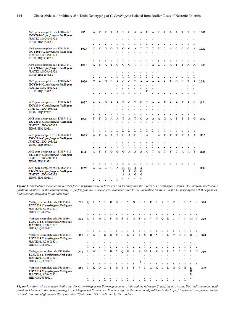
3.6. Sequencing of Net B and Cpb2 Toxin Genes of C.
perfringens
Sequencing of net B toxin gene in this study revealed that
it was highly conserved in both nucleotide and amino acid
sequence. Only one difference in this gene was identified
where a replacement of an amino acid was occurred at codon
379 (glutamate GAA → arginine AGG), while a silent
mutation was detected at nucleotide 1134 (GAG→GAA,
both are glutamate) (Figure 6, 7). In addition to very few
changes in nucleotides of eight strains in which alanine
changed into threonine at position 168 attributing that into
the use of different strains that obtained from different
countries. A more systematic nucleotide variation of net B
gene (A replaced by G) was recorded in 6 isolates in CDS
position 502 leading to a shift from threonine (ACT) to
alanine (GCT) in amino acid position 168 of NET B protein.
The nucleotide and amino acid sequences of C. perfringens
net B toxin gene were deposited into GenBank under
accession number (KJ724530). Additionally, phylogenetic
tree of nucleotides and amino acids based on net B toxin gene
sequences of the C. perfringens isolate is shown (Figure 8).
The difference in nucleotide sequence and amino acids
replacement of (NET B) toxin in this study opens significant
opportunities for further studies in Egypt for the development
of novel vaccines against NE. On the other hand, no
mutations were recorded in cpb2 gene when compared with
its identical mutant sequence (accession number
FJ493474.1). The nucleotide and amino acid sequences of C.
perfringens cpb2 toxin gene were deposited into GenBank
under accession number (KJ874348) (Figure 9). Additionally,
distance and standard error between net B and cpb2 toxin
genes of C. perfringens strains under study indicated that
identity percentage of both toxin genes was 86.7% (Table
11). According to nucleotide sequencing of the consensus
cpb2 gene in this study, frame shift mutations were recorded
as 21 bp deletions and 4 bp additions when it was compared
with the complete wild genome (accession number
AY609161.1) however, no mutations were recorded when it
was compared with its identical mutant sequence (accession
number FJ493474.1).
Table 11. Distance and standard error between net B and beta2 toxin genes
of C. perfringens strains under study.
C. Perfringens toxin gene Distance Standard error
Net B 0.098
Beta2 0.867
114 Ghada Abdelaal Ibrahim et al.: Toxin Genotyping of C. Perfringens Isolated from Broiler Cases of Necrotic Enteritis
Figure 6. Nucleotides sequence similarities for C. perfringens net B toxin gene under study and the reference C. perfringens strains. Dots indicate nucleotides
positions identical to the corresponding C. perfringens net B sequences. Numbers refer to the nucleotide positions in the C. perfringens net B sequences.
Mutations are indicated by the solid bars.
Figure 7. Amino acids sequence similarities for C. perfringens net B toxin gene under study and the reference C. perfringens strains. Dots indicate amino acid
positions identical to the corresponding C. perfringens net B sequence. Numbers refer to the amino acid positions in the C. perfringens net B sequence. Amino
acid substituation of glutamate (E) by arginine (R) at codon 379 is indicated by the solid bar.

Animal and Veterinary Sciences 2017; 5(6): 108-120 115
Figure 8. Phylogenetic tree of net B toxin gene sequence of a C. perfringens strain.
Figure 9. Nucleotides sequence similarities for C. perfringens cpb2 toxin gene under study and the reference C. perfringens strain. Dots indicate nucleotides
positions identical to the corresponding C. perfringens cpb2 sequences. Numbers refer to the nucleotide positions in the C. perfringens cpb2 sequences. No
mutations were recorded.=
C. perfringens NetB gene complete cds accession No FJ189484.1
C. perfringens NetB gene partial cds accession No HQ585982.1
C. perfringens EGY2014 NetB gene partial cds
C. perfringens NetB gene partial cds accession No KC460332.198
0.002

116 Ghada Abdelaal Ibrahim et al.: Toxin Genotyping of C. Perfringens Isolated from Broiler Cases of Necrotic Enteritis
4. Discussion
Clostridium perfringens organisms are of an economic
concern in poultry production. They constitute a risk for
transmission to humans through the food chain. Colonization
of poultry by clostridia is a very early event in the animals’
life and can be transmitted within the broiler chicken
operation.
The percentage of C. perfringens positive isolates in NE
diseased broilers was 47.8% while it was isolated in a higher
percentage (52.7%) from the healthy broilers. This attributed
to a large number of C. perfringens could be found in healthy
broilers but the proliferation of C. perfringens or increase of
its number in the gut depends on many factors like
contaminated soil, dust, feed, litter and also induced by
nutrition, pH and coccidial infection. All these factors might
cause hindering of the digestion and decreased feed
consumption that lead to low absorption, growth retardation
and so appearance of the disease [33]. In the similar trend,
higher percentages (41.6%, 58.4%, 75% and 40%) of C.
perfringens isolation in chickens were recorded with many
authors [34, 35, 36, 37]. Meanwhile in previous studies [38,
39] a lower prevalence rate (8 and 5%) of NE diseased cases
from the intestinal broiler chickens, respectively were
recorded. This variation might be due to the different
methodologies used for isolation, classifying the
microorganism or using of growth promoting in poultry
farms [7].
An acute form of NE disease could be seen from about two
weeks of age however, the subclinical form was observed at
varying ages of birds, but it was first detected most commonly
in birds at 21 to 23 days of age [18]. In current study, the
incidence of C. perfringens according to the age of the
chickens was higher (52.8%) in 2-3 weeks of age as shown in
(table 3). These results were in line with many authors [33, 40,
41] who stated that NE disease is most common in broiler
chickens causing high mortality rate at 2–3 or 4 weeks of age.
The pathogencity of C. perfringens is associated with their
ability to secrete major and minor toxins which play
important role in pathogenesis and induction of the disease.
Multiplex PCR technique showed that all ten isolates in this
study harboured cpa gene which give characteristic bands at
400 bp confirming that all of C. perfringens type. This result
goes hand in hand with several anthers [42, 43, 44].
For long time, α-toxin or phospholipase C enzyme of C.
perfringens was considered the main virulence factor in NE
disease. A new discovered virulence determinant (net B)
toxin recently was discovered and studied [9, 45, 46]. In this
paper, net B toxin of C. perfringens was studied and detected
in NE diseased broilers in a percentage of (46.7%) but didn’t
found in the isolates from healthy birds. These results were in
accordance with a study [47] in which they stated that net B
gene was only detected in Candian isolates that were
associated with NE outbreaks but it wasn’t found in isolates
from healthy birds. In addition, net B gene was found in
77.8%, 74.4% and 70% in chickens derived NE C.
perfringens strains [9, 18, 48]. However the latter study
showed also, that 2/15 isolates carried net B toxin gene from
healthy chickens and they explained the cause for the
negative NET B strains from the diseased birds (didn’t not
carry net B gene) were that alternative virulence factors may
constitute complex associations with other microflora that
were required for disease production.
Throughout the last decade, several epidemiological
studies showed wide distribution of beta2 (β2) toxigenic C.
perfringens strains among human and other animal species
[49] but its exact role in pathogenesis would still to be
further elucidated [50]. In this study, it was discovered in
both diseased and healthy birds in percentages of 75% and
71.4%. Similarl studies [51, 47, 36] detected cpb2 toxin
gene in 75%, 74.2% and 62.6% of C. perfringens type A
isolates in NE affected chickens. C. perfringens isolates
were not capable of causing disease without net B gene
especially it is linked with the health condition of the bird
while a weak or no relationship between β2 toxin and NE
disease in birds [8, 46].
The experimental study of the pathogencity of both toxins
in chicks revealed post mortem enlargement of the small
intestine in NE affected chicks due to gas accumulation that
could lead to thinning of the wall of the intestine. Similar
macroscopic lesions were also detected by [52, 53, 40].
Eleven net B positive strains were able to induce lesions
typical of NE in induction chickens models [8]. Importantly
in vitro, all of C. perfringens isolates that carried net B gene
expressed also NET B protein but only 54.5% of positive
strains of cbp2 gene, produced β2 toxin [51]. Alpha toxin of
C. perfringens from healthy birds was confirmed to be failed
to induce the disease while 33% of broilers that were
inoculated with NET B diseased isolates, developed NE
specific intestinal lesions [54].
DNA sequencing has been used to investigate the genetic
variation in individual genes, such as those encoding alpha
and NetB toxins. NE affected birds fall into three distinct
sequence based clades while non-pathogenic isolates from
healthy birds tend to be more genomically diverse [14].
Nucleotide sequencing of net B in this study identified that
glutamate amino acid was replaced with arginine at codon
379 in addition a silent mutation was detected at nucleotide
1134. In a similar way, a single nucleotide variation was
observed in net B gene of four isolates at CDS position 10 (T
replaced by with no AA shift) and in 2 isolates in CDS
position 497 (C replaced by T with shift from Ala to Val in
AA position 166) [55].
The gene sequencing of cpb2 didn’t show mutations in this
paper. Differently, the difference of nucleotide sequences at
positions 6, 10, 12, 20 and 198 of two Iranian C. perfringens
isolates was recorded [49] with 99% similarity to each other
and 73 % identity with the cpb2 sequences of C. perfringens
strains. An absence of β2 toxin expression where almost half
of the non-porcine consensus cpb2 genes (44.4%) carried a
frameshift mutation was also, reported [56]. However, 88.5%
of 78 non-porcine isolates carried atypical cpb2, but β2 toxin

Animal and Veterinary Sciences 2017; 5(6): 108-120 117
was not expressed. Atypical β2 toxin displayed 62.3%
identity and 80.4% similarity to consensus β2 toxin.
The hematological examination of experimentally infected
broilers with NET B and B2 toxins of C. perfringens revealed
a decrease in erythrocytic count, Hb concentration and PCV
values. While blood indices didn't show any changes after 5
days of infection. These results could be observed in the
hemolytic type of anemia and could be attributed to action of
α toxin which causes the breakdown of phospholipids of
erythrocytes membrane and cause hemolysis by damaging
circulating erythrocytes. Hemolytic anemia which was
associated with excessive destruction of erythrocyte might be
caused by variety of diseases like bacterial infection like
Clostridium [22]. Also, C. perfringens bacteremia is
commonly associated with intravascular hemolysis [57].
A significant reduction in RBCs, Hb, and PCV values
were recorded in infected broiler chicks than normal ones.
Such results might be attributed to the sequestration of iron in
the bone marrow macrophages and hepatocytes during the
infection, thus become unavailable to be utilized in
hemoglobin synthesis, resulting in inhibition of
erythropoiesis [23]. Group (G1) which was infected by NET
B toxin showed a significant decrease in RBCs count, Hb
concentration and PCV in the affected birds. This result
indicated microcytic hypochromic anemia as showed by the
erythrocytic indices that were proportionally correlated with
the severity of infection. These results are in accordance with
some researches [58].
Concerning to leucogram revealed neutrophilia and
lymphopenia after 5 days post infection in both G1 and G2
groups. In addition, neutrophilia and lymphocytosis were
shown after 12 days of infection by β2 infected group (G3),
but lymphocytosis and neutropenia were observed in G1
(NET B infected group). These results were common in
acute inflammatory response because the inflammatory
mediators stimulated the movement of neutrophil during
acute inflammation, also stimulated the movement of
lymphocytes from the blood to the inflamed tissue and
lymphoid tissues. The severity of lymphopenia reflects the
severity of systemic inflammatory response [59, 60, 61].
There was an increased TLC (Lymphocytosis) which might
be due to the antigenic stimulation of C. perfringens that
could lead to an increase in the thymus dependent
lymphocytes (T lymphocytes) production as reported
[22].The results of biochemical tests indicated that a
significant increase in ALT and AST transaminase
enzymes, uric acid and creatinine were noticed in both
infected groups (G1, G2) at 5th
and 10th
days post infection.
This increased in serum AST level had been associated with
hepatocellular damage in chickens, turkeys and ducks as
well as the worse effect of microorganism or its toxin in the
liver and kidney as described by [62]. These results agreed
with a study [63] which reported that, a significant
elevation in the activities of AST and ALT due to invasion
of the liver by pathogenic bacteria which causes liver cell
damage. Similar results were obtained by [60, 64]. Also,
some authors [61, 65] reported a significant increase in liver
and kidney enzymes in broiler chickens post C. perfringens
infection. Hypoprotienemia and hypoalbuminemia in the
infected broiler chicken might be due to cease feeding and
diarrhea. Similarly, similar studies [22, 66] mentioned that
bacterial toxins, increase the capillary permeability and
permitted the escape of plasma proteins into tissue resulting
in hypoprotienemia. A Significant increase in gamma and
alpha globulins could be associated with bacterial
septicemia [22]. The increase in uric acid and creatinine
could be due to the effect of the microorganisms and their
toxins on the kidneys. Our results were completely agreed
with many studies [67, 68, 69] in which the increased levels
of creatinine and uric acid in case of renal disease were
reported. Hypocalcemia and hyperphosphatemia could be
due to decrease calcium resorption by damaged renal
tubules and associated with hypoalbuminemia as reported
[62, 70]. Decreased calcium level lead to hypoalbuminemia
where decreased albumin concentration lowers the total
calcium level, while both ionized and complex calcium
levels remain normal. Also the metabolism of calcium and
phosphorus were closely linked in the body [62, 70]. These
results agreed with [61] who reported that the significant
decrease in calcium and chloride as well as a significant
increase in phosphorus in Guinea pig experimentally
infected with C. perfringens type A. Additionally, the
serum electrolytes showed significant decrease in serum
sodium and chloride levels of infected groups while there is
no significant variance in the serum potassium level.
Similar results reported that sodium and chloride are
particularly exposed to loss in diarrhea stools as they are
components of the gastrointestinal secretions [61, 70].
5. Conclusion
In summary, C. perfringens NET B toxin harbouring
isolates exhibited more lethal, pathogenic and virulent effects
than β2 toxin harbouring isolates in broilers. Vaccine
preparations that include NET B toxoid can protect chickens
against disease. A series of single amino acid substitution
derivatives of NET B have potential value for vaccine
formulations. It is likely that NET B will be an important
antigen to include in an effective, commercially viable,
necrotic enteritis vaccine.
References
[1] Osman, K. M. and Elhariri, M. (2013): Antibiotic resistance of Clostridium perfringens isolates from broiler chickens in Egypt. Rev. sci. tech. Off. int. Epiz., 32 (3): 841-850.
[2] Asaduzzaman, M.; Miah, M. S.; Siddika, A.; Popy, N. and Hossain, M. M. (2011): Experimental production of necrotic enteritis in broiler chickens. Bangl. J. Vet. Med., 9 (1): 33–41.
[3] Kaldhusdal, M. and Hofshagen, M. (1992): Barley inclusion and avoparcin supplementation in broiler diets. 2. Clinical, pathological, and bacteriological findings in a mild form of necrotic enteritis. Poult. Sci., (71): 1145-1153.
118 Ghada Abdelaal Ibrahim et al.: Toxin Genotyping of C. Perfringens Isolated from Broiler Cases of Necrotic Enteritis
[4] Lee, K. W.; Lillehoj, H. S.; Jeong, W.; Jeoung, H. Y. and An, D. J. (2011): Avian necrotic enteritis: experimental models, host immunity, pathogenesis, risk factors, and vaccine development. Poult. Sci., 90 (7): 1381–1390.
[5] Skinner, J. T.; Bauer, S.; Young, V.; Pauling, G. and Wilson, J. (2010): An economic analysis of the impact of subclinical (mild) necrotic enteritis in broiler chickens. Avian Dis., 54 (4):1237–1240.
[6] Thomas, P.; Arun, T. R..; Karthik, K.; Berin, P. V.;Asok Kumar, M.; Neetu Singh.; Usharani, J.; Palanivelu, M.; Gupta, S. K.; Dhama, K. and Viswas, K. N.(2014):Molecular Characterization and Toxinotyping of a Clostridium perfringens Isolate from a Case of Necrotic Enteritis in Indian Kadaknath Fowl. Asian Journal of Animal and Veterinary Advances., 9: 385-394.
[7] Craven, S. E.; Stern, N. S.; Bailey, J. S. and Cox, N. A. (2001): Incidence of C. perfringens in broiler chickens and their environment during production and processing. Avian Dis., 45: 887–896.
[8] Miyamoto, K.; Wen, Q.; and McClane, B. A. (2004): Multiplex PCR genotyping assay that distinguishes between isolates of Clostridium perfringens type A carrying a chromosomal enterotoxin gene (cpe) locus, a plasmid cpe locus with an IS1470-like sequence, or a plasmid cpe locus with an IS1151 sequence. J. Clin. Microbiol., 42: 1552–1558.
[9] Keyburn, A. L.; Boyce, J. D.; Vaz, P.; Bannam, T. L.; Ford, M. E.; Parker, Rubbo, A. D.; Rood, J. I. and Moore, R. J. (2008): NetB, New Toxin That Is Associated with Avian Necrotic Enteritis Caused by Clostridium perfringens. PLOS Patholog., 4 (2):e26.
[10] Timbermont, L.; De Smet, L.; Van Nieuwerburgh, F.; Parreira, V. R.; Van Driessche, G.; Haesebrouck, F.; Ducatelle, R.; Prescott, J.; Deforce, D.; Devreese, B. and Van Immerseel, F. (2014): Perfrin, a novel bacteriocin associated with netB positive Clostridium perfringens strains from broilers with necrotic enteritis. Vet Res., 45:40.
[11] Bueschel, D. M.; Jost, B. H.; Billington, S. J.; Trinh, H. T. and Songer, J. G. (2003): Prevalence of cpb2, encoding beta2 toxin, in C. perfringens field isolates: correlation of genotype with phenotype. Vet. Microbiol., 94: 121 -129.
[12] Gibert, M.; Jolivet, R. C. and Popoff, M. R. (1997): Beta2 toxin, a novel toxin produced by C. perfringens. Gene., 203 (1): 65-73.
[13] Petit, L.; Gibert, M. and Popoff, M. R. (1999): Clostridium perfringens: toxinotype and genotype. Trends Microbiol., 7(3):104-10.
[14] Lacey, J. A.; Johanesen, P. A.; Lyras, D. and Moore, R. J. (2016): Genomic diversity of necrotic enteritis-associated strains of Clostridium perfringens: a review. Avian Pathology., 45 (3).
[15] Willis, A. T. (1977): Anaerobic Bacteriology-Clinical and Laboratory Practice. 3rd ed.
[16] Cruickshank, R.; Duguid, J. P.; Marmion, B. R. and Swain, R. H. A. (1975): Medical Microbiology, 12th Ed., Living stone, London, New York, 812-825.
[17] Koneman, E. W.; Allen, S. D.; Dowell, V. R. and Summers, H. W. (1983): Colour atlas and text book of diagnostic
microbiology. 2nd Ed. J. B. LippinCott, New York, London.
[18] Keyburn, A. L.; Yan, X.; Bannam, T. L.; Immerseel, F. V.; Rood, J. I. and Moore, R. J. (2010): Association between avian necrotic enteritis andClostridium perfringens strains expressing NetB toxin. Vet. Res., 41(2): 21.
[19] Shojadoost, B.; Vince, A. R. and Prescott, J. F. (2012): The successful experimental induction of necrotic enteritis in chickens by Clostridium perfringens: a critical review. Veterinary Research., 43: (74).
[20] Natt, M. P. and Herrick, A. C. (1952). A new blood diluent for counting the erythrocytes and leucocytes of chickens. Poul. Sci. 31: 735-738.
[21] Van Kempen, E. J. and Zijlstra, W. G. (1961): Colorimetric determination of hemoglobin. Clin. Chem. Acta.6: 538.
[22] Coles, E. H. (1986): Veterinary Clinical Pathology.4th ed. W. B. Sounders Company, Philadelphia, London, Toronto, Mexico, Sydney, Tokyo, Hong Kong.
[23] Jain, N. C. (2000). Schalm's veterinary hematology.8th. Ed. Lea and Febiger, Philadelphia, U. S. A.
[24] Reitman, S. and Frankel, S. (1957): A colorimetric method for the determination of AST and ALT. Am. J. Clin. Path.25:56.
[25] Doumas, B. T. and Bigs, H. G. (1972): Determination of serum globulin. In: Standard Methods of Clinical chemistry, Vol. 7, ed. G. R. Cooper. New York Academic press.
[26] Henry, R. J. (1979): Calorimetric methods for determination of serum creatinine. Clinical Chemistry, Principles and techniques, 2nd ed. Harper and Row, 525.
[27] Caraway, W. T. (1963): Uric acid. Standard Methods of Clin. Chem. 1963; 4:239-47.
[28] Laemmli, U. K. (1970): Cleavage of structural proteins during the assembly of the head of bacteriophage T4. Nature. 227: 680-685.
[29] Glinder, E. M. and King, J. D. (1972): Rapid calorimetric determination of calcium in biological fluids with methylene blue. Am. J. Clin, Path. 58: 376–382.
[30] EI-Merzabani, M. M.; Anwer-El-Aaser, A. and Zakhary, N. H. (1977): A New Method for Determination of Inorganic Phosphorus in Serum without Deproteinization. J. Clin. Chem. Clin. Biochem., 15: 715-718.
[31] Oser, B. L. (1979): Hawk's physiological chemistry. 14th ed. MC Graw Hill Company Ltd., London.
[32] Vaikosen, E. S. and Muller, W. (2001): Evaluating biochemical tests for isolation and identification of C. perfringens in faecal samples of small ruminants in Nigeria. Bulletin of animal Health and Production in Africa., 49(4):244-248.
[33] Johanssons, A. (2006): C. perfringens the causal agent of necrotec enteritis in poultery Ph. D. Thesis, Biomedical Sciences, Fac. Vet. Med. Swedish Univ.
[34] Mustafa, M. G. (2000): Some studies on Clostridial infection in birds. M. V. Sc. Thesis, Fac. of Vet. Med., Zagazig Univ.
[35] Manfreda, G.; Bondoioli, V.; De Cesare, A. and Franchini, A. (2005): quantitative evaluation of Clostridium perfringens in Italian broilers. Poult. Sci., 62 (Suppl): 91-92.
Animal and Veterinary Sciences 2017; 5(6): 108-120 119
[36] Osman, K. M.; Soliman, Y. A.; Amin, Z. M. S. and Aly, M. A. K. (2012): Prevalence of Clostridium perfringens type A isolates in commercial broiler chickens and parent broiler breeder hens in Egypt. Rev. sci. tech. Off. int. Epiz., 31 (3): 931-941.
[37] EI-Jakee, J.; Nagwa, S. A.; Mona, A. E.; Azza S. M.; Abu Elnaga; Riham, H. H; Shawky, N. M. and Shawky, H. M. (2013): Characterization of Clostridium perfringens Isolated from Poultry. Global Veterinaria., 11 (1) 1992-6197: 88-94.
[38] Miah, M. S.; Asaduzzaman.; Sufian, M. A. and Hossain, M. M.(2011): Isolation of Clostridium perfringens, Causal agents of necrotic enteritis in chickens. Journal of the Bangladesh Agricultural University., 9 (1).
[39] Kalender, H. and Ertas, H. B. (2005): Isolation of Clostridium perfringens from chickens and detection of the alpha toxin gene by polymerase chain reaction (PCR). Turk J Vet Anim Sci 29, 847–851.
[40] Cooper, K. K. and Songer, J. G (2010): Virulence of Clostridium perfringens in an experimental model of poultry necrotic enteritis. Vet Microbiol., 142 (3-4): 323-328.
[41] Das, A.; Mazumder, Y.; Dutta, B. K.; Shome, B. R.; Bujarbaruah, K. M. and Kumar, A. (2008): Clostridium perfringens Type A from Broiler Chicken with Necrotic Enteritis. International Journal of Poultry Science., 7 (6) 1682-8356: 601-609.
[42] Shanmugasamy, M. and Rajeswar, J. (2012): Alpha toxin specific PCR for detection of toxigenic strains of Clostridium perfringens in poultry. Vet. World, 5: 365-368.
[43] Sarkar, M.; Ray, J. P.; Mukhopadhayay, S. K.; Niyogi, D. and Ganguly, S. (2013): Study on Clostridium perfringens type A infection in broilers of West Bengal, INDIA. IIOABJ., 4: (4): 1–3.
[44] Doosti, A., Pasand, M.; Mokhtari-Farsani, A.; Ahmadi, R. and Chehelgerdi, M. (2015): Prevalence of Clostridium perfringens type A isolates in different tissues of broiler chickens. Bulg. J. Vet. Med., (In Press).
[45] Johansson, A.; Aspan, A.; Kaldhusdal, M. and Engstrom, B. E. (2010): Genetic diversity and prevalence of netB in Clostridium perfringens isolated from a broiler flock affected by mild necrotic enteritis. Vet. Microbiol., 144: 87–92.
[46] Ezatkhah, M.; Alimolaei, M. and Shahdadnejad, N. (2016): The Prevalence of netB Gene in Isolated Clostridium perfringens from organic broiler farms suspected to necrotic enteritis. Int J Enteric Pathog., (In press).
[47] Chalmers, G.; Martin, S. W.; Hunter, D. B.; Prescott, J. F.; Weber, L. J. and Boerlin, P. (2008): Genetic diversity of Clostridium perfringens isolated from healthy broiler chickens at a commercial farm. Vet. Microbiol., 127: 116–127.
[48] Mohamed, M. A.; Ahmed, S. O. and Abdelmotelib, T. Y. (2009): Associated of virulence gene markers in Clostridium perfringens strains isolated from healthy and diseased broiler chickens with necrotic enterits. Assiut Vet. Med. J., 55: 123 (10).
[49] Tolooe, A.; Shojadoost, B. and Peighambari, S. M. (2011): Molecular detection and characterization of cpb2 gene in Clostridium perfringens isolates from healthy and diseased
chickens. The Journal of Venomous Animals and Toxins including Tropical Diseases., 1678-9199 (17 1): 59-65.
[50] Svobdova, I.; Steinhouserova, I.; and Nebola, M. (2007): Incidence of C. perfringens in broiler chickens in Czech Repuplic. Acta-Veterinaria-Brno., 76 (supplementum8): s25-s30.
[51] Crespo R., Fisher D. J.; Shivaprasad H. L.; Fernandez-Miyakawa M. E. and Uzal F. A. (2007): Toxinotypes of Clostridium perfringens isolated from sick and healthy avian species. J. Vet. Diagn. Invest., 19: 329–333.
[52] Van Immerseel, F.; De Buck, J.; Pasmans, F.; Huyghebaert, G.; Haesebrouck, F. and Ducatelle.; R. N. (2004): Clostridium perfringens in poultry: an emerging threat for animal and public health. Avian Pathol., 33: 537–549.
[53] Broussard, C. T., Hofacre, C. L., Page, R. K. and Fletcher, O. J., (1986): Necrotic enteritis in cage reared commercial layer pullets. Avian Dis., 30: 617–619.
[54] Timbermont, L.; Lanckriet, A.; Gholamiandehkordi, A. R.; Pasmans, F.; Martel, A. and Haesebrouck, F. (2009): Origin of Clostridium perfringens isolates determines the ability to induce necrotic enteritis in broilers, Comp. Immunol. Microbiol. Infect. Dis., (32):503–512.
[55] Abildgaard, L.; Sondergaard, T. E.; Engberg, R. M.; Schramm, A. and Højberg, O. (2010): In vitro production of necrotic enteritis toxin B, NetB, by netB-positive and netB-negative Clostridium perfringens originating from healthy and diseased broiler chickens. Veterinary Microbiology., 144: 231–235.
[56] Jost, B. H., Billington, S. J., Trinh, H. T., Bueschel, D. M., Songer, J. G. (2005): Atypical cpb2 genes, encoding beta2-toxin in Clostridium perfringens isolates of nonporcine origin., Infect. Immun. 73:652-656.
[57] Topley, Y. and Wilson, T. (1998): Microbiology and microbial infections. Ninth Edition. v.3., systemic bacteriology oxford university press, USA.
[58] Jain, N. C. (1986). Schalm’s Veterinary Hematology. 4thEdn., Lea and Febiger, Philadelphia.
[59] Imhof, B. A. and Dunon, D. (1995): "Leukocyte migration and adhesion." Adv. Immunol., 58:345-416.
[60] Heba, H. E. and Hala A. M. (2009): Pathological and bacteriological studies on Clostridium perfringens infection in kidney of cattle, camel and sheep. Egypt. J. Comp. Path. & Clinic. Path., 22 (2): 88–108.
[61] Fatma, M. Youssef, Heba, E. Farhan and Soliman, A. (2013): Biochemical alterations associating clostridium perferingens infections in sheep. 12th Sci. Cong., Egyptian Society For Cattle Diseases, 3-6 Dec. Hurgada, Egypt. 1-10.
[62] Campbell, T. and Coles, E. (1986): Avian clinical Pathology,'in "Veterinary Clinical pathology ".4th Ed., W. B Saunders Company. Philadelphia. London and Toronto.
[63] Burtis, C. A. and Ashwood, E. R. (1999): Tietz textbook of clinical chemistry. 3rd ed. W. B. Saunders Company, Philadelphia: 617-721.
[64] Amany, A. M. and Morsi, A. A. (1995): "Clinico and histopathological studies on the effect of Clostridium chauvoei in guinea pigs. Egypt." J. Comp. Pathol. and Clinc. Pathol., 8( 2): 15-26.

120 Ghada Abdelaal Ibrahim et al.: Toxin Genotyping of C. Perfringens Isolated from Broiler Cases of Necrotic Enteritis
[65] Fatma, M. Yousseff and Hala, M. El-Genaidy (2012): Clinicopathological studies on chickens infected by anaerobic bacteria at Ismailia Governorate.
[66] Ostroff, S., Kobayashi, J. and Lewis, J. (1989). Infections with Escherichia coli 0157:H7 an emerging gastrrointestinal pathogen. Results of a one-year, prospective, population-based study. JAMA. PP 262:355-359.
[67] Pai C.; Gordon R.; Sims, H. and Bryan, L. (1984). Sporadic cases of hemorrhagic colitis associated with Escherichia coli 0157:H7. Clinical. Epidemiologic and bacteriologic features. Ann Intern Med. 101:738-742.
[68] Obrig, T.; Del Vecchio, P.; Karmali, M.; Petric, M.; Moran, T.
and Judge, T. (1987). Pathogenesis ofhemolyticuremic syndrome (Letter). Lancet. Vol 2, 687-689.
[69] Mona S. Zaki, Olfat, A. Fawzy and Osfor, M. H. (2012): Effect of E. coli O: 157 on Baladi Broiler Chicken and some Biochemical studies. Life Science Journal, 9(1): 91-94.
[70] Duncan, J. R., Orasse, K. W. and Mohaffy, A. (1994): Veterinary laboratory medicine; clinical pathology 3rd. Ed., Ames, Iowa state, University press.
[71] Radostits, O. M.; Gay, C. C.; Blood, D. C. and Hincheliff, K. W. (2000): "Veterinary Medicine. A textbook of the Diseases of Cattle, Sheep, Goats and Horses." J. Clin. Microbiol., 31 (2): 467-469.