toxicology mechanisms and methods · lipid signaling and lipidomics laboratory, division of...
TRANSCRIPT

This article was downloaded by:[Nationwide Childrens Hospital]On: 1 December 2007Access Details: [subscription number 770237668]Publisher: Informa HealthcareInforma Ltd Registered in England and Wales Registered Number: 1072954Registered office: Mortimer House, 37-41 Mortimer Street, London W1T 3JH, UK
Toxicology Mechanisms andMethodsPublication details, including instructions for authors and subscription information:http://www.informaworld.com/smpp/title~content=t713396575
Mercury Activates Phospholipase A2 and InducesFormation of Arachidonic Acid Metabolites in VascularEndothelial CellsJessica N. Mazerik a; Himabindu Mikkilineni a; Vivek A. Kuppusamy a; EmilySteinhour a; Alon Peltz a; Clay B. Marsh a; Periannan Kuppusamy a; NarasimhamL. Parinandi aa Lipid Signaling and Lipidomics Laboratory, Division of Pulmonary, Allergy, CriticalCare, and Sleep Medicine, Department of Internal Medicine, The Ohio StateUniversity College of Medicine, Columbus, OH, United States
Online Publication Date: 01 November 2007To cite this Article: Mazerik, Jessica N., Mikkilineni, Himabindu, Kuppusamy, Vivek A., Steinhour, Emily, Peltz, Alon,Marsh, Clay B., Kuppusamy, Periannan and Parinandi, Narasimham L. (2007) 'Mercury Activates Phospholipase A2 andInduces Formation of Arachidonic Acid Metabolites in Vascular Endothelial Cells', Toxicology Mechanisms and Methods,17:9, 541 - 557To link to this article: DOI: 10.1080/15376510701380505URL: http://dx.doi.org/10.1080/15376510701380505
PLEASE SCROLL DOWN FOR ARTICLE
Full terms and conditions of use: http://www.informaworld.com/terms-and-conditions-of-access.pdf
This article maybe used for research, teaching and private study purposes. Any substantial or systematic reproduction,re-distribution, re-selling, loan or sub-licensing, systematic supply or distribution in any form to anyone is expresslyforbidden.
The publisher does not give any warranty express or implied or make any representation that the contents will becomplete or accurate or up to date. The accuracy of any instructions, formulae and drug doses should beindependently verified with primary sources. The publisher shall not be liable for any loss, actions, claims, proceedings,demand or costs or damages whatsoever or howsoever caused arising directly or indirectly in connection with orarising out of the use of this material.
Dow
nloa
ded
By:
[Nat
ionw
ide
Chi
ldre
ns H
ospi
tal]
At:
00:1
0 1
Dec
embe
r 200
7 Toxicology Mechanisms and Methods, 17:541–557, 2007
Copyright ©c Informa Healthcare USA, Inc.
ISSN: 1537-6516 print; 1537-6524 online
DOI: 10.1080/15376510701380505
Mercury Activates Phospholipase A2 andInduces Formation of Arachidonic Acid
Metabolites in Vascular Endothelial CellsJessica N. Mazerik,
Himabindu Mikkilineni,
Vivek A. Kuppusamy,
Emily Steinhour, Alon Peltz,
Clay B. Marsh,
Periannan Kuppusamy, and
Narasimham L. Parinandi
Lipid Signaling and Lipidomics
Laboratory, Division of
Pulmonary, Allergy, Critical
Care, and Sleep Medicine,
Department of Internal
Medicine, The Ohio State
University College of Medicine,
Columbus, OH, United States
ABSTRACT Currently, mercury has been identified as a risk factor in
cardiovascular diseases among humans. Here, we tested our hypothesis that
mercury modulates the activity of the vascular endothelial cell (EC) lipid
signaling enzyme phospholipase A2 (PLA2), which is an important player in the
EC barrier functions. Monolayers of bovine pulmonary artery ECs (BPAECs) in
culture, following labeling of membrane phospholipids with [3H]arachidonic
acid (AA), were exposed to the inorganic form of mercury, mercury chloride,
and the release of free AA (index of PLA2 activity) and formation of AA
metabolites were determined by liquid scintillation counting and enzyme
immunoassay, respectively. Mercury chloride significantly activated PLA2 in
BPAECs in a dose-dependent (0 to 50 µM) and time-dependent (0 to 120 min)
fashion. Metal chelators significantly attenuated mercury-induced PLA2 acti-
vation, suggesting that cellular mercury–ligand interaction is required for the
enzyme activation and that chelators are suitable blockers for mercury-induced
PLA2 activation in ECs. Sulfhydryl (thiol-protective) agents, calcium chelating
agents, and cPLA2-specific inhibitor also significantly attenuated the mercury-
induced PLA2, suggesting the role of thiol and calcium in the activation
of cPLA2 in BPAECs. Significant formation of AA metabolites, including
the release of total prostaglandins, thromboxane B2, and 8-isoprostane, were
observed in BPAECs following their exposure to mercury chloride. Mercury
chloride induced cytotoxicity as observed by the altered cell morphology and
enhanced trypan blue uptake, which was attenuated by the cPLA2 inhibitor
AACOCF3. The results of this study revealed that inorganic mercury-induced
PLA2 activation through the thiol and calcium signaling and the formation of
bioactive AA metabolites further demonstrated the association of PLA2 with
the cytotoxicity of mercury in ECs. Overall, the results of the current study
underscore the importance of PLA2 signaling in mercury-induced endothelial
dysfunctions.
KEYWORDS Arachidonic Acid; Isoprostane; Lipid Signaling; Mercury; Phospholipase A2;
PLA2; Prostaglandin; Thromboxane B2; Vascular Endothelial Cells
Received 17 January 2007;
accepted 3 April 2007.
This work was supported by the
National Institutes of Health grant HL
067176-05, EB 004031, and Dorothy M.
Davis Heart and Lung Research
Institute funds.
Address correspondence to
Narasimham L. Parinandi, Ph.D., Room
611-A, Division of Pulmonary, Critical
Care, and Sleep Medicine, Dorothy M.
Davis Heart and Lung Research
Institute, The Ohio State University,
473 W. 12th Avenue, Columbus,
OH 43210. E-mail:
541

Dow
nloa
ded
By:
[Nat
ionw
ide
Chi
ldre
ns H
ospi
tal]
At:
00:1
0 1
Dec
embe
r 200
7
INTRODUCTION
Mercury (Hg), a heavy metal belonging to the
transition element series of the periodic table, has
been used in industrial processes and medical practice
(Clarkson et al. 2003; Pleva 1994; Mutter et al. 2004)
resulting in accidental and occupational exposures to
mercury. Anthropogenic activities cause the increased
release of the element into the environment leading
to the pollution of air, water, and soil (Clarkson et al.
2003; Sarkar 2005; Kuehn 2005). Inorganic mercury
is toxic to many organisms including humans and is
converted to more toxic organic forms (methylmercury)
through biomethylation by microorganisms (bacteria)
(Boening 2000; Dopp et al. 2004). Consumption of
contaminated fish has been shown as a major source
of environmental mercury in humans that could lead
to suppression of the beneficial effects of omega-3 fatty
acids on coronary artery disease (Landmark and Aursnes
2004; Chan and Egeland 2004; Clarkson 2002).
The role of mercury toxicity as a possible risk factor
in cardiovascular disease has been emphasized (Kosta
1991). Elevated body levels of mercury, due to fish
consumption by humans, have been hypothesized as a
risk factor in coronary heart disease (Yoshizawa et al.
2002). Increased levels of urinary mercury have been
shown to be associated with elevated cholesterol levels
in humans and mercury has been suggested as a risk
factor of myocardial infarction, coronary disease, and
cardiovascular disease (Kim et al. 2005). An association
between the occupational exposure to mercury in
mining and refining and risk of cardiovascular diseases
has been shown (Boffetta et al. 2001).
Although mercury has been shown to be associated
with cardiovascular diseases among humans, detailed
studies leading to the understanding of mechanisms of
mercury-induced cardiovascular diseases are currently
lacking. Vascular endothelium plays a pivotal role in
the structure and function of the blood vessel and
maintains the homeostasis of the circulatory system
and the entire body in general. Methylmercury has
been shown to cause hypertension in rats (Wakita
1987). Mercury-induced vascular endothelial damage
and vasculitis in humans upon autopsy have been
documented (Egermayer 2000). Therefore, it is con-
ceivable to hypothesize that mercury exerts its toxic
effects on the vascular endothelium, which in turn
may contribute to the mercury-induced cardiovascular
diseases. Phospholipids of cellular membranes play an
important role in the cell as structural and functional
entities. Phospholipases are enzymes that specifically
hydrolyze the membrane phospholipids and generate
bioactive lipid second messengers, which play a vital
role in cell signaling (Dennis et al. 1991; Divecha and
Irvine 1995).
Phospholipase A2 (PLA2) is an important membrane
phospholipid hydrolyzing enzyme that catalyzes the
hydrolysis of the membrane phospholipids at the
sn-2 position generating free unsaturated fatty acid
and lysophospholipid (Dennis et al. 1991). Thus, the
unsaturated fatty acid released from the membrane
phospholipids upon the action of PLA2, usually
arachidonic acid, is a substrate for cyclooxygenases
(COXs) and lipoxygenases (LOXs), which mediate the
formation of potentially bioactive arachidonic acid
metabolites such as prostaglandins and leukotrienes
(Chakraborti 2003). These arachidonic metabolites of
COXs and LOXs have been identified to play crucial
roles in inflammatory cascades and are tightly regulated
by the activity of PLA2 (Dennis et al. 1991). PLA2 is
also a very important housekeeping enzyme involved
in membrane formation and repair (Balsinde et al.
2000). PLA2 has been shown to be activated by several
different agonists in different systems both in vitro
and in vivo (Chakraborti 2003). Roles of PLA2 and
arachidonic acid metabolites in cardiovascular diseases
have been emerging (Lambert et al. 2006). Therefore,
unregulated PLA2 activation mediated by agonists
such as environmental toxicants can jeopardize the
endothelial function and eventually the vessel function.
As environmental mercury has been implicated in
cardiovascular disease and earlier we have shown
that mercury activates PLD in vascular endothelial
cells (ECs) in vitro, here we have hypothesized that
inorganic mercury activates PLA2, induces the release
of arachidonic acid from the membrane phospho-
lipids, and mediates the formation of arachidonic acid
metabolites, and that the activation of PLA2 regulates
the mercury-induced cytotoxicity in ECs. To test our
hypothesis, we investigated whether inorganic mercury,
in the form of mercury chloride, could induce PLA2-
mediated release of arachidonic acid, formation of
the arachidonic acid metabolites, and regulation of
mercury-induced cytotoxicity by PLA2 in our well-
established bovine pulmonary artery ECs (BPAECs)
in vitro. For the first time, our current study revealed
that inorganic mercury induced the activation of PLA2,
release of arachidonic acid, formation of arachidonic
J. N. Mazerik et al. 542
Dow
nloa
ded
By:
[Nat
ionw
ide
Chi
ldre
ns H
ospi
tal]
At:
00:1
0 1
Dec
embe
r 200
7
acid metabolites, and regulation of cytotoxicity of
mercury by PLA2 in BPAECs.
MATERIALS AND METHODS
Materials
Bovine pulmonary artery endothelial cells (BPAECs)
(passage 2) were obtained from Cell Applications Inc.
(San Diego, CA). Fetal bovine serum (FBS), trypsin,
and nonessential amino acids were obtained from
Gibco Invitrogen Corp. (Grand Island, NY). Minimum
essential medium (MEM), mercury chloride, ethylene-
diaminetetracetic acid (EDTA), D-pencillamine, dithio-
threitol (DTT), N-acetyl-L-cysteine (NAC), meso-2,3-
dimercapto-succinic acid (DMSA), ethylene glycol-
bis (β-aminoethyl ether)-N,N,N′N′-tetraacetic acid
(EGTA), trypan blue (0.4%), and BAPTA-AM (BAPTA)
were obtained from Sigma Chemical Co. (St. Louis,
MO). [3H]arachidonic acid was obtained from Amer-
ican Radiolabeled Chemicals, Inc. (St. Louis, MO).
Arachidonly trifluoromethyl ketone (AACOCF3) and
enzyme immunoassay kits for total prostaglandins,
thromboxane B2, and 8-isoprostane were obtained from
Cayman Chemical Co. (Ann Arbor, MI).
Cell Culture
BPAECs were cultured in MEM supplemented with
10% FBS, nonessential amino acids, antibiotics, and
growth as described previously (Varadharaj et al. 2006).
Cells in culture were maintained at 37◦C in a humid-
ified environment of 5% CO2-95% air and grown to
contact-inhibited monolayers with typical cobblestone
morphology. When confluence was reached, cells were
trypsinized and subcultured in T 75-cm2 flasks or 35 ×
10-mm or 100-mm tissue culture dishes. Confluent
cells showed cobblestone morphology under light
microscope and stained positive for factor VIII. All
experiments were conducted between 8 and 20 passages
(75% to 80% confluence).
Assay of Release of Arachidonic Acidand PLA2 Activation
Release of arachidonic acid from cellular membrane
phospholipids is widely assayed as an index of PLA2
activity (Balsinde et al. 2000). BPAECs in 35-mm
dishes (5 × 10⁵ cells/dish) were labeled with carrier-
free [3H]arachidonic acid (5 µCi/ml) in complete EC
media containing 10% FBS, nonessential amino acids,
antibiotic, and growth factor for 12 h at 37◦C in 5%
CO2-95% air. The radioactive medium was removed
by aspiration and cells were incubated in serum-free
MEM or MEM containing mercury chloride (HgCl2)
at the chosen concentrations (1 to 50 µM) for specified
lengths of time (0 to 120 min). When required, cells
prelabeled with [3H]arachidonic acid were pretreated
with selected pharmacological agents/inhibitors for 1 h
and then exposed to mercury chloride in the absence
or presence of the pharmacological inhibitors for
specified lengths of time. At the end of the incubation
period the amount/extent of arachidonic acid released
into the medium, as an index of PLA2 activity, was
determined by liquid scintillation counting. The extent
of arachidonic acid released was expressed as DPM of
[3H]/dish.
Determination ofCyclooxygenase-Mediated Formation
of Arachidonic Acid Metabolites
The COX-mediated formation of arachidonic acid
metabolites in BPAECs following their exposure to
mercury chloride was determined by utilizing the com-
mercially available EIA kits (Cayman Chemical Co.,
Ann Arbor, MI). Release of total prostaglandins, throm-
boxane B2, and 8-isoprostane by cells was determined
according to the manufacturer’s recommendations.
The extent of arachidonic acid metabolites released by
cells was expressed as pg/mL medium.
Cellular Total Thiol Determination
Total cellular thiol content was measured by
DTNB-coupled spectrophotometric assay according to
Parinandi et al. (1999) and Hagele et al. (2006). BPAECs
were grown to 100% confluence in 100-mm dishes and
then treated with MEM or MEM containing mercury
chloride (25 µM) for 60 min. After incubation, cells
were detached by gentle scraping and centrifuged at
5,000 × g for 10 min at 4◦C. The cell pellets were
then lysed using Triton × 100. Cell lysates were treated
with 5,5′-dithiobis (DTNB) and the absorbance was
determined at 412 nm on a Spectromax plate reader.
Total thiol values were obtained from a standard curve
prepared with GSH and expressed as µg thiols/mg
protein.
543 Mercury Induces Endothelial Arachidonate Release

Dow
nloa
ded
By:
[Nat
ionw
ide
Chi
ldre
ns H
ospi
tal]
At:
00:1
0 1
Dec
embe
r 200
7
Assay of Cytotoxicity
Morphological changes in BPAECs grown in 35-mm
dishes up to 70% confluence, following their exposure
to different concentrations of mercury chloride (0,
5, 10, and 25 µM) in MEM for designated lengths
of time (5, 15, 30, and 60 min) at 37◦C in a
humidified environment of 5% CO2-95% air, were
examined as an index of cytotoxicity. The role of
cPLA2 in the mercury-induced morphological changes
was investigated in cells pretreated with the cPLA2-
specific inhibitor AACOCF3 (1 µM) for 1 h in MEM
and then subjecting the cells to mercury chloride
(10 µM) treatment in MEM for 60 min at 37◦C
in a humidified environment of 5% CO2-95% air.
Simultaneous controls were established with MEM
alone or MEM containing the inhibitor alone under
identical conditions. Images of cell morphology were
digitally captured with the Nikon Eclipse TE2000-S
at either 10× or 100× magnification. Additionally,
the trypan blue exclusion assay was also performed
to assess the cPLA2 inhibitor (AACOCF3) effect on
mercury-induced cytotoxicity in BPACEs according to
Verity et al. (1994). Following pretreatment of cells with
AACOCF3 (1, 5, 10 µM) in MEM for 60 min, the cells
were treated with mercury chloride (10 µM) in MEM
for 60 min at 37◦C in a humidified environment of 5%
CO2-95% air. Simultaneous controls were established
with MEM alone or MEM containing AACOCF3
alone. At the end of incubation, treatment media
were removed and cells were treated with 1.0 mL
trypan blue solution (1:10 dilution of stock 0.4%
trypan blue solution in 0.85% saline) for 3 min at
37◦C in a humidified environment of 5% CO2-95%
air. Following this, the solution was removed, cells
were gently washed three times with PBS containing
0.5% glucose, and the total number of cells and the
number of cells that did not exclude trypan blue were
determined under Nikon Eclipse TE2000-S microscope
at 20× magnification in an observed field. Mercury-
induced cytotoxicity was determined from the total
number of cells and the number of cells that had taken
up trypan blue and expressed as % cells that had taken
up trypan blue in an observed field.
Protein Determination
Cellular protein levels were determined by BCA
protein assay (Pierce, Rockford, IL).
Statistical Analysis of Data
Standard deviation (SD) for each data point was
calculated from triplicate samples. Data were subjected
to one-way analysis of variance, and pair-wise multiple
comparisons were done by Dunnett’s method with P <
0.05 indicating significance.
RESULTS
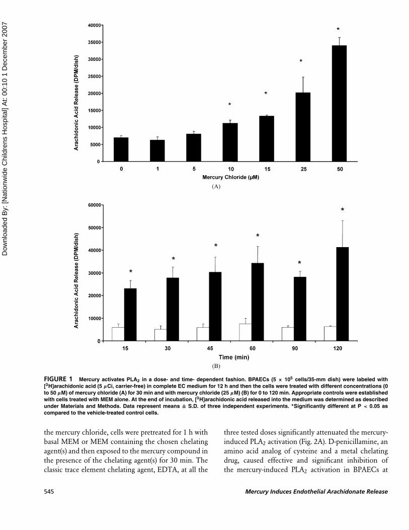
Mercury Activates PLA2 in a Dose-and Time-Dependent Fashion
As no reports have been made so far on mercury-
induced activation of PLA2 in the vascular ECs and
earlier we have shown that mercury activates PLD
in ECs (Hagele et al. 2006), here we investigated
whether inorganic mercury (mercury chloride) would
induce the release of arachidonic acid as an index
of PLA2 activation in BPAECs in a dose-dependent
(0 to 50 µM) fashion following incubation of cells
for 30 min with mercury chloride. Mercury chloride
significantly caused the activation of PLA2 at 10, 15,
25, and 50 µM concentrations upon treatment of cells
for 30 min, as compared to that in the cells treated with
vehicle alone (Fig. 1A). The time-dependant activation
of PLA2 in BPAECs upon their treatment with mercury
chloride was also evident. At 15 min of treatment,
mercury chloride caused a significant activation of
PLA2, which further increased at 30, 45, 60, 90, and
120 min of treatment with the metal as compared
to the cells treated with the vehicle alone (Fig. 1B).
Overall, these results revealed that inorganic mercury
(mercury chloride) was effective in causing a significant
and dose- and time-dependent activation of PLA2 in
BPAECs.
Metal Chelating Agents AttenuateMercury-Induced PLA2 Activation
Chelating agents complex with transition metals and
have been shown to protect against metal-mediated
adverse effects and metal toxicity (Blanusa et al. 2005).
However, the modulatory effects of chelating agents
on mercury-induced activation of PLA2 have not
been reported so far. Therefore, here, the effects of
well-established chelating agents including EDTA and
D-pencillamine (0.1, 0.5, and 1 mM) were examined
on the PLA2 activation induced by mercury chloride
(25 µM) in BPAECs. Prior to the treatment of cells with
J. N. Mazerik et al. 544
Dow
nloa
ded
By:
[Nat
ionw
ide
Chi
ldre
ns H
ospi
tal]
At:
00:1
0 1
Dec
embe
r 200
7
FIGURE 1 Mercury activates PLA2 in a dose- and time- dependent fashion. BPAECs (5 × 10⁵ cells/35-mm dish) were labeled with
[3H]arachidonic acid (5 µCi, carrier-free) in complete EC medium for 12 h and then the cells were treated with different concentrations (0
to 50 µM) of mercury chloride (A) for 30 min and with mercury chloride (25 µM) (B) for 0 to 120 min. Appropriate controls were established
with cells treated with MEM alone. At the end of incubation, [3H]arachidonic acid released into the medium was determined as described
under Materials and Methods. Data represent means ± S.D. of three independent experiments. ∗Significantly different at P < 0.05 as
compared to the vehicle-treated control cells.
the mercury chloride, cells were pretreated for 1 h with
basal MEM or MEM containing the chosen chelating
agent(s) and then exposed to the mercury compound in
the presence of the chelating agent(s) for 30 min. The
classic trace element chelating agent, EDTA, at all the
three tested doses significantly attenuated the mercury-
induced PLA2 activation (Fig. 2A). D-penicillamine, an
amino acid analog of cysteine and a metal chelating
drug, caused effective and significant inhibition of
the mercury-induced PLA2 activation in BPAECs at
545 Mercury Induces Endothelial Arachidonate Release

Dow
nloa
ded
By:
[Nat
ionw
ide
Chi
ldre
ns H
ospi
tal]
At:
00:1
0 1
Dec
embe
r 200
7
FIGURE 2 Metal chelating agents attenuate mercury-induced PLA2 activation. BPAECs (5 × 10⁵ cells/35-mm dish) were labeled with
[3H]arachidonic acid (5 µCi, carrier-free) in complete EC medium for 12 h following which the cells were pretreated for 1 h with MEM alone
or MEM containing EDTA (0.1, 0.5, and 1 mM) (A) or D- penniclillamine (0.1, 0.5, and 1 mM) (B) and then subjected to treatment with vehicle
alone or mercury chloride (25 µM) for 30 min. At the end of incubation, [3H]arachidonic acid released into the medium was determined
as described under Materials and Methods. Data represent means ± S.D. of three independent experiments. ∗Significantly different at
P < 0.05 as compared to cells treated with vehicle alone. ∗∗Significantly different at P < 0.05 as compared to cells treated with mercury
chloride alone.
two chosen doses, 0.5 and 1 mM (Fig. 2B). Overall,
these results showed that EDTA and D-penicillamine
were effective chelating agents in causing significant
attenuation of PLA2 activation in BPAECs induced by
mercury chloride.
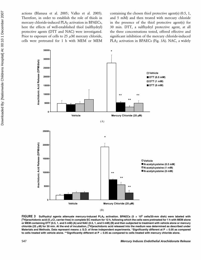
Sulfhydryl Agents AttenuateMercury-Induced PLA2 Activation
Reports have been made that the thiols (nonprotein
and protein) are the targets for heavy metal cellular
J. N. Mazerik et al. 546
Dow
nloa
ded
By:
[Nat
ionw
ide
Chi
ldre
ns H
ospi
tal]
At:
00:1
0 1
Dec
embe
r 200
7
actions (Blanusa et al. 2005; Valko et al. 2005).
Therefore, in order to establish the role of thiols in
mercury chloride-induced PLA2 activation in BPAECs,
here the effects of well-established thiol (sulfhydryl)
protective agents (DTT and NAC) were investigated.
Prior to exposure of cells to 25 µM mercury chloride,
cells were pretreated for 1 h with MEM or MEM
containing the chosen thiol protective agent(s) (0.5, 1,
and 5 mM) and then treated with mercury chloride
in the presence of the thiol protective agent(s) for
30 min. DTT, a sulfhydryl protective agent, at all
the three concentrations tested, offered effective and
significant inhibition of the mercury chloride-induced
PLA2 activation in BPAECs (Fig. 3A). NAC, a widely
FIGURE 3 Sulfhydryl agents attenuate mercury-induced PLA2 activation. BPAECs (5 × 10⁵ cells/35-mm dish) were labeled with
[3H]arachidonic acid (5 µCi, carrier-free) in complete EC medium for 12 h, following which the cells were pretreated for 1 h with MEM alone
or MEM containing DTT (0.5, 1, and 5 mM) (A) and NAC (0.5, 1, and 5 mM) (B) and then subjected to treatment with vehicle alone or mercury
chloride (25 µM) for 30 min. At the end of incubation, [3H]arachidonic acid released into the medium was determined as described under
Materials and Methods. Data represent means ± S.D. of three independent experiments. ∗Significantly different at P < 0.05 as compared
to cells treated with vehicle alone. ∗∗Significantly different at P < 0.05 as compared to cells treated with mercury chloride alone.
547 Mercury Induces Endothelial Arachidonate Release
Dow
nloa
ded
By:
[Nat
ionw
ide
Chi
ldre
ns H
ospi
tal]
At:
00:1
0 1
Dec
embe
r 200
7
used thiol protector and antioxidant, caused effective
and significant attenuation of PLA2 activation in ECs
induced by mercury (Fig. 3B). Collectively, these results
revealed that thiol-protective agents effectively atten-
uated PLA2 activation induced by mercury chloride,
suggesting the involvement of cellular thiols.
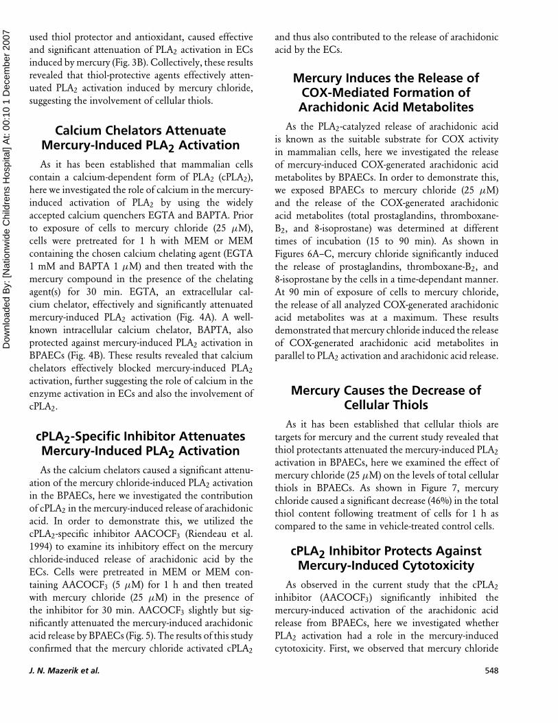
Calcium Chelators AttenuateMercury-Induced PLA2 Activation
As it has been established that mammalian cells
contain a calcium-dependent form of PLA2 (cPLA2),
here we investigated the role of calcium in the mercury-
induced activation of PLA2 by using the widely
accepted calcium quenchers EGTA and BAPTA. Prior
to exposure of cells to mercury chloride (25 µM),
cells were pretreated for 1 h with MEM or MEM
containing the chosen calcium chelating agent (EGTA
1 mM and BAPTA 1 µM) and then treated with the
mercury compound in the presence of the chelating
agent(s) for 30 min. EGTA, an extracellular cal-
cium chelator, effectively and significantly attenuated
mercury-induced PLA2 activation (Fig. 4A). A well-
known intracellular calcium chelator, BAPTA, also
protected against mercury-induced PLA2 activation in
BPAECs (Fig. 4B). These results revealed that calcium
chelators effectively blocked mercury-induced PLA2
activation, further suggesting the role of calcium in the
enzyme activation in ECs and also the involvement of
cPLA2.
cPLA2-Specific Inhibitor AttenuatesMercury-Induced PLA2 Activation
As the calcium chelators caused a significant attenu-
ation of the mercury chloride-induced PLA2 activation
in the BPAECs, here we investigated the contribution
of cPLA2 in the mercury-induced release of arachidonic
acid. In order to demonstrate this, we utilized the
cPLA2-specific inhibitor AACOCF3 (Riendeau et al.
1994) to examine its inhibitory effect on the mercury
chloride-induced release of arachidonic acid by the
ECs. Cells were pretreated in MEM or MEM con-
taining AACOCF3 (5 µM) for 1 h and then treated
with mercury chloride (25 µM) in the presence of
the inhibitor for 30 min. AACOCF3 slightly but sig-
nificantly attenuated the mercury-induced arachidonic
acid release by BPAECs (Fig. 5). The results of this study
confirmed that the mercury chloride activated cPLA2
and thus also contributed to the release of arachidonic
acid by the ECs.
Mercury Induces the Release ofCOX-Mediated Formation ofArachidonic Acid Metabolites
As the PLA2-catalyzed release of arachidonic acid
is known as the suitable substrate for COX activity
in mammalian cells, here we investigated the release
of mercury-induced COX-generated arachidonic acid
metabolites by BPAECs. In order to demonstrate this,
we exposed BPAECs to mercury chloride (25 µM)
and the release of the COX-generated arachidonic
acid metabolites (total prostaglandins, thromboxane-
B2, and 8-isoprostane) was determined at different
times of incubation (15 to 90 min). As shown in
Figures 6A–C, mercury chloride significantly induced
the release of prostaglandins, thromboxane-B2, and
8-isoprostane by the cells in a time-dependant manner.
At 90 min of exposure of cells to mercury chloride,
the release of all analyzed COX-generated arachidonic
acid metabolites was at a maximum. These results
demonstrated that mercury chloride induced the release
of COX-generated arachidonic acid metabolites in
parallel to PLA2 activation and arachidonic acid release.
Mercury Causes the Decrease ofCellular Thiols
As it has been established that cellular thiols are
targets for mercury and the current study revealed that
thiol protectants attenuated the mercury-induced PLA2
activation in BPAECs, here we examined the effect of
mercury chloride (25 µM) on the levels of total cellular
thiols in BPAECs. As shown in Figure 7, mercury
chloride caused a significant decrease (46%) in the total
thiol content following treatment of cells for 1 h as
compared to the same in vehicle-treated control cells.
cPLA2 Inhibitor Protects AgainstMercury-Induced Cytotoxicity
As observed in the current study that the cPLA2
inhibitor (AACOCF3) significantly inhibited the
mercury-induced activation of the arachidonic acid
release from BPAECs, here we investigated whether
PLA2 activation had a role in the mercury-induced
cytotoxicity. First, we observed that mercury chloride
J. N. Mazerik et al. 548
Dow
nloa
ded
By:
[Nat
ionw
ide
Chi
ldre
ns H
ospi
tal]
At:
00:1
0 1
Dec
embe
r 200
7
FIGURE 4 Calcium chelators attenuate mercury-induced PLA2 activation. BPAECs (5 × 10⁵ cells/35-mm dish) were labeled with
[3H]arachidonic acid (5 µCi, carrier-free) in complete EC medium for 12 h, following which the cells were pretreated for 1 h with MEM
alone or MEM containing EGTA (1 mM) (A) or BAPTA (1 µM) (B) and then subjected to treatment with vehicle alone or mercury chloride
(25 µM) for 30 min. At the end of incubation, [3H]arachidonic acid released into the medium was determined as described under Materials
and Methods. Data represent means ± S.D. of three independent experiments. ∗Significantly different at P < 0.05 as compared to cells
treated with vehicle alone. ∗∗Significantly different at P < 0.05 as compared to cells treated with mercury chloride alone.
induced cytotoxicity in BPAECs in both a dose- and
time-dependent manner as observed from the alter-
ations in cell morphology (Fig. 8). With both the
increase in time of exposure and dose, mercury chloride
induced the loss of cell morphology leading to the
formation of circular cellular shape. AACOCF3 (1 µM)
restored the mercury chloride (10 µM)-induced loss
of cell morphology (Fig. 9). As seen in Figure 10,
mercury chloride (10 µM) also significantly enhanced
the uptake of trypan blue, which was significantly
attenuated by the cPLA2 inhibitor AACOCF3 (5 and
10 µM) in a dose-dependent manner. Collectively,
549 Mercury Induces Endothelial Arachidonate Release

Dow
nloa
ded
By:
[Nat
ionw
ide
Chi
ldre
ns H
ospi
tal]
At:
00:1
0 1
Dec
embe
r 200
7
FIGURE 5 cPLA2-specific inhibitor attenuates mercury-induced PLA2 activation. BPAECs (5 × 10⁵ cells/35-mm dish) were labeled with
[3H]arachidonic acid (5 µCi, carrier-free) in complete EC medium for 12 h, following which the cells were pretreated for 1 h with MEM
alone or MEM containing the cPLA2-specific inhibitor AACOCF3 (5 µM) and then subjected to treatment with vehicle alone or mercury
chloride (25 µM) for 30 min. At the end of incubation, [3H]arachidonic acid released into the medium was determined as described under
Materials and Methods. Data represent means ± S.D. of three independent experiments. ∗Significantly different at P < 0.05 as compared
to cells treated with vehicle alone. ∗∗Significantly different at P < 0.05 as compared to cells treated with mercury chloride alone.
these results revealed that mercury chloride induced
cytotoxicity in BPAECs, which was attenuated by the
cPLA2 inhibitor suggesting the role of cPLA2 in the
inorganic mercury-induced cytotoxicity in ECs.
DISCUSSION
The current study revealed that inorganic mercury
(mercury chloride) activated PLA2 (release of arachi-
donic acid) from the membrane phospholipids of
FIGURE 6 Mercury induces release of COX-mediated formation of arachidonic acid metabolites. BPAECs (5 × 10⁵ cells/35-mm dish)
were treated with MEM alone or MEM containing mercury chloride (25 µM) for different time periods (15 to 90 min). At the end of the
incubation period, the release of total prostaglandins (A), thromboxane-B2 (B), and 8-isoprostane (C) was determined as described under
Materials and Methods. Data represent means ± S.D. of three independent experiments. ∗Significantly different at P < 0.05 as compared
to cells treated with vehicle alone. (Continued)
J. N. Mazerik et al. 550

Dow
nloa
ded
By:
[Nat
ionw
ide
Chi
ldre
ns H
ospi
tal]
At:
00:1
0 1
Dec
embe
r 200
7
FIGURE 6 (Continued)
BPAECs in a dose- and time- dependent fashion. The
study also showed that the mercury-induced PLA2
activation in BPAECs was attenuated by metal chelat-
ing agents, thiol protectants, and calcium chelators,
indicating the complexing of mercury with the cellular
targets, involvement of cellular thiols, and the role of
calcium in the mercury-induced release of arachidonic
acid from the cells. This study also demonstrated that
the cPLA2-specific inhibitor (AACOCF3) (Riendeau
et al. 1994) attenuated the mercury-induced release
of arachidonic acid by BPAECs, further suggesting
the activation of cPLA2 by inorganic mercury (mer-
cury chloride). Furthermore, it was also evident from
the current results that mercury chloride induced
the formation of COX-generated arachidonic acid
metabolites in a time-dependent manner. Overall, this
study demonstrated that inorganic mercury induced
the activation of PLA2 resulting in the release of
arachidonic acid and formation of its metabolites in
the vascular ECs.
551 Mercury Induces Endothelial Arachidonate Release

Dow
nloa
ded
By:
[Nat
ionw
ide
Chi
ldre
ns H
ospi
tal]
At:
00:1
0 1
Dec
embe
r 200
7
FIGURE 7 Mercury causes the decrease of cellular thiols. BPAECs (2 × 10⁶ cells/100-mm dish) were treated with MEM alone or MEM
containing mercury chloride (25 µM) for 60 min. After incubation, total thiols were determined spectrophotometrically as described in
Materials and Methods. Data represent means ± S.D. of three independent experiments. ∗Significantly different at P < 0.05 as compared
to cells treated with vehicle alone.
FIGURE 8 Mercury induces alterations in cell morphology. BPAECs (5 × 10⁵ cells/35-mm dish) were treated with MEM alone or MEM
containing mercury chloride (5, 10, and 25 µM) for different time periods (5 to 60 min). At the end of the incubation period, the medium was
replaced with PBS containing 0.5% glucose and the cells were examined under light microscope at a magnification of 10× as described
under Materials and Methods. Each micrograph is a representative picture of three independent experiments conducted under identical
conditions.
J. N. Mazerik et al. 552
Dow
nloa
ded
By:
[Nat
ionw
ide
Chi
ldre
ns H
ospi
tal]
At:
00:1
0 1
Dec
embe
r 200
7
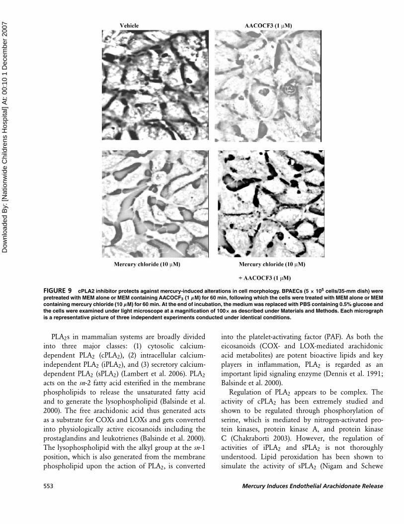
FIGURE 9 cPLA2 inhibitor protects against mercury-induced alterations in cell morphology. BPAECs (5 × 10⁵ cells/35-mm dish) were
pretreated with MEM alone or MEM containing AACOCF3 (1 µM) for 60 min, following which the cells were treated with MEM alone or MEM
containing mercury chloride (10 µM) for 60 min. At the end of incubation, the medium was replaced with PBS containing 0.5% glucose and
the cells were examined under light microscope at a magnification of 100× as described under Materials and Methods. Each micrograph
is a representative picture of three independent experiments conducted under identical conditions.
PLA2s in mammalian systems are broadly divided
into three major classes: (1) cytosolic calcium-
dependent PLA2 (cPLA2), (2) intracellular calcium-
independent PLA2 (iPLA2), and (3) secretory calcium-
dependent PLA2 (sPLA2) (Lambert et al. 2006). PLA2
acts on the sn-2 fatty acid esterified in the membrane
phospholipids to release the unsaturated fatty acid
and to generate the lysophospholipid (Balsinde et al.
2000). The free arachidonic acid thus generated acts
as a substrate for COXs and LOXs and gets converted
into physiologically active eicosanoids including the
prostaglandins and leukotrienes (Balsinde et al. 2000).
The lysophospholipid with the alkyl group at the sn-1
position, which is also generated from the membrane
phospholipid upon the action of PLA2, is converted
into the platelet-activating factor (PAF). As both the
eicosanoids (COX- and LOX-mediated arachidonic
acid metabolites) are potent bioactive lipids and key
players in inflammation, PLA2 is regarded as an
important lipid signaling enzyme (Dennis et al. 1991;
Balsinde et al. 2000).
Regulation of PLA2 appears to be complex. The
activity of cPLA2 has been extremely studied and
shown to be regulated through phosphorylation of
serine, which is mediated by nitrogen-activated pro-
tein kinases, protein kinase A, and protein kinase
C (Chakraborti 2003). However, the regulation of
activities of iPLA2 and sPLA2 is not thoroughly
understood. Lipid peroxidation has been shown to
simulate the activity of sPLA2 (Nigam and Schewe
553 Mercury Induces Endothelial Arachidonate Release

Dow
nloa
ded
By:
[Nat
ionw
ide
Chi
ldre
ns H
ospi
tal]
At:
00:1
0 1
Dec
embe
r 200
7
FIGURE 10 cPLA2 inhibitor protects against mercury-induced cytotoxicity. BPAECs (5 × 10⁵ cells/35-mm dish) were pretreated with
MEM alone or MEM containing AACOCF3 (1, 5, and 10 µM) for 60 min, following which the cells were treated with MEM alone or MEM
containing mercury chloride (10 µM) for 60 min. At the end of incubation, cytotoxicity was assayed as described under Materials and
Methods. Cytotoxicity was calculated as the% cells that had taken up trypan blue. Data represent means ± S.D. of three independent
experiments. ∗Significantly different at P < 0.05 as compared to cells treated with vehicle alone. ∗∗Significantly different at P < 0.05 as
compared to cells treated with mercury chloride alone.
2000). Reactive oxygen species have been shown to
activate iPLA2 and cause release of arachidonic acid in
macrophages (Martinez and Moreno 2001). Oxidant
(hydrogen peroxide)-mediated release of arachidonic
acid by astrocytes has been demonstrated due to
activation of cPLA2 and iPLA2 (Xu et al. 2003). Taken
together, these studies have revealed that PLA2 activity
is regulated by cellular signaling cascades, ROS, and
oxidative stress.
Among the toxic mercury compounds, the ability of
only methylmercury in activating PLA2 in neurons and
astrocytes has been reported (Verity et al. 1994; Aschner
2000; Shanker et al. 2002, 2003). Dysregulation of
calcium and increases in intracellular calcium in cere-
bellar granule cells (neurons) induced by methylmer-
cury have been reported (Limke et al. 2004; Sarafian
1993; Marty and Atchison 1998). Methylmercury has
been shown to increase intracellular concentrations of
calcium in NG108–15 cells (Hare et al. 1999). Both
the elevation of intracellular calcium and activation
of phosphatidylcholine-specific phospholipase C and
cPLA2 by methylmercury in MDCK cells have been
demonstrated (Kang et al. 2006). However no reports
have been made so far on the mercury-induced acti-
vation of PLA2 in the vascular ECs. The results of the
present study had shown that mercury chloride induced
the release of arachidonic acid, which was attenuated
by calcium chelators in BPAECs and cPLA2-specific
inhibitors, suggesting the inorganic mercury-induced
activation of cPLA2 in the ECs. In addition, the partial
inhibition of the inorganic mercury-induced arachi-
donic acid release from BPAECs as observed in the
present study also suggested the activation of calcium-
independent intracellular PLA2 (iPLA2) in ECs.
Methylmercury has been shown to cause neurotox-
icity in astrocytes and neurons through the generation
of ROS, induction of oxidative stress, and loss of
cellular thiols including GSH (Shanker and Aschner
2001; Shanker et al. 2005). Recently, we have shown
that both inorganic (mercury chloride) and organic
(methylmercury and thimerosal) forms of mercury
cause the activation of phospholipase D through the
loss of cellular thiols in BPAECs (Hagele et al. 2006).
Also, the results of the present study demonstrated the
complete attenuation of the mercury chloride-induced
arachidonic acid release from BPAECs by the thiol
J. N. Mazerik et al. 554
Dow
nloa
ded
By:
[Nat
ionw
ide
Chi
ldre
ns H
ospi
tal]
At:
00:1
0 1
Dec
embe
r 200
7
protectants (DTT and NAC) and loss of cellular thiols
induced by mercury chloride. This observation strongly
suggested the role of cellular thiols in mercury chloride-
induced release of arachidonic acid and activation of
PLA2 in ECs. Heavy metals including mercury have
been shown to react with the cellular thiols (Hagele
et al. 2006). The interaction of inorganic mercury
with cellular thiols causing the release of arachidonic
acid, as noticed in the present study, might have
involved the thiol-dependant signaling events or the
direct interaction of inorganic mercury with the enzyme
or both.
Arachidonic acid metabolites including the COX-
generated prostanoids (prostaglandins, thromboxane,
and prostacyclin) have been identified as important
inflammatory mediators in vascular endothelial dys-
function and atherosclerosis (Reiss and Edelman 2006;
Bogatcheva et al. 2005). Influx of calcium, activation of
cPLA2, and release of arachidonic acid have been shown
to play a crucial role in COX-mediated generation
of arachidonic acid metabolites in the vascular ECs
(Antoniotti et al. 2003). The results of the current study
clearly revealed the mercury chloride-induced forma-
tion of COX-generated arachidonic acid metabolites
in BPAECs and further suggested the activation of
COXs and formation of arachidonic acid-derived in-
flammatory mediators in ECs under inorganic mercury
exposure. Nevertheless, the present study also suggested
the activation of COXs in addition to the activation of
PLA2 in ECs by inorganic mercury.
Our current study clearly revealed that mer-
cury chloride-induced cytotoxicity in BPAECs (alter-
ations in cell morphology and membrane damage)
was protected by the cPLA2 inhibitor (AACOCF3),
thus establishing a role of cPLA2 in the inorganic
mercury-induced cytotoxicity in ECs. Cytotoxicity of
methylmercury has been reported in neurons and
astrocytes (Verity et al. 1994; Aschner 2000; Shanker
et al. 2002, 2003). Mepacrine, a well-established
PLA2 inhibitor, has been shown to protect against
methylmercury-induced cytotoxicity in the cerebel-
lar granule cells as observed from the trypan blue
uptake study, suggesting an association between the
methylmercury-induced PLA2 activation and cytotox-
icity in those cells (Verity et al. 1994). These reports
further support our current findings that cPLA2 activa-
tion also regulated the induction of cytotoxicity in ECs
under mercury exposure.
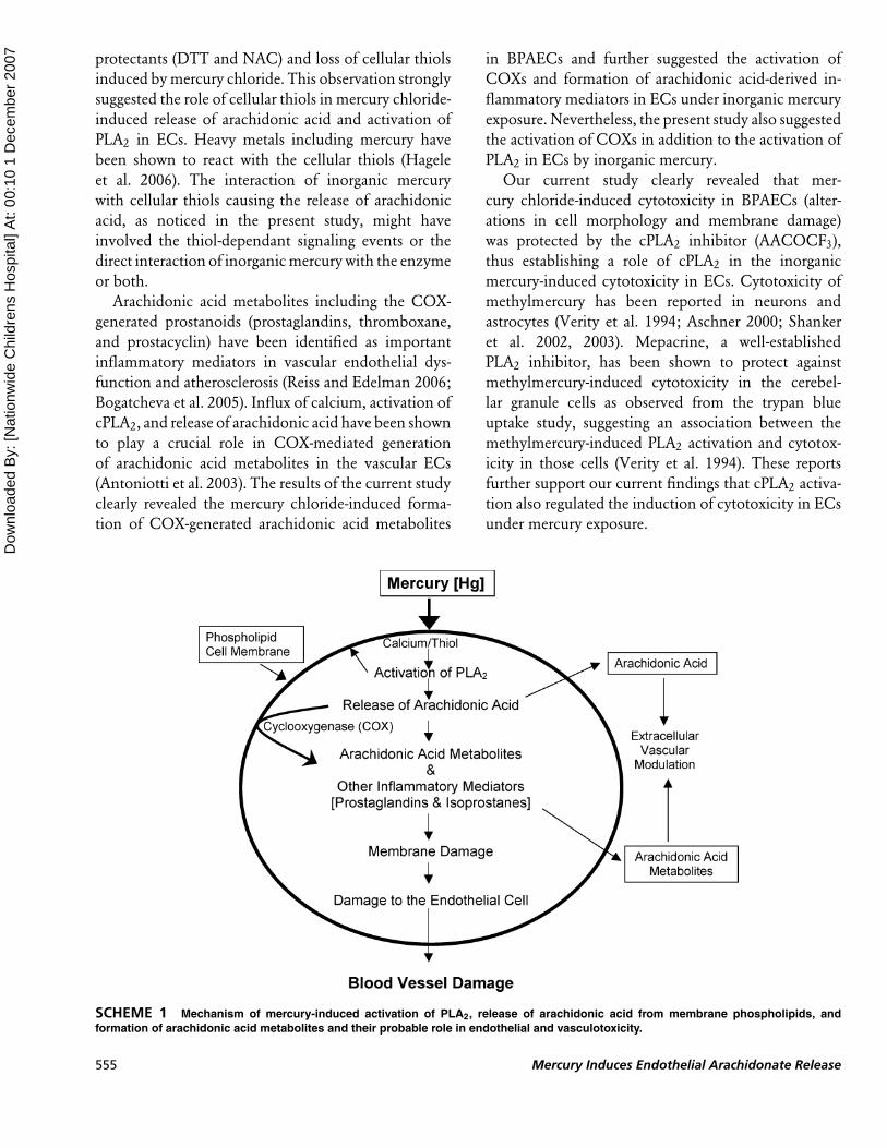
SCHEME 1 Mechanism of mercury-induced activation of PLA2, release of arachidonic acid from membrane phospholipids, and
formation of arachidonic acid metabolites and their probable role in endothelial and vasculotoxicity.
555 Mercury Induces Endothelial Arachidonate Release

Dow
nloa
ded
By:
[Nat
ionw
ide
Chi
ldre
ns H
ospi
tal]
At:
00:1
0 1
Dec
embe
r 200
7
The role of phospholipases including PLA2 and
COXs in vascular diseases and ischemic tissue injury
is becoming increasingly evident (Hurt-Camejo et al.
2001; Phillis and O’Regan 2003). Furthermore, mercury
has been implicated as a risk factor in myocardial
infarction, coronary disease, and cardiovascular disease
among humans (Yoshizawa et al. 2002; Kim et al. 2005;
Nash 2005). Therefore, the results of the current study
that inorganic mercury (mercury chloride) induced
the release of arachidonic acid through activation of
PLA2 and mediated the formation of COX-mediated
inflammatory arachidonic acid metabolites in ECs
have profound implications in the understanding of
mechanisms of mercury-induced cardiovascular dis-
eases (Scheme 1).
REFERENCES
Antoniotti, S., Fiorio, P. A., Pregnolato, S., Mottola, A., Lovisolo, D.,
and Munaron, L. 2003. Control of endothelial cell proliferation by
calcium influx and arachidonic acid metabolism: a pharmacological
approach. J. Cell Physiol. 197(3):370–378.
Aschner, M. 2000. Astrocyte swelling, phospholipase A2, glutathione and
glutamate: interactions in methylmercury-induced neurotoxicity.
Cell Mol Biol. (Noisy-le-grande) 46(4):843–854.
Balsinde, J., Winstead, M. V., and Dennis, E. A. 2000. Phospholipase A2
regulation of arachidonic acid mobilization. FEBS Lett 531:2–6.
Blanusa, M., Varnai, V. M., Piasek, M., and Kostial, K. 2005. Chelators as
antidotes of metal toxicity: therapeutic and experimental aspects.
Curr. Med. Chem. 12(23):2771–2794.
Boening, D. W. 2000. Ecological effects, transport, and fate of mercury:
a general review. Chemosphere 40:1335–1351.
Boffetta, P., Sallsten, G., Garcia-Gomez, M., Pompe-Kirn, V., Zaridze,
D., Bulbulyan, M., Caballero, J. D., Ceccarelli, F., Kobal, A. B.,
and Merler, E. 2001. Mortality from cardiovascular disease and
exposure to inorganic mercury. Occup. Environ. Med. 58:461–
466.
Bogatcheva, N. V., Sergeeva, M. G., Dudek, S. M., and Verin, A. D. 2005.
Arachidonic acid cascade in endothelial pathobiology. Microvasc.
Res. 69:107–127.
Chakraborti, S. 2003. Phospholipase A2 isoforms: a perspective. Cell
Signal 15:637–665.
Chan, H. M., and Egeland, G. M. 2004. Fish consumption, mercury
exposure, and heart disease. Nutr. Rev. 62:68–72.
Clarkson, T. W. 2002. The three modern faces of mercury. Environ. Health
Perspect. 110:11–23.
Clarkson, T. W., Magos, L., and Myers, G. J. 2003. The toxicology of
mercury-current exposures and clinical manifestations. N. Engl. J.
Med. 349:1731–1737.
Dennis, E. A., Rhee, S. G., Billah, M. M., and Hannun, Y. A. 1991. Role
of phospholipases in generating lipid second messengers in signal
transduction. FASEB J 5:2068–2077.
Divecha, N., and Irvine, R. F. 1995. Phospholipid signaling. Cell 80:269–
278.
Dopp, E., Hartman, L. M., Florea, A. M., Rettenmeier, A. W., and Hirner,
A. V. 2004. Environmental distribution, analysis, and toxicity of
organometal (loid) compounds. Crit. Rev. Toxicol. 34:301–333.
Egermayer, P. 2000. Epidemics of vascular toxicity and pulmonary
hypertension: what can be learned?. J. Intern. Med. 247:11–17.
Hagele, T. J., Mazerik, J. N., Gregory, A., Kaufman, B., Magalang, U.,
Kuppusamy, M., Marsh, C. B., Kuppusamy, P., and Parinandi, N. L.
2006. Mercury activates vascular endothelial cell phospholipase D
through thiols and oxidative stress. Int. J. Toxicol. 26(1): 57–69.
Hare, M. F., McGinnis, K. M., and Atchison, W. D. 1993. Methylmercury
increases intracellular concentrations of Ca++ and heavy metals in
NG108–15 cells. J. Pharmacol. Exp. Ther. 266(3):1626–1635.
Hurt-Camejo, E., Camejo, G., Peilot, H., Oorni, K., and Kovanen, P. 2001.
Phospholipase A2 in vascular disease. Circ. Res. 89:298–304.
Kang, M. S., Jeong, J. Y., Seo, J. H., Jeon, H. J., Jung, K. M., Chin,
M. R., Moon, C. K., Bonventre, J. V., Jung, S. Y., and Kim, D.
K. 2006. Methylmercury-induced toxicity is mediated by enhanced
intracellular calcium through activation of phosphatidylcholine-
specific phospholipase C. Toxicol. Appl. Pharmacol. 216(2):206–
215.
Kim, D. S., Lee, E. H., Yu, S. D., Cha, J. H,., and Ahn, S. C. 2005. Heavy
metal as risk factor of cardiovascular disease—an analysis of blood
lead and urinary mercury. J. Prev. Med. Pub. Health 38:401–407.
Kostka, B. 1991. Toxicity of mercury compounds as a possible risk for
cardiovascular diseases. Br. J. Ind. Med. 48:845.
Kuehn, B. 2005. Medical Groups Sue EPA Over Mercury Rule. JAMA.
294:415–416.
Lambert, I. H., Pedersen, S. F., and Poulsen, K. A. 2006. Activation of
PLA2 isoforms by cell swelling and ischemia/hypoxia. Acta Physiol.
187:75–85.
Landmark, K., and Aursnes, I. 2004. Mercury, fish, fish oil and the risk of
cardiovascular disease. Tidsskr. Nor. Laegeforen. 124:198–200.
Limke, T. L., Bearss, J. J., and Atchison, W. D. 2004. Acute exposure
to methylmercury causes Ca2 +dysregulation and neuronal death
in rat cerebellar granule cells through an M3 muscarinic receptor-
linked pathway. Toxicol. Sci. 80:60–68.
Martinez, J., and Moreno, J. 2001. Role of Ca2+-independent phospho-
lipase A2 on arachidonic acid release induced by reactive oxygen
species. Arch. Biochem. Biophys. 392(2):257–262.
Marty, M. S., and Atchison, W. D. 1998. Elevations of intracellular Ca2+
as a probable contributor to decreased viability in cerebellar granule
cells following acute exposure to methylmercury. Toxicol. Appl.
Pharmacol. 150(1):98–105.
Mutter, J., Naumann, J., Sadaghiani, C., Walach, H., and Drasch, G. 2004.
Amalgam studies disregarding basic principles of mercury toxicity.
Int. J. Hyg. Environ. Health 207:391–397.
Nash, R. A. 2005. Metals in medicine. Altern. Ther. Health Med. 11:18–25.
Nigam, S., and Schewe, T. 2000. Phospholipase A2s and lipid peroxida-
tion. Biochim. Biophys. Acta 1488:167–181.
Parinandi, N. L., Scribner, W. M., Vepa, S., Shi, S., and Natarajan, V. 1999.
Phospholipase D activation in endothelial cells is redox sensitive.
Antioxid. Redox. Signal 1(2):193–210.
Phillis, J. W., and O’Regan, M. H. 2003. The role of phospholipases,
cyclooxygenases, and lipoxygenases in cerebral ischemic/traumatic
injuries. Crit. Rev. Neurobiol. 15:61–90.
Pleva, J. 1994. Dental mercury—a public health hazard. Rev. Environ.
Health 10:1–27.
Reiss, A. B., and Edelman, S. D. 2006. Recent insights into the role
of prostanoids in atherosclerotic vascular disease. Curr. Vasc.
Pharmacol. 4:395–408.
Riendeau, D., Guay, J., Weech, P. K., Laliberte, F., Yergey, J., Li, C.,
Desmarais, S., Perrier, H., Liu, S., Nicoll-Griffith, D., and Street, I.
P. 1994. Arachidonyl trifluoromethyl ketone, a potent inhibitor of
85-kDa Phospholipase A2, blocks production of arachidonate and
12-hydroxyeicosatetraenoic acid by calcium ionophore-challenged
platelets. J. Biol. Chem. 269(22):15619–15624.
Sarafian, T. A. 1993. Methylmercury increases intracellular Ca2+ and
inositol phosphate levels in cultured cerebellar granule neurons. J.
Neurochem. 61(2):648–657.
Sarkar, B. A. 2005. Mercury in the environment: effect on health and
reproduction. Rev. Environ. Health 20:39–56.
Shanker, G., and Aschner, M. 2001. Identification and characterization
of uptake systems for cystine and cysteine ion cultured astrocytes
and neurons: evidence for methylmercury-targeted disruption of
astrocyte transport. J. Neurosci. Res. 66:998–1002.
J. N. Mazerik et al. 556
Dow
nloa
ded
By:
[Nat
ionw
ide
Chi
ldre
ns H
ospi
tal]
At:
00:1
0 1
Dec
embe
r 200
7
Shanker, G., Mutkus, L. A., Walker, S. J., and Aschner, M. 2002.
Methylmercury enhances arachidonic acid and cytosolic phospholi-
pase A2 expression in primary cultures of neonatal astrocytes. Mol.
Brain Res. 106:1–11.
Shanker, G., Syversen, T., and Aschner, M. 2003. Astrocyte-mediated
methylmercury Neurotoxicity. Biol. Trace Elem. Res. 95(1):1–
10.
Shanker, G., Syversen, T., Aschner, J. L., and Aschner, M. 2005.
Modulatory effect of glutathione status and antioxidants on
methylmercury-induced free radical formation in primary cultures
of cerebral astrocytes. Mol. Brain Res. 137:11–22.
Valko, M., Morris, H., and Cronin, M. T. 2005. Metals, toxicity and
oxidative stress. Curr. Med. Chem. 12(10):1161–1208.
Varadharaj, S., Steinhour, E., Hunter, M. G., Watkins, T., Baran, C. P.,
Magalang, U., Kuppusamy, P., Zwier, J. L., Marsh, C. B., Natarajan,
V., and Parinandi, N. L. 2006. Vitamin C-induced activation of
phospholipase D in lung microvascular endothelial cells: regulation
by MAP kinases. Cell Signal 18:1396–1407.
Verity, M. A., Sarafian, T., Pacifici, E. H. K., and Sevanian, A. 1994.
Phospholipase A2 stimulation by methylmercury in neuron culture.
J. Neurochem. 62:705–714.
Wakita, Y. 1987. Hypertension induced by methylmercury in rats. Toxicol.
Appl. Pharmacol. 89:144–147.
Xu, J., Yu, S., Sun, A. Y., and Sun, G. Y. 2003. Oxidant-mediated AA
release from astrocytes involves cPLA2 and iPLA2. Free Radic. Biol.
Med. 34(12):1531–1543.
Yoshizawa, K., Rimm, E. B., Morris, J. S., Spate, V. L., Hsieh, C. C.,
Spiegelman, D., Stampfer, M. J., and Willett, W. C. 2002. Mercury
and the risk of coronary heart disease in men. N. Engl. J. Med.
347:1755–1760.
557 Mercury Induces Endothelial Arachidonate Release