toxicological profile for silver profile for silver agency for toxic substances and disease registry...
TRANSCRIPT
TOXICOLOGICAL PROFILE FORSILVER
Agency for Toxic Substances and Disease RegistryU.S. Public Health Service
December 1990


1
1. PUBLIC HEALTH STATEMENT
This Statement was prepared to give you information about silver and toemphasize the human health effects that may result from exposure to it. TheEnvironmental Protection Agency (EPA) has identified 1177 sites on itsNational Priorities List (NPL). Silver has been found at 27 of these sites.However, we do not know how many of the 1177 NPL sites have been evaluated forsilver. As EPA evaluates more sites, the number of sites at which silver isfound may change. The information is important for you because silver maycause harmful health effects and because these sites are potential or actualsources of human exposure to silver.
When a chemical is released from a large area, such as an industrialplant, or from a container, such as a drum or bottle, it enters theenvironment as a chemical emission. This emission, which is also called arelease, does not always lead to exposure. You can be exposed to a chemicalonly when you come into contact with the chemical. You may be exposed to itin the environment by breathing, eating, or drinking substances containing thechemical or from skin contact with it.
If you are exposed to a hazardous substance such as silver, severalfactors will determine whether harmful health effects will occur and what thetype and severity of those health effects will be. These factors include thedose (how much), the duration (how long), the route or pathway by which youare exposed (breathing, eating, drinking, or skin contact), the otherchemicals to which you are exposed, and your. individual characteristics suchas age, sex, nutritional status, family traits, life style, and state ofhealth.
1.1 WHAT IS SILVER?
Silver is one of the basic elements that make up our planet. Silver israre, but occurs naturally in the environment as a soft, "silver" coloredmetal. Because silver is an element, there are no man-made sources of silver.People make jewelry, silverware, electronic equipment, and dental fillingswith silver in its metallic form. It also occurs in powdery white (silvernitrate and silver chloride) or dark-gray to black compounds (silver sulfideand silver oxide). Silver could be found at hazardous waste sites in the formof these compounds mixed with soil and/or water. Therefore, these silvercompounds will be the main topic of this profile. Throughout the profile thevarious silver compounds will at times be referred to simply as silver.
Photographers use silver compounds to make photographs. Photographicmaterials are the major source of the silver that is released into theenvironment. Another source is mines that produce silver and other metals.

2
1. PUBLIC HEALTH STATEMENT
The natural wearing down of silver-bearing rocks and soil by the wind and rainalso releases large amounts of silver into the environment.
Silver that is released into the environment may be carried longdistances in air and water. Rain washes silver compounds out of many soils sothat it eventually moves into the groundwater. Silver is stable and remainsin the environment in one form or another until it is taken out again bypeople. Because silver is an element, it does not break down, but it canchange its form by combining with other substances. Over time it may changefrom the form first released, to metallic silver, and then back to the same orother compounds. The form it is found in depends on environmental conditions.More information on the chemical and physical properties of silver compoundscan be found in Chapter 3, on its production, use, and disposal in Chapter 4,and on silver in the environment in Chapters 4 and 5.
1.2 HOW MIGHT I BE EXPOSED TO SILVER?
Most people are exposed daily to very low levels of silver mainly in foodand drinking water, and less in air. The silver in these sources is at leastpartially due to naturally occurring silver in water and soil. Skin contactand breathing in air containing silver compounds also occurs in the workplace.Other sources of exposure include the use of silver in medicines, and inactivities such as jewelry-making, soldering, and photography. Exposure fromeveryday use, such as wearing jewelry or eating with silver-coated flatware,is not expected to result in silver being taken into the body.
Silver levels of less than 0.000001 mg silver per cubic meter of air(mg/m3), 0.2-2.0 parts silver per billion parts water (ppb) in surface waters,such as lakes and rivers, and 0.20-0.30 parts silver per million parts soil(ppm) in soils are found from naturally occurring sources. Silver compoundsare also found in groundwater and at hazardous waste sites throughout theUnited States. Drinking water supplies in the United States have been foundto contain silver levels of up to 80 ppb. Surveys show that one-tenth to one-third of samples taken from drinking water supplies (both groundwater andsurface water) contain silver at levels greater than 30 ppb. For moreinformation on exposure to silver see Chapter 5.
1.3 HOW CAN SILVER ENTER AND LEAVE MY BODY?
Silver may enter your body through the mouth, throat, or digestive tractafter eating food or drinking water that contains silver, or through yourlungs after breathing air containing silver. It can also enter your bodythrough your skin when you put your hands into solutions containing silvercompounds, such as those used in photography, or when you come in contact withsilver-containing powders. Silver is also known to enter the body whenmedicines containing it are taken or applied to the skin or gums. Generally,much less silver will enter the body through the skin than through the lungs orstomach.

3
1. PUBLIC HEALTH STATEMENT
Because many silver compounds dissolve in water and do not evaporate, themost common way that silver may enter the body of a person near a hazardouswaste site is by drinking water that contains silver or eating food grown nearthe site in soil that contains silver. Silver can also enter the body whensoil that has silver in it is eaten. Most of the silver that is eaten orbreathed in leaves the body in the feces within about a week. Very littlepasses through the urine. It is not known how much of the silver that entersthe body through the skin leaves the body. Some of the silver that is eaten,inhaled, or passes through the skin may build up in many places in the body.More information on how silver enters and leaves the body can be found inChapter 2.
1.4 HOW CAN SILVER AFFECT MY HEALTH?
Since at least the early part of this century, doctors have known thatsilver compounds can cause some areas of the skin and other body tissues toturn gray or blue-gray. Doctors call this condition "argyria." Argyriaoccurs in people who eat or breathe in silver compounds over a long period(several months to many years). A single exposure to a silver compound mayalso cause silver to be deposited in the skin and in other parts of the body;however, this is not known to be harmful. It is likely that many exposures tosilver are necessary to develop argyria. Once you have argyria it ispermanent. However, the condition is thought to be only a "cosmetic" problem.Most doctors and scientists believe that the discoloration of the skin seen inargyria is the most serious health effect of silver.
Exposure to dust containing relatively high levels of silver compoundssuch as silver nitrate or silver oxide may cause breathing problems, lung andthroat irritation and stomach pain. These effects have been seen in workersin chemical manufacturing facilities that make silver nitrate and silveroxide. One man developed severe breathing problems shortly after working withmolten silver. Skin contact with silver compounds has been found to causemild allergic reactions, such as rash, swelling, and inflammation, in somepeople.
Studies of the health effects of silver in animals commonly use silvernitrate. Doctors and scientists assume that effects seen using silver nitratein animals will be very similar to effects in humans caused by any silvercompound. While this is likely to be true, it is still possible that somesilver compounds will be more harmful, or toxic, than silver nitrate.
One animal study suggests that long-term exposure (125 days) tomoderately high levels of silver nitrate in drinking water may have a slighteffect on the brain because exposed animals were less active than animalsdrinking water without silver. Another study found that some of the animalsthat drank water containing moderately high levels of silver for most of theirlives (9 months or longer) had hearts that were larger than normal. It is notyet known whether these effects would occur in humans. There have been

4
1. PUBLIC HEALTH STATEMENT
suggestions in some occupational studies in humans that silver can causekidney problems; however, more people exposed to silver need to be studied tofind out if silver causes these effects.
No studies of cancer or birth defects in animals from eating, drinking,or breathing in silver compounds were found. Therefore, it is not known ifthese effects would occur in humans. One study of animals drinking silvercompounds mixed with water for most of their life found no effect onfertility. Another study found that reproductive tissues were damaged inanimals after they received injections of silver nitrate. However, thetissues recovered even while the animals received more injections of silvernitrate. Tests in animals show that silver compounds are likely to be life-threatening for humans only when large amounts (that is, grams) are swallowerand that skin contact with silver compounds is very unlikely to belifethreatening.
Silver does have helpful uses. For example, silver nitrate was used formany years as drops in newborns' eyes to prevent blindness caused bygonorrhea, and is also used in salves for burn victims. Some water treatmentmethods (including water filters) also use a form of silver to kill bacteria.More information on the health effects from exposure to silver is presented inChapter 2. More information on the helpful uses of silver is presented inChapter 4.
1.5 WHAT LEVELS OF EXPOSURE HAVE RESULTED IN HARMFUL HEALTH EFFECTS?
Reports of cases of argyria suggest that gram amounts of a silvercompound taken in medication in small doses over several months may causeargyria in some humans. People who work in factories that manufacture silvercompounds can also breathe in the compounds. In the past, some of theseworkers have become argyric. However, the level of silver in the air and thelength of exposure that caused argyria in these workers is not known. It isalso not known what level of silver causes breathing problems, lung and throatirritation, or stomach pain in people.
Studies in rats show that drinking water containing very large amounts ofsilver (2589 parts of silver per million parts of water, or about 2.6 gramsper liter) is likely to be life-threatening.
There is very little information about health effects following skincontact with silver compounds. Argyria that covers the entire body is notseen following skin contact with silver compounds, although the skin maychange color where it touches the silver. However, many people who have usedskin creams containing silver compounds such as silver nitrate and silversulphadiazine have not reported health problems from the silver in themedicine. In one animal study, a strong solution of silver nitrate (about 41grams of silver nitrate per liter of water which is equal to 41 parts of

5
1. PUBLIC HEALTH STATEMENT
silver nitrate per thousand parts of water) applied to the skin of guinea pigsfor 28 days did not cause the animals to die; however, it did cause the guineapigs to stop gaining weight normally. It is not known if this would happen topeople if they were exposed the same way.
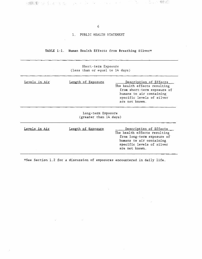
Tables l-1 through l-4 present the information that is availableconcerning specific levels of exposure and health effects. The amount ofsilver that has caused death in rats, and that has caused mice to be lessactive are shown in Table 1-4.
1.6 IS THERE A MEDICAL TEST TO DETERMINE WHETHER I HAVE BEEN EXPOSED TO SILVER?
There are reliable and accurate ways of measuring silver in the body.Silver can be measured in the blood, urine, feces, and body tissues of exposedindividuals. Because urine and blood samples are easy to get, these fluidsare most often used to find out if a person has been exposed to silver in thelast week or so. Silver builds up in the body, and the best way to learn ifpast exposure has occurred is to look for silver in samples of skin. Testsfor silver are not commonly done at a doctor's office because they requirespecial equipment. Although doctors can find out if a person has been exposedto silver by having blood or skin samples examined, they can not tell whetherany health effects will occur. Information about tests for measuring silverin the body is in Chapters 2' and 6.
1.7 WHAT RECOMMENDATIONS HAS THE FEDERAL GOVERNMENT MADE TO PROTECT HUMANHEALTH?
The federal government has developed regulations and guidelines toprotect people from the possible health effects from long-term exposure tosilver in drinking water. The Environmental Protection Agency (EPA) suggeststhat the level of silver in drinking water not be more than 0.05 milligramsper liter of water (mg/L) (which is equal to 50 parts of silver per billionparts of water or ppb). However, in May, 1989, the EPA announced that thisrestriction on silver levels in drinking water might be removed. For shorttermexposures (l-10 days), EPA suggests that drinking water levels of silvernot be more than 1.142 mg/L (which is equal to 1.142 parts of silver permillion parts of water or ppm).
Any release to the environment of more than 1 pound silver nitrate or1000 pounds of silver alone should be reported to the National ResponseCenter. To limit the amount silver workers are exposed to during an 8-hourshift for a 40-hour work week, the Occupational Safety and HealthAdministration (OSHA) has set a legal limit (Permissible Exposure Limit orPEL) of 0.01 milligrams of silver per cubic meter of air (mg/m3) in workroomair.


10
1. PUBLIC HEALTH STATEMENT
For more information on criteria and standards for silver exposure, seeChapter 7.
1.8 WHERE CAN I GET MORE INFORMATION?
If you have any more questions or concerns not covered here, pleasecontact your State Health or Environmental Department or:
Agency for Toxic Substances and Disease RegistryDivision of Toxicology1600 Clifton Road, E-29Atlanta, Georgia 30333
This agency can also give you information on the location of the nearestoccupational and environmental health clinics. Such clinics specialize inrecognizing, evaluating, and treating illnesses that result from exposure tohazardous substances.

11
2. HEALTH EFFECTS
2.1 INTRODUCTION
This chapter contains descriptions and evaluations of studies andinterpretation of data on the health effects associated with exposure tosilver. Its purpose is to present levels of significant exposure for silverbased on toxicological studies, epidemiological investigations, andenvironmental exposure data. This information is presented to provide publichealth officials, physicians, toxicologists, and other interested individualsand groups with (1) an overall perspective of the toxicology of silver and (2)a depiction of significant exposure levels associated with various adversehealth effects.
Silver occurs naturally in several oxidation states. The most common areelemental silver (0 oxidation state) and the monovalent silver ion (+loxidation state). Most of the toxicological studies of silver haveinvestigated these chemical forms of the element. Other possible oxidationstates of silver are +2 and +3, however, no toxicological studies were locatedthat researched the health effects of silver compounds with these oxidationstates. Most occupational exposures to silver occur through inhalation ofsilver-containing dusts or dermal exposure to photographic compounds.Published studies on human inhalation of silver are based predominantly onexposure to elemental silver, silver nitrate, and silver oxide. Human oraldata come from information on medicines containing silver, such as silveracetate-containing antismoking lozenges, breath mints coated with silver, andsilver nitrate solutions for treating gum disease. Animal studies usually arebased on exposure to silver nitrate and silver chloride in drinking water.Humans may be dermally exposed to silver through the use of silver-containingprocessing solutions for radiographic and photographic materials, dentalamalgams, and medicines (e.g., silver sulphadiazine cream and solutions fortreating burns).
2.2 DISCUSSION OF HEALTH EFFECTS BY ROUTE OF EXPOSURE
To help public health professionals address the needs of persons livingor working near hazardous waste sites, the data in this section are organizedfirst by route of exposure -- inhalation, oral, and dermal -- and then byhealth effect -- death, systemic, immunological, neurological, developmental,reproductive, genotoxic, and carcinogenic effects. These data are discussedin terms of three exposure periods -- acute, intermediate, and chronic.Levels of significant exposure for each exposure route and duration (forwhich data exist) are presented in tables and illustrated in figures. The
12
2. HEALTH EFFECTS
points in the figures showing no-observed-adverse-effect levels (NOAELS) orlowest-observed-adverse-effect levels (LOAELS) reflect the actual doses(levels of exposure) used in the studies. LOAELs have been classified into"less serious" or "serious" effects. These distinctions are intended to helpthe users of the document identify the levels of exposure at which adversehealth effects start to appear, determine whether or not the intensity of theeffects varies with dose and/or duration, and place into perspective thepossible significance of these effects to human health.
The significance of the exposure levels shown on the tables and graphsmay differ depending on the user's perspective. For example, physiciansconcerned with the interpretation of clinical findings in exposed persons orwith the identification of persons with the potential to develop such diseasemay be interested in levels of exposure associated with "serious" effects.Public health officials and project managers concerned with response actionsat Superfund sites may want information on levels of exposure associated withmore subtle effects in humans or animals (LOAEL) or exposure levels belowwhich no adverse effects (NOAEL) have been observed. Estimates of levelsposing minimal risk to humans (Minimal Risk Levels, MRLs) are of interest tohealth professionals and citizens alike.
Estimates of exposure posing minimal risk to humans (MRLs) have beenmade, where data were believed reliable, for the most sensitive noncancer endpoint for each exposure duration. MRLs include adjustments to reflect humanvariability and, where appropriate, the uncertainty of extrapolating fromlaboratory animal data to humans. Although methods have been established toderive these levels (Barnes et al. 1987; EPA 1989a), uncertainties areassociated with the techniques. Furthermore, ATSDR acknowledges additionaluncertainties inherent in the application of these procedures to derive lessthan lifetime MRLs. As an example, acute inhalation MRLs may not beprotective for health effects that are delayed in development or are acquiredfollowing repeated acute insults, such as hypersensitivity reactions, asthma,or chronic bronchitis. As these kinds of health effects data become availableand methods to assess levels of significant human exposure improve, these MRLswill be revised.
2.2.1 Inhalation Exposure
2.2.1.1 Death
No studies were located regarding death in humans or animals afterinhalation exposure to silver or silver compounds.
2.2.1.2 Systemic Effects
No studies were located regarding cardiovascular or musculoskeletaleffects in humans or animals after inhalation exposure to silver or silvercompounds.

13
2. HEALTH EFFECTS
Respiratory Effects. Respiratory effects have been observed infrequentlyin humans following inhalation of silver compounds. In one case report of aworker who had become ill 14 hours after he had been working with moltensilver ingots, symptoms were limited primarily to the respiratory system(Forycki et al. 1983). Unfortunately, the concentration and chemicalcomposition of the silver in the work room air were not known, and the historyof exposure to silver prior to this incident was not reported. The initialsymptoms seen in this patient included audible crackles during breathing,rapid pulse, low oxygen content of capillary blood, and scattered thickeningof the lungs observed in chest radiograms. The patient's symptoms progressedto acute respiratory failure, from which the patient eventually recoveredfully.
Occupational exposure to silver dusts can also lead to respiratoryirritation (Rosenman et al. 1979, 1987). One occupational study describes agroup of 30 employees of a manufacturing facility involved in the productionof silver nitrate and silver oxide (Rosenman et al. 1979). The average airlevel of these silver compounds over the duration of the workers' exposure wasnot estimated. However, personal air monitoring conducted 4 months previousto the study determined an 8 hour time-weighted average (TWA) concentrationrange of 0.039 to 0.378 mg silver/m3. Duration of employment ranged from lessthan one, to greater than ten years. Twenty-five of the 30 workers complainedof upper respiratory irritation (sneezing, stuffiness, and running nose orsore throat) at some time during their employment, with 20 out of 30complaining of cough, wheezing, or chest tightness. Chest radiograms andresults of clinical examination of respiratory function were predominantlynormal, with no demonstrated relationships between abnormalities and durationof employment. Similar complaints were recorded for workers involved in themanufacture of silver metal powders, although the workers were concurrentlyexposed to acids, hydroquinone, formaldehyde, caustics, solvents, and cadmium(Rosenman et al. 1987).
Acute (2-8 hours) inhalation of an aerosol containing colloidal silver byrabbits (whole body exposure, concentrations not given) has been reported tolead to ultrastructural damage and disruption of cells of the trachealepithelium (Konradova 1968).
Gastrointestinal Effects. Abdominal pain has also been reported byworkers exposed to silver nitrate and oxide in the workplace (Rosenman et al.1979). The pain was described as "burning in quality and relieved byantacids" and was reported in 10 out of 30 workers examined. Exposure levelswere estimated to be between 0.039 and 0.378 mg silver/m3. No information onchemical form or particle size was provided. Duration of employment rangedfrom less than one, to greater than ten years. This symptom correlatedsignificantly with blood silver levels, indicating that those workers exposedto higher levels of airborne silver nitrate and/or oxide are more likely tosuffer gastrointestinal pain.

14
2. HEALTH EFFECTS
No studies were located regarding gastrointestinal effects in animalsfollowing inhalation exposure to silver or silver compounds.
Hematological Effects. Blood counts were reported to be normal in allindividuals observed in the occupational study of silver-exposed workersconducted by Rosenman et al (1979) with the exception of one individual withan elevated hemoglobin level. In a study by Pifer et al. (1989), silverreclamation workers chronically exposed to insoluble silver compounds (e.g.,the silver halides) exhibited a marginal decrease in red blood cell count, aswell as an increase in mean corpuscular volume. However, the toxicologicalsignificance of these findings is unclear.
No studies were located regarding hematological effects in animalsfollowing inhalation exposure to silver or silver compounds. Despite the lackof supportive animal data, occupational exposure findings suggest thathematological effects are not a sensitive indicator of silver toxicity.
Hepatic Effects. A study that measured levels of several liver enzymes(alanine amino transferase, aspartate amino transferase, gamma glutamyltransferase, and alkaline phosphatase) found no significant differencesbetween workers exposed to silver and insoluble silver compounds and thosewith no history of silver exposure (Pifer et al. 1989).
No studies were located regarding hepatic effects in animals followinginhalation exposure to silver or silver compounds.
Renal Effects. Occupational exposure to silver metal dust has beenassociated with increased excretion of a particular renal enzyme (N-acetyl-β-Dglucosaminidasej, and with decreased creatinine clearance (Rosenman et al.1987). Both of these effects are diagnostic of marginally impaired renalfunction. However, the workers in this study were also exposed to cadmium,which was detected in the urine of 5 of the 27 workers studied. Cadmium isknown to be nephrotoxic; differentiation of the effects of the two metals inthe kidney is not possible with the data presented. Therefore, no conclusioncan be drawn regarding renal effects of silver based on this study.
No studies in animals were located which support the observation of renaleffects in the Rosenman et al (1987) study. Studies in animals have focusedonly on the deposition of silver in the kidney following oral exposure (Olcott1947; 1948) and renal function tests were not conducted.
Dermal/Ocular Effects. Skin and ocular burns, caused by contact withsilver nitrate, have been reported in workers (Moss et al. 1979; Rosenman etal 1979).
Granular deposits were observed in the conjunctiva and cornea of the eyesof 20 out of the 30 workers in the occupational study of Rosenman et al.

15
2. HEALTH EFFECTS
(1979), and subjective determination of the degree of silver deposition in theconjunctiva correlated with the duration of employment (see also Moss et al.1979). Furthermore, the amount of deposition in the eyes was found tocorrelate significantly with reports of changes in skin color and decreasednight vision. The complaint of decreased night vision was also recorded in astudy of workers involved in the manufacture of metal silver powders (Rosenmanet al. 1987).
An investigation of silver reclamation workers found that 21% and 25%exhibited conjunctival and cornea1 argyrosis (silver staining or deposition),respectively (Pifer et al. 1989). Moreover, 74% of the subjects exhibitedsome degree of internal nasal-septal pigmentation. However, no associationwas observed between silver deposition and ocular impairment.
In another report describing the same cohort of workers as studied byRosenman et al. (1979), Moss et al. (1979) conducted electrophysiological andpsychophysiological studies of the eyes of 7 of the 10 workers who hadcomplained of decreased night vision. No functional deficits were found inthe vision of these workers.
The relative contributions of dermal/ocular absorption, ingestion, andinhalation of silver compounds to the development of these ocular deposits andskin color changes are not known. However, granular deposits containingsilver have been observed to-develop in various ocular tissues of animalsfollowing ingestion of silver compounds, and it is likely that systemicabsorption following inhalation exposure also results in silver deposition(Matuk et al 1981; Olcott 1947; Rungby 1986). The possibility remains thatthe deposits were in some proportion caused by direct exposure of the eyes toairborne silver compounds.
No studies were located regarding dermal or ocular effects in animalsfollowing inhalation exposure to silver or silver compounds.
No studies were located regarding the following health effects in humansor animals after inhalation exposure to silver or silver compounds.
2.2.1.3 Immunological Effects
2.2.1.4 Neurological Effects
2.2.1.5 Developmental Effects
2.2.1.6 Reproductive Effects
2.2.1.7 Genotoxic Effects
2.2.1.8 Cancer

16
2. HEALTH EFFECTS
2.2.2 Oral Exposure
2.2.2.1 Death
No studies were located regarding death in humans following oral exposureto silver or silver compounds.
Death has been observed in rats following ingestion of colloidal silverand inorganic silver compounds. In each case the level of silver was very high.For example, death was reported in rats (number not specified) following acuteoral ingestion of silver colloid (Dequidt et al. 1974). In another study,Walker (1971) reported deaths in 3 of 12 rats during a 2-week exposure tosilver nitrate in drinking water. Cause of death was not reported in either ofthese studies. However, the rats in the Walker (1971) study were observed todecrease their water intake "precipitously" beginning on the 1st day ofexposure, and survivors were generally described as "poorly groomed andlistless" at the end of the exposure. No lethality occurred in a lower dosegroup.
Death was also reported in an unspecified number of rats receiving 222.2mgsilver/kg/day as silver nitrate in drinking water over a longer duration (Matuket al. 1981). The deaths began occurring approximately 23 weeks into 37-weekexperiment during which the exposed animals also showed a decreased weight gaincompared to animals receiving only water. The highest NOAEL values and allreliable LOAEL values for death in each species and durationare recorded in Table 2-l and plotted in Figure 2-l.
2.2.2.2 Systemic Effects
No studies were located regarding respiratory, gastrointestinal,hematological, musculoskeletal, hepatic, or renal effects in humans or animalsafter oral exposure to silver or silver compounds.
Cardiovascular Effects. No studies were located regarding cardiovasculareffects in humans following oral exposure to silver or silver compounds.
One study reported enlargement of the left ventricle in rats following 9-29 months of oral exposure to silver nitrate or silver chloride in drinkingwater (Olcott 1950). Left ventricle size (expressed as a ratio of ventricleweight to body weight) increased with exposure,duration, and showed a tendencyto increase with dose of silver. The authors suggest that the increase inventricle size could be caused by hypertension, but no blood pressuremeasurements were performed. Gross and histopathological examination of thetissues revealed only a few scattered granular deposits in the heart. Theeffect on left ventricle size was seen at a dose of 88.9 mg silver/kg/day;


19
2. HEALTH EFFECTS
however, limitations of the study such as poor experimental design andinadequate reporting of methods preclude use of these data to predictequivalent levels of exposure in humans.
Dermal/Ocular Effects. Gray or blue-gray discoloration of the skin hasbeen observed in individuals that have ingested both metallic silver and silvercompounds in small doses over periods of months to years. Silver containinggranules have been observed during histopathologic examination of the skin ofthese individuals. The condition is termed "argyria." Unfortunately, onlyrough estimates of the amount of silver ingested were located, and thereforeprecise levels of exposure resulting in discoloration cannot be established.
Case histories of argyria have been published concerning individuals whohad ingested silver through excessive use of antismoking lozenges containingsilver acetate, silver nitrate solutions for the treatment of gum disease,breath mints coated with metallic silver, and capsules containing silvernitrate for the relief of gastrointestinal "discomfort" (Aaseth et al. 1981;Blumberg and Carey 1934; East et al. 1980; MacIntyre et al 1978; Marshall andSchneider 1977; Shelton and Goulding 1979; Shimamoto and Shimamoto 1987). Ingeneral, quantitative data were nonexistent or unreliable and could not be usedto establish LOAELs. The only common symptom among these cases was theresulting gray pigmentation of the skin of primarily sun-exposed regions.Examination of skin biopsies. from these individuals at the light microscopiclevel revealed granular deposits in the dermis. The granules were distributedthroughout the dermis, but were particularly concentrated in basement membraneand elastic fibers surrounding sweat glands. The granules have been observed tocontain silver (Bleehen et al. 1981; MacIntyre et al. 1978).
Ingestion of silver nitrate and silver chloride will also cause depositionof silver granules in the skin of animals (Olcott 1948; Walker 1971). However,skin discoloration in animals following exposure to these silver compounds hasnot been studied specifically, and the level of deposition that leads to skindiscoloration in humans cannot be established based on existing animal data.Granules are also observed in the eyes of rats exposed to silver nitrate indrinking water at doses that cause general deposition in other tissues (Matuket al. 1981; Olcott 1947; Rungby 1986). The number of deposits in the eyes isrelated to the degree of yellow-to-darkgray pigmentation observed at grossexamination, which in turn is related to the duration of exposure.
Other Systemic Effects. Rats receiving 222.2 mg silver/kg/day in theirdrinking water lost weight over a 37 week exposure period. Weight loss firstappeared about 23 weeks into the experiment, and the authors observed thatseveral animals that lost weight rapidly died. Body weight in the survivingexperimental animals was an average of 50% less than that of control rats
20

20
2. HEALTH EFFECTS
drinking only distilled water over the same exposure period (Matuk et al.1981).
2.2.2.3 Immunological EffectsNo studies were located regarding immunological effects in humans oranimals following oral exposure to silver or silver compounds.
2.2.2.4 Neurological EffectsSeveral reports describe the deposition of what are assumed to be
silvercontaining granules in tissues of the central nervous system. One reportdescribes such granules in certain areas of the brain of an argyric woman atautopsy (Landas et al. 1985) who had used nose drops containing silver nitrate(concentration not specified) for an unspecified duration. The areas of thebrain described as containing silver in the Landas et al (1985) study are knownto have more direct exposure to blood-borne agents than other areas (e.g., the"circumventricular organslt, and the paraventricular and supraoptic nuclei ofthe hypothalamus). Unfortunately, the study examines only these specializedareas, and so does not provide complete information on the distribution ofsilver throughout the brain. There is no evidence that clearly relates theexistence or deposition of these granules to a neurotoxic effect of silverexposure.
However, one study has found that 20 female mice exposed to silver nitratein drinking water for 4 months, and observed to have such deposits in thecentral nervous system, were less active (hypoactive) than unexposed controls(Rungby and Danscher 1984). Activity was easured using a blind assay. Thehighest concentration of granular deposits occurred in certain areas involvedin motor control (i.e., red nucleus, deep cerebellar nuclei, and motor nucleiof the brainstem), with lesser amounts observed in the basal ganglia, theanterior olfactory nucleus, and in the cortex in general. A specificrelationship between the deposition of granules in these brain areas followingsilver ingestion and the decrease in gross activity has not been established.The highest NOAEL values and all reliable LOAEL values for neurological effectsin each species and duration are recorded in Table 2-l and plotted in Figure 2-1.
2.2.2.5 Developmental EffectsNo studies were located regarding developmental effects in humans or
animals after oral exposure to silver or silver compounds.
2.2.2.6 Reproductive EffectsNo studies were located regarding reproductive effects in humans after
oral exposure to silver or silver compounds.
21
21
2. HEALTH EFFECTS
No diminution of fertility was observed in male rats exposed,. for up to2 years, to 88.9 mg silver/kg/day as silver nitrate or silver chloride indrinking water (Olcott 1948). Appearance of spermatozoa was normal, and nosilver deposits were observed in the testes. Unfortunately, poor experimentaldesign and reporting of methods preclude use of these data in determining a noeffect level for male reproductive effects.
2.2.2.7 Genotoxic Effects
No studies were located regarding genotoxic effects in humans or animalsafter oral exposure to silver or silver compounds.
2.2.2.8 Cancer
No studies were located regarding cancer in humans or animals after oralexposure to silver or silver compounds.
2.2.3 Dermal Exposure
2.2.3.1 Death
No studies were located regarding death in humans following dermalexposure to silver or silver compounds.
Mortality following dermal application of silver nitrate has beeninvestigated in guinea pigs (Wahlberg 1965). The investigators applied 2.0 mLof a 0.239 molar solution of silver nitrate, in water by skin depot to 3.1 cm2
of skin for 8 weeks. No deaths were recorded; however, during the exposureperiod the guinea pigs ceased to gain weight. In concurrent investigations ofequimolar amounts of other metal salts using the same methods, mercuricchloride and cobalt chloride caused the death of more than half of the testanimals.
The NOAEL value for death is recorded in Table 2-2.
2.2.3.2 Systemic Effects
No studies were located regarding respiratory, cardiovascular,gastrointestinal, hematological, musculoskeletal, hepatic, renal, or oculareffects in humans or animals after dermal exposure to silver or silvercompounds.
Dermal. Medical case histories indicate that dermal exposure to silver orsilver compounds for extended periods of time can lead to local skindiscoloration similar in nature to the generalized pigmentation seen afterrepeated oral exposure. However, the amount of silver and the duration of timerequired to produce this effect cannot be established with the existing

23
2. HEALTH EFFECTS
information (Buckley 1963; McMahon and Bergfeld 1983). Moreover, adverseeffects such as argyria have not been associated with the use of silversulphadiazine as a bactericidal agent (Fox et al. 1969). No studies werelocated regarding dermal effects in animals after dermal exposure to silver orsilver compounds.
Other Systemic Effects. Decreased body weight gain was observed inguinea pigs following application of 81 mg silver nitrate (2 mL of a 0.239 Msolution) to 3.1 cm2 of skin. At the end of 8 weeks, the silver nitrate-exposedguinea pigs weighed approximately 10-20% less than unexposed controls andcontrols exposed to distilled water (Wahlberg 1965).
2.2.3.3 Immunological Effects
Medical case histories describe mild allergic responses attributed torepeated dermal contact with silver and silver compounds (Catsakis and Sulica1978; Hey1 1979; Marks 1966). Sensitization occurred in response to contactwith powdered silver cyanide, radiographic processing solutions, and apparentlyto silver in dental amalgam. The duration of exposure ranged from 6 months in aworker exposed to silver cyanide, 10 years for a woman employed as a radiographprocessor, to 20 years for a woman whose allergy had apparently been caused bydental fillings. The concentration of silver that caused these allergicresponses is not known. No studies were located' regarding immunologicaleffects in animals after dermal exposure to silver or silver compounds.
No studies were located regarding the following health effects in humansand animals after dermal exposure to silver or silver compounds.
2.2.3.4 Neurological Effects
2.2.3.5 Developmental Effects
2.2.3.6 Reproductive Effects
2.2.3.7 Genotoxic Effects
2.2.3.8 Cancer
2.3 TOXICOKINETICS
2.3.1 Absorption
2.3.1.1 Inhalation Exposure
Studies in humans regarding the absorption of silver following inhalationexposure are limited to occupational studies and a case study. It is assumed

24
2. HEALTH EFFECTS
that the predominant routes of exposure to silver in the workplace areinhalation and dermal, with the dermal route being more important whenprolonged contact with silver in solution occurs (as in photographicprocessing). Given this assumption, existing studies suggest that silver andsilver compounds can be absorbed when inhaled, although the degree ofabsorption, both absolute and relative to the degree of dermal absorption, isnot known.
A case study involving an accidental exposure of one worker toradiolabeled silver metal during a nuclear reactor mishap supports theassumption that absorption of silver metal dust can occur following inhalationexposure (Newton and Holmes 1966). Radioactive silver was measured using whole-body gamma-ray spectrometry beginning two days after a one-time inhalationexposure and continued for up to 200 days. Localization of silver in the liver,and detection in feces indicated that passage through the lungs had occurred.Unfortunately this study did not measure exposure, and therefore absorptioncould not be quantitated.
Twelve out of 30 workers in a chemical manufacturing facility whichproduced silver nitrate and silver oxide were found to have blood silver levelsgreater than the detection limit of 0.6 µg silver/100 mL blood (Rosenman et al.1979). Exposure levels were estimated to range from 0.039 to 0.378 mgsilver/m3. DiVincenzo et al. (1985) examined the silver content ofblood, urine, and feces of workers exposed to TWA levels of 0.001 to 0.1 mg/m3
insoluble silver in a photographic materials manufacturing facility. Theidentity of the specific silver compounds to which the workers were exposed wasnot reported. In exposed workers, silver was detected in 80% of the bloodsamples and in 100% of the fecal samples (mean concentrations of 0.011 µg/mland 15 µg/g, respectively). Silver was detected in 2 of 35 (6%) urine samplesfrom exposed workers with a mean concentration of 0.009 µg/g. Silver was alsodetected in the feces of controls (not exposed occupationally) at a meanconcentration of 1.5 µg/g. Although these studies suggests that silvercompounds are absorbed from the lungs, unknown exposure levels and lack ofcompound identification prevent estimation of extent or rate.
A study in dogs indicates that absorption of inhaled metallic silverparticles with a median aerodynamic diameter of approximately 0.5 µm isextensive, and is not dependent upon particle size (Phalen and Morrow 1973).Absorption was measured in one dog that remained anesthetized during the entireperiod between exposure and sacrifice. In this dog, 3.1% (0.8 µg) of thedeposited material was dissolved, transported out of the lungs, and was foundmostly in liver and blood 6 hours after exposure; a 1 µg/cm2/day absorption ratefor metallic silver was estimated by the authors. up to 90% of the depositedsilver was estimated to be absorbed into the systemic circulation based on allexperimental data. Clearance from the lung to the blood was triphasic, withhalf-lives of 1.7, 8.4, and 40 days.

25
2. HEALTH EFFECTS
2.3.1.2 Oral Exposure
Based on medical case studies and experimental evidence in humans, manysilver compounds, including silver salts and silver-protein colloids, are knownto be absorbed by humans across mucous membranes in the mouth and nasalpassages, and following ingestion. Absorption of silver acetate occurredfollowing ingestion of a 0.08 mg/kg/day dose of silver acetate containingradiolabeled silver (110mAg). Approximately 21% of the dose was retained inthe body at 1 week (East et al. 1980; MacIntyre et al. 1978). Furthermore, theoccurrence of generalized argyria in a woman who repeatedly applied silvernitrate solution to her gums (Marshall and Schneider 1977) suggests thatabsorption across the oral mucosa can occur. Information concerning the rate oforal absorption in humans was not located.
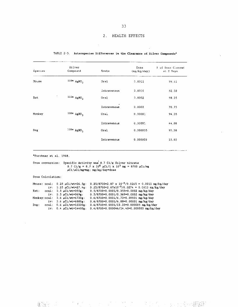
The extent of absorption of an administered dose has been found to beassociated with transit time through the gastrointestinal tract; the authorsreport that this may explain some of the interspecies differences in silverretention observed 1 week after exposure (see Table 2-3). The faster thetransit time, the less silver is absorbed (Furchner et al. 1968). Transit timesvary from about 8 hours in the mouse and rat to approximately 24 hours in themonkey, dog, and human (Furchner et al. 1968).
2.3.1.3 Dermal Exposure
Several silver compounds appear to be absorbed through the intact skin ofhumans, although the degree of absorption is thought to be low. For example,silver thiosulfate penetrated the intact skin of a photochemical worker via theeccrine sweat glands and deposited in the dermis, leading to the development oflocalized argyria within 6 months of exposure (Buckley 1963). Silver compoundsalso are absorbed through the damaged skin of humans. Silver was detected inthe urine, blood, and body tissues of humans with seriously burned skinfollowing treatment with topical preparations containing 0.5% silver nitrate toprevent bacterial infection (Bader 1966). The levels of silver found in one ofthe individuals studied by Bader (1966) were 0.038 and 0.12 ppm for urine andblood, respectively, and ranged from below detection in lung and brain to 1,250ppm in skin. Snyder et al. (1975) estimated that less than 1% of dermally-applied silver compounds are absorbed through the intact skin of humans.
Absorption of silver nitrate across intact skin has been demonstrated inguinea pigs and is similar to that of intact human skin (Wahlberg 1965). Theamount absorbed was estimated to be approximately 1% of the applied dose within5 hours of exposure. Silver administered in the form of silver sulphadiazinecream was minimally absorbed through both the intact and burnedskin of rats and distributed throughout the body (Sano et al. 1982). Theabsorption of silver increased through burned skin after blister removal. Theauthors did not determine the percentage of the applied dose that was absorbed(Sano et al. 1982).


27
2. HEALTH EFFECTS
2.3.2 Distribution
2.3.2.1 Inhalation Exposure
Limited information was located concerning the distribution of silver inhumans following inhalation of elemental silver or silver compounds. Usingwhole-body spectrometer measurements obtained from a person accidently exposedto radiolabeled silver, Newton and Holmes (1966) estimated that 25% of thedetectable 110mAg was distributed to the liver between 2 and 6 days afterexposure.
Phalen and Morrow (1973) reported that 96.9%, 2.4%, and 0.35% of the doseinitially deposited in the lungs of a dog following intratrachealadministration was detected in the lungs, liver, and blood, respectively, 6hours after exposure. The remaining silver was detected in the gall bladder andbile (0.14%), intestines (0.10%), kidneys (0.06%), and stomach (0.02%).The distribution of metallic silver (expressed as a percentage of the initialamount deposited) 225 days after exposure differed from that at 6 hours, withthe majority of the metal detected in the liver (0.49%), brain (0.035%), gallbladder and bile (0.034%), intestines (0.028%), lungs and trachea (0.019%),bone (0.014%), stomach and contents (0.012%), heart (0.009%), and muscle(0.007%). The distribution to tissues other than the lungs is similar at 6hours and 225 days if silver in the lungs is not considered. At both timepoints the majority of the silver is found in the liver (approximately 77% ofthe total body silver excluding lung content).
2.3.2.2 Oral ExposureThe distribution of silver to various body tissues depends upon the route
and quantity of silver administered and its chemical form. An oral dose ofsilver, following absorption, undergoes a first pass effect through the liverresulting in excretion into the bile, thereby reducing systemic distribution tobody tissues (Furchner et al. 1968). The subsequent distribution of theremaining silver is similar to the distribution of silver absorbed followingexposure by the inhalation and dermal routes and following intramuscular orintravenous injection.
Silver distributes widely in the rat following ingestion of silverchloride (in the presence of sodium thiosulfate) and silver nitrate in drinkingwater (at 88.9 mg silver/kg/day for silver nitrate) (Olcott 1948); The amountof silver in the various tissues was not measured, although qualitativedescriptions of the degree of pigmentation were made. High concentrations wereobserved in the tissues of the reticuloendothelial system in the liver, spleen,bone marrow, lymph nodes, skin, and kidney. Silver was also distributed toother tissues including the tongue, teeth, salivary glands, thyroid,parathyroid, heart, pancreas, gastrointestinal tract, adrenal glands, andbrain. Within these tissues advanced accumulation of silver

28
2. HEALTH EFFECTS
particles was found in the basement membrane of the glomeruli, the walls ofblood vessels between the kidney tubules, the portal vein and other parts ofthe liver, the choroid plexus of the brain, the choroid layer of the eye, andin the thyroid gland (Olcott 1948; Moffat and Creasey 1972; Walker 1971).
Approximately 18-19% of a single oral dose of silver acetate was retainedin the body of a human 8-30 weeks after exposure (East et al. 1980; Macintyreet al. 1978). This amount is 10% greater than that retained in dog tissues 20weeks after a single oral dose (Furchner et al. 1968).
2.3.2.3 Dermal Exposure
Following the topical application of silver nitrate for the treatment ofburns in two humans, silver was distributed to the muscles (0.03-2.3 ppm),liver (0.44 ppm), spleen (0.23 ppm), kidney (0.14 ppm), heart (0.032-0.04 ppm),and bones (0.025 ppm) (Bader 1966). No studies were located that quantitatedthe distribution of silver in animals following dermal exposure to silver orits compounds. However, Sano et al. (1982) detected silver in thesame tissues of rats following topical application of silver sulphadiazinecream.
2.3.2.4 Other Routes of Exposure
In rats, silver was unevenly distributed in organs and tissues followingintravenous or intramuscular injection of radiolabeled metallic silver and/orsilver nitrate, respectively. The highest concentrations were found, indecreasing order, in the gastrointestinal tract, liver, blood, kidney, muscle,bone, and skin following intramuscular injection (Scott and Hamilton 1950).Following intravenous injection the highest concentrations were found, indecreasing order, in the liver, pancreas, spleen, and plasma (Klaassen 1979a).As is shown in Table 2-4, the proportion of the dose distributed to the tissuesis positively correlated with the dose administered (Scott andHamilton 1950).
Silver is cleared from the system via the liver (Furchner et al. 1968;Scott and Hamilton 1950). Deposition of uncleared silver can occur along therenal glomerular basement membrane (Creasey and Moffat 1973; Danscher 1981; Hamand Tange 1972; Moffat and Creasey 1972) and mesangium (Day et al. 1976), andin the Kupffer cells and the sinusoid endothelium cells of the liver (Danscher1981). Silver has also been detected intra- and extracellularly in the skin andmucosa of the tongue, in the chromaffin cells, cells of the zona glomerulosa,and zona fasciculata of the adrenal glands, and in the exocrine and endocrinesections of the pancreas (Danscher 1981).
In rodents, silver has been shown to cross the placenta and to enter thefetuses following an intraperitoneal injection of silver lactate to the mothers(Rungby and Danscher 1983a). Silver was detected in the liver and brain tissuesof rat fetuses (Danscher 1981; Rungby and Danscher 1983a).


30
2. HEALTH EFFECTS
2.3.3 MetabolismThe deposition of silver in tissues is the result of the precipitation of
insoluble silver salts, such as silver chloride and silver phosphate. Theseinsoluble silver salts appear to be transformed into soluble silver sulfidealbuminates, to bind to or form complexes with amino or carboxyl groups in RNA,DNA, and proteins, or to be reduced to metallic silver by ascorbic acid orcatecholamines (Danscher 1981). The blue or gray discoloration of skin exposedto ultraviolet light in humans with argyria may be caused by the photoreductionof silver chloride to metallic silver. The metallic silver is then oxidized bytissue and bound as black silver sulfide (Danscher 1981). Buckley et al.(1965) identified silver particles deposited in the dermis of awoman with localized argyria as being composed of silver sulfide.
In rats, silver deposits in internal organs such as the kidney, have alsobeen identified as the sulfide (Berry and Galle 1982). Under conditions ofexposure to high doses of selenium, the sulfur can be replaced by selenium(Berry and Galle 1982). The deposition of silver in the kidney was increasedunder conditions of high selenium exposure. This may be important in thedevelopment of argyria in people exposed to silver who ingest foods thatcontain large amounts of selenium (See Section 2.7).
2.3.4 Excretion2.3.4.1 Inhalation Exposure
The clearance of radioactive silver metal dust in a man who wasaccidentally exposed illustrated the rapid removal of silver from the lungsprimarily by ciliary action, with subsequent ingestion and ultimate eliminationin the feces (Newton and Holmes 1966). Lung clearance fit a biexponentialprofile, with biological half-lives of 1 and 52 days. Radioactive silver wasdetected in the feces up to 300 days after exposure, but was not detected inurine samples (collected up to 54 days after exposure).
Chronic exposure of workers to unidentified silver compounds resulted inthe detection of silver in 100% of the fecal samples and 6% of the urinesamples (DiVincenzo et al. 1985). This occupational exposure is assumed to haveoccurred primarily by the inhalation route.
In dogs, lung clearance of metallic silver particles (average aerodynamicdiameter of 0.5µ) following intra-tracheal intubation was accompanied by anincrease in silver concentration in the area of the stomach and liver. Theincrease in silver concentration in the stomach suggests that some proportionof the silver particles are cleared by the mucociliary escalator and swallowed.However, the predominant route of clearance from the lung appearedto be through dissolution of the silver and transport through the blood. The
31

31
2. HEALTH EFFECTS
silver was apparently carried by the blood to the liver, with little clearedvia the mucociliary passages (Phalen and Morrow 1973). Approximately 90% of theinhaled dose was excreted in the feces within 30 days of exposure. Clearanceof deposited silver particles from the lung fit a triexponential profile, withbiological half-lives of 1.7, 8.4, and 40 days, accounting for 59, 39, and 2%of the radioactivity excreted, respectively. Clearance of absorbed silver fromthe liver fit a biexponential profile with biological half-lives of 9.0 and 40days accounting for 97% and 3% of the radioactivity excreted, respectively(Phalen and Morrow 1973).
2.3.4.2 Oral Exposure
Following oral exposure to silver acetate in humans, silver is eliminatedprimarily in the feces, with only minor amounts eliminated in the urine (Eastet al. 1980). The rate of excretion is most rapid within the first week after asingle oral exposure (East et al. 1980). Whole-body retention studies in miceand monkeys following oral dosing with radiolabeled silver nitrateindicate that silver excretion in these species follows a biexponential profilewith biological half-lives of 0.1 and 1.6 days in mice and 0.3 and 3 days inmonkeys. In similarly exposed rats and dogs, silver excretion followed atriexponential profile with biological half-lives of 0.1, 0.7, and5.9 days in rats and 0.1, 7.6, and 33.8 days in dogs (Furchner et al. 1968).Data for whole body clearance of silver at two days after exposure for thesefour species are presented in Table 2-5 (Furchner et al. 1968). Transit timethrough the gut may explain some of these interspecies differences in silverexcretion. Transit time is approximately 8 hours in mice and rats, andapproximately 24 hours in dogs and monkeys (Furchner et al. 1968). Animalsexcrete from 90% to 99% of an administered oral dose of silver in the feceswithin 2 to 4 days of dosing (Furchner et al. 1968; Jones and Bailey 1974;Scott and Hamilton 1950). Excretion in the feces is decreased and deposition intissues, such as the pancreas, gastrointestinal tract, and thyroid, isincreased when saturation of the elimination pathway in the liver occurs as aresult of chronic or high level acute exposure to silver (see Table 2-4)(Constable et al. 1967; Olcott 1948; Scott and Hamilton 1950).
2.3.4.3 Dermal Exposure
No studies were located concerning the excretion of silver by humans oranimals following dermal exposure to elemental silver or silver compounds.Once absorption through the skin and distribution to bodily tissues occurs, itcan be expected that elimination would be similar to that of silver absorbedvia oral or inhalation exposure, that is, primarily via the feces, withminimal amounts excreted in the urine.
2.3.4.4 Other Routes of Exposure
Whole body retention studies in mice, rats, monkeys, and dogs followingintravenous injection of radiolabeled silver nitrate indicate that silver

32
2. HEALTH EFFECTS
excretion in these species follows a triexponential profile. (Furchner et al.1968). For mice and monkeys, this differs from the biexponential profile seenfollowing oral exposure. Whole body clearance following intravenous exposurewas slower than clearance following oral exposure in each of the four speciesobserved. In addition, the difference in clearance rate between species wasmore dramatic. Clearance at 2 days post-exposure ranged from 15% in the dog to82% in the mouse (see Table 2-5) (Furchner et al. 1968).
Silver removal from the liver by biliary excretion was demonstrated byScott and Hamilton (1950). Control rats and rats with ligated bile ducts wereadministered radioactive metallic silver by intramuscular injection. In ratswith ligated bile ducts, excretion of silver in the feces was 19%, compared to97% in controls. Deposition in the liver of rats with ligated bile ducts was48% and 2.5% in the gastrointestinal tract compared to 0.36% and 1.12%,respectively in the controls (Scott and Hamilton 1950). Klaassen (1979b)determined that biliary excretion accounted for between 24% and 45% of thesilver administered to rats. The concentration of silver in the bile wasestimated to be between 16 and 20 times greater than that in plasma. Anincrease in the bile/liver tissue ratio (µg/ml per µg/g) from 4.2 to 6.4indicates that more silver is concentrated in the bile as the dose of silverincreases. It is believed that active transport is involved in the transfer ofsilver from the plasma to the bile (Klaassen 1979b). There are apparentlyinterspecies differences in this transport process. The variability in theextent of biliary silver excretion appears to be related to the ability of theliver to excrete silver into the bile, not to the ability of the silver to passbetween the plasma and the liver. Rats excreted silver in the bile at 10 timesthe rate of rabbits. Dogs excreted silver in the bile at a rate lower than thatof rabbits (Klaassen 1979b). Dogs had the highest amount of silver retained inthe liver (2.9 µg silver/g), as compared to the rabbit (2.13 ,µg silver/g) andrat (1.24 µg silver/g).
2.4 RELEVANCE TO PUBLIC HEALTH
The one clinical condition that is known in humans to be attributable tolong-term exposure to silver and silver compounds is a gray or blue-graydiscoloring of the skin (argyria). Argyria may occur in an area of repeated orabrasive dermal contact with silver or silver compounds, or more extensivelyover widespread areas of skin and the conjunctiva of the eyesfollowing long-term oral or inhalation exposure. Argyria was common around theturn of the century when many pharmaceutical preparations contained silver(Hill et al. 1939). It is much less common today, probably because most currentmedications containing silver are for dermal application only. Case reports inhumans have reported that repeated dermal contact with silvercompounds may in some cases lead to contact dermatitis, and a generalizedallergic reaction to silver.
Evidence from both human and animal studies indicates that inhalation ofsilver compounds can irritate the respiratory pathway. Occupational studies

34
2. HEALTH EFFECTS
and reports of cases where individuals have accidentally swallowed solutions ofsilver nitrate show that both inhalation and ingestion may cause gastricdiscomfort as well.
Studies in humans and animals indicate that silver compounds are absorbedreadily by the inhalation and oral routes and poorly by the dermal route, andare distributed widely throughout the body. Observations made during surgery onsilver exposed individuals and histopathologic studies of animals exposed tosilver compounds demonstrate that within certain tissues of the body (mostnotably liver, kidney, pancreas, skin, conjunctiva of the eyes, and, to alesser degree, certain brain areas) silver is deposited in the form of granulesvisible with the light microscope. However, with the exception of one report ofdecreased activity in mice exposed to silver nitrate, and one report ofenlarged hearts in rats exposed to silver nitrate or silverchloride, there is no evidence that suggests that the silver deposits mightinterfere with the normal functioning of these organs in humans.
Death. There is no information concerning death in humans followingexposure to silver compounds by any route.
Data concerning death observed in animals following oral and dermalexposure to silver compounds suggest that levels of exposure would have to bequite high to cause death in humans. High levels of colloidal silver wereobserved to cause death in rats when administered in drinking water for acuteand intermediate exposure durations. The cause of death was unknown. Thecorresponding daily oral dose for a 70-kg man based on the dose levels testedwould be approximately 12 grams. Death caused by silver has not been observedto occur in humans or animals following dermal exposure to silver compounds,nor is it expected to occur.
Systemic Effects. Silver nitrate and/or silver oxide have been reportedto cause upper and lower respiratory tract irritation in humans when inhaled.In one case, inhalation of an unknown amount and chemical form of silver duringwork with molten silver ingots produced respiratory failure the day afterexposure (Forycki et al. 1983). Without treatment the worker may have died.However, exposures such as this are not expected to be common and should beexamined on a case by case basis.
Upper respiratory irritation has been observed in humans at estimatedexposure levels of between 0.039 and 0.378 mg silver/m3 for less than 1 togreater than 10 years. Evidence that silver colloid can act as an irritant isprovided by the fact that ultrastructural damage was seen in the trachealepithelium of rabbits following inhalation exposure to an unknown concentrationof silver colloid. However, these effects are likely to be related to thecaustic properties of the compounds, not to the presence of silver. The effectsare not expected to persist when exposure to air containing silver compoundshas stopped.

35
2. HEALTH EFFECTS
The same exposure conditions can also cause gastric discomfort in humans.Again, this effect is likely to be caused by the caustic effects of the silvercompounds, and not the presence of silver. There is no evidence that suggeststhat dermal exposure to silver can cause gastric effects.
Occupational exposure to silver compounds has not been observed to affectblood counts. Although no supportive studies were located regardinghematological effects in other species or by other routes, the occupationalexposure findings suggest that hematological effects are not a sensitiveindicator of silver toxicity.
Silver is deposited in the glomerular basement membrane of the kidney ofanimals, and therefore might be expected to affect renal function. However, nostudies of renal function in animals were located, and occupational studies inhumans are not adequate for establishing a clear relationship between exposureto silver and renal impairment.
No human studies were located that indicate that exposure to silver orsilver compounds will affect the cardiovascular system. However, an animalstudy did show an increase in the relative size of the left ventricle of ratsthat had been chronically exposed to silver nitrate or silver chloride indrinking water. Despite the suggestion by the authors that the increase in leftventricle size may be caused by vascular hypertension, ‘this effect has notbeen observed in animals or in humans. These endpoints have not beenspecifically addressed in reliable studies to date.
The predominant effect of exposure to silver in humans is the developmentof a characteristic, irreversible pigmentation of the skin. This condition iscalled argyria. Clinicians describe the pigmentation as slate-gray, bluegray,or gray in color and report it as most noticeable in areas of skin exposed tolight. The pigmentation is not a toxic effect per se, nor is it known to bediagnostic of any other toxic effect. However, the change in skin color can besevere enough to be considered a cosmetic disfigurement in some cases.
The discoloring is likely to be caused by the photoreduction of silverchloride and/or silver phosphate in the skin. X-ray dispersive analysis of skinand other tissues reveals that the granules consist of silver complexed withsulfur and/or selenium. The photoreduced deposits are not removed by the body,and there are no clinical means of removing them.
Levels of silver exposure that have led to argyria in humans in the pastare poorly documented, and it is not possible to establish minimum risk levelsfor this effect based on these data. Hill and Pillsbury (1939) in their reviewof cases of argyria report that total doses of silver that have resulted inargyria can be as low as a total of 1.4 grams of silver (as silver nitrate)ingested in small unspecified doses over several months.

36
2. HEALTH EFFECTS
An animal model for studying the pigmentation changes seen in humans doesnot exist. Therefore existing experimental animal data are of limited use inpredicting the exposure levels that would result in argyria in humans.Granular deposits that contain silver have been observed in both pigmented andunpigmented skin of silver-exposed humans. Similar granules have been observedin various tissues in animals following silver exposure (see Section2.2 and below). However, a direct correlation has not been established betweenthe granular deposits seen in animals following exposure to silver and thedeposition leading to skin discoloration in humans.
Immunological Effects. No studies were located that investigated toxiceffects on the immune system in humans or animals exposed to silver, or thatindicate that immune-related disease can be affected by silver exposure.Silver has been observed to elicit a mild allergic response (contactdermatitis) in humans following dermal exposure to various silver compounds.
Neurological Effects. Neurological effects attributable to silver havenot been reported in humans nor have existing case or occupational studiesfocused on this endpoint. Exposure to silver has been observed to result in thedeposit of silver in neurons of the central nervous system of a woman who hadused nasal drops containing silver nitrate and in animals exposed byintraperitoneal injection and through drinking water. However, this effect isnot known to be toxic. As measured using a controlled, blind assay, theactivity of mice with silver deposits in their brain was less than that ofcontrols. The decrease in activity could be attributable to other factorsunrelated to central nervous system function (such as loss of appetite due togastric effects, or general malaise) and the relevance to humans is not known.
Exposure to silver has been observed to affect the volume of hippocampalcell groups within the brain of animals. Several cell groups within thehippocampus (a well defined structure of the brain involved in some aspects ofmemory) are reduced in overall volume in rats exposed during their first 4weeks of life to subcutaneously injected silver lactate (0.137 mgsilver/kg/day) (Rungby et al. 1987). Unfortunately, the study is limited inthat only one small region of the brain was examined. It is prudent to assumethat similar effects would be observed in humans; however, the implications ofthe altered volume of these cell groups are not known.
Developmental Effects. Based on the existing information, it is not knownwhether silver causes developmental toxicity in humans. No studies were foundconcerning developmental effects in humans after exposure to silver. However, ahuman study by Robkin et al. (1973) did investigate the possibility of arelationship between the concentration of this heavy metal in the tissue offetuses and the occurrence of developmental abnormalities. These authorsreported that the concentration of silver in the fetal liver of 12 anencephalichuman fetuses was higher (0.75±0.15 mg/kg) than the values from 12 fetusesobtained either through therapeutic abortions

37
2. HEALTH EFFECTS
(0.23±0.05 mg/kg), or in 14 spontaneously aborted fetuses (0.21±0.05 mg/kg).The concentration in 9 premature infants was 0.68±0.22 mg/kg. The authorscould not determine if the higher concentrations of silver in anencephalicfetuses were associated with the malformation, or with fetal age.
Silver has been demonstrated in the brains of neonatal rats whose mothersreceived injections of silver lactate on days 18 and 19 of gestation (Rungbyand Danscher 1984). As mentioned above, treatment of neonatal rats has alsobeen found to reduce the volumes of certain cell groups within the hippocampus(Rungby et al. 1987). However, functional tests were not performed on theserats, and therefore, neither the significance of the silver accumulation, northe decrease in regional hippocampal volume can be determined.
Reproductive Effects. The existing evidence does not point to a strongeffect of silver on reproduction. However, no multigeneration reproductivestudies were located, and therefore a firm conclusion regarding reproductivetoxicity can not be made.
There is no historical evidence in humans to suggest that silver affectsreproduction, although studies specifically designed to address this endpointin humans were not located. One study in five male rats found that singlesubcutaneous injections of 0..04 millimole/kg silver nitrate caused temporaryhistopathological damage to testicular tissue (Hoey 1966). Eighteen hours aftera single injection, silver caused shrinkage, edema, and deformation of theepididymal tubules. All affected tissues showed gradual recovery from damagefollowing the initial injection, in spite of continued daily injections.Although treatment over a 30-day period had no effect on spermatogenesis,spermatozoa were observed with separated and pyknotic heads. A separatedrinking water study in male rats did not observe changes in spermatozoa ordiminution in fertility.
Finally, direct intrauterine injection of silver nitrate terminatedpregnancies in monkeys (Dubin et al. 1981). Single dose intrauterine injectionsof 1% silver nitrate solution (0.78 mg/kg) resulted in vaginal bleeding for 1or 2 days following treatment. The bleeding lasted for an average of 5.3 days.Pregnancy was terminated in all these cases. In subsequent pregnancies, thesemonkeys produced normal offspring. The relevance of direct uterine injection tohuman exposure conditions from NPL site contamination must be evaluated on acase by case basis since this effect has not been studied by the more commonexposure pathways.
Genotoxic Effects. No studies were located that examined the mutagenicityor genotoxicity of silver in human cells in vivo or in vitro. Existing data onmutagenicity are inconsistent, but data on genotoxicity suggest that the silverion is genotoxic. Table 2-6 presents the results of in vitro genotoxicitystudies using bacteria and nonhuman mammalian cell cultures. From these studiesand others it is evident that the silver ion

38
2. HEALTH EFFECTS
does bind with DNA in solution in vitro, and that it can interact with DNA inways that cause DNA strand breaks and affect the fidelity of DNA replication(Goff and Powers 1975; Loeb et al. 1977; Luk et al. 1975; Mauss et el. 1980;Robison et al. 1982; Scicchitano and Pegg 1987). However, silver has not beenfound to be mutagenic in bacteria (Demerec et al. 1951; Kanematsu et al. 1980;McCoy and Rosenkranz 1978; Nishioka 1975; Rossman and Molina 1986).
Cancer. No studies were located regarding cancer in humans followinginhalation, oral, or dermal exposure to silver or silver compounds.Fibrosarcomas have been induced in rats following subcutaneous imbedding ofsilver foil (Oppenheimer et al. 1956). In this study, imbedded silver metalfoils appeared to produce fibrosarcomas earlier (latent period as short as 275days compared to 364-714 days) and more frequently (32% of implantation sitescompared to O-S%) than other metal foils (steel, tantalum, tin, and vitallium)tested. However, experiments on several metals (steel, tantalum, andvitallium) were not complete at the time of publication so adequate comparisonscould not be made. In addition, it should be noted that several material areknown to regularly produce such tumors when implanted subcutaneously inanimals, and the relevance to carcinogenesis in humans is uncertain (Coffin andPalekar 1985). Both positive (Schmahl and Steinhoff 1960) and negative (Furstand Schlauder 1977) results for tumorigenesis have been reported followingsubcutaneous and intramuscular injection, respectively, of colloidal silver inrats. However, the relevance of these routes of exposure to exposure conditionsat hazardous waste sites has not been clearly established. Animal toxicity andhuman occupational studies using normal routes of exposure have not providedindications of carcinogenicity, and silver is not expected to be carcinogenicin humans.
2.5 BIOMARKERS OF EXPOSURE AND EFFECT
Biomarkers are broadly defined as indicators signaling events in biologicsystems or samples. They have been classified as markers of exposure, markersof effect, and markers of susceptibility (NAS/NRC 1989).
A biomarker of exposure is a xenobiotic substance or its metabolite(s) orthe product of an interaction between a xenobiotic agent and some targetmolecule or cell that is measured within a compartment of an organism (NAS/NRC1989). The preferred biomarkers of exposure are generally the substance itselfor substance-specific metabolites in readily obtainable body fluid or excreta.However, several factors can confound the use and interpretation ofbiomarkers of exposure. The body burden of a substance may be the result ofexposures from more than one source. The substance being measured may be ametabolite of another xenobiotic (e.g., high urinary levels of phenol canresult from exposure to several different aromatic compounds). Depending on theproperties of the substance (e.g., biologic half-life) and environmentalconditions (e.g., duration and route of exposure), the substance and all ofits metabolites may have left the body by the time biologic samples can be


40
2. HEALTH EFFECTS
taken. It may be difficult to identify individuals exposed to hazardoussubstances that are commonly found in body tissues and fluids (e.g., essentialmineral nutrients such as copper, zinc and selenium). Biomarkers of exposure tosilver are discussed in Section 2.5.1.
Biomarkers of effect are defined as any measurable biochemical,physiologic, or other alteration within an organism that, depending onmagnitude, can be recognized as an established or potential health impairmentor disease (NAS/NRC 1989). This definition encompasses biochemical or cellularsignals of tissue dysfunction (e.g., increased liver enzyme activityor pathologic changes in female genital epithelial cells), as well asphysiologic signs of dysfunction such as increased blood pressure or decreasedlung capacity. Note that these markers are often not substance specific. Theyalso may not be directly adverse, but can indicate potential health impairment(e.g., DNA adducts). Biomarkers of effects caused by silver are discussed inSection 2.5.2.
A biomarker of susceptibility is an indicator of an inherent or acquiredlimitation of an organism's ability to respond to the challenge of exposure toa specific xenobiotic. It can be an intrinsic genetic or other characteristicor a preexisting disease that results in an increase in absorbed dose,biologically effective dose, or target tissue response. If biomarkers ofsusceptibility e xist, they are discussed in Section 2.7, "POPULATIONS THAT AREUNUSUALLY SUSCEPTIBLE."
2.5.1 Biomarkers Used to Identify or Quantify Exposure to Silver
Silver can be detected in blood, urine, feces, hair, and biopsy specimensusing standard analytic techniques, as well as whole body analysis using invivo neutron activation. The presence of silver in these samples can be used,with varying degrees of accuracy depending on the sample, as a biomarker ofexposure to silver compounds. Analysis of hair has been used to monitor forsilver exposure (DiVincenzo et al. 1985). However, silver can be adsorbed ontohair surfaces as well as deposited during hair formation, and since currenttesting procedures cannot differentiate between the two modes, hair monitoringis an unreliable biomarker of exposure (DiVincenzo et al. 1985). Levels ofsilver in feces, blood, and urine have been associated with recentexposure via inhalation, oral, and dermal routes. Levels in these biologicalmedia may serve as more reliable, primary biomarkers of exposure to silver thanlevels in hair (DiVincenzo et al. 1985; Rosenman et al. 1979, 1987). Thesebiomarkers appear to be independent of the route of exposure, but have not beenquantitatively correlated with level and duration of exposure. Theprevalence and estimated magnitude of silver deposition in the skin, however,were associated with duration of occupational exposure.
Because silver is eliminated primarily through the feces, recent exposureis most easily monitored through fecal analysis. Measurements of silver in theblood are also significant and indicate exposure to the metal. However,

41
2. HEALTH EFFECTS
silver is not always detected in the urine samples of workers with knownexposure to the chemical, and is not as reliable a biomarker as feces andblood. DiVincenzo et al. (1985), for example, detected silver in 100% of fecessamples and only 6% of urine samples from workers chronically exposed to silvercompounds in air. Increased blood silver levels, above the detection limit forsilver (0.6 µg/l00 mL blood), have been associated with inhalation exposure tothe metal in a study by Rosenman et al. (1979).
Levels in biopsy specimens (e.g., of skin) provide information concerningrepeated exposure (Blumberg and Carey 1934; East et al. 1980). After a burnvictim had been dermally exposed to silver nitrate (as a bactericidal agent),Bader (1966) found silver primarily in the patient's skin as well as in theblood and urine. Further information can be found in Section 2.3.
2.5.2 Biomarkers Used to Characterize Effects Caused by Silver
Several effects associated with silver exposure have been reported inhumans which may be useful as biomarkers of effects. The significance of thesebiomarkers, however, is in doubt, because they do not appear consistently inexposed individuals and do not seem to correlate well with levels and durationof exposure.
One easily observed effect of silver exposure is argyria which is a slate-gray or blue-gray discoloration of the conjunctivae, cornea, skin, and otherepithelial surfaces. Oral, inhalation, or dermal absorption of silver may causeargyria in humans. A potential biomarker of silver deposition that could leadto this effect would be the presence of insoluble silver salts(e.g., silver chloride, sulfide, or phosphate) in skin biopsy, especially thatassociated with basement membrane (Danscher 1981). The granular deposition ofsilver in the cornea of workers has been loosely associated with complaints ofdecreas.ed night vision (Moss et al. 1979; Rosenman et al. 1979). However,Pifer et al. (1989) studied various ophthalmological end points in workersexposed to silver and silver compounds and could find no significant ocularimpairments associated with the metal.
Low oxygen content in capillary blood, scattered thickening of lungs (asobserved in chest radiograms), and upper respiratory irritation have beenobserved in studies of workers exposed intensely or chronically to moltensilver or silver dusts (Forycki et al. 1983; Rosenman et al. 1979, 1987).Inhalation exposure also led to decreased red blood cell count and anincreased mean corpuscular volume (Pifer et al. 1989). However, these potentialhematologic biomarkers are not specific for silver exposure, and do notindicate or predict significant clinical sequelae.
Rosenman et al. (1987) found that inhalation exposure to silver causedchanges in two renal end points which could be biomarkers of mildnephrotoxicity. In this study exposed workers exhibited lower creatinineclearance and higher excretion of the urinary enzyme N-acetyl-β-D

42
2. HEALTH EFFECTS
glucoseaminidase. However, workers in the study were also exposed to cadmium,a known nephrotoxic agent, which may have been responsible for the observedchanges. Therefore, these biochemical effects cannot be considered reliablebiomarkers of silver exposure. Occupational exposure to silver nitrate andsilver oxide, leading to blood silver levels above 0.6 µg/100 mL, correlatedstrongly with increased complaints of abdominal pain (Rosenman et al. 1979).Moreover, dermal exposure to silver and silver compounds has been associatedwith a mild allergic reaction in humans which may be a biomarker ofimmunological effects (Catsakis and Sulica 1978; Hey1 1979; Marks 1966).Please refer to Section 2.2 of Chapter 2 for a more detailed discussion of theeffects caused by silver and its compounds.
2.6 INTERACTIONS WITH OTHER CHEMICALS
As with other metals, relationships exist through which silver caninfluence the absorption, distribution, and excretion of one or more othermetals. These influences are not known to increase the toxicity of othermetals, nor are other metals known to add to any toxic effects of silver.
However, high intake of selenium (e.g., as sodium selenite or seleniumoxide) may lead to increased deposition of insoluble silver salts in bodytissues through the formation of silver selenide (Alexander and Aaseth 1981;Berry and Galle 1982; Nuttall 1987). Exposure to silver nitrate in drinkingwater concurrent with intraperitoneal injections of selenium dioxide resultsin a higher rate of deposition of granular deposits in the kidneys of rats thanthat seen with exposure to silver nitrate alone (Berry and Galle 1982). Higherdeposition rates are likely to accelerate the development of argyria, althoughno data were located to confirm this.
No other studies were located regarding additive or synergistic toxicinteractions of silver with any other substance. However, exposure to moderate-to-high silver levels (130-1000 ppm) in rats with dietary deficiencies such asvitamin E alone (Bunyan et al. 1968; Grass0 et al. 1969) or vitamin E andselenium (Van Vleet 1976; Van Vleet et al. 1981) can cause moderate-to-severeliver necrosis.
It should be noted that selenium plays a dual role in the toxicity ofsilver. On the one hand, it increases the silver deposition rate in bodytissues, which suggests that humans exposed to both high selenium and highsilver may be more likely to develop argyria. On the other hand, aseleniumdeficient diet combined with high silver intake can cause livernecrosis.
2.7 POPULATIONS THAT ARE UNUSUALLY SUSCEPTIBLE
Populations that are unusually susceptible to toxic effects of silverexposure are those that have a dietary deficiency of vitamin E or selenium, orthat may have a genetically based deficiency in the metabolism of theseessential nutrients. Individuals with damaged livers may also be more

43
2. HEALTH EFFECTS
susceptible to the effects of silver exposure. In addition, populations withhigh exposures to selenium may be more likely to develop argyria.Furthermore, some individuals may exhibit an allergic response to silver.
2.8 ADEQUACY OF THE DATABASE
Section 104(i)(5) of CERCLA, directs the Administrator of ATSDR (inconsultation with the Administrator of EPA and agencies and programs of thePublic Health Service) to assess whether adequate information on the healtheffects of silver is available. Where adequate information is not available,ATSDR, in conjunction with the National Toxicology Program (NTP), is requiredto assure the initiation of a program of research designed to determine thehealth effects (and techniques for developing methods to determine such healtheffects) of silver.
The following categories of possible data needs have been identified by ajoint team of scientists from ATSDR, NTP, and EPA. They are defined assubstance-specific informational needs that, if met would reduce or eliminatethe uncertainties of human health assessment. In the future, the identifieddata needs will be evaluated and prioritized, and a substance-specific researchagenda will be proposed.
2.8.1 Existing Information on Health Effects of Silver
The existing data on health effects of inhalation, oral, and dermalexposure of humans and animals to silver are summarized in Figure 2-2. Thepurpose of this figure is to illustrate the existing information concerning thehealth effects of silver. Each dot in the figure indicates that one or morestudies provide information associated with that particular effect. The dotdoes not imply anything about the quality of the study or studies. Gaps in thisfigure should not be interpreted as "data needs" information.
The majority of literature reviewed concerning the health effects ofsilver in humans described case reports of individuals who developed argyriafollowing oral exposure to silver. Occupational studies describing two separateworker populations were also located. The predominant route of exposure in theoccupational studies is believed to have been inhalation, butthe possibility of some degree of oral or dermal exposure cannot be ruled out.Information on human exposure is limited in that the possibility of concurrentexposure to other toxic substances cannot be excluded, and the duration andlevel of exposure to silver generally cannot be quantitated from theinformation presented in these reports.
As can be seen in Figure 2-2, very little information exists on theeffects of dermal or inhalation exposure to silver in animals. Despite the needto evaluate NPL site exposure on a case by case basis, these routes are notexpected to be significant sources of silver exposure. Furthermore, the oralexposure route has been examined primarily in regards to silver


45
2. HEALTH EFFECTS
deposition in various tissues. The studies were not designed to examine otherend points.
2.8.2 Identification of Data Needs
Acute-Duration Exposure. Information exists on the effects ofacuteduration inhalation exposures to silver in humans and experimentalanimals. The information located is limited to one case report and an animalstudy that examined a single end point. Information concerning acute-durationexposure by the oral and dermal routes was not located. Insufficient data existto establish a target organ or an MRL. Pharmacokinetic data that would supportthe identification of target organs across routes for acute-duration exposureswere also not located. A more general data need is a comparative analysis ofthe toxicity of various silver compounds. Comparative toxicity data of silvercompounds would allow a more accurate analysis of variations in toxicity causedby site-specific conditions, as may occur at NPL sites, or oxidizing andreducing conditions associated with specific exposure routes. Acutedurationexposure information would be useful because there may be populations adjacentto hazardous waste sites that might be exposed to silver for brief periods.
Intermediate-Duration Exposure. Information exists on the effects ofintermediate dose exposures in both humans (inhalation and oral) andexperimental animals (oral only). However, sufficient data do not exist toidentify a target organ or establish an MRL for intermediate exposuredurations. The exact duration and level of exposure in the human studiesgenerally cannot be quantitated because the information is derived fromanecdotal case reports rather than controlled epidemiological studies.Moreover, the animal studies predominantly describe deposition in the nervoussystem, eyes, and skin. One animal study has implicated the heart as a targetorgan. Controlled epidemiological studies in which exposure duration andlevel are quantified could be useful in identifying target organs in humans andfor estimating the risk associated with intermediate-duration exposures.Additional animal studies investigating possible functional changes in organsin which silver deposition has been observed could also be used to identifypossible health effects in humans. Animal studies that report deposition ofsilver in the skin employ intermediate or chronic exposure levels that are wellabove those estimated to cause argyria in humans. A reliable animal model ofsilver deposition rates and the occurrence of argyria may not be possiblebecause of the photoreduction role that light plays.and the difficulty inproviding similar conditions for laboratory animals. However, a dose-responsestudy in which the deposition of silver in the skin is examined would behelpful in deriving MRLs for the development of argyria. Pharmacokinetic datathat would support the identification of target organs across routes forintermediate-duration exposures were also not located. Little or no reliableinformation exists for other end points. Intermediateduration exposureinformation would be useful because there may be mobile or

46
2. HEALTH EFFECTS
migratory populations adjacent to hazardous waste sites that might be exposedto silver for similar durations.
Chronic-Duration Exposure and Cancer. No controlled epidemiologicalstudies have been conducted in humans. Although argyria has been known to occurfollowing chronic silver exposure, the general lack of quantitative informationconcerning this effect in humans or animals precludes the derivation of an MRLfor this duration. Occupational studies weakly suggest that impairment ofvision, gastrointestinal distress, or renal histopathology may result fromchronic exposure to silver in humans. Additional information would be useful inconfirming or denying these possibilities, and in establishing an MRL forchronic exposure. Pharmacokinetic data that would be useful in theidentification of target organs or carcinogenic potential across routes forchronic duration exposures were also not located. Predominantlynegative genotoxicity studies and the lack of reports of cancer associated withsilver in humans, despite long-standing and varied usage, suggest that silverdoes not cause cancer. However, no chronic toxicity/carcinogenicity bioassayshave been conducted in animals, and one study has reported an increase intumors in rats following subcutaneous injections (the tumors occurredpredominantly at the site of injection). A combined chronictoxicity/carcinogenicity study would be useful to address uncertainties in thedatabase. Chronic-duration exposure information would be useful because theremay be populations adjacent to hazardous waste sites that might be exposed tosilver for long periods of time.
Genotoxicity. No studies were located that address the genotoxic effectsof silver in humans. All information on silver genotoxicity comes from in vitrostudies (predominantly microbial assays). These studies indicate that, whilesilver ions do interact with DNA in vitro, silver is not mutagenic. However,there is evidence that silver can cause DNA strand breaks and influence thefidelity of DNA replication. Better characterization of this evidence ofgenotoxicity, especially in in vivo test systems, would assist in theevaluation of silver genotoxicity.
Reproductive Toxicity. No information on the reproductive effects ofsilver in humans was located. Limited information in one study in laboratoryanimals suggests that chronic oral exposure to levels of silver high enough tocause widespread granular deposition has a low potential to induce adversereproductive effects in either sex. However, this study did not examine allrelevant reproductive end points. Furthermore, no studies were located thatexamined reproductive effects following silver exposure by inhalation or dermalroutes. One subcutaneous injection study in animals demonstrated an effect ontesticular tissue and sperm morphology (Hoey 1966). Examination of reproductivepathology in a go-day study would be useful to determine whether or not amultigeneration reproductive study is warranted to clarify the issue ofreproductive effects of silver.

47
2. HEALTH EFFECTS
Developmental Toxicity. No information concerning developmental effectsof silver in humans resulting from ingestion, inhalation, or dermal contactwith silver was found. One study did investigate the relationship betweensilver levels in fetal tissue and the occurrence of deformities. However, acausal relationship was not established between exposure to silver and thedeformities. Limited data in neonatal rats indicate that silver in drinkingwater can reduce the volume of certain well-defined brain regions. However, thefunctional significance of changes in volume of these small brain areas is notknown. Data from pharmacokinetic studies that would support cross-routeextrapolation were not located. Studies that further investigate the aboveend points for all routes of exposure would be useful to characterize thedevelopmental effects of silver.
Immunotoxicity. Information on immunological effects of silver in humansis limited to clinical observations of allergic reactions to silver compoundsafter repeated dermal exposure in humans. No animal studies were located thatexamine immunologic end points, or that provide additional informationregarding the allergic response to the silver ion. Information concerning theallergic potential of silver by the dermal, oral, and inhalation routes wouldbe useful in identifying potential sensitive populations. A battery of immunefunction tests (e.g., ratio of T cells to B lymphocytes, levels of antibodyclasses, macrophage function, etc.) would be useful to determine whether silvercompounds adversely affect the immune system.
Neurotoxicity. Existing studies show that silver can be deposited inanatomically defined regions of the brain .in both humans and animals followingrepeated oral exposure to silver. Other studies indicate that neuroanatomicalchanges can occur in young rats, and that the general activity level of exposedmice is less than that of unexposed mice. The significance of theneuroanatomical changes is not clear, and the study investigated only one smallarea that was not reported as an area of high silver deposition. Studies ofthe neuroanatomical areas that concentrate silver, and more specificneurobehavioral tests, would assist in defining the neurotoxic potential ofsilver for all routes of exposure.
Epidemiological and Human Dosimetry Studies. Most of the existinginformation on the effects of silver in humans comes from cases of individualsdiagnosed with argyria following the intentional ingestion of medicinal silvercompounds (silver nitrate and silver acetate) and from exposure of smallnumbers of worker populations in chemical manufacturing industries. Inherentstudy limitations include unquantified exposure concentrations and durations,as well as possible concomitant exposure to other toxic substances.Wellcontrolled epidemiological studies of communities living in close proximityto areas where higher than background levels of silver have been detected insoil and surface and/or groundwater, such as might occur near hazardous wastesites, and occupationally exposed groups would help supply information neededto clarify speculation regarding human health effects caused by silver.

48
2. HEALTH EFFECTS
Biomarkers of Exposure and Effect. Silver can be detected in blood,urine, feces, hair, and skin biopsy specimens. The best indictor of recentexposure to silver or silver compounds is detection of silver levels in fecesand blood. Intermediate as well as long-term exposures are best monitored bymeasuring silver in blood or skin biopsy specimens. Argyria, the change in skincolor associated with silver exposure, is also an indicator of chronicexposure. No other biomarkers for silver have been developed. Development ofalternative biomarkers capable of detecting early exposure to low levels ofsilver would be useful in determining the possible toxic effects of this metal.
The only biomarkers of effect that have been reliably associated withsilver exposure are argyria and granular deposits in the dermis and eyes.These are normally observed only in cases of intermediate and long-termexposure. Some clinical symptoms (e.g., gastrointestinal distress andrespiratory discomfort) have been loosely associated with exposure, but are notdefinitive for exposure. No good quantitative correlations have been drawnbetween body levels of silver and these observed effects. Development ofadditional biomarkers of effect, especially for short-term and low-level silverexposure would be useful in determining the potential of silver to cause healthimpairment or disease. More information on the body burden of individuals withargyria, including skin biopsies, would help clinicians determine the risk ofargyria for individuals with a history of silver exposure. If exposure levelsof silver can be shown to correlate with specific adverse health effects, itmay be possible to determine quantitative relationships between changes intissue and/or body levels of silver.
Absorption, Distribution, Metabolism, and Excretion. The database forinhalation and dermal absorption of silver compounds in humans consistsprimarily of qualitative evidence from occupational case studies. Limitedquantitative information exists on the oral absorption of silver compounds inhumans. Research into the quantitative absorption of various silver compoundsfollowing relevant exposure routes would be useful to better predict thepotential for toxic responses to particular silver compounds in humans.
Additional research into the comparative absorption, distribution,metabolism, and excretion of different silver compounds would allow a moreaccurate determination of the effects of silver exposure under specificenvironmental conditions. The current database primarily provides informationconcerning silver nitrate. Certain compounds that may exist at hazardouswaste sites, such as silver oxide, silver thiosulfate, silver chloride, silverphosphate, and silver sulfide, have not been studied.
Studies were located for oral and dermal absorption in animals, but arelacking for absorption from inhalation exposure. Additional animal data wouldbe useful in predicting the rate and extent of the inhalation absorption ofvarious silver compounds in humans.

49
2. HEALTH EFFECTS
The only information that exists regarding distribution of silver inhumans comes from an accidental exposure to an unknown quantity of radiolabeledsilver metal dust. The distribution of various silver compounds is known inanimals following inhalation and intravenous exposure; only qualitativeinformation exists for oral or dermal exposure. Quantitative data on thedistribution of various silver compounds following oral and dermal exposurewould be useful when predicting the distribution of silver following exposureto specific silver compounds in humans.
There are data to assess the metabolic fate of silver compounds in humansand animals. Additional studies may shed light on possible variation insusceptibility to silver-related toxic effects. Elucidating the mechanism bywhich silver exerts toxicity in mammalian cells would assist in evaluating howthis affects the health of the whole organism.
The kinetics of the excretion of various silver compounds are wellcharacterized in animals and limited human data exist for inhalation and oralexposure. Further study into (1) the underlying basis for observed speciesdifferences; (2) quantitation of the elimination of dermally absorbed silvercompounds; and (3) the basis for observed interpersonal differences intolerance would aid in identification of human subpopulations with varyingsusceptibilities to the toxic effects of silver.
Comparative Toxicokinetics. A limited number of studies exist regardingthe comparative toxicokinetics of orally administered silver compounds in rats,dogs, monkeys, and humans. A more complete comparison of the absorption andelimination of silver in humans and rats may be warranted given that much ofthe toxicokinetic data comes from rats. It would also be useful to acquire dataon the comparative toxicokinetics of various silver compounds in severalspecies of experimental animals and in humans following inhalation and dermalexposure in order to model the kinetics of silver deposition across differentexposure scenarios and within sensitive populations.
2.8.3 On-going Studies
No on-going studies were identified that explore the health effects ortoxicokinetics of silver or that attempt to associate silver levels in humantissues with effects.


51
3. CHEMICAL AND PHYSICAL INFORMATION
3.1 CHEMICAL IDENTITY
The synonyms and identification numbers for silver and selected silvercompounds are listed in Tables 3-1 through 3-6.
3.2 PHYSICAL AND CHEMICAL PROPERTIES
Important physical and chemical properties of silver and selected silvercompounds are given in Tables 3-7 through 3-12.


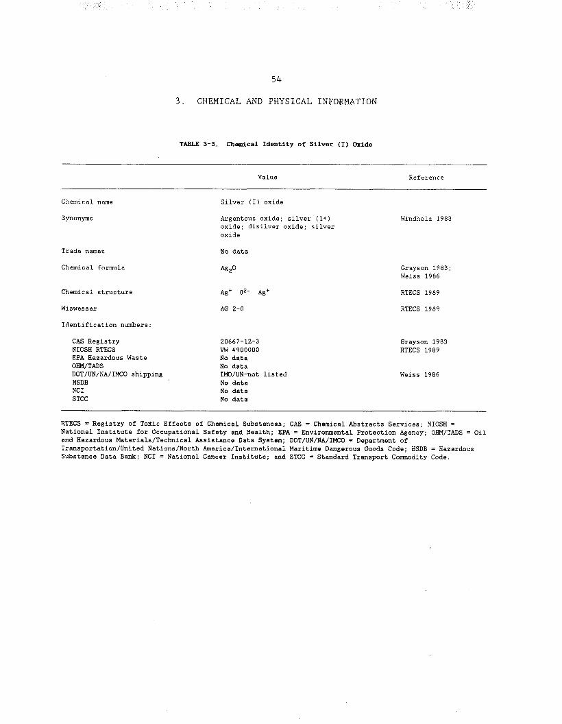

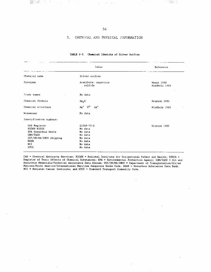
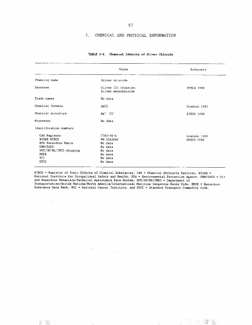


65
4. PRODUCTION, IMPORT, USE AND DISPOSAL
4.1 PRODUCTION
Silver is a rare, but naturally occurring, element. It is often founddeposited as a mineral ore in association with other elements. It is acquiredprimarily as a by-product during the retrieval of copper, lead, zinc, and goldores (Grayson 1983). The primary silver mines of the United States are locatedin the Coeur d'Alene mining district in the northern Idaho panhandle (Smith andCarson 1977). This area accounts for approximately 71% of domestic mineproduction (Drake 1980). It is mined using either open pit or undergroundmethods, and the ore is then upgraded through a series of processes includingflotation and smelting. The silver is finally extracted electrolytically by theMoebius process, the Balbach-Thum process, or the Parkers process (Grayson1983; Smith and Carson 1977).
World mine production in 1986 was 419.8 million troy ounces (forconversion: troy ounces x 31.1034768 = grams) (Reese 1986). Mine production inthe United States declined from 1978 to 1986, reaching a low of 34.2 milliontroy ounces in 1986, due to a combination of falling silver prices and risingproduction costs (Reese 1986). This trend appeared to continue according to asurvey conducted by The Silver Institute in 1988 and 1989. The United Statesproduction of silver from ores and concentrates was 3.4 and 4.2 million troyounces in 1988 and 1989, respectively. However, when recovered silver isincluded in the production figures, total production was 8.8 and 9.3 milliontroy ounces for 1988 and 1989, respectively (The Silver Institute 1990). UnitedStates consumption in 1986 reached a high of 126.4 million troy ounces,-largelydue to increased industrial consumption and use in special issue coinage (Reese1986). In 1987, the estimated consumption was 63.7 million troy ounces for theUnited States and 172 million troy ounces worldwide (The Silver Institute 1988)
Since 1951, silver consumption has exceeded its extraction from ore.Secondary silver production involves the recovery of silver from new and oldscrap, resulting from silver-containing wastes generated by industry and theconsumer. Recycled silver accounted for 40% of U.S. refinery production in 1971and had increased to 67% by 1974 (Smith and Carson 1977). It was estimated tobe 61% and 56% in 1988 and 1989, respectively (The Silver Institute 1990). Theestimated world-wide recovery of silver from the photographic industry is about67% of the total used (The Silver Institute 1988). It has been estimated that80%, 68%, and 75% of today's annual consumption by the electrical, industrial-alloy, and art industries, respectively, is recycled silver, but theseestimates may be high.

66
4. PRODUCTION, IMPORT, USE AND DISPOSAL
4.2 IMPORT
The United States 1986 net import reliance approximated 60% of apparentdomestic consumption. Despite this, the 1986 U.S. dependence on foreign importsdecreased. Import levels fell from 152.6 million troy ounces in 1985 to 144.9million troy ounces in 1986 (Reese 1986).
The largest decrease in imported silver was from the United Kingdom andSwitzerland. For these two countries import levels fell by 18.1 million ounces,primarily in the form of refined bullion. A total of 125.4 million troy ouncesof refined silver were imported in 1986 with only 9.5 million troy ouncesaccounted for in other forms.
U.S. exports of silver decreased slightly from 28.8 million troy ounces in1985 to 25.1 million troy ounces in 1986 (Reese 1986).
4.3 USE
Silver metal and silver compounds have been and still are used in a widevariety of ways. In the past, silver was used for surgical prostheses and/orsplints, fungicides (both of which are now obsolete), and coinage (discontinuedfrom general circulation within the United States in 1970). Although silverstill serves some of the above functions, the current uses are even morevaried. Photographic materials accounted for 45% of the U.S. consumption in1986. Electrical and electronic products, such as electrical contacts, silverpaints, and batteries, consumed approximately 25%. Silver has been an importantcomponent in the manufacture of bearings in the past, although today its use inthis area is limited by cost and availability. Silver is also an importantcomponent in brazing alloys and solders, which represent approximately 5% ofthe 1986 silver consumption. More aesthetic uses of silver includeelectroplated ware, sterling ware, and jewelry; in 1986, they accounted for 11%of recorded uses.
Other uses account for the remaining 14%; these include use in mirrors,dental amalgam, and medical supplies for treatment of burns, use as a catalystin the manufacture of formaldehyde and ethylene oxide, as an active agent forpurification and disinfection of drinking water and water in swimming pools, incertain chemical analyses involving titration, and in cloud seeding(Grayson 1983; HSDB 1988; NRC 1977; Smith and Carson 1977). Silver ions arealso used medically as an antibacterial agent (Becker 1987; Becker et al.1978;Fox et al. 1969; Webster et al. 1981).
4.4 DISPOSAL
Treatment of air emissions containing silver is not a concern asatmospheric emissions rarely approach the federal threshold limit value foroccupational exposure of 0.01 mg/m3 (Smith and Carson 1977).
67
4. PRODUCTION, IMPORT, USE AND DISPOSAL
Moreover, as consumption of silver-containing products outweighs supply,these products tend to be recycled whenever feasible. The largest source ofnonrecycled silver in the waste stream is attributable to photographic materialuse by small-scale consumers (Smith and Carson 1977). This tends to be releasedin the form of silver thiosulfate, which is converted intoinsoluble silver forms by micro-organisms during wastewater treatment (Grayson1983). Several methods have been suggested for recovering silver from variouswaste media, including waste water, solid waste, and gas effluents. Theseinclude electrolytic recovery, agglomeration, and metal concentration (CHMR1989). At present the criteria for land disposal practices are undergoingsignificant revision, and consultation with environmental regulatory agenciesis advised (HSDB 1988).


69
5. POTENTIAL FOR HUMAN EXPOSURE
5.1 OVERVIEW
Silver is a rare element, which occurs naturally in its pure form as awhite, ductile metal, and in ores. It has an average abundance of about 0.1 ppmin the earth's crust and about 0.3 ppm in soils. There are four oxidationstates (0, l+, 2+, and 3+); the 0 and 1+ forms are much more common than the 2+and 3+ forms. Silver occurs primarily as sulfides, in association with iron(pyrite), lead (galena), and tellurides, and with gold. Silver is found insurface waters in various forms: (1) as the monovalent ion (e.g., sulphide,bicarbonate, or sulfate salts); (2) as part of more complex ions with chloridesand sulfates; and (3) adsorbed onto particulate matter.
Silver is released to air and water through natural processes such as theweathering of rocks and the erosion of soils. Important sources of atmosphericsilver from human activities include the processing of ores, steel refining,cement manufacture, fossil fuel combustion, municipal waste incineration, andcloud seeding. The total U.S. annual release of silver tothe environment as a result of human activities in 1978 was estimated to beapproximately 2 million kg. ,Of this amount, 77% was from,land disposal ofsolid waste, 17% was discharged to surface waters, and 6% emitted to theatmosphere. Ore smelting and fossil fuel combustion emit fine particles ofsilver that may be transported long distances and deposited with precipitation.The major source of release to surface waters is effluent from photographicprocessing. Releases from the photographic industry and from disposal of sewagesludge and refuse are the major sources of soil contamination with silver.Sorption is the dominant process controlling partitioning in water and movementin soil. Silver may leach from soil into groundwater; acidic conditions andgood drainage increase the leaching rate. Silver is bioconcentrated to amoderate extent in fish and invertebrates.
The general population is exposed to silver primarily through theingestion of drinking water and food. The most recent estimate by NIOSHindicates that about 70,000 people are potentially exposed to silver inworkplace environments in the United States. Inhalation is probably the mostimportant route of occupational exposure. Populations with exposure to higherthan background levels of silver include workers in industries processing orusing the compound and members of the general public who consume drinking wateror food containing elevated levels of silver. Sources of elevated dietarysilver include seafood from areas near sewage outfalls or industrial sourcesand crops grown in areas with high ambient levels of silver in the airor soil.
According to the VIEW Database (1989), silver has been found at 27 siteson the National Priority List of 1,177 sites. The frequency of these sites

70
5. POTENTIAL FOR HUMAN EXPOSURE
within the United States can be seen in Figure 5-l. EPA's Contract LaboratoryProgram (CLP) statistical database indicates that silver has been detected at100% of the 2,783 Superfund hazardous waste sites that have had samples of allmedia analyzed by the CLP (CLP 1988).
5.2 RELEASES TO THE ENVIRONMENT
5.2.1 Air
The total U.S. annual anthropogenic release of silver to the atmospherefrom production processes and consumptive uses in 1978 was estimated at 77,700kg (Scow et al. 1981). Of this amount, an estimated 30,000 kg were releasedfrom metals production, 22,000 kg from use in electrical contacts andconductors, 9,000 kg from coal and petroleum combustion, 7,000 kg from iron andsteel production, 2,000 kg from cement manufacture, and the remainder frommiscellaneous uses. Urban refuse was the source of an additional 10,000 kg.Smith and Carson (1977) estimated that cloud seeding with silver iodidecontributed 3,100 kg annually (based on data from the early 1970s).
5.2.2 Water
The total U.S. annual release of silver to surface waters in 1978 fromproduction processes and consumptive uses was estimated to be 125,000 kg (Scowet al. 1981). Of this amount, an estimated 65,000 kg were released fromphotographic developing, 54,000 kg from photographic manufacture, 5,000 kg frommetals production, and the remainder from miscellaneous uses. An additional70,000 kg were estimated to be released from sewage treatment plants, 72,000 kgfrom urban runoff, and 438,000 kg from natural sources(e.g., soil erosion). Silver released in precipitation as a result of cloudseeding has decreased and is not expected to contribute significant amounts towater (Scow et al. 1981). Leachates containing silver may enter ground waterswhen tailing ponds or piles are situated in areas with high water tables orwhen abandoned mines or sections of mines are saturated (Letkiewicz et al.1984).
Other sources of silver release to surface waters include textile plantwastewater effluent (Rawlings and Samfield 1979); petroleum refinery effluents(Snider and Manning 1982); and quench water and fly ash scrubber watereffluents from municipal incinerators (Law and Gordon 1979). Silver wasdetected in 7 of 58 (12%) samples from the National Urban Runoff Programsurvey (Cole et al. 1984).
5.2.3 Soil
The total U.S. annual release of silver to land from production processesand consumptive uses in 1978 was estimated at 1.01 million kg (Scow et al.1981). Of this amount, an estimated 630,000 kg were released from thephotographic industry (in manufacture and developing), 165,000 kg from metals

72
5. POTENTIAL FOR HUMAN EXPOSURE
production, 150,000 kg from uses in electrical contacts and conductors, 60,000kg from uses in brazing alloys and solders, and the remainder frommiscellaneous uses. An additional 370,000 to 520,000 kg were estimated to bereleased from urban refuse and 220,000 kg from sewage treatment. Smith andCarson (1977) estimated that the use of silver containing photographicmaterials contributed an annual 370,000 kg in sewage sludge; of this amount anestimated 52.5% was placed in landfills, 26.7% was lagooned, and 20.8% wasspread on land.
The major source of elevated silver levels in cultivated soils is from theapplication of sewage sludge and sludge effluents as agricultural amendments.Additional anthropogenic sources of silver in soil include atmosphericdeposition (especially from ore processing); landfilling of household refuse,sewage sludge, or industrial wastes; and leaching of metaltailings (Smith and Carson 1977).
5.3 ENVIRONMENTAL FATE
5.3.1 Transport and Partitioning
The global biogeochemical movements of silver are characterized byreleases to the atmosphere, water, and land by natural and man-made sources,possible long-range transport of fine particles in the atmosphere, wet and drydeposition, and sorption to soil and sediments. The major forms of silver inthe atmosphere are probably metallic silver, silver sulfide, silver sulfate,silver carbonate, and silver halides (Smith and Carson 1977). Silver isreleased to the atmosphere as an aerosol (suspension of solid or liquidparticles in a gas such as air). Mining operations such as grinding emit largeparticles (more than 20 µ diameter) that settle near the source whileparticles emitted from smelting, fossil-fuel fired power plants, and solidwaste incinerators are smaller and are likely to be transported away from thesource of release (Scow et al. 1981). Fine particles (less than 20 µ diameter)in the aerosol tend to be transported long distances in theatmosphere and are deposited with precipitation. Long-range atmospherictransport of silver is indicated by several studies in which atmosphericparticulate concentrations were elevated above background levels in areasremoved from cloud seeding or mining activities (Davidson et al. 1985;Struempler 1975). Scow et al. (1981) estimated that about 50% of the silverreleased into the atmosphere from industrial operations will be transportedmore than 100 km and will eventually be deposited by precipitation.
The transport and partitioning of silver in surface waters and soils isinfluenced by the particular form of the compound. Lindsay and Sadiq (1979)stated that under oxidizing conditions the primary silver compounds would bebromides, chlorides, and iodides, while under reducing conditions the freemetal and silver sulfide would predominate. In water, the major forms ofsilver are as the monovalent ion in the form of sulfate, bicarbonate, orsulfate salts; as part of more complex ions with chlorides and sulfates; and

73
5. POTENTIAL FOR HUMAN EXPOSURE
as an integral part of, or adsorbed onto, particulate matter (Boyle 1968). Inone study, silver in river water was primarily found in the following forms:silver ion (Ag+) -- 53-71%, silver chloride (Ag Cl°) -- 28-45%, silver chlorideion (AgCl2-) -- 0.6-2.0% (Whitlow and Rice 1985). Callahan et al. (1979) statedthat sorption is the dominant process leading to the partitioning of silver insediments. Significant quantities of silver in water are sorbed by manganesedioxide; pH and oxidation-reduction conditions affect sorption (Anderson et al.1973). Kharkar et al. (1968) reported that approximately 90% of the silver inrivers was in a dissolved form and 10% was a suspended solid. Concentrations inlake sediments were reported to be 1000 times that of the overlying waters; thehighest content was associated with fine-grained sediments (Freeman 1977).
The mobility of silver in soils is affected by drainage (silver tends tobe removed from well-drained soils); oxidation-reduction potential and pHconditions (which determine the reactivity of iron and manganese complexeswhich tend to immobilize silver); and the presence of organic matter (whichcomplexes with silver and reduces its mobility) (Boyle 1968). Thedistribution coefficient (Kd: ratio of the concentration in soil to theconcentration in water) for silver in a number of soils ranged from 10 to 1,000(Baes and Sharp 1983). Factors that affect the Kd include soil pH, clay contentand particle size distribution, organic matter content, and free iron andmanganese oxide content. The enhanced ability of organic matter to immobilizesilver is demonstrated by the increased levels of silver found in peat and bogsoils and in marshes (Boyle 1968). In pasture plants growing in the vicinity ofan airborne source of silver such as a smelter, silver in the leaves isapparently derived from deposition ,of airborne silver, while concentrations inthe roots are from soil uptake (Ward et al. 1979). Silver levels in the leaveswere slightly greater than levels in the roots.
Silver accumulation in marine algae appears to result from adsorptionrather than uptake; bioconcentration factors of 13,000-66,000 have beenreported (Fisher et al. 1984).
Data on the potential for accumulation of silver has been studied inseveral aquatic species. Several of these studies do not conform to currentbioconcentration test procedures in terms of numbers of fish, duration ofexposure, and measurement of concentrations in aquaria. EPA (1980a) reported abioconcentration factor of less than 1 in bluegills (Lepomis macrochirus)exposed to silver nitrate for 28 days. Approximate bioaccumulation factors of4-6 for bluegill were calculated based on a 6-month study and 2-10 for largemouth bass (Micropterus salmoides) exposed to silver nitrate for 4 months (bothdry weight) (Coleman and Cearley 1974).
Terhaar et al. (1977) studied bioconcentration (uptake from water) andbioaccumulation (uptake from food and water) of silver thiosulfate complexes inalgae (Scenedesmus sp.), water flea (Daphnia magna), mussels (Ligumia sp. andMargaritifera sp.), and fathead minnow (Pimephales promelas) in 10-week
74

74
5. POTENTIAL FOR HUMAN EXPOSURE
exposures. Bioconcentration indices were 96-150 for algae, 12.2-26 for Daphnia,5.9-8.5 for mussels, and 1.8-28 for fish. Bioaccumulation indices were 9-26 forDaphnia, 6.6-9.8 for mussels, and 4.0-6.2 for fish. These indices, which arebased on measured wet weight concentrations in biota and nominal concentrationsin water, indicate little potential for silver biomagnification (systematicincrease in residue concentrations moving up a food chain) in the testedaquatic food chain.
Bioconcentration factors of 1,055-7,650 (wet weight) were estimated in a21-month study with the mussel (Mytilus edulis) in salt water (Calabrese et al.1984). The clam, Macoma balthica, contained silver at 32-133 µg/g (dry weighttissue) in an area of San Francisco Bay near a sewage outfall; backgroundconcentrations in this species in the bay were less than 1 µg/g (Thomson et al.1984). These data indicate that inputs of silver to an estuary are available tosediment-dwelling animals. Silver from sewage sludge at an ocean disposal sitewas bioaccumulated by the sea scallop (Placopecten magellanicus). Maximumconcentrations in scallops located near the disposal site were 9.08 ppm (dryweight tissue) while scallops located away from the site had levels less than 1ppm (Pesch et al. 1977). The estimated biological half-lives for theelimination of silver were 26.4 days for the Pacific oyster (Crassostrea gigas)and 149.1 days for the American oyster (C. virginica) (Okazaki and Panietz1981).
5.3.2 Transformation and Degradation
5.3.2.l Air
Particulates of metallic silver emitted from the burning of fossil fuelsand municipal refuse are likely to become coated with silver oxide, silversulfide, and silver carbonate as the particles cool and undergo deposition(Smith and Carson 1977).
5.3.2.2 Water
In fresh water, silver may form complex ions with chlorides, ammonium (inareas of maximum biological activity), and sulfates; form soluble organiccompounds such as the acetate and the tartrate; become adsorbed onto humiccomplexes and suspended particulates; and become incorporated into, or adsorbedonto, aquatic biota (Boyle 1968). Where decaying animal and plantmaterial are abundant, silver strongly precipitates as the sulfide or combineswith humic materials (Smith and Carson 1977).
5.3.2.3 Soil
Silver tends to form complexes with inorganic chemicals and humicsubstances in soils (Boyle 1968). Since silver is toxic to soil microorganismsand inhibits bacterial enzymes (Domsch 1984), biotransformation is not expectedto be a significant process.

75
5. POTENTIAL FOR HUMAN EXPOSURE
5.4 LEVELS MONITORED OR ESTIMATED IN THE ENVIRONMENT
5.4.1 Air
Silver was measured in particulate samples from rural and urban areas bothadjacent to and removed from activities such as metal smelting, refining, andsilver iodide cloud seeding. Background levels appear to be less than 1 ng/m3
as evidenced by the measurement of average silver concentrations of 0.018 ng/m3
at Great Smoky Mountains National Park; 0.012 ng/m3 at Olympic National Park;and less than 0.19 ng/m3 at Glacier National Park (Davidson etal. 1975). The highest particulate levels (mean -- 10.5 ng/m3; range -- 0.936-36.5 ng/m3) were measured in Kellogg, Idaho (in the Coeur d'Alene River Basin)near a large smelter complex (Ragaini et al. 1977). In an industrialized areaof northwest Indiana, silver was measured at less than l-5 ng/m3 (Harrison etal. 1971). A level of 1 ng/m3 was reported by Douglas (1968) in a rural cloud-seeding target area. In a rural area of Nebraska where no cloud seeding wasknown to have occurred, Struempler (1975) found particulate silverconcentrations averaged 0.04-0.15 ng/m3 during three sampling periods. Thisresearcher theorized that anthropogenic sources, such as long-range transportfrom cloud seeding, were responsible for the enrichment of silver by factors of326-355 over its average concentration in the earth's crust. Silverconcentrations in precipitation resulting from seeding clouds with silveriodide were 10-4500 ng/L compared with concentrations of 0-20 ng/L withoutcloud seeding (Cooper and Jolly 1970).
5.4.2 Water
Boyle (1968) reported average (background) ambient concentrations ofsilver in fresh waters of 0.2 µg/L and in sea water of 0.25 µg/L. Waters thatleach silver-bearing deposits (e.g., in mining areas) may carry up to 100 timesmore silver than other fresh waters (Scow et al. 1981). Leaching is enhanced bylow pH (Smith and Carson 1977). In samples of 170 lakes inCalifornia, silver concentrations averaged 0.1 µg/L with a maximum of 6.0 µg/L(Bradford et al. 1968). Kharkar et al. (1968) reported that the average silverconcentration of 10 U.S. rivers was 0.30 µg/L (range: 0.092-0.55 µg/L). Inanother survey, Kopp (1969) found silver in 6.6% of 1,577 surface waterssampled with a mean detected concentration of 2.6 µg/L (range: 0.1-38 µg/L).For 1970-1979, according to U.S. surface water sampling data from EPA's STORETdatabase, the annual mean levels ranged from 1 µg/L to 9 µg/L and annualmaximum concentrations were 94 µg/L to 790 µg/L (Scow et al. 1981). In 10 outof 13 major U.S. river basins, silver concentrations decreased from 1975-1979as compared with 1970-1974. Concentrations increased in the North Atlantic,Southeast, and Lower Mississippi basins. In the U.S. Geological Survey, WaterResources Division portion of the database (from the early 1960s to mid-1988),silver was detected in 2,195 of over 10,000 surface water samples; the meanand median concentrations in these samples were 1.9 µg/L and 2.0 µg/L,respectively (Eckel and Jacob 1988).

76
5. POTENTIAL FOR HUMAN EXPOSURE
Hem (1970) reported a median silver concentration of 0.23 µg/L in U.S.drinking water. Letkiewicz et al. (1984) analyzed the results of three surveysof U.S. groundwater and surface water used as drinking water supplies. Thesesurveys were the 1969 U.S. Public Health Service Community Water Supply Survey(CWSS 1969), the 1978 EPA Community Water Supply Survey (CWSS 1978), and the1978 through 1980 EPA Rural Water Survey (RWS). In CWSS 1969, silver wasdetected (minimum positive value was 0.1 µg/L) in 309 of 677 groundwatersupplies, (mean 1.7 µg/L, median 1.3 µg/L, and range 0.1 to 9 µg/L). Silver wasdetected in 59 of 109 surface water supplies with a mean and median of 1.3 µg/Land a range of 0.1 to 4 µg/L. In CWSS 1978, silver was detected (minimumpositive value was 30 µg/L) in 8 of 81 groundwater supplies (range 30-40 µg/L,mean 31.9 µg/L, and median 30 µg/L). Silver was found in 4 of 25 surface watersupplies (range 30-40 µg/L, mean 36.2 µg/L, and median 37.5 µg/L). In the RWSconducted between 1978 and 1980, silver was detected (minimum quantifiableconcentration apparently was 20 µg/L) in 10 of 71 groundwater supplies (meanand median 40 µg/L and range 20-80 µg/L). Silver was detected in 8 of 21surface water supplies. The range, mean, and median of these 8 supplies were20-60 , µg/L, 36.2 µg/L, and 35 µg/L, respectively. Letkiewicz et al. (1984)also summarized information from EPA's Federal Reporting Data System as of1984, which indicated that 14 public water supplies (13 from groundwater) inthe United States reported silver levels above 50 µg/L. Letkiewicz et al.(1984) stated that it is not possible to determine which of these surveys isrepresentative of current levels of silver in the U.S. water supply. The largerange in apparent detection limits further limits the usefulness of these datain estimating silver levels in U.S. water supplies.
Silver has been detected with a geometric mean concentration of 6.0 µg/Lin groundwater samples from 613 of the 2,783 (22%) hazardous waste sitesincluded in EPA's Contract Laboratory Program (CLP) statistical database (CLP1988). It has also been detected in surface water samples from 552 of the 2,783(20%) sites in the CLP statistical database with a geometric mean concentrationof 9.0 µg/L (CLP 1988).
5.4.3 SoilFrom a series of measurements in Canada, Boyle (1968) estimated that the
average silver content of soils (except for mineralized zones such as miningareas) was 0.30 ppm and the average abundance in the earth's crust was 0.10 ppm. The major source of elevated silver levels in cultivated soils is from theapplication of sewage sludge and sludge effluents (Smith and Carson 1977). Theaverage silver concentration in soils near a lead smelting complex in Kellogg,Idaho (in the Coeur d'hlene River Basin) was 20 ppm (range: 3.2-31 ppm)(Ragaini et al. 1977). Klein (1972) measured soil metal concentrations in theGrand Rapids, Michigan area in order to examine possible relationships betweenconcentrations and land use. Silver concentrations in soils that wereclassified by land use were 0.13 ppm (residential), 0.19 ppm (agricultural),and 0.37 ppm (industrial) (Klein 1972).

77
5. POTENTIAL FOR HUMAN EXPOSURE
The Contract Laboratory Program (CLP) statistical database indicates thatsilver has been detected with a geometric mean concentration of 4.5 ppm in soilsamples from 1,807 of 2,783 (65%) hazardous waste sites that have had samplesanalyzed by the CLP (CLP 1988).
5.4.4 Other Media
Coal has been reported to contain silver at concentrations of up to 10 ppm(Boyle 1968). Klusek et al. (1983) measured the following silver concentrationsat a bituminous coal-fired electric generating station: coal -- 0.29 mg/kg; flyash -- 1.6 mg/kg; and bottom ash -- <0.l mg/kg. In the combustible portions ofmunicipal solid waste, mean silver concentrations were3 ppm (range: <3-7 ppm) (Law and Gordon 1979). A municipal incinerator was.found to emit particles containing 390 ppm silver (Law and Gordon 1979). Themean and maximum silver concentrations i.n U.S sewage sludge were 225 mg/kg and960 mg/kg (dry weight), respectively (Bunch 1982). Sludge silver concentrations(mg/kg dry weight) were reported as follows: from sewage treatment plants withindustrial or municipal wastes -- 15-120 mg/kg; from plants withphotoprocessing effluents as a source -- 450-27,000 mg/kg (Lytle 1984) .
Scow et al. (1981) reported that the median silver concentrations insewage treatment plant influent and effluent were 0.008 mg/L and 0.002 mg/L,respectively. Treated effluents from a large photographic processing plantcontained an average of 0.07 mg/L silver (range: <0.02-0.30 mg/L) in the formof silver thiosulfntc: complexes, silver bromide, and silver sulfide (Bard etal. 1976).
Cunningham and Stroube (1987) collected samples of various foods in 20U.S. cities between 1979 and 1980. Silver concentrations (mg/kg wet weight) incomposite samples of the following food groups were: dairy products -- <0.061;meat, fish, and poultry -- mean 0.015, range 0-87; cereal and grain products --mean 0.008, range o-0.140; leafy vegetables .-- mean 0.007, range 0-0.039;fruits -- <0.050; oils and fats -- <0.030. The average silver concentration ofa mixture of 201. foods prepared to represent the typical U.S. diet was 0.0091mg/kg dry weight (Iyengar et al. 1987). The average concentration in cow's milkin the United States has been reported to be 0.047 ppm (range:0.037-0.059 ppm)(Murthy and Rhea 1968), EPA (1980a) summarized data on silver content in foodas follows: beef -- 0.004-0.024 mg/kg; pork-- 0.007-0.012 mg/kg; mutton and lamb -- 0.006-0.011 mg/kg; tea -- 0.20-2.00mg/kg (dry weight); mollusks -- 0.1-10.0 mg/kg.
Mean silver concentrations in one brand of nonfilter and filter cigaretteswere reported to be 0.18 mg/kg and 0.27 mg/kg, respectively (Nadkarni et al.1970).
In a summary of 1975-1979 data on fish tissue from EPA's STORET database,the mean concentration of silver in 221 samples was 0.225 mg/kg (wet weight
78
5. POTENTIAL FOR HUMAN EXPOSURE
total fish), with a range of 0.004-1.900 mg/kg (Scow et al. 1981). In Lake Pontchartrain, Louisiana (which is likely to receive substantial inputs of metals from municipal and agricultural activities) silver concentrations in clams and American oyster tissues were 0.4-2.4 mg/kg and 5.5 mg/kg (all dry weight), respectively (Byrne and DeLeon 1986)
5.5 GENERAL POPULATION AND OCCUPATIONAL EXPOSURE
Food and water are the most likely major sources of exposure to natural and anthropogenic silver for the general U.S. population (Letkiewicz et al. 1984). airborne silver and the dental and medical uses of silver. Letkiewicz et al. (1984) estimated that about 50% of the 214 million people in the United States who use public drinking water supplies had silver present in their water at 0.01-10 pg/L; 10-30% may receive water with levels greater than 30 pg/L. They estimated that 46,000 people in the U.S. receive drinking water with silver concentrations exceeding the current U.S. Safe Drinking Water Act maximum contaminant limit of 50 pg/L. containing systems is another possible source of exposure to silver.
The general population is also exposed through the inhalation of
Swimming pool water purified with silver-
The averaged daily dietary intake (including fluids) of silver has been estimated to be 70 pg/day (Snyder et al. 1975) and 88 pg/day (Kehoe et al. 1940). The average daily dietary intake of two subjects over 30 days was determined to be 35-44 pg/day (Tipton et al. 1966). The silver content of food was estimated at 4.5 pg/day based on the content of a mixture of 201 foods prepared to represent the typical U.S. diet (Iyengar et al. 1987). Most of the U.S. population breathes air containing a maximum o f 1.0 ng/m3 silver, which contributes a maximum of 0.023 pg/day. Drinking water supplies containing 10 pg/L would provide an estimated 20 pg/day of the 70-88 pg/day estimated daily intake. 60-100 ,ug/day (based on an estimated daily water intake of 2 L) and constitutes the major source of silver intake (Letkiewicz et al. 1984). Although silver has been detected in cigarettes, the average daily intake from smoking has not been determined. A very limited use of silver salts is in purification systems in isolated locations (such as mountain cabins and in space missions) (Silver Institute 1975).
At levels of 30-50 pg/L, drinking water contributes
The 1972-1974 National Occupational Hazard Survey (NOHS), conducted by NIOSH estimated that 19,343 workers in 2,163 plants were potentially exposed to silver in 1970 (NIOSH 1976). The largest number of exposed workers were in special trade contracting, primary metal industries, and industries using electrical machinery and electrical equipment and supplies. groups with the largest number o f exposed workers were air conditioning, heating and refrigeration mechanics and repairmen; plumbers and pipefitters; miscellaneous assemblers; welders and flamecutters; and miscellaneous machine operators.
The occupational
79
5. POTENTIAL FOR HUMAN EXPOSURE
Preliminary data from a second workplace survey, the 1980-1983 NationalOccupational Exposure Survey (NOES) conducted by NIOSH, indicated that 67,054workers, including 15,763 women, in 3,123 plants were potentially exposed tosilver in the workplace in 1980 (NIOSH 1984a). These estimates were derivedfrom observations of the actual use of silver (67% of total estimate) and theuse of trade name products known to contain the compound (33%). The largestnumber of workers were exposed in the primary metal industries, businessservices, health services, instruments and related products industries, andfabricated metal products industries.
Neither the NOHS nor the NOES databases contain information on thefrequency, concentration, or duration of exposure of workers to any of thechemicals listed therein. These surveys provide only estimates of the number ofworkers potentially exposed to chemicals in the workplace.
Additional industrial processes which act as potential sources ofoccupational exposure to silver include the processing of silver chemicals suchas silver nitrate and silver oxide for uses such as photography, and smeltingand refining of silver-containing ores (DiVincenzo et al. 1985).
5.6 POPULATIONS WITH POTENTIALLY HIGH EXPOSURES
The most likely sources of higher than background levels of silver for thegeneral population are ingestion of contaminated food and drinking water, Theestimated 46,000 persons in the United States whose drinking water containsmore than 50 µg/L silver (attributable to natural and/or anthropogenic sources)would have an estimated daily intake of at least 100 µg/day from water alone(Letkiewicz et al. 1984). Higher levels of silver have been detected inshellfish near industrial or sewage inputs (Byrne and DeLeon 1986; Pesch et al.1977; Thomson et al. 1984) and are likely to occur in crops grown on sludge-amended soils, in the vicinity of smelters or mining operations, or in areaswith naturally high background silver levels.
Elevated atmospheric silver concentrations have been attributed tosmelting and refining of silver and other metals, and the use of silver iodidein cloud seeding (Scow et al. 1981). Populations living close to mines may havehigher exposures. Approximately 71% of domestic mine production occurs inIdaho, Arizona, and Colorado; the Coeur d'Alene River Basin in Idahosupplies the greatest amount of silver (Drake 1980). Crops grown on soils withelevated silver concentrations (either from anthropogenic sources or fromnaturally high background levels) or exposed to high ambient atmosphericconcentrations are likely to become enriched with silver (Ragaini et al. 1977;Ward et al, 1979).
Silver has been used in lozenges and chewing gums designed to aid thecessation of smoking. Silver acetate in chewing gum has been classified as anover-the-counter smoking deterrent by the Food and Drug Administration (Malcolmet al. 1986). Several cases of high body levels of silver have been

80
5. POTENTIAL FOR HUMAN EXPOSURE
reported (Malcolm et al. 1986). A skin silver concentration thousands of timeshigher than would be expected as a normal value was found in a patient after anestimated 6 month exposure to silver acetate lozenges (East et al. 1980;MacIntyre et al. 1978).
Scow et al. (1981) estimated that a person developing six rolls of filmcould be exposed to up to 16 grams of silver through dermal contact withphotographic solutions. However, many people use implements or wear glovesduring film developing and therefore this is not expected to result inwidespread, high level exposures. Inhalation was not expected to be asignificant route of uptake during film processing because of the lowvolatility of silver in solution.
5.7 ADEQUACY OF THE DATABASE
Section 104 (i) (5) of CERCLA, directs the Administrator of ATSDR (inconsultation with the Administrator of EPA and agencies and programs of thePublic Health Service) to assess whether adequate information on the healtheffects of silver is available. Where adequate information is not available,ATSDR, in conjunction with the National Toxicology Program (NTP), is requiredto assure the initiation of a program-of research designed to determine thehealth effects (and techniques for developing methods to determine such healtheffects) of silver.
The following categories of possible data needs have been identified by ajoint team of scientists from ATSDR, NTP, and EPA. They are defined assubstance-specific informational needs that, if met would reduce or eliminatethe uncertainties of human health assessment. In the future, the identifieddata needs will be evaluated and prioritized, and a substance-specific researchagenda will be proposed.
5.7.1 Identification of Data Needs
Physical and Chemical Properties. No data exist on the partitioncoefficients and Henry's law constant for silver and its compounds. A vaporpressure has been determined for silver at very high temperatures (greater than900°C), but not for any of its compounds. Generally, the fate of silver in theenvironment is fairly well understood; however, a determination of theseenvironmentally relevant values for silver compounds might provide amore complete estimation of the fate of silver in the environment. Tables 3-7to 3-12 contain information on the known physical and chemical properties ofsilver and several important silver compounds.
Production, Use, Release, and Disposal. The production, use, release, anddisposal of silver is well characterized and indicates that risk of exposurefor the general population is potentially high. Silver and silver compounds areproduced and used for a wide variety of common products and
81
5. POTENTIAL FOR HUMAN EXPOSURE
applications, including photographic materials, jewelry, tooth amalgams,medical supplies, and water purification. The extensive production and use ofsilver leads to a high risk of release to the environment, particularly to soiland water. Silver has been detected in various food products, with the highestlevels detected in fish. Silver is both rare and valuable, and consumptionexceeds production. Therefore, manufacturers attempt to conserve the metal bylimiting releases and recycling instead of disposing of the metal. Methodsexist for recovering silver from several waste media. Improvements in capturingreleased silver before it reaches the environment would be beneficial for botheconomic and health reasons.
According to the Emergency Planning and Community Right-to-Know Act of1986, 42 U.S.C. Section 11023, industries are required to submit chemicalrelease and off-site transfer information to EPA. The Toxics Release Inventory(TRI), which contains this information for 1987, became available in May of1989. This database will be updated yearly and should provide a list ofindustrial production facilities and emissions.
Environmental Fate. The factors governing the environmental fate ofsilver are not well characterized. While silver and its compounds aretransported in the air, water, and soil, and are partitioned between thesemedia, the mechanisms of transport and partitioning are not well-defined. Nopartition coefficients or constants have been determined for silver or itscompounds. Little information was found in the available literature ontransformation of silver in water or soil. Some microorganisms present in thesemedia may be able to transform si1ver and silver compounds; however, silver isnot expected to be significantly transformed in the environmentbecause it is toxic to microorganisms. Further information on the size and fluxof environmental compartments and the transport and transformations of silverand silver compounds in the environment would be useful in defining pathwaysfor potential human exposure.
Bioavailability from Environmental Media. Silver is known to be absorbedfrom the lungs following inhalation exposure to silver dust or air contaminatedwith silver compounds, but data on the extent and rate of absorption arelimited. Silver is also absorbed following oral or dermal exposure to drinkingwater, solutions and medical products containing silver compounds. No data werelocated on bioavailability of silver from soil, plant material, or foods.However, silver is found in all these environmental media and it is likely thatsome silver might be absorbed from these sources. Further information on thebioavailability of silver from contaminated air, water, soil, plants, and otherfoods would help in assessing the health riskassociated with increased exposures that might occur in populations in thevicinity of hazardous waste sites.
Food Chain Bioaccumulation. The data available indicate that silver canbioconcentrate to a limited extent in algae, mussels, clams, and other aquatic

82
5. POTENTIAL FOR HUMAN EXPOSURE
Food Chain Bioaccumulation. The data available indicate that silver canbioconcentrate to a limited extent in algae, mussels, clams, and other aquaticorganisms. However, many of the studies that were performed do not conform tothe current state of the art in terms of sample size, duration, and analysis ofcontaminant levels in aquaria. Reliable data would be useful in determining thepossibility of biomagnification and in defining pathways forgeneral population exposure, as well as in estimating exposures from NPL sitecontamination.
Exposure Levels in Environmental Media. Silver has been detected in allenvironmental media, but most of the data are not current. Current data fromEPA's CLP indicate silver is found at levels above background in ground water,surface water and soil near hazardous waste sites. Elevated levels of silverhave been detected in shellfish located near sources of silver pollution.Estimates of average daily human intake from air, drinking water, food, andtotal diet have been calculated. More current information, that better definesmajor sources and forms of silver, would increase the accuracy of estimates ofdaily exposure to silver. This information could be used to develop a morethorough representation of the contribution of silver exposure fromcontamination at hazardous waste sites. Data that better characterize levels infish and shellfish would aid in identifying populations with potentially highexposures to silver from these sources.
Exposure Levels in Humans. Silver has been detected in the blood,tissues, urine, and feces of humans. The only biological monitoring studieslocated consisted of small numbers of worker populations in chemicalmanufacturing industries. Studies that better characterize important sources ofgeneral population exposure and define populations with potentially highexposure, such as those located near hazardous waste sites, would be helpful.More specific information concerning the chemical form of silver present athazardous waste sites would also be useful. These data would assist indeveloping a more accurate estimate of the potential for silver exposure fromhazardous waste sites contaminated with the metal.
Exposure Registries. No exposure registries for silver were located.This compound is not currently one of the compounds for which a subregistry hasbeen established in the National Exposure Registry. The compound will beconsidered in the future when chemical selection is made for subregistries tobe established. The information that is amassed in the National ExposureRegistry facilitates the epidemiological research needed to assess adversehealth outcomes that may be related to the exposure to this compound.
5.7.2 On-going Studies
No long-term research studies on the environmental fate of silver wereidentified. However, environmental monitoring being conducted in conjunctionwith remedial investigation/feasibility studies at NPL sites where silver has

83
5. POTENTIAL FOR HUMAN EXPOSURE
been found should add useful information regarding environmentalconcentrations, chemical species, fate, and transport of the compounds.
No on-going studies or long-term research concerning occupational orgeneral population exposures to silver were identified.


85
6. ANALYTICAL METHODS
The purpose of this chapter is to describe the analytical methods that areavailable for detecting and/or measuring and monitoring silver in environmentalmedia and in biological samples. The intent is not to provide an exhaustivelist of analytical methods that could be used to detect and quantify silver.Rather, the intention is to identify well-established methods that are used asthe standard methods of analysis. Many of the analytical methods used to detectsilver in environmental samples are the methods approved by federal agenciessuch as EPA and the National Institute for Occupational Safety and Health(NIOSH). Other methods presented in thischapter are those that are approved by a trade association such as theAssociation of Official Analytical Chemists (AOAC) and the American PublicHealth Association (APHA). Additionally, analytical methods are included thatrefine previously used methods to obtain lower detection limits, and/or toimprove accuracy and precision.
The analytical methods used to quantify silver in biological andenvironmental samples are summarized in two tables. Applicable analyticalmethods for determining silver in biological fluids and tissues are listed inTable 6-1, and those used'for determining silver in various environmentalsamples are listed in Table 6-2.
6.1 BIOLOGICAL MATERIALS
Trace levels (10-6 to 10-9 g/g of sample) of silver can be accuratelydetermined in biological samples by several different analytical techniques,provided that the analyst is well acquainted with the specific problemsassociated with the chosen method. These methods include high frequency plasmatorch-atomic emission spectroscopy (HFP-AES), neutron activationanalysis (NAA)., graphite furnace (flameless) atomic absorption spectroscopy(GFAAS), flame atomic absorption spectroscopy (FAAS), and micro-cup atomicabsorption spectroscopy (MCAAS).
Atomic absorption spectroscopy equipped with various atomizers is the bestand most prevalent analytical method used to analyze trace amounts of silver inbiological tissues and fluids. GFAAS offers high detectability(subnanogram/gram of sample) and requires relatively small samples for analysisof biological tissues (DiVincenzo et al. 1985; Segar and Gilio 1973).Background absorption from sample matrix components can be a problem, butcorrection using a deuterium continuum light source is adequate if cautiouslyapplied (Segar and Gilio 1973). The detection limit of silver in biologicaltissues was 2x10⁻⁵ µg/g of sample.


90
6. ANALYTICAL METHODS
Recently, Starkey et al. (1987) modified the GFAAS technique fordetermining trace levels of silver in the blood of exposed and unexposedindividuals. Ethylene diamine tetraacetic acid (anticoagulant) and ammoniumhydrogen phosphate buffer (matrix modifier) were added to blood samples priorto analysis. Starkey and co-workers indicated that the GFAAS technique ishighly selective and sensitive and does not require a complex samplepretreatment (ashing and digestion with strong acids). A detection limit of15x10-3 µg/l00 mL of sample was reported.
Howlett and Taylor (1978) used an atomic absorption spectroscopy fittedwith a micro-cup assembly (MCAAS) for determining silver levels in human wholeblood. The MCAAS technique affords a rapid, precise, and relatively simplemethod for the measurement of silver in blood. Furthermore, this techniquerequires no sample preparation prior to analysis except pipetting and drying.A detection limit level of 0.27 µg/l00 mL of blood sample was measured.Howlett and Taylor (1978) noted that repeated measurement of silver in bloodusing a single nickel cup showed a gradual decrease in sensitivity.
FAAS technique has been successfully used to detect levels of silver inpost-mortem human liver; the detection limit for this method was 0.34 µg/g(Johnson 1976).
HFP-AES can determine ng amounts of silver in a small sample of humanblood. Prepared human blood sample was introduced into the atomizer chamber asan aerosol, formed by nebulization of the sample solution (Nakashima et al.1975). The authors noted that the sensitivity of the HFP-AES technique wasimproved by eliminating moisture in the aerosol with a second condenser at -3to -5°C. The use of bismuth as a coprecipitate showed an enhancing effect onthe silver emission at 328.06 nm. A detection limit of 0.25 µg/l00 mL of samplewas attainable. Advantages of the HFP-AES methodology include freedom from mosttypes of chemical interference, high sensitivity, and multielementalcapability. However, this technology might have to be adapted to currentlyavailable instrumentation in order to be useful. The presence ofspectral interferences is a disadvantage of plasma emission spectroscopy.These interferences are caused when a sample contains elements that haveanalytical emission lines that overlap the line chosen for the analyte. Bloodis particularly troublesome because of high concentration of iron. Iron has avery complex emission spectrum. Also, the analytical line for silver used inthe Nakashima et al. paper has interference from manganese. For this reason,the blood is subjected to dangerous perchloric acid/nitric acid digestion andpreconcentration of silver ion prior to analysis. Other inherent disadvantagesof HFP-AES include the employment of time-consuming procedure, the need forstandard additions for accurate quantification, and its high costs whencompared to GFAAS. Unless a laboratory is already furnished with theinstrumentation, purchase of HFP-AES is not recommended for the analysis ofsilver alone. GFAAS or even DCP-AES could be employed for the determination ofsilver in biological samples.

91
6. ANALYTICAL METHODS
Owing to its high sensitivity, the NAA technique has been widely employedfor determination of trace elements (including silver) in biological andenvironmental samples. The NAA technique is based on interaction of the nucleiof individual silver atoms of the sample with neutron irradiation, resulting inthe emission of γ-rays (photons). The radioactivity of the irradiated sample ismeasured with a high-resolution lithium-drifted germanium detector. The long-lived, metastable 110mAg isotope of silver was formed following irradiation ofhuman hair samples. A half-life of 250.4 days for 110mAg gives ample time toinitiate counting after an irradiation and cooling period (Dutkiewicz et al.1978). The authors noted a detection limit for silver of 0.69 ppm in humanhair. (See Section 2.5 for a discussion of the disadvantages of using hairsamples for monitoring exposure to silver.) A disadvantage of NAA is that it isa very expensive technique and may not be readily available in mostlaboratories.
DiVincenzo et al. (1985) employed the GFAAS technique to evaluate humansamples for biological monitoring of silver exposure levels in the workplace.The authors determined the total silver concentration in urine, blood, feces,and hair with detection limits of 0.005 µg/L,0.5 µg/100 mL, 0.2 µg/g, and 0.02µg/g, respectively.
Scanning electron microscopy (SEM) in concert with x-ray energyspectrometry (XES) has been used to detect silver in pulmonary, lacrimal sac,and skin tissues of individuals with diffuse interstitial lung disease, chronicdacryocystitis, and skin disorders, respectively (Brody et al. 1978; Loefflerand Lee 1987; Tanita et al. 1985). Brody et al. (1978) observed particles ofpreselected lesions of human pulmonary tissue magnified to 300x by SEM, and thesilver content was analyzed by XES. The authors noted that SEM and XEStechniques permit a rapid and conclusive determination of silver, silvercompounds, and complexes in tissue lesions.
6.2 ENVIRONMENTAL SAMPLES
Atomic absorption and plasma emission spectroscopy are perhaps the mostwidely used analytical techniques for the determination of silver levels inair, soil, and water. Rains et al. (1984) employed atomic absorptionspectroscopy with flame atomization (FAAS) and direct current plasma-atomicemission spectroscopy (DCP-AES) to determine silver levels in solid-wasteleachate. In the FAAS technique, a diluted solution of the sample followingashing and digestion is sprayed into a flame by means of a nebulizer. The hightemperature causes formation of atoms, which can be observed (at 328.1 nmresonance line) by absorption spectroscopy. The authors noted that interferenceencountered by the FAAS technique was largely alleviated by the use of 1%solution of ammonium dibasic phosphate buffer as a matrix modifier. In the DCP-AES technique, Rains and co-workers observed silver as a broad band emission at328.068 nm resonance line. Addition of lithium carbonate to sample solution

92
6. ANALYTICAL METHODS
reduces the inter-element interferences observed in unbuffered direct-currentplasmas, but does not significantly degrade DCP-AES detection limits.Detection limits of silver in solid-waste leachate sample by FAAS and DCP-AEStechniques were 0.473x10⁻⁶ g/mL sample and 0.38x10⁻⁶ g/L sample, respectively(Rains et al. 1984).
GFAAS technique is more sensitive than FAAS methodology for determinationof silver in water samples. Rain and stream water have been analyzed by GFAAStechnique to detect silver at ng/mL levels (Rattonetti 1974).
Inductively coupled argon plasma with atomic emission spectroscopy(ICP-AES) has been recommended by NIOSH (method 7300) for determining silverin air. ICP-AES offers multi-element capabilities and high sensitivity butspectral (background) interference can be a problem (NIOSH 1984b). TheEPAestablished analytical test procedure (method 200.7) to analyze dissolved,suspended or total silver in drinking water, surface water, and domestic andindustrial wastewaters employs the ICP-AES technique (EPA 1987a). Anestimated detection limit of 7.0x10⁻⁶ g silver/L sample was measured.
Neutron activation analysis (NAA) methodology has been used to determinesilver levels in environmental samples. Bogen (1973) reported a detection limitof 8x10⁻⁹ g silver/l0 cm2 filter. The author indicated that the use of high-resolution lithium-drifted-germanium detection allows multi-elemental analysisto be performed in a single measurement without any chemical pretreatment ofthe air sample. A highly precise, sensitive, andnondestructive computer-assisted NAA technique for the determination in air ofmulti-element particulate matter has been designed by Dams et al. (1970). Theauthors reported a detection limit of 1x10⁻⁷ g silver/sample. The NAA techniqueby Bogen (1973) and Dams et al. (1970), utilizes the long-lived isotope ofsilver (110mAg ) for quantifying silver levels in air. The fasternondestructive NAA technique developed by Dams et al. (1970) utilizes theshort-lived isotope 110Ag (half-life = 24.6 seconds) to detect silver in airfollowing an 18-second neutron irradiation of air sample. Hence, counting canbe initiated after an irradiation and cooling period of a few minutes.
Hung et al. (1982) developed a sensitive and selective method for silveranalysis by reacting silver (I) with 2(-3,5-dibromo-2-pyridylazo)-5-diethylamino phenol in the presence of an anionic surfactant, sodium lauryl sulfate.The ternary complex formed is red and exhibits an absorption peak at 570 nm.Hung and his co-workers employed EDTA as a chelating agent, thereby reducingthe interference of common ions. Recoveries were good, and a detection limit of0.39 ppm of silver was achieved.
Paper chromatographic, micro thin-layer chromatographic (TLC) andphotodensitometric (PD) methods have also been successfully used to determinelevels of silver compounds in freshwater and eye lotion samples (Devi and Kumar1981; Massa 1969). Simple paper and micro thin layer chromatographic (TLC)techniques were employed by Devi and Kumar (1981) to detect and quantify
93
6. ANALYTICAL METHODS
trace (40 ppm) levels of silver nitrate in fresh water. Devi and Kumar reacteda prepared silver nitrate sample with succinate dehydrogenase enzymechromogenicreagent complex solution prior to paper chromatographic or micro TLC analysis.The metals are recognized by their ability to inhibit the enzymatic formationof a pink reaction product.
Soil samples have been analyzed for silver by AAS (Klein 1972), NAA, andx-ray fluorescence analysis (Ragaini et al. 1977). No statements on thesensitivity, accuracy, or precision of these methods for soil analysis werepresented in the brief description of these methods.
6.3 ADEQUACY OF THE DATABASE
Section 104 (i) (5) of CERCLA, directs the Administrator of ATSDR (inconsultation with the Administrator of EPA and agencies and programs of thePublic Health Service) to assess whether adequate information on the healtheffects of silver is available. Where adequate information is not available,ATSDR, in conjunction with the National Toxicology Program (NTP), is requiredto assure the initiation of a program of research designed to determine thehealth effects (and techniques for developing methods to determine such healtheffects) of silver.
The following categories of possible data needs have been identified by ajoint team of scientists from ATSDR, NTP, and EPA. They are defined assubstance-specific informational needs that, if met would reduce or eliminatethe uncertainties of human health assessment. In the future, the identifieddata needs will be evaluated and prioritized, and a substance-specificresearch agenda will be proposed.
6.3.1 Identification of Data Needs
Methods for Determining Biomarkers of Exposure and Effect. Existingmethods of measuring levels of silver in blood, urine, feces, hair, and tissuesare extremely sensitive and can measure levels in the low ppm to ppt. Thesemethods are accurate and reliable and can be used to measure both backgroundlevels of exposure and levels at which biological effects occur. No additionalanalytical methods for determining trace levels of silver inbiological materials are needed.
Highly sensitive methods exist to measure silver concentrations in blood,urine, hair, and skin samples of individuals showing the few health effectsthat have been associated with silver exposure. These methods are also able toaccurately measure background levels in the population. No additionalanalytical methods appear to be needed for the known biomarkers of effect.
Methods for Determining Parent Compounds and Degradation Products inEnvironmental Media. Sophisticated and highly refined methods are available

94
6. ANALYTICAL METHODS
to detect trace levels of silver and its compounds in air, solid wasteleachate, water (the medium of most concern for human exposure), food, andother environmental media. These methods can accurately measure backgroundlevels in environmental samples, as well as levels at which health effectsoccur. There are no known deficiencies in the analytical methods fordetermining silver in environmental media, and no additional analytical methodsappear to be necessary.
6.3.2 On-Going Studies
No on-going studies concerning techniques for measuring and determiningsilver in biological and environmental samples were located.

95
7. REGULATIONS AND ADVISORIES
Silver is on the list of chemicals appearing in "Toxic Chemicals Subjectto Section 313 of the Emergency Planning and Community Right-to-Know Act of1986" (EPA 1987b).
No international regulations pertaining to silver were found. The national andstate regulations and guidelines regarding silver in air, water, and othermedia are summarized in Table 7-1.


99
8. REFERENCES
*Aaseth J, Olsen A, Halse J, et al. 1981. Argyria-tissue deposition ofsilver as selenide. Stand J Clin Lab Invest 41:247-251.
Abdel Rahim AG. 1985. The effects of dietary L-ascorbic acid on theabsorption and utilization of Na75Se03 of silver-treated rats. Comp BiochemPhysiol 81C:131-132.
*ACGIH. 1986. Documentation of the threshold limit values and biologicalexposure indices. 5th ed. American Conference of Governmental IndustrialHygienists, Inc., Cincinnati, OH, 529.
*Alexander J, Aaseth J. 1981. Hepatobiliary transport and organ distributionof silver in the rat as influenced by selenite. Toxicology 21:179-186.
*Alexander J, Aaseth J, Refsvik T. 1980. Biliary excretion of glutathione inthe rat-significance for the biliary excretion of heavy metals [Abstract].Naunyn-Schmiedeberg Arch Pharmacol 313:R66.
Andelman JB. 1973. Incidence, variability and controlling factors for traceelements in natural, fresh waters. In: Singer PC, ed. Trace metals andmetal-organic interactions in natural waters. Ann Arbor, MI: Ann ArborScience Publishers Inc., 57-88.
*Anderson JB, Jenne EA, Chao TT. 1973. The sorption of silver by poorlycrystallized manganese oxides. Gebchimica et Cosmochimica Acta 37:611-622.
Anghileri LJ. 1968. In vivo and in vitro deiodination of silver iodide.Experientia 24:895.
Anghileri LJ. 1969. Studies on the in vivo breakdown of insoluble halides.Acta Isotope 9:347-356.
Aoki K, Hori J, Kawashima K. 1967. Effect of metallic cations on humanserum: Study by starch-gel electrophoresis. II Effect of Hg++, Cr+++, Ag+,Ni++, Cd++, Zn++, Ba++, Mg++, Al+++ and Fe+++. Arch Biochem Biophys120:255-267.
__________________________
*Cited in text.

100
8. REFERENCES
Aronstam RS, Eldefrawi ME. 1979. Transition and heavy metal inhibition ofligand binding to muscarinic acetylcholine receptors from rat brain. ToxicolAppl Pharmacol 48:489-496.
Baba S, Shinohara Y, Sano H, et al. 1984. Application of high-performanceliquid chromatography with synchronized accumulating radioisotope detector toanalysis of glyceryl trinitrate and its metabolites in rat plasma. JChromatog 305:119-126.
*Bader KF. 1966. Organ deposition of silver following silver nitrate therapyof burns. Plast Reconstr Surg 37:550-551.
*Baes CF III, Sharp RD. 1983. A proposal for estimation of soil leaching andleaching constants for use in assessment models. J Environ Qua1 12:17-28.
Bailey JA, Jones AM, Roy DR. 1973. Effects of silver from cloud seeding onmicroflora of animal digestive systems. Report to US Bureau ofReclamation, Division of Atmospheric Water Resources Management, Denver, CO,by Department of Fishery and Wildlife Biology, Colorado State University, FortCollins, CO. NTIS PB226-062.
Ballinger PM, Brown BS, Griffin MM, et al. 1982. Evidence for carriage ofsilver by sulphadimidine: Hemolysis of human erythrocytes. Br J Pharmacol77:141-145.
*Bard CC, Murphy JJ, Stone DL, et al. 1976. Silver in photoprocessingeffluents. Water Pollut Cont Fed 48:389-394.
*Barnes D, Bellin J, DeRosa C, et al. 1987. Reference dose (RfD):Description and use in health risk assessments. Appendix A: Integrated riskinformation system supportive documentation. Washington, DC: USEnvironmental Protection Agency, Office of Health and EnvironmentalAssessment. EPA/600/8-86-032a.
Barrie HJ, Harding HE. 1947. Argyro-siderosis of the lungs in silverfinishers. Brit J Industr Med 4:225-232.
*Becker RO. 1987. The effect of electrically generated silver ions on humancells. In: Proceedings of the First International Conference on Gold andSilver in Medicine, Bethesda, Maryland, May 13-14, 1987. Washington, DC: TheGold and Silver Institutes, 227-243.
*Becker RO, Spadaro & 1978. Treatment of orthopedic infections withelectrically generated silver ions. The Journal of Bone and Joint Surgery 60-A:871-881.

101
8. REFERENCES
Bergmann M, Engel C. 1968. [Silver deposits in the gastric mucosa and liverin universal argyrosis due to treatment with tragesine]. Dtsch Gesundheitsw23:629-31. (German).
*Berry JP, Galle P. 1982. Selenium and kidney deposits in experimentalargyria. Electron microscopy and microanalysis. Pathol Biol (Paris)30:136-140.
Bertine KK, Goldberg ED. 1971. Fossil fuel combustion and the majorsedimentary cycle. Science 1731233-235.
Bertine KK, Goldberg ED. 1972. Trace elements in clams, mussels, and shrimp.Limnology Oceanography 17:877-884.
Bessing C, Kallus T. 1987. Evaluation of tissue response to dental alloys bysubcutaneous implantation. Acta Odontol Stand 45:247-255.
Biddinger GR, Gloss SP. 1984. The importance of trophic transfer in thebioaccumulation of chemical contaminants in aquatic ecosystems. ResiRev 91:103-145.
Blaha K Jr, Havrdova J, Rosina J, et al. 1987. The effect of 110mAg onceruloplasmin oxidase activity in rats. J Hyg Epidemiol Microbial Immunol31139-43.
*Bleehen SS, Gould DJ, Harrington CI, et al. 1981. Occupational argyria;light and electron microscopic studies and x-ray microanalysis. Br J Dermatol104:19-26.
*Blumberg H, Carey TN. 1934. Argyremia: Detection of unsuspected andobscure argyria by the spectrographic demonstration of high blood silver. JAm Med Assoc 103:1521-1524.
*Bogen J. 1973. Trace elements in atmospheric aerosol in the Heidelbergarea, measured by instrumental neutron activation analysis. Atmos Environ7:1117-1125.
Boissevain CH, Drea WF. 1936. Relation between the occurrence of endemicgoiter and the presence of traces of silver and barium in drinking water.Endocrinology 26:686-687.
Bourdon-Ranisteano S, Bourdon R, Prouillet F. 1977. [Determination of silverin biological materials]. Ann Biol Clin 35:397-400. (French).
*Boyle RW. 1968. Geochemistry of silver and its deposit notes on geochemicalprospecting for the element. Geological Survey of Canada. Ottawa, Ont:Canada, Department of Energy, Mines and Resources. 160., l-96.

102
8. REFERENCES
Bradford GR, Bair FL, Hunsaker V. 1968. Trace and major element content of170 High Sierra Lakes in California. Limnology and Oceanography 13:526-530.
Braidech MM, Emery FH. 1935. The spectrographic determination of minorchemical constituents in various water supplies in the United States. J AmerWater Works Assoc 27:557-580.
Brar SS, Nelson DM, Kline JR, et al. 1970. Instrument analysis for traceelements present in Chicago area surface air. Journal of Geophysics Research75:2939-2945.
Braunstein HM, Pack DJ, Oakes TW. 1982. Intercomparison of stable elementcontent of foods by statistical methods. Proc Univ MO Annu Conf Trace SubstEnviron Health. 16:377-390.
Breton J, Foncin JF, Caroff J, et al. 1971. [Argyria and argyrism: Twounusual cases. Value of electron microscopy and derived methods]. Med LegDommage Corp 4:245-248. (French).
*Brody AR, Vallyathan NV, Craighead JE. 1978. Use of scanning electronmicroscopy and x-ray energy spectrometry to determine the elemental content ofinclusions in human tissue lesions. Scanning Microsc 2:615-622.
Brooks SM. 1981. Lung disorders resulting from the inhalation of metals.Clin Chest Med 2:235-254.
Brooks SM. 1986. Pulmonary reactions to miscellaneous mineral dusts,man-made mineral fibers, and miscellaneous pneumoconioses. In: Merchant JA,ed. Occupational respiratory diseases. Morgantown, WV: US Department ofHealth and Human Services, Division of Respiratory Disease Studies, 401-458.
Brozman M, Chorvath D, Jakubovsky J. 1976. [Localization of silver inglomerular basement membranes in experimental argyrosis]. Bratisl Lek Listy66:253-263. (Polish).
Bruevich TS, Bogomolets NN, Berezovskii AD. 1980. [Sensitizing.action ofprecious metal compounds: Gold, platinum, ruthenium, rhodium and silver].Gig Tr Prof Zabol:42-44. (Russian).
Brulaud KW, Bertine K, Koide M, et al. 1974. History of metal pollution inthe Southern California coastal zone. Environ Sci Technol 8:425-432.
Brune D, Beltesbrekke H. 1979. Levels of mercury and silver in dust from thetrimming of amalgam dies. Scand J Dent Res 87:462-465.

103
8. REFERENCES
Brune D, Nordberg G, Wester PO. 1980. Distribution of 23 elements in thekidney, liver and lungs of workers from a smeltery and refinery in northSweden exposed to a number of elements and of a control group. The Science ofthe Total Environment 16:13-35.
*Buckley WR. 1963. Localized argyria. Arch Dermatol 88:531-539.
Buckley WR, Terhaar CJ. 1973. The skin as an excretory organ in argyria.Trans St John's Hosp Dermatol Sot 59:39-44.
*Buckley WR, Oster CF, Fassett DW. 1965. Localized argyria: II. Chemicalnature of the silver containing particles. Arch Dermatol 92:697-705.
*Bunch RL. 1982. Sewage sludge dilemma of the eighties. In: Pawlowski L,ed. Physiochemical methods for water and wastewater treatment. Amsterdam:Elsevier. 69-81.
*Bunyan J, Diplock AT, Cawthorne MA, et al. 1968. Vitamin E and stress. 8.Nutritional effects of dietary stress with silver. Br J Nutr 22:165-182.
Butt EM, Nusbaum RE, Gilmour TC, et al. 1964. Trace metal levels in humanserum and blood. Arch Environ Health 8:60-65.
*Byrne CJ, DeLeon JR. 1986. Trace metal residues in biota and sediments fromLake Pontchartrain, Louisiana. Bull Environ Contam Toxicol 37:151-158.
Cabe PA, Carmichael NG, Tilson HA. 1979. Effects of selenium, alone and incombination with silver or arsenic, in rats. Neurobeh Toxicol 1:275-278.
Caffee HH, Bingham HG. 1982. Leukopenia and silver sulfadiazine. J Trauma22:586-587.
*Calabrese A, MacInnes JR, Nelson DA, et al. 1984. Effects of long-termexposure to silver or copper on growth, bioaccumulation and histopathology inthe blue mussel Mytilus edulis. Marine Environmental Research 11:253-274.
*Callahan MA, Slimak MW, Gabel NW, et al. R; 1979. Water-relatedenvironmental fate of 129 priority pollutants. EPA-440/4-79-029a,b.
Camner P, Lundborg M, Hellstrom P. 1974. Alveolar macrophages and 5 pmparticles coated with different metals. Arch Environ Health 29:211-213.
Camner P, Hellstrom PA, Lundborg M, et al. 1977. Lung clearance of 4-µmparticles coated with silver, carbon, or beryllium. Arch Environ Health32:58-62.

104
8. REFERENCES
Carson BL, Ellis HV 111, McCann JL. 1986. Toxicology and biological monitoring of metals in humans. Chelsea, MI: Lewis Publishers, Inc., 219-225.
*Casto BC, Meyers J , DiPaolo JA. 1979. Enhancement of viral transformation for evaluation of the carcinogenic or mutagenic potential of inorganic metal salts. Cancer Res 39:193-198.
*Catsakis LH, Sulica VI. 1978. Allergy to silver amalgams. Oral Surg 46:371-375.
CELDS. 1989. Computer-Environment Legislative Data Systems. The Environmental Technical Information System (ETIS). Department of Urban and Regional Planning, Planning Information Program, University o f Illinois, Urbana-Champaign, IL. January 6, 1989.
Cerrai E, Ghersini G. 1966. Reversed-phase paper chromatography of some cations with two nitrophenylthiophosphate derivatives as stationary phases.Chromatogr 22:425-430.
Charley RC, Bull AT. 1979. Bioaccumulation of silver by a multispecies community of bacteria. Arch Microbial 123:239-244.
*CHEMLINE 1988. National Library of Medicine, National Toxicology Information Program, Bethesda, MD. December 5, 1988.
Cherian MG, Goyer RA, 1978. Role of metallothioneins in disease. Ann ClinLab Sci 8:91-94.
Chien PT. 1979. Metallic copper and silver dust hazards . . . [editorial]. Am Ind Hyg Assoc J 40:747-748.
*CHMR 1989. Hazardous waste minimalization manual for small quantity generators. Center for Hazardous Materials Research, University of Pittsburgh Applied Research Center, Pittsburgh, PA.
Chorvath D, Brozman M, Jakubosky J. 1976. [Morphology of experimental argyrosis in kidneys]. Cesk Patol 4:161-165. (Czech).
CCRIS. 1988. Chemical Carcinogenesis Research Information System. Chemical Information System (CIS). December 5, 1988.
Clemente GF, Cigna Rossi L, Santaroni GP. 1977. Trace element intake and excretion in the Italian population. J of Radioanal Chem 37:549-558.

105
8. REFERENCES
*Coffin DL, Palekar LD. 1985. Bioassays for asbestos and other solidmaterials. In: Milman HA and Weisburger EK, ed. Handbook of CarcinogenTesting. Park Ridge, NJ: Noyes Publications, 384-419.
Coglianese MP, Martin M. 1981. Individual and interactive effects ofenvironmental stress on the embryonic development of the pacific oyster,Crassostrea gigas. I. The toxicity of copper and silver. Mar Environ Res5:13-27.
*Cole RJ, Frederick RE, Healy RP, et al. 1984. Preliminary findings of thepriority pollutant monitoring project of the nationwide urban runoff program.Water Pollut Cont Fed 56:898-908.
*Coleman RL, Cearley JE. 1974. Silver toxicity and accumulation inlargemouth bass and bluegill. Bull Environ Contam Toxicol 12:53-61.
Connors RC, Daniels F Jr. 1973. Generalized argyria. Cutis 11:796-801.
*Constable J, Morris P, Burke J. 1967. Absorption pattern of silver nitratefrom open wounds. Plast Reconst Surg 39:342-348.
*Contract Laboratory Program Statistical Database. 1988. US EnvironmentalProtection Agency, Contract Laboratory Program, Washington, DC.
*Cooper CF, Jolly WC. 1970. Ecological effects of silver iodide and otherweather modification agents. Water Resource Research 6:88-98.
Costello J. 1983. Mortality of metal miners a retrospective cohort and casecontrol- study. In: Wagner WL, Rom WN, Merchant JA, eds. Health issuesrelated to metal and nonmetallic mining. Boston, MA: Butterworth Publishers,227-240.
*Creasey M, Moffat DB. 1973. The deposition of ingested silver in the ratkidney at different ages. Experientia 29:326-327.
Creason JP, Hinners TA, Bumgarner JE, et al. 1975. Trace elements in hair,as related to exposure in metropolitan New York. Clin Chem 21:603-612.
*Creason JP, Svendsgaard D, Bumgarner J, et al. 1976. Maternal-fetal tissuelevels of 16 trace elements in 8 selected continental United Statescommunities. Proc Univ MO Annu Conf Trace Subst Environ Health. 10:53-62.
*Cunningham WC, Stroube WB Jr. 1.987. Application of an instrumental neutronactivation analysis procedure to analysis of food. Sci Total Environ63:29-43.

106
8. REFERENCES
Curtin GC, King HD, Mosier EL. 1974. Movement of elements into theatmosphere from coniferous trees in subalpine forests of Colorado and Idaho.Journal of Geochemical Exploration 3:245-263.
*Dams R, Robbins JA, Rahn KA, et al. 1970. Non destructive neutronactivation analysis of air pollution particulates. Anal Chem 42:861-867.
*Dams R, Billet J, Hoste J. 1975. Neutron activation analysis of F, SC, Se,Ag, and Hf, in aerosols using short-lived isotopes. Int J Environ Anal Chem4:141-153.
*Danscher G. 1981. Light and electron microscopic localization of silver inbiological tissue. Histochemistry 71:177-186.
*Davidson CI, Goold WD, Mathison TP, et al. 1985. Airborne trace elements inGreat Smoky Mountains, Olympic, and Glacier National Parks. Environ SciTechnol 19:27-35.
Davies PH, Goettl JP Jr, Sinley JR. 1978. Toxicity of silver to rainbowtrout (Salmo gairdneri). Water Research 12:113-117.
*Day WA, Hunt JS, McGiven AP. 1976. Silver deposition in mouse glomeruli.Pathology 8:201-204.
Deby C, Bacq ZM, Simon D. 1973. In vitro inhibition of the biosynthesis of aprostaglandin by gold and silver. Biochem Pharmacol 22:3141-3143.
*Demerec M, Bertani G, Flint J. 1951. A survey of chemicals for mutagenicaction on E. coli. The American Naturalist 85:119-136.
*Dequidt J, Vasseur P, Gromez-Potentier J. 1974. [Experimental toxicologicalstudy of some silver derivatives]. Bulletin de la Societe de Pharmacie deLille 1:23-35. (French).
*Devi YP, Kumar NV. 1981. Simple paper and micro thin layer chromatographicmethod for separation and detection of mercuric chloride, copper sulfate,cadmium sulfate, and silver nitrate in fresh water. J Assoc Off Anal Chem64:1301-1304.
Dick WA, Bonta JV, Haghiri F, et al. 1983. Stream water quality of two smallwatersheds as affected by surface coal mining. J Environ Qua1 12:351-358.
Dillard CJ, Tappel AL. 1986. Mercury, silver, and gold inhibition ofselenium-accelerated cysteine oxidation. J Inorg Biochem 28:13-20.

107
8. REFERENCES
Diplock AT, Green J, Bunyan J, et al. 1967. Vitamin E and stress. 3. Themetabolism of D-alpha-tocopherol in the rat under dietary stress with silver.Br J Nutr 21:115-125.
Diplock AT, Baum H, Lucy JA. 1971. The effect of vitamin E on the oxidationstate of selenium in rat liver. Biochem J 123:721-729.
Dissanayake CB, Tobschall HJ, Palme H, et al. 1983. The abundance of somemajor and trace elements in highly polluted sediments from the Rhine rivernear Mainz, West Germany. Sci Total Environ 29:243-260.
*DiVincenzo GD, Giordano CJ, Schriever LS. 1985. Biologic monitoring ofworkers exposed to silver. Int Arch Occup Environ Health 56:207-215.
*Domsch KH. 1984. Effects of pesticides and heavy metals on biologicalprocesses in soil. Plant Soil 76:367-378.
*Douglas WJ. 1968. Toxic properties of materials used in weathermodification. Proceedings of the first national conference on weathermodification. Boston, MA: American Meteorological Society, 351-360.
*Drake HJ. 1980. Silver. Mineral facts and problems. Washington, DC: U.S.Department of the Interior, Bureau of Mines. Preprint from Bulletin 671.
Dreisback RH, Robertson WO, ed. 1977. Handbook of poisoning: Prevention,diagnosis and treatment. 12th ed. Los Altos, CA: Lang Medical Publications,373-376.
*Dubin NH, Parmley TH, Cox RT, et al. 1981. Effect of silver nitrate onpregnancy termination in cynomolgus monkeys. Fertil Steril 36:106-109.
Dunn JA, Holland KB, Jezorek JR. 1987. Argentation liquid chromatography ofpolynuclear aromatic hydrocarbons on a silver(I)-loaded mercaptopropyl silicagel stationary phase. J Chromatog 394:375-381.
DuPont T, Gomez J, Cuvillier P, et al. 1984. [Drug-induced generalizedargyria. Value of blood and urine analysis. Apropos of 2 cases]. IARCMedical 4:103-105. (French).
*Dutkiewicz T, Paprotny W, Sokolowska D, et al. 1978. Trace element contentof human hair determined using neutron activation analysis as monitor ofexposure effects to environmental metals. Chemia Analityczna 23:261-272.
Dyck W. 1967. Adsorption and coprecipitation of silver on hydrous ferricoxides. Can J Chem 46:1441-1444.

108
8. REFERENCES
Dymond AM, Kaechele LE, Jurist JM, et al. 1970. Brain tissue reactions tosome chronically implanted metals. J Neurosurg 33:574-580.
*East BW, Boddy K, Williams ED, et al. 1980. Silver retention, total bodysilver and tissue silver concentrations in argyria associated with exposure toan anti-smoking remedy containing silver acetate. Clin Exp Dermatol5:305-311.
Eaton DL. 1985. Effects of various trace metals on the binding of cadmium torat hepatic metallothionein determined by the cd/hemoglobin affinity assay.Toxicol Appl Pharmacol 78:158-162.
Eaton DL, Stacey NH, Wong K-L, et al. 1980. Dose-response effects ofvarious metal ions on rat liver metallothionein, glutathione, heme oxygenaseand cytochrome P-450. Toxicol Appl Pharmacol 55:393-402.
*Eckel WP, Jacob TA. 1988. Ambient levels of 24 dissolved metals in USsurface and ground waters. American Chemical Society, Annual Meeting, LosAngeles, CA, September 25-30, 1988. Preprint Extended Abstract.
Eley BM, Garrett JR. 1983. Tissue reactions to the separate implantation ofindividual constituent phases of dental amalgam, including assessment byenergy dispersive x-ray microanalysis. Biomaterials 4:73-80.
El-Yazigi A, Al-Saleh I, Al-Mefty 0. 1984. Concentrations of Ag, Al, Au, Bi,Cd, Cu, Pb, Sb, and Se in cerebrospinal fluid of patients with cerebralneoplasms. Clin Chem 30:1358-1360.
EPA. 1977. Heavy metals in gardens near the ASARCO Smelter, Tacoma,Washington. US EPA/OTS Public Files. 40-7748027.
EPA. 1979. US Environmental Protection Agency. Federal Register44:15926-15981.
*EPA. 1980a. Ambient water quality criteria for silver. Cincinnati, OH: USEnvironmental Protection Agency, Environmental Criteria and Assessment Office.PB81-117822.
*EPA. 1980b. U.S. Environmental Protection Agency. Federal Register45:79318-79319.
EPA. 1982. Mortality in employees of the ASARCO Lead Refinery, Omaha,Nebraska with cover letter. US EPA/OTS Public Files.
*EPA. 1985a. Drinking water criteria document for silver. Cincinnati, OH:US Environmental Protection Agency, Environmental Criteria and AssessmentOffice. PB86-118288.

109
8. REFERENCES
*EPA. 1985b. Silver. Chemical, physical and biological properties ofcompounds present at hazardous waste sites. Washington, DC: USEnvironmental Protection Agency, Office of Waste Programs and Enforcement.PB89-132203.
*EPA 1986. Reference values for risk assessment. Final draft. Cincinnati,OH: US Environmental Protection Agency, Office of Solid Waste. ECAO-CIN-477
*EPA. 1987a. US Environmental Protection Agency. Code of FederalRegulations. 40 CFR 136.1.
*EPA. 1987b. US Environmental Protection Agency. Federal Register 52:21152.
*EPA. 1987c. US Environmental Protection Agency. Code of FederalRegulations. 40 CFR 116.4.
*EPA. 1987d. US Environmental Protection Agency. Code of FederalRegulations. 40 CFR 141.
EPA. 1987. US Environmental Protection Agency. Code of FederalRegulations. 40 CFR 117.3.
*EPA. 1988a. Health Advisory Draft for Silver. US Environmental ProtectionAgency, Office of Drinking Water, Washington, DC. (Prepublication draft).
*EPA. 1988b. US Environmental Protection Agency. Code of FederalRegulations. 40 CFR 302.4.
EPA. 1988. Drinking water criteria document for silver (final draft).Cincinnati, OH: US Environmental Protection Agency, Environmental Criteriaand Assessment Office. ECAO-CIN-026.
*EPA. 1989a. Interim Methods for Development of Inhalation Reference Doses.US Environmental Protection Agency, Office of Health and EnvironmentalAssessment. Washington, DC. EPA 600/8-88/066F.
*EPA. 1989b. US Environmental Protection Agency. Federal Register 54:22062.
Eturska M, Obreshkova E. 1979. [Argyria in the prolonged use of adsorgan].Vutr Boles 18:121-3.
Evans WH, Read JI, Caughlin D. 1985. Quantifications of results forestimating elemental dietary intakes of lithium, rubidium, strontium,molybdenum, vanadium and silver. Analyst 110:873-877.

110
8. REFERENCES
Fallon ME, Horvath FJ. 1985. Preliminary assessment of contaminants in softsediments of the Detroit River. J Great Lakes Res 11:373-378.
Farhan FM, Hibibi N, Mofidi J, et al. 1979. Direct determination of tracesof heavy metals in canned vegetables by arc spectrography. J Agric Food Chem27:637-638.
*FDA. 1988a. US Food and Drug Administration. Code of Federal Regulations.21 CFR 103.35.
*Fisher NS, Bohe M, Teyssie J-L. 1984. Accumulation and toxicity of Cd, Zn,Ag, and Hg in four marine phytoplankters. Mar Ecol Prog Ser 18:201-213.
Flessel CP, Furst A, Radding SB. 1980. A comparison of carcinogenic metals.In: Sigel H, ed. Metal ions in biological systems. Vol. 10, New York, NY:M. Dekker, 23-54.
*Forycki Z, Zegarski W, Bardzik J, et al. 1983. Acute silver poisoningthrough inhalation. Bulletin of the Institute of Maritime ,and TropicalMedicine in Gdynia 34:199-203.
Fowler BA, Nordberg PF. 1986. Specific metals. In: Friberg L, Nordberg GF,Vouk VB, eds. Handbook on the toxicology of metals. Vol. 2, 2nd ed. NewYork, NY: Elsevier Science Publishing Co., Inc., 521-531.
*Fox CL, Rappole BW, Stanford W. 1969. Control of Pseudomonas infection inrats. Surg Gynecol and Obstet 128:1021-1026.
Frank R, Stonefield KI, Luyken H, et al. 1986. Survey of elemental contentsin two organs of slaughtered bovine, porcine and avian specimens, Ontario,Canada 1980-83. Environmental Monitoring and Assessment 6:259-265.
Franz H. 1968. [Demonstration of small amounts of tissue-bound silver].Histochemie 12:227-229.
*Freeman RA. 1979. Ecological kinetics of silver in an alpine lakeecosystem. In: Marking LL, Kimerle RA, eds. Aquatic toxicology.Philadelphia, PA: American Society for Testing and Materials, 342-358.
Frei JV, Stephens P. 1968. The correlation of promotion of tumor growth andof induction of hyperplasia in epidermal two-stage carcinogenesis. Br JCancer 22:83-92.
Fujita S. 1971. Silver-palladium-gold alloys carcinogenicity and acidmucopolysacharides in the induced tumors. Chem Abstracts 77:136075a.

111
8. REFERENCES
*Furchner JE, Richmond CR, Drake GA. 1968. Comparative metabolism ofradionuclides in mammals-IV. Retention of silver-110m in the mouse, rat,monkey, and dog, Health Physics 15:505-514.
Furst A. 1981. Bioassay of metals for carcinogenesis: Whole animals.Environ Health Perspect 40:83-91.
Furst A, Schlauder MC. 1977. Inactivity of two noble metals as carcinogens.J Environ Path01 Toxicol 1:51-57.
Galloway JN, Likens GE. 1979. Atmospheric enhancement of metal deposition inAdirondack lake sediments. Limnology Oceanography 24:427-433.
Gallyas F. 1979. Factors affecting the formation of metallic silver and thebinding of silver ions by tissue components. Histochemistry 64:97-109.
Gammill JC, Wheeler B, Carothers EL, et al. 1950. Distribution ofradioactive silver colloids in tissues of rodents following injection byvarious routes. Proc Sot Exp Biol Med 74:691-695.
Ganther HE. 1980. Interactions of vitamin E and selenium with mercury andsilver. Ann NY Acad Sci 355:212-226.
Ganther HE, Wagner PA, Sunde ML, et al. 1973. Protective effects of seleniumagainst heavy metal toxicities. Proc Univ MO Annu Conf Trace Subst EnvironHealth. 7:247-252.
*Gaul LE, Staud AH. 1935. Clinical spectroscopy. Seventy cases ofgeneralized argyrosis following organic and colloidal silver medication,including a biospectrometric analysis. J Am Med Assoc 104:1387-1390.
George SG, Pirie BJS, Calabrese A, et al. 1986. Biochemical andultrastructural observations of long-term silver accumulation in the mussel,Mytilus edulis. Marine Environmental Research 18:255-265.
Gerritse RG, Vriesema R, Dalenberg JW, et al. 1982. Effect of sewage sludgeon trace element mobility in soils. J Environ Qua1 11:359-364.
Gjerdet NR, Kallus T, Hensten-Pettersen A. 1987. Tissue reactions toimplanted orthodontic wires in rabbits. Acta Odontol Stand 45:163-169.
Goebel HH, Muller J. 1973. Ultrastructural observations on silver depositionin the choroid plexus of a patient with argyria. Acta Neuropathol (Berl)26:247-251.
Goff H, Powers EL. 1975. Effects of X-rays on Ag-DNA complexes. Int JRadiat Biol 27:503-507.

112
8. REFERENCES
Goldberg ED. 1986. The mussel watch concept. Environmental Monitoring andAssessment 7:91-103.
Goldberg RL, Kaplan SR, Fuller GC. 1983. Effect of heavy metals on humanrheumatoid synovial cell proliferation and collagen synthesis. BiochemPharmacol 32:2763-2766.
Goldschmidt PR, Cogen RB, Taubman SB. 1976. Effects of amalgam corrosionproducts on human cells. J Periodontal Res 11:108-115.
Goodman Gilman A, Goodman LS, Rall TW, et al., ed. 1985. Goodman andGilman's the pharmacological basis of therapeutics. New York, NY: MacmillanPublishing Company, 951; 966-967; 1107.
Gould GW, Colyer J, East JM, et al. 1987. Silver ions trigger Ca2+ releaseby interaction with the (Ca2+-Mg2+)-ATPase. J Biol Chem 262:7676-7679.
Grabowski BF, Haney WG Jr. 1972. Characterization of silver deposits intissue resulting from dermal application of a silver-containingpharmaceutical. J Pharm Sci 61:1488-1490.
Granati A, Lenzi R, Poggini G, et al. 1982. [Evaluation of the impact ofsilver on human health]. Difesa Sociale 61:90-112. (Italian).
*Grass0 P, Abraham R, Hendy R, et al. 1969. The role of dietary silver inthe production of liver necrosis in vitamin E-deficient rats. Exp Mol Pathol11:186-199.
Grasso P, Abraham R, Hendy R, et al. 1973. Hepatocellular necrosis fromdietary silver in vitamin E-deficient rats [Abstract]. J Path01 100:ix.
*Grayson M, ed. 1978. Silver and silver alloys; Silver and compounds.Kirk-Othmer encyclopedia of chemical technology. Vol. 21, 3rd ed. l-32.
Green B. 1967. Effects of silver ions on deoxyribonucleic acid-polycyclichydrocarbon complexes. Biochem J 104:63-64.
Greenberg RR, Zoller WH, Gordon GE. 1978. Composition and size distributionsof particles released in refuse incineration. Environ Sci Technol 12:566-573.
Greene RM, Su WPD. 1987. Argyria. American Family Physician 36:151-154.
Gregus Z, Klaassen CD. 1986. Disposition of metals in rats: A comparativestudy of fecal, urinary, and biliary excretion and tissue distribution ofeighteen metals. Toxico1 Appl Pharmacol 85:24-38.

113
8. REFERENCES
Greig RA, Wenzloff DR, Adams A, et al. 1977. Trace metals in organisms fromocean disposal sites of the middle eastern United States. Arch Environ ContamToxicol 6:395-409.
Greig RA, Schurman S, Pereira J, et al. 1983. Metals and PCB concentrationsin Windowpane Flounder from Long Island Sound. Bull Environ Contam Toxicol31:257-262.
Habu T. 1968. Histopathological effects of silver-palladium-gold alloyimplantation on the oral submucous membranes and other organs [Abstract].Shika Igaku 31:17-48.
*Ham KN, Tange JD. 1972. Silver deposition in rat glomerular basementmembrane. Aust J Exp Bio Med Sci 50:423-434.
Hamilton A, Hardy HL. 1974. Silver. Industrial toxicology. 3rd ed. Acton,MA: Publishing Sciences Group, Inc., 171-172.
Hamilton EI, Minski MJ. 1972/1973. Abundance of the chemical elements inman's diet and possible relations with environmental factors. Sci TotalEnviron 1:375-394.
Hamilton EI, Minski MJ, Cleary JJ. 1972/1973. The concentrations anddistribution of some stable elements in healthy tissues from the UnitedKingdom. An environmental study. Sci Total Environ 1:341-374.
Hanna C, Fraunfelder FT, Sanchez J. 1974. Ultrastructural study of thecornea and conjunctiva. Arch Ophthalmol 92:18-22.
Hanzlik PJ, Presho E. 1923. Comparative toxicity of metallic lead and otherheavy metals for pigeons. J Pharmacol Exp Ther 21:145-150.
Harding HE. 1951. Fibrosis in the lungs of a silver finisher. Br J Ind Med8:256-263.
Harker JM, Hunter D. 1935. Occupational argyria. Br J Dermatol Syph47:441-455.
*Harrison PR, Rahn KA, Dams R, et al. 1971. Areawide trace metalconcentrations measured by multielement neutron activation analysis. Journalof the Air Pollution Control Association 21:563-570.
Harvey RW, Luoma SN. 1985. Effect of adherent bacteria and bacterialextracellular polymers upon assimilation by Macoma balthica of sediment-boundcadmium, zinc and silver. Mar Ecol Prog Ser 22:281-289.

114
8. REFERENCES
Hauser TR, Bromberg SM. 1982. EPA's monitoring program at Love Canal 1980.Environmental Monitoring and Assessment 2:249-272.
Heit M. 1979. Variability of the concentrations of seventeen trace elementsin the muscle and liver of a single striped bass, Marone saxatilis. BullEnviron Contam Toxicol 23:1-5.
Heit M, Tan Y, Klusek C, et al. 1981. Anthropogenic trace elements andpolycyclic aromatic hydrocarbon levels in sediment cores from two lakes in theAdirondack acid lake region. Water Air Soil Pollut 15:441-464.
*Hem JD. 1970. Study and interpretation of the chemical characteristics ofnatural waters. US Geological Survey Paper 1473. Washington, DC: USGeological Service., 202-203.
Henry SA. 1950. Cutaneous cancer in relation to occupation. Annals of theRoyal College of Surgeons of England 7:425-454.
Henry WM, Knapp KT. 1980. Compound forms of fossil fuel fly ash emissions.Environmental Science and Technology 14:450-456.
Hershelman GP, Schafer HA, Jan T-K, et al. 1981. Metals in marine sedimentsnear a large California municipal outfall. Marine Pollution Bulletin12:131-134.
*Hey1 T. 1979. Contact dermatitis from silver coat. Contact Dermatitis5:197.
Hildebrand SG, Cushman RM, Carter JA. 1976. The potential toxicity andbioaccumulation in aquatic systems of trace elements present in aqueous coalconversion effluents. Proc Univ MO Annu Conf Trace Subst Environ Health.10:305-313.
*Hill WR, Pillsbury DM. 1939. Argyria, the pharmacology of silver.Baltimore, MD: The Williams and Wilkins Co.
Hill CH, Matrone G. 1970. Chemical parameters in the study of in vivo and invitro interactions of transition elements. Fed Proc 29:1474-1481.
Hirakawa K. 1983. Determination of silver and cerium in the liver and thekidney from a severely burned infant treated with silver sulfadiazine andcerium nitrate. Radioisotopes 32:59-65. (Japanese).
Hodge VF, Folson TR. 1972. Estimate of the world budget of fallout silvernuclides. Nature 237:98-99.

115
8. REFERENCES
Hoekstra WG. 1975. Biochemical function of selenium and its relation tovitamin E. Fed Proc 34:2083-2089.
*Hoey MJ. 1966. The effects of metallic salts on the histology andfunctioning of the rat testis. J Reprod Fertil 12:461-471.
*Holland MK, White IG. 1980. Heavy metals and spermatozoa. 1. Inhibitionof the motility and metabolism of spermatozoa by metals related to copper.Fertil Steril 34:483-489.
Holland MK, Suter DAI, White IG. 1976. Possible mechanisms involved in thereduction in motility of human spermatozoa by copper zinc and silver. JReprod Fertil 46:507-508.
Housley M, Heckingbottom R, Todd CJ. 1977. The interaction of Ag withSi(II1). Surface Science 68:179-188.
*Howlett C, Taylor A. 1978. Measurement of silver in blood byatomic-absorption spectrophotometry using the micro-cup technique. Analyst103:916-920.
*HSDB. 1988. Hazardous Substances Databank. National Library of Medicine,National Toxicology Information Program, Bethesda, MD. December 5, 1988.Hudson E. 1970. A survey of silver recovery. Part I. The mechanics ofsilver recovery. Radiography 36:263-269.
Huff B, ed. 1988. Physicians desk reference. 42nd ed. Oradell, NJ:Medical Economics Co., 122, 409, 416, 961.
Huggins HA. 1983. Mercury--a factor in mental disease? Part 1. Canmercury-silver amalgams cause psychiatric symptoms. Oral Health 73:42-45.
*Hung S-C, Qu C-L, Wu S-S. 1982. Spectrophotometric determination of silverwith 2-(3,5-dibromo-2-pyridylazo)-5-diethylaminophenol in the presence ofanionic surfactant. Talanta 29:85-88.
Hunt JS, McGiven AR, Day WA. 1976. Immune complex glomerular disease inargyric mice. Pathology 8:205-210.
Hunter D. 1983. Silver and compounds. In: Parmeggiani L, ed.Encyclopaedia of occupational health and safety. Vol. 2, 3rd ed. Geneva:International Labour Office, 2047-2048.
Indraprasit S, Alexander GV, Gonick HC. 1974. Tissue composition of majorand trace elements in uremia and hypertension. J Chronic Dis 27:135-161.

116
8. REFERENCES
Ireland MP. 1988. A comparative study of the uptake and distribution ofsilver in a slug, Arion water and a snail, Achatina fulica. Comp BiochemPhysiol 9OC:189-194.
*IRIS. 1989. Integrated Risk Information System. US EnvironmentalProtection Agency, Cincinnati, OH.
*Iyengar GV, Tanner JT, Wolf WR, et al. 1987. Preparation of a mixed humandiet material for the determination of nutrient elements, selected toxicelements and organic nutrients: A preliminary report. Sci Total Environ235-252.
Jensen LS. 1975. Modification of a selenium toxicity in chicks by dietarysilver and copper. 3 Nutr 105:769-775.
Jensen LS, Peterson RP, Falen L. 1974. Inducement of enlarged hearts andmuscular dystrophy in turkey poults with dietary silver. Poultry Science53:57-64.
John W, Kaifer R, Rahn K, et al. 1973. Trace element concentrations inaerosols from the San Francisco Bay area. Atmospheric Environment 7:107-118.
*Johnson CA. 1976. The determination of some toxic metals in human liver asa guide to normal levels in New Zealand. Part I. Determination of Bi, Cd,Cr, Co, Cu, Pb, Mn, Ni, Ag, Ti and Zn. Analytica Chemica Acta 81:69-74.
Johnson JS, Kilburn KH. 1983. Cadmium induced metal fume fever: Results ofinhalation challenge. Am J Ind Med 4:533-540.
*Jones AM, Bailey JA. 1974. Effect of silver from cloud seeding on cecalflora of rabbits. Water Air Soil Pollut 3:353-363.
Jones CJ, McGugan PJ, Lawrence PF. 1978. An investigation of the degradationof some dry cell batteries under domestic waste landfill conditions. Journalof Hazardous Material 2:259-290.
Just J, Szniolis A. 1938. Germicidal properties of silver in water. JournalAmerican Water Works Association 28:492-506.
*Kaczmar SW, D'Itri FM, Zabik MJ. 1984. Volatilization rates of selectedhaloforms from aqueous environments. Environmental Toxicology and Chemistry3:31-35.
Kalistratova VS, Kogan AG, Kozlova MD. 1966. [Behavior of 111Ag in animals].In: Moskalev I, ed. Raspredel Biol Deistria Radioacktiv Izotop, Sb Statei.Moscow, Russia: Atomizdat, 146-151. (Russian).
117
8. REFERENCES
Kallus T, Hensten-Pettersen A, Mjor I A. 1983. Tissue response to allergenicleachables from dental material. J Biomed Mater Res 17:741-755.
*Kanematsu N, Hara M, Kada T. 1980. Ret assay and mutagenicity studies onmetal compounds. Mutation Research 77:109-116.
Kargov SI, Korolev NI, Stanislavskii OB, et al. 1986. [Interaction ofimmobilized DNA with silver ions]. Mol Biol (Mosk) 20:1499-1505. (Russian),
Kehoe JC. 1984. Intracanal corrosion of a silver cone producing a localizedargyria: Scanning electron microscope and energy dispersive x-ray analyzeranalyses. Journal of Endodontics 10:199-201.
*Kehoe RA, Cholak J, Story RV. 1940. A spectrochemical study of the normalranges of concentrations of certain trace metals in biological materials. JNutr 19:579-592.
Kempton S, Sterritt RM, Lester JN. 1987. Heavy metal removal in primarysedimentation. I. The influence of metal solubility. Sci Total Environ63:231-247.
Kempton S, Sterritt RM, Lester JN. 1987. Heavy metal removal in primarysedimentation II. The influence of metal speciation and particle sizedistribution. Sci Total Environ 63:247-258.
Kent NL, McCance RA. 1941. The absorption and excretion of 'minor' elementsby man. I. Silver, gold, lithium, boron and vanadium. Biochemical Journal35:837-844.
Kesseru E, Leon F. 1974. Effect of different solid metals and metallic pairson human sperm motility. Int J Fertil 19:81-84.
Kesseru E, Hurtado H, Muhe B. 1974. Copper IUD: Enhancement of its efficacyby the addition of silver and nickel. Contraception 9:141-151.
Kettner VW, Vogel K, Kruger G. 1970. Contribution to the clinical picture ofargyrosis. Deut Gesundheitsw 25:1746-1747.
Kharchenko PD, Stepanenko PZ. 1972. [Peculiarities of disturbances of albinorat higher nervous activity during the action of silver electrolyticsolutions]. Fiziol Zh (Kiev) 18:596-600. (Ukranian).
Kharchenko PD, Berdyshev GD, Stepanenko PZ, et al. 1973. [Change in thenucleic acid level in rat brain and liver during long-term introduction ofsilver ions in drinking water]. Fiziol Zh (Kiev) 19:362-368. (Ukranian).

118
8. REFERENCES
*Kharkar DP, Turekian KK, Bertine KK. 1968. Stream supply of dissolvedsilver, molybdenum, antimony, selenium, chromium, cobalt, rubidium and cesiumto the oceans. Geochimica et Cosmochimica Acta 32:285-298.
King JS. 1966. A comparative investigation of neuroglia in representativevertebrates: A silver carbonate study. J Morph01 119:435-465.
Klaassen CD. 1978. Effect of metallothionein on hepatic disposition ofmetals. Am J Physiol 234:E47-E53.
*Klaassen CD. 1979a. Effect of spironolactone on the biliary excretion anddistribution of metals. Toxicol Appl Pharmacol 50:41-48.
*Klaassen CD. 1979b. Biliary excretion of silver in the rat, rabbit and dog.Toxicol Appl Pharmacol 50:49-56.
*Klein DH. 1972. Mercury and other metals in urban soils. EnvironmentalScience and Technology 6:560-562.
Klein DH, Russell P. 1973. Heavy metals: Fallout around a power plant.Environmental Science and Technology 7:357-358.
*Klusek CS, Miller KM, Heit M. 1983. Trace element and radionuclide massbalances at a coal-fired electric generating station. Environ Int 9:139-144.
Kober GM. 1916. The gold, silver, jewelry and allied industries. In: KoberGM, Hanson WC, eds. Diseases of occupation and vocational hygiene.Philadelphia, PA: P. Blakiston's Son and Co., 580-585.
Kojo T, Kaneko T, Ito A, et al. 1986. [Experimental studies of the effect ofmetal cores (silver alloy) on the gingival tissue. 2nd report. Extensiveinvestigation of gingival discoloration]. J Jpn Prosthoclort Sot30:1278-1286. (Japanese).
Kone BC, Kaleta M, Gullans SR. 1988. Silver ion (Ag+)-induced increases incell membrane potassium and sodium permeability in the renal proximal tubule:Reversal by thiol reagents. J Membr Biol 102:11-19.
Konga M, Nakamura T, Jyoyama T, et al. 1982. Industrial hygienic approach tothe workers exposed to inorganic silver. Proceedings of the tenth Asianconference on occupational health, 2:584-587. September 5-10, Singapore.
*Konradova V. 1968. The ultrastructure of the tracheal epithelium in rabbitsfollowing inhalation of aerosols of colloidal solutions of heavy metals. II.Signs of cell alteration in the epithelium after 8 hour inhalation ofcolloidal solutions of iron and silver. Folia Morph01 (Praha) 16:265-271.

119
8. REFERENCES
Konradova V. 1968. The ultrastructure of the tracheal epithelium in rabbitsfollowing the inhalation of aerosols of colloidal solutions of heavy metals.1. Changes in the ultrastructure of the tracheal epithelium after 2-hourinhalation of colloidal solutions of iron and silver. Folia Morphol (Praha)16:258-264.
*Kopp JF. 1969. The occurrence of trace elements in water. Proc Univ MOAnnu Conf Trace Subst Environ Health. 3:59-73.
Kopp JF, Kroner RC. 1967. Tracing water pollution with an emissionspectrograph. J Water Pollut Cont Fed 39:1659-1668.
Kosarek W. 1981. Removal of various toxic heavy metals and cyanide fromwater by membrane processes. In: Cooper WJ, ed. Chemistry in water reuse.Vol. 1, Ann Arbor, MI: Ann Arbor Science Publishers, Inc., 261-280.
Krebs A. 1983. [Drug induced nail disorders]. Deutsch Apotheker Zeitung123:557-561. (German).
Kroner RC, Kopp JF. 1965. Trace elements in six water systems of the UnitedStates. Journal American Water Works Association 57:150-156.
Kulskii LA, Kharchenko PD, Stepanenko PZ. 1972. [Dynamics of corticalactivity changes in albino rats after chronic silver intoxication]. DopovAkad Nauk Ukr RSR Ser B 34:660-662. (Ukranian).
*Landas S, Fischer J, Wilkin LD, et al. 1985. Demonstration of regionalblood-brain barrier permeability in human brain. Neurosci Lett 57:251-256.
Larsen PF, Zdanowicz V, Johnson AC. 1983. Trace metal distribution in thesurficial sediments of Penobscat Bay, Maine. Bull Environ Contam Toxicol31:566-573.
La Torraca F. 1962. [Anatomic, histopathological, and histochemical aspectsof acute experimental intoxication with silver salts]. Folia Medica45:1065-1069. (Italian).
*Law SL, Gordon GE. 1979. Sources of metals in municipal incineratoremissions. Environmental Science and Technology 13:432-438.
Lee RE Jr, Von Lehmden DJ. 1973. Trace metal pollution in the environment.Journal of the Air Pollution Control Association 23:853-857.
Leirskar J. 1974. On the mechanism of cytotoxicity of silver and copperamalgams in a cell culture system. Scand J Dent Res 82:74-81.

120
8. REFERENCES
Leirskar J, Heigeland K. 1972. A methodologic study of the effect of dentalmaterials on growth and adhesion of animal cells in-vitro. Stand J Dent Res80:120-133.
Leirskar J, Helgeland K. 1981. Mechanism of toxicity of dental materials.International Endodontic Journal 14:42-48.
Lemez L. 1980. Sites for experimental production of tracheal and/oroesophageal malformations in 4-day-old chick embryos. Folia Morphol (Praha)28:52-55.
Leroux-Robert C, Benevent J, Benevent D, et al. 1982. [Renal argyriadiscovered in nephrotic syndrome]. Nephrologie 3:101. (French).
Lester JN. 1983. Significance and behavior of heavy metals in waste watertreatment processes 1. Sewage treatment and effluent discharge. Sci TotalEnviron 30:1-44.
*Lester JN, Sterritt RM, Kirk PWW. 1983. Significance and behavior of heavymetals in waste water treatment processes II. Sludge treatment and disposal.Sci Total Environ 30:45-83.
*Letkiewicz F, Spooner C, Macaluso C, et al. 1984. Occurrence of silver indrinking water, food, and air. Report to US Environmental Protection Agency,Office of Drinking Water, Criteria and Standards Division, Washington, DC, byJRB Associates, McLean, VA.
Lindh U, Brune D, Nordberg G, et al. 1980. Levels of antimony, arsenic,cadmium, copper, lead, mercury, selenium, silver, tin and zinc in bone tissueof industrially exposed workers. Sci Total Environ 16:109-116.
*Lindsay WL, Sadiq M. 1979. Theoretical solubility relationships of silverin soils. In: Klein DA, ed. Environmental impacts of artificial icenucleating agents. Dowden, Hutchinson, and Ross, Inc.
Lockhart SP, Rushworth A, Azmy AAF, et al. 1983. Topical silversulphadiazine: Side effects and urinary excretion. Burns 10:9-12.
*Loeb IA, Sirover MA, Weymouth LA, et al.' 1977. Infidelity of DNA synthesisas related to mutagenesis and carcinogenesis. J Toxicol Environ Health2:1297-1304.
*Loeffler KU, Lee WR. 1987. Argyrosis of the lacrimal sac. Graefe's ArchClin Exp Ophthalmol 225:146-150.

121
8. REFERENCES
Luis AS, Duarte CS. 1960. Experimental study on the pathogenesis of acutepulmonary edema. I. Pulmonary edema induced by silver nitrate [Abstract]. Ma1Cardiovasc 56:39-49.
*Luk KFS, Maki AH, Hoover RJ. 1975. Studies of heavy metal binding withpolynucleotides using optical detection. J Am Chem Sot 97:1241-1242.
Lukanov J, Atmadjov P. 1979. Investigating the effect of silver ions on thecontractile function of smooth-muscle preparations from guinea pig stomach, invitro. Folia Med (Plovdiv) 21:11-19.
Luoma SN, Jenne EA. 1977. The availability of sediment-bound cobalt, silverand zinc to a deposit-feeding clam. In: Drucker H, Wilding RE, eds.Biological implications of metals in the environment. Springfield, VA:National Technical Information Service.
Lyons WMB, Fitzgerald WMF. 1980. Trace metal fluxes to nearshore Long IslandSound sediments. Mar Pollut Bull 11:157-161.
Lytle PE. 1984. Fate and speciation of silver in publicly owned treatmentworks. Environmental Toxicology and Chemistry 3:21-30.
MacIntyre EH, McClatchy JK, Rudolph H, et al. 1973. Identification of silverin a periapical lesion of a tooth. Am J Clin Path01 60:613-615.
*MacIntyre D, McLay ALC, East BW, et al. 1978. Silver poisoning associatedwith an antismoking lozenge. Br Med J 2:1749-1750.
Mackison FW, Stricoff RS, Partridge LJ Jr. 1980. Pocket guidelines tochemical hazards. Washington, DC: US Government Printing Office, 1661.Magos L, Webb M. 1978. Theoretical and practical considerations on theproblem of metal-metal interaction. Environ Health Perspect 25:151-154.
*Malcolm R, Currey HS, Mitchell MA, et al. 1986. Silver acetate gum as adeterrent to smoking. Chest 90:107-111.
Malins DC, McCain BB, Brown DW, et al. Chemical pollutants in sediments anddiseases of bottom-dwelling fish in Puget Sound, Washington. EnvironmentalScience and Technology 18:705-713.
Mangal PC, Verma KB. 1979. Effect of induced skin cancer on theconcentration of some trace elements. Indian J Med Res 69:290-295.
Marie J, Leveque B, Watchi JM, et al. 1966. [Argyria in a child followingpharyngeal spracy of silver salts repeated for 6 years]. Ann Pediatr13:2657-2659. (French).

122
8. REFERENCES
Marino AA, Berger TJ, Becker RO, et al. 1974. The effect of selected metalson marrow cells in culture. Chem Biol Interact 9:217-223.
*Marks R. 1966. Contact dermatitis due to silver. Br J Dermatol 78:606-607.
*Marshall JP II, Schneider RP. 1977. Systemic argyria: Secondary to topicalsilver nitrate. Arch Dermatol 113:1077-1079.
Martin M, Castle W. 1984. Petrowatch: Petroleum hydrocarbons, syntheticorganic compounds and heavy metals in mussels from the Monterey Bay area ofcentral California. Marine Pollution Bulletin 15:259-266.
Masiak M, Owczarek H, Skowron S, et al. 1982. Serum levels of certain traceelements (Ag, Co, Cr) in healthy subjects (part II). Acta Physiol Pol33165-73.
Maslenko AA. 1976. [Effects of "silver water" and drinking water treatedwith silver on the digestive organs]. Vrach Delo, Issue 5, 88-90. (Russian).
*Massa V. 1969. [Analysis of silver, in the presence of other metals, bymeans of dithizonate chromatography and photodensitometry. Drug controlapplications]. Trav Sot Pharm Montpellier 29:221-224. (French).
Matuk Y. 1983. Distribution of radioactive silver in the subcellularfractions of various tissues of the rat and its binding to low molecularweight proteins. Can J Physiol Pharmacol 61:1391-1395.
*Matuk Y, Ghosh M, McCulloch C. 1981. Distribution of silver in the eyes andplasma proteins of the albino rat. Can J Ophthalmol 16:145-150.
*Mauss Y, Poulet P, Steibel J, et al. 1980. Interaction of proflavine-calfthymus DNA complexes with Ag+ and Hg++ ions. Studia Biophysics 81:95-96.
McCabe LJ, Symons JM, Lee RD, et al. 1970. A survey of community watersupply systems. Journal of the American Water Works Association 62:670-687.
*McCoy EC, Rosenkranz HS. 1978. Silver sulfadiazine: Lack of mutagenicactivity. Chemotherapy (Basel) 24:87-91.
McDermot DJ, Alexander GV, Young DR, et al. 1976. Metal contamination offlatfish around a large submarine outfall. Journal of the Water PollutionControl Federation 48:1913-1918.
McDowell LR, Forseth JA, Piper RC. 1978. Influence of arsenic, sulfur,cadmium, tellurium, silver and selenium on the selenium-vitamin E deficiencyin the pig. Nutrition Reports International 17:19-34.

123
8. REFERENCES
McGinnis JP Jr, Greer JL, Daniels DS. 1985. Amalgam tattoo: Report of anunusual clinical presentation and the use of energy dispersive x-ray analysisas an aid to diagnosis. J Am Dent Assoc 110:52-54.
McLaughlin AIG, Grout JLA, Barrie HJ, et al. 1945. Iron oxide dust and thelungs of silver finishers. Lancet, March 17, 337-341.
*McMahon JT, Bergfeld WF. 1983. Metallic cutaneous contaminant mimickingmalignant melanoma. Cleveland Clinic Quarterly 50:177-181.
Mehregan AH, Faghri B. 1974. Implantation dermatoses. Acta Dermatovener(Stockholm) 54:61-64.
*Mehta AC, Dawson-Butterworth K, Woodhouse MA. 1966. Argyria. Electronmicroscopic study of a case. Br J Dermatol 78:175-179.
Mertz DP, Koschnick R, Wilk G, et al. 1968. [Studies on the metabolism oftrace elements in humans. I. Serum values for cobalt, nickel, silver, cadmium,chromium, molybdenum, manganese]. Z Klin Chem Klin Biochem 6:171-174.(German).
Mertz DP, Wilk G, Koschnick R. 1975. [Conditions affecting renal excretionof silver by humans. Studies on metabolism of trace elements. VIII]. Z KlinChem Klin Biochem 13:13-15. (German).
Minoia C, Oppezzo MC, Pozzoli L, et al. 1985. [Environmental and biologicalmonitoring of subjects occupationally exposed]. G Ital Med Lav 7:65-73.(Italian).
*Mitteldorf AJ, Landon DO. 1952. Spectrochemical determination of themineral-element content of beef. Analytical Chemistry 24:469-472.
*Moffat DB, Creasey M. 1972. The distribution of ingested silver in thekidney of the rat and of the rabbit. Acta Anat 83:346-355.
Mogilnicka EM, Piotrowski JK, Tomaszewski A. 1976. Effect of some metals(Rb, Cs, Ag, Ti, Sb, U) on metallothionein level in the liver and kidney ofthe rat. Bromat Chem Toksykol 9:357-359. (Polish)
Mogilnicka EM, Milaszewicz M, Piotrowsk JK. 1978. [Binding of silver in theliver of the rat]. Bromat Chem Toksykol 11:59-65. (Polish).
Moller B. 1979. Reactions of human dental pulp to silver amalgamrestorations. A study with emphasis on source material characteristics.Swedish Dent [Suppl] 2:1-37.

124
8. REFERENCES
Moore JW, Sutherland DJ. 1981. Distribution of heavy metals andradionuclides in sediments, water, and fish in an area of Great Bear Lakecontaminated with mine wastes. Arch Environ Contam Toxicol 10:329-338.
Morgan WKC. 1984. Other pneumoconioses. In: Morgan WKC, Seaton A, eds.Occupational lung disease. 2nd ed. Philadelphia, PA: W.B. Saunder Company,449-497.
*Moss AP, Sugar A, Hargett NA, et al. 1979. The ocular manifestations andfunctional effects of occupational argyrosis. Arch Ophthalmol 97:906-908.
Muroma A. 1961. Skin reactions produced by certain metallic salts. AnnalesMedicine Experimentalis et Biologiae Fenniae 39:277-279.
*Murthy GK, Rhea U. 1968. Cadmium and silver content of market milk.J Dairy Science 51:610-613.
*Nadkarni RA, Ehmann WD, Burdick D. 1970. Investigations on the relativetransference of trace elements from tobacco into smoke condensate. Tobacco170:25-27.
Naganuma A, Tanaka T, Maeda K, et al. 1983. The interaction of selenium withvarious metals in vitro and in vivo. Toxicology 29:77-86.
Nakamuro K, Sayato Y. 1986. Studies on the toxicity of silver ion in waterfiltered through a household water purifier equipped with a silver-coatedcharcoal filter. Eisei Kagaku 32:28-33.
*Nakashima R, Sasaki S, Shibata S. 1975. Determination of silver inbiological materials by high-frequency plasma-torch emission spectrometry.Analytica Chimica Acta 77:65-70.
*NAS/NRC. 1989. Biological markers in reproductive toxicology. NationalResearch Council. Board of Environmental Studies and Toxicology. Committeeon Biological Markers. 15-35.
Nathanson JA, Bloom FE. 1976. Heavy metals and adenosine cyclic3',5'-monophosphate metabolism: Possible relevance to heavy metal toxicity.Mol Pharmacol 12:390-398.
Nechay BR, Saunders JP. 1984. Inhibition of adenosine triphosphates in vitroby silver nitrate and silver sulfadiazine. Journal of the American College ofToxicology 3137-42.
Nehring RB. 1976. Aquatic insects as biological monitors of heavy metalpollution. Bull Environ Contam Toxicol 15:147-154.

125
8. REFERENCES
Neri LC, Hewitt D, Schreiber GB, et al. 1975. Health aspects of hard andsoft waters. Journal American Water Works Association 67:403-409.
*Newton D, Holmes A. 1966. A case of accidental inhalation of zinc-65 andsilver-110m. Radiat Res 29:403-412.
Nilner K, Glantz P-O. 1982. The prevalence of copper-, silver-, tin-, andzinc-ions in human saliva. Swed Dent J 6:71-77.
*NIOSH. 1976. National Occupational Hazard Survey (1970) Database. USDepartment of Health and Human Services, National Institute for OccupationalSafety and Health, Cincinnati, OH
*NIOSH. 1984a. National Occupational Exposure Survey (1980-1983) Database.US Department of Health and Human Services, National Institute forOccupational Safety and Health, Cincinnati, OH.
*NIOSH. 1984b. NIOSH, manual of analytical methods. Washington, DC: USGovernment Printing Office., 73001-73005.
*NIOSH. 1985. Pocket guide to chemical hazards. Washington, DC: USDepartment of Health and Human Services. DHEW (NIOSH) Publication No 78-210.
Nishimura M. 1978. [A study on the cytotoxicity of Au-Pd-Ag alloyscontaining Cd (in vitro)]. Shika Rikogaku Zasshi 19:1-7. (Japanese).
*Nishioka H. 1975. Mutagenic activities of metal compounds in bacteria.Mutat Res 31:185-189.
Nordberg GF, Gerhardsson L. 1988. Silver. In: Seiler HG, Sigel H, eds.Handbook on toxicity of inorganic compounds. New York, NY: Marcel Dekker,Inc., 619-624.
Norden B, Matsuoka Y, Kurucsev T. 1986. Nucleic acid-metal interactions: V.The effect of silver(I) on the structures of A- and B-DNA forms. Biopolymers25:1531-1545.
Nriagu JR. 1979. Global inventory of natural and anthropogenic emissions oftrace metals to the atmosphere. Nature 279:409-4.11.
*NRC. 1977. Silver. In: Drinking water and health. Vol. 2. Washington,DC: National Academy of Sciences, National Research Council, 102-106.
*Nuttall KL. 1987. A model for metal selenide formation under biologicalconditions. Med Hypotheses 24:217-221.

126
8. REFERENCES
Oh SH, Whanger PD, Deagen JT. 1981. Tissue metallothionein: Dietaryinteraction of cadmium and zinc with copper, mercury, and silver. J ToxicolEnviron Health 7:547-560.
OHM, TADS. 1988. Oil and Hazardous Materials Technical Assistance DataSystem. Chemical Information System (CIS). December 5, 1988.
*Okazaki RK, Panietz MH. 1981. Depuration of twelve trace metals in tissuesof the oysters Crassostrea gigas and Crassostrea virginica. Marine Biology63:113-120.
*Olcott CT. 1947. Experimental argyrosis. III. Pigmentation of the eyes ofrats following ingestion of silver during long periods of time. Am J Pathol23:783-789.
*Olcott CT. 1948. Experimental argyrosis. IV. Morphologic changes in theexperimental animal. Am J Path 24:813-833.
*Olcott CT. 1950. Experimental argyrosis. V. Hypertrophy of the leftventricle of the heart. Archives of Pathology 49:138-149.
Olson BH, Guinn VP. 1978. Accumulations of trace elements in soil and plantsfrom land disposal of secondary domestic wastewater. In: Land treatment ofwastewater international symposium, Hanover, NH. 2:289-299
Oppenheimer BS, Oppenheimer ET, Danishefsky I, et al. 1956. Carcinogeniceffect of metals in rodents. Cancer Res 16:439-441.
Orentreich N, Pearlstein HH. 1969. Traumatic tattoo due to silver salt.Arch Dermatol 100:107-108.
Orstavik D, Hongslo JK. 1985. Mutagenicity of endodontic sealers.Biomaterials 6:129-132.
Osborn JF, Santhanam S, Davidson CI, et al. 1984. Characterization ofairborne trace metal and trace organic species from coal gasification.Environmental Monitoring and Assessment 4:317-333.
*OSHA. 1988a. US Department of Labor, Occupational Safety and HealthAdministration. Code of Federal Regulations. 29 CFR 1910.20
*OSHA. 1988b. US Department of Labor, Occupational Safety and HealthAdministration. Code of Federal Regulations. 29 CFR 1910.1000Owens CJ, Yarbrough DR III, Brackett NC. 1974. Nephrotic syndrome followingtopically applied sulfadiazine silver therapy. Arch Intern Med 134:332-335.

127
8. REFERENCES
Page AL. 1974. Fate and effects of trace elements in sewage sludge whenapplied to agricultural lands. A literature review study. Ultimate DisposalResearch Program. Cincinnati, OH: US Environmental Protection Agency, Officeof Research and Development. NTIS PB-231 171.
Palmer GR, Weine FS, Palmer MJ, et al. 1979. A study of the tissue reactionto silver cones and Ti-6Al-4V in the rhesus monkey. Journal of Endodontics5:116-120.
*Pariser RJ. 1978. Generalized argyria: Clinicopathologic features andhistochemical studies. Arch Dermatol 114:373-377.
Parkki MG. 1981. Inhibition of rat hepatic microsomal epoxide hydrase byheavy- and organometals in vitro. Adv Exp Med Biol 136:729-738. Part A.
Perry RMA. 1947. Diseases of the lung resulting from occupational dustsother than silica. Thorax 2:91-120.
*Pesch G, Reynolds B, Rogerson P. 1977. Trace metals in scallops from withinand around two ocean disposal sites. Marine Pollution Bulletin 8:224-228.
Petering HG. 1976. Pharmacology and toxicology of heavy metals: Silver.Pharmacol Ther 1:127-130. Part A.
Peterson RP, Jensen LS, Harrison PC. 1973. Effect of silver-induced enlargedhearts during the first four weeks of life on subsequent performance ofturkeys. Avian Diseases 17:802-806.
Pezzarossa E, Alinovi A, Ferrari C. 1983. Generalized argyria. J CutanPath01 10:361-363.
Pfitzer EA. 1975. Toxicology. In: Cralley LV, Atkins PR, eds. Industrialenvironmental health. The worker and the community. New York, NY: AcademicPress, 67-128.
*Phalen RF, Morrow PE. 1973. Experimental inhalation of metallic silver.Health Phys 24:509-518.
*Pickston L, Lewin JF, Drysdale JM, et al. 1983. Determination ofpotentially toxic metals in human livers in New Zealand. J Anal Toxicol7:2-6.
*Pierce FD, Gortatowski MJ, Mecham HD, et al. 1975. Improved automatedextraction method for atomic adsorption spectrometry. Analytical Chemistry47:1132-1135.

128
8. REFERENCES
*Pifer JW, Friedlander BR, Kintz RT, et al. 1989. Absence of toxic effectsin silver reclamation workers. Stand J Work Environ Health 15:210-221.
Pokorny J. 1983. [A model of interaction of metal ions with the phosphategroup] . Biologia (Bratisl) 38:289-292. (German).
Polachek AA, Cope CB, Williard RF, et al. 1960. Metabolism of radioactivesilver in a patient with carcinoid. J Lab Clin Med 56:499-505.
Pratt JR, McCormick PV, Pontasch KW, et al. 1988. Evaluating solubletoxicants in contaminated soils. Water Air Soil Pollut 37:293-307.
Prose PH. 1963. An electron microscopic study of human generalized argyria.Am J Path01 42:293-299
Quijada S, Soza X, Croxatto HB. 1978. Fertility inhibition by intraoviductalcopper beads in rabbits. Contraception 17:553-562.
*Ragaini RC, Ralston HR, Roberts N. 1977. Environmental trace metalcontamination in Kellogg, Idaho, near a lead smelting complex. Environ SciTechnol 11:773-781.
*Rains TC, Watters RL Jr, Epstein MS. 1984. Application of atomic absorptionand plasma emission spectrometry for environmental analysis. EnvironmentInternational 10:163-168.
Ramelow GJ, Maples RS, Thompson RL, et al. 1987. Periphyton as monitors forheavy metal pollution in the Calcasieu River estuary. Environ Poll43:247-263.
Raskin RB. 1984. Toxicity of silver amalgam: Fact or fiction. NY StateDent J 50:582, 585, 587.
*Rattonetti A. 1974. Determination of soluble cadmium, lead, silver, andindium in rainwater and stream water with the use of flameless atomicabsorption. Anal Chem 46:739. .
Rauber A, Bruner B. 1987. Ingestion of concentrated silver nitrate: Areport of two cases. Vet Hum Toxicol 29:321-322.
*Rawlings GD, Samfield M. 1979. Textile plant wastewater toxicity.Environmental Science and Technology 13:160-164.
*Reese RG Jr. 1986. The minerals year book. Vol. 1, Washington, DC: USDepartment of the Interior, Bureau of Mines, 837-856.

129
8. REFERENCES
Reichenbach DD. 1985. Cardiovascular system. In: Mottet NK, ed.Environmental pathology. New York, NY: Oxford University Press, 356-367.
Reuss M. 1983. Comparison of different methods for estimating the leachingof heavy metals from coal combustion wastes. Nat Sci Tech 15:193-205.
Reymond J-L, Stoebner P, Amblard P. 1980. [Cutaneous argyria: An electronmicroscopic study of four cases with microanalysis X study of one case]. AnnDermatol Venereol 107:251-255. (French).
Ribadeau Dumas JL, Larmande P, Bourin M, et al. 1978. [Is there such a thingas encephalopathy related to organic compounds of silver]. Nouv Presse Med7:1956. (French),
Ribarov SR, Benov LC. 1981. Relationship between the hemolytic action ofheavy metals and lipid peroxidation. Bioch Biophys Acta640:721-726.
Ribarov S, Benov L. 1985. Glutathione reductase and glucose-6-phosphatedehydrogenase in erythrocytes treated with heavy metals. Acta PhysiolPharmacol Bulg 11:51-54.
Ribarov S, Benov L, Benchev I. 1986. The mechanism of AgNO3 induced lipidperoxidation in erythrocytes. Biomed Biochim Acta 45:321-330.
Ridlington JW, Whanger PD. 1981. Interactions of selenium and antioxidantswith mercury, cadmium and silver. Fundam Appl Toxicol 1:368-375.
Rimerman RA, Buhler DR, Whanger PD. 1977. Metabolic interactions of seleniumwith heavy metals. In: Lee SD, ed. Biochemical effects of environmentalpollutants. Ann Arbor, MI: Ann Arbor Science Publishers, Inc., 377-396.
*Robison SH, Cantoni O, Costa M. 1982. Strand breakage and decreasedmolecular weight of DNA induced by specific metal compounds. Carcinogenesis(Lond) 3:657-662.
*Robkin MA, Swanson DR, Shepard TH. 1973. Trace metal concentrations inhuman fetal livers. Trans Am Nucl Sot 17:97.
Roe FJC, Lancaster MC. 1964. Natural, metallic and other substances, ascarcinogens. Brit Med Bull 20:127-133.
Roediger WEW. 1973. The nature of silver binding in the- canine thyroid "C"cell. S Afr J Med Sci 38:17-22.

130
8. REFERENCES
Roesijadi G, Young JS, Drum AS, et al. 1984. Behavior of trace metals inMytilus edulis during a reciprocal transplant field experiment. Mar Ecol ProgSer 18:155-170.
*Rosenman KD, Moss A, Kon S. 1979. Argyria: Clinical implications ofexposure to silver nitrate and silver oxide. J Occup Med 21:430-435.
*Rosenman KD, Seixas N, Jacobs I. 1987. Potential nephrotoxic effects ofexposure to silver. Br J Ind Med 44:267-272.
Roshchin AV, Ordzhonikidze EK. 1986. [Metal toxicokinetics and itssignificance for the prevention of occupational poisoning]. Gig Tr ProfZabol, Issue 3, l-6. (Russian).
*Rossman TG, Molina M. 1986. The genetic toxicology of metal compounds: II.Enhancement of ultraviolet light-induced mutagenesis in Escherichia coli WP2.Environ Mutagen 8:263-271.
Roy DR, Bailey JA. 1974. Effect of silver from cloud seeding on rumenmicrobial function. Water Air Soil Pollut 3:343-351.
*RTECS. 1988. Registry of Toxic Effects of Chemical Substances. NationalLibrary of Medicine, National Toxicology Information Program, Bethesda, MD.December 5, 1988.
*Rungby J. 1986. Experimental argyrosis: Ultrastructural localization ofsilver in rat eye. Exp Mol Path01 45:22-30.
Rungby J. 1986. Exogenous silver in dorsal root ganglia, peripheral nerve,enteric ganglia, and adrenal medulla. Acta Neuropathol (Berl) 69:45-53.
Rungby J. 1986. The silver nitrate prophylaxis of crede causes silverdeposition in the cornea of experimental animals. Exp Eye Res 42:93-94.
*Rungby J. 1987. Silver-induced lipid peroxidation in mice: Interactionswith selenium and nickel. Toxicology 45:135-142.
*Rungby J, Danscher G. 1983a. Neuronal accumulation of silver in brains ofprogeny from argyric rats. Acta Neuropathol (Berl) 61:258-262.
*Rungby J, Danscher G. 1983b. Localization of exogenous silver in brain andspinal cord of silver exposed rats. Acta Neuropathol (Berl) 60:92-98.
*Rungby J, Danscher G. 1984. Hypoactivity in silver exposed mice. ActaPharmacol Toxicol 55:398-401.

131
8. REFERENCES
*Rungby J, Slomianka L, Danscher G, et al. 1987. A quantitative evaluationof the neurotoxic effect of silver on the volumes of the components of thedeveloping rat hippocampus. Toxicology 43:261-268.
Rungby J, Ellermann-Eriksen S, Danscher G. 1987. Effects of selenium ontoxicity and ultrastructural localization of silver. Arch Toxicol 61:40-45.
Rungby J, Hultman P, Ellermann-Eriksen S. 1987. Silver affects viability andstructure of cultured mouse peritoneal macrophages and peroxidative capacityof whole mouse liver. Arch Toxicol 59:408-412.
Ryan DE, Holzbecher J, Stuart DC. 1978. Trace elements in scalp-hair ofpersons with multiple sclerosis and of normal individuals. Clin Chem24:1996-2000.
Saffiotti U, Shubik P. 1963. Studies on promoting action in skincarcinogenesis. National Cancer Institute Monograph 10:489-506.
Sakai K, Umeda T, Yamane Y. 1985. In vitro DNA methylation bymethylnitrosourea in isolated copper- or silver-preloaded rat liver nuclei.Biochemical Pharmacology 34:4071-4073.
*Sano S, Fujimori R, Takashima M, et al. 1982. Absorption, excretion andtissue distribution of silver sulphadiazine. Burns 8:278-285.
Saxena J, Howard PH. 1977. Environmental transformation of alkylated andinorganic forms of certain metals. In: Perlman D, ed. Advances in appliedmicrobiology. Vol. 21, New York, NY: Academic Press, 185-226.
Schatz H. 1982. Unusual cases. Retina 2:189-90.
Schelenz R. 1977. Dietary intake of 25 elements by man estimated by neutronactivation analysis. J Radioan Chem 37:539-548.
Schmahl D, Steinhoff D. 1960. [Experimental carcinogenesis in rats withcolloidal silver and gold solutions]. Z Krebsforsc 63:586-591. (German)
Schopf E, Schulz KH, Isensee I. 1969. [Investigations on lymphocytetransformation in mercury sensitivity. Non specific transformation due tomercury compounds]. Arch Klin Exp Derm 234:420-433. (German).
Schultka R, Schmidt R, Drosner H-P. 1971. [Histochemical localization ofheavy metal in the kidneys of white rats during embryonal and postnataldevelopment]. Acta Histochem (Jena) 40:123-130. (German).
Schulze A, Bingas B. 1968. [Meningioma development induced by a foreign body].Beitr Neurochir 15:297-301. (German).

132
8. REFERENCES
*Scicchitano DA, Pegg AE. 1987. Inhibition of 06-alkylguanine-DNA-alkyltransferaseby metals. Mutat Res 192:207-210.
Scott WL Jr. 1967. Silver uptake in brains of chronically gamma-irradiatedrats: A study by neutron activation analysis. Radiat Res 31:522-528.
*Scott KG, Hamilton JG. 1950. The metabolism of silver in the rat withradio-silver used as an indicator. University of California Publications inPharmacology 2:241-262.
Scott KG, Hamilton JG. 1948. The metabolism of silver [Abstract]. J ClinInvest 27:555-556.
Scott R, Norman PM. 1980. Silver deposition in arteriolar basal laminae inthe cerebral cortex of argyric rats. Acta Neuropathol (Berl) 52:243-246.
*Scow K, Goyer M, Nelken L, et al. 1981. Exposure and risk assessment forsilver. Report to US Environmental Protection Agency, Office of WaterRegulations and Standards, Washington, D.C., by Arthur D. Little, Inc.,Cambridge, MA. PB85-211993.
*Segar DA, Gilio JL. 1973. The determination of trace transition elements inbiological tissues using flameless atom reservoir atomic absorption. Int JEnviron Anal Chem 2:291-301.
Sharma DC, Sharma M, Rathore AS, et al. 1980. Effect of silver, gold, andmercury colloids on erythrocyte and iron metabolism. Indian J Exp Biol18:1309-1311.
Shaver SL, Mason KE. 1951. Impaired tolerance to silver in vitamin Edeficient rats. Anat Ret 109:382.Shaw EB. 1980. Questions the need for prophylaxis with silver nitrate.Pediatrics 59:792.
*Shelton D, Goulding R. 1979. Silver poisoning associated with anantismoking lozenge [letter]. Br Med J, January 27; 1(6158):267.
*Shimamoto Y, Shimamoto H. 1987. Systemic argyria secondary to breathfreshener "Jintan Silver Pills". Hiroshima J Med Sci 36:245-247.
*Shouse SS, Whipple GH. 1931. I. Effects of the intravenous injection ofcolloidal silver upon the hematopoietic system in dogs. Jour Exp Med53:413-420.

133
8. REFERENCES
Siemiatycki J, Gerin M, Hubert J. 1981. Exposure-based case control approachto discovering occupational carcinogens: Preliminary findings. In: Peto R,Schneiderman M, eds. Quantification of occupational cancer, Banbury ReportNo. 9. Cold Spring Harbor, NY: Cold Spring Harbor Laboratory, 471-481.Silver Institute. 1975. Silver guards good health. Silver Inst. Lett.5(5):1.
Sirover MA, Loeb LA. 1976. Infidelity of DNA synthesis in vitro: Screeningfor potential metal mutagens or carcinogens. Science 194:1434-1436.
Sittig M. 1985. Silver and Compounds. Handbook of toxic and hazardouschemicals and carcinogens. 2nd ed. Park Ridge, NJ: Noyes Data Corporation,789-790.
Skouby SO, Tabor A. 1978. [Experience with a copper-silver IUD, Cu-T 2200WI . Ugeskr Laeg 140:1882-1885. (Danish).
Smirnov VG. 1983. [Experimental studies of toxicology of silver andestablishment of maximum permissible levels for its compounds in the air ofworksites]. Gig Tr Prof Zabol, Issue 12, 33-37. (Russian).
Smith DR, Stephenson MD, Flegal AR. 1986. Trace metals in musselstransplanted to San Francisco Bay. Environmental Toxicology and Chemistry5:129-138.
*Smith IC, Carson BL. 1977. Trace metals in the environment, Vol. 2. Silver.Ann Arbor, MI: Ann Arbor Science Publishers, Inc.
Smith TJ, Blough S. 1982. Chromium, manganese, nickel,and other elements.In: Rom WN, ed. Environmental and occupational medicine. Boston, MA:Little, Brown and Co., 491-510.
*Snider EH, Manning FS. 1982. A survey of pollutant emission levels in wastewaters and residuals from the petroleum refining industry. EnvironmentInternational 7:237-258.
*Snyder WS, et al. 1975. Report of the task group on reference man. Oxford,England: Pergamon Press, 407-708.
*Soldatenkova NA, Smirnov VG. 1983. [Atomic absorption method of determiningsilver in the air]. Gig Tr Prof Zabol, Issue 6, 53-54. (Russian).
Spiegel L. 1931. A discoloration of the skin and mucous membranes resemblingargyria, following the use of bismuth and silver arsphenamine. Archives ofDermatology and Syphilology 23:266-286.

134
8. REFERENCES
Spielman A, Gutman D, Laufer D. 1981. Anesthesia following endodonticoverfilling with AH26. Report of a case. Oral Surg Oral Med Oral Pathol52:554-556.
Splittgerber AG, Tappel AL. 1979. Inhibition of glutathione peroxidase bycadmium and other metal ions. Arch Biochem Biophys 197:534-542.
Stammberger H. 1982. [Argyrosis of the nasal mucosa]. Laryng Rhino1 Otol(Stuttg) 61:234-237. (German).
Stanley JS. 1986. Broad scan analysis of the FY82 national human adiposetissue survey specimens. Volume 1. Executive Summary. Washington, DC: USEnvironmental Protection Agency, Office of Toxic Substances, Design andDevelopment Branch, National Human Monitoring Program. EPA 560/5-86-035.
*Starkey BJ, Taylor AP, Walker AW. 1987. Measurement of silver in blood byelectrothermal atomic absorption spectrometry (ET-AAS). Ann Clin Biochem24:SI91-SI93.
Starzynska T, Mandat A. 1987. [A case of argyria after peroral treatment ofpeptic ulcer with silver albuminate]. Wiad Lek 40:691-693. (Polish).
Stephenson T, Lester JN. 1987. Heavy metal behavior during the activatedsludge process I. Extent of soluble and insoluble metal removal. Sci TotalEnviron 63:199-214.
Stephenson T, Lester JN. 1987. Heavy metal behavior during the activatedsludge process II. Insoluble metal removal mechanisms. Sci Total Environ63:215-230.
*Stokinger HE. 1981. The metals. In: Clayton GD, Clayton FE, eds. Patty'sindustrial hygiene and toxicology. Vol. 2A, 3rd ed. New York, NY: JohnWiley and Sons, Inc., 1881-1894.
Strassburg M, Schubel F. 1967. [Generalized allergic reaction caused bysilver amalgam fillings]. Deutsch Zahnarztuche Zeitschrift 22:3-g. (German).
Strauch B, Buch W, Grey W, et al. 1969. Methemoglobinemia: A complication ofsilver nitrate therapy used in burns. AORN' Journal 10:54-56.
Strauch B, Buch W, Grey W, et al. 1969. Successful treatment ofmethemoglobinemia secondary to silver nitrate therapy. New England Journal ofMedicine 281:257-258.
Strong CR, Luoma SN. 1981. Variations in the correlation of body size withconcentrations of copper and silver in the bivalve Macoma balthica. Can JFish Aquat Sci 38:1059-1064.

135
8. REFERENCES
*Struempler AW. 1975. Trace element composition in atmospheric particulatesduring 1973 and the summer of 1974 at Chadron, Neb. Environ Sci Technol9:1164-1168.
Sugawara N, Sugawara C. 1984. Effect of silver on ceruloplasmin synthesis inrelation to low-molecular-weight protein. Toxicology Letters 20:99-104.
Suissa M. 1983. Spectrophotometric quantitation of silver grains eluted fromautoradiograms. Anal Biochem 133:511-514.
Sujari ANA, Bowen HJM. 1986. Interactions of silver with humates and otherspecies in natural waters. 3 Radioanal Nucl Chem 106:213-222.
Sunderman FW Jr. 1987. Metal induction of heme oxygenase. In: SilbergeldEK, Fowler RA, eds. Annals of the New York Academy of Sciences. Vol. 514,New York, NY: New York Academy of Sciences, 65-80.
Swanson AB, Wagner PA, Ganther HE, et al. 1974. Antagonistic effects ofsilver and tri-o-cresyl phosphate on selenium and glutathione peroxidase inrat liver and erythrocytes [Abstract]. Fed Proc 33:693.
Tan E-L, Williams MW, Schenley RL, et al. 1984. The toxicity of sixteenmetallic compounds in Chinese hamster ovary cells. Toxicol Appl Pharmacol74:330-336.
*Tanita Y, Kato T, Hanada K, et al. 1985. Blue macules of localized argyriacaused by implanted acupuncture needles. Electron microscopy androentgenographic microanalysis of deposited metal. Arch Dermatol121:1550-1552.
Temple RM, Farooqi AA. 1985. An elderly, slate-grey woman. Practitioner229:1053-1054.
Teraok H. 1981. Distribution of 24 elements in the internal organs of normalmales and the metallic workers in Japan. Archives of Environmental Health36:155-165.
*Terhaar CJ, Ewe11 WS,. Dziuba SP, et al. 1977. A laboratory model forevaluating the behavior of heavy metals in an aquatic environment. WaterResearch 11: 101-110.
Terner K, Javor R. 1982. The effect of PGF2a on human oral mucous membrane.Pharmacol Res Commun 14:511-522.
*The International Technical Information Institute (ITII). 1982. Toxic andhazardous industrial chemicals safety manual. Tokyo, Japan: InternationalTechnical Information Institute, 464-465.

136
8. REFERENCES
Theodossiadis G, Kouris T, Papadopoulou C. 1982. Determination of traceelement Ag and SC concentrations in human cataractous lenses. Ophthalmic Res14:436-441.
*The Silver Institute. 1988. Silver consumption in the photographicindustry. The Silver Institute, Washington, DC. 20008.
*The Silver Institute. 1990. Latest Silver Institute refining statistics.The Silver Institute, Inc., Washington, DC. 20008.
Thompson JD, Nechay BR. 1981. Inhibition by metals of a canine renalcalcium, magnesium activated adenosine triphosphatase. J Toxicol EnvironHealth 7:901-908.
*Thomson EA, Luoma SN, Johansson CE, et al. 1984. Comparison of sedimentsand organisms in identifying sources of biologically available trace metalcontamination. Water Research 18:755-766.
Thorlacius-Ussing 0, Rungby J. 1984. Ultrastructural localization ofexogenous silver in the anterior pituitary gland of the rat. Exp Mol Pathol41:58-66.
Thorlacius-Ussing 0, Graabaek PM. 1986. Simultaneous ultrastructuraldemonstration of heavy metals (silver, mercury) and acid phosphatase.Histochemical Journal 18:639-646.
Tichy P, Rosina J, Blaha K Jr, et al. 1986. Biliary excretion of 110mAg andits kinetics in the isolated perfused liver. J Hyg Epidemiol MicrobialImmunol 30:145-148.
Timperley MH. 1978. Collaborative tests of water analysis (the CHEMAQUAprogram). New Zealand Journal of Science 21:557-564.
Tipton IH, Cook MJ. 1963. Trace elements in human tissue. Part II. Adultsubjects from the United States. Health Phys 9:103-145.
*Tipton IH, Stewart PL, Martin PG. 1966. Trace elements in diets andexcreta. Health Phys 12:1683-1689.
Tomokuni K. 1979. Interaction of zinc and other metals on the activity oferythrocyte delta- aminolevulinic acid dehydratase in vitro. J Toxicol Sci4:11-18.
Tomokuni K, Ogata M. 1980. Comparative study of effects of lead on theactivity of erythrocyte pyrimidine 5' -nucleotidase and delta-aminolevulinatedehydratase in vivo and in vitro. Arch Toxicol 45:197-201.

137
8. REFERENCES
Tomokuni K, Ogata M. 1980. [In-vitro effect of heavy metals on the activityof pyrimidine 5' nucleotidase and gamma amino levulinate dehydratase in thehuman erythrocyte]. Jap J Ind Health 22:282-283. (Japanese).
Tomza U, Janicki T, Kossman S. 1983. Instrumental neutron activationanalysis of trace elements in hair: A study of occupational exposure to anon-ferrous smelter. Radiochem and Radioanal Letters 58:209-220.
*Ulitzur S, Barak M. 1988. Detection of genotoxicity of metallic compoundsby bacterial bioluminescence test. Journal of Bioluminescence andChemiluminescence 2:95-99.
Underwood EJ. 1979. Interactions of trace elements. In: Oehme FW, ed.Hazardous and toxic substances. Vol. 2, New York, NY: Marcel Dekker, Inc.,641-668.
Valente P, Axelrod JL. 1978. Acute leukopenia associated with silversulfadiazine therapy. J Trauma 18:146-147.
Van Campen DR. 1966. Effects of zinc, cadmium, silver and mercury on theabsorption and distribution of copper-64 in rats. J Nutr 88:125-130.
Van Vleet JF. 1976. Induction of lesions of selenium-vitamin E deficiency inpigs fed silver. Am J Vet Res 37:1415-1420.
*Van Vleet JF. 1977. Protection by various nutritional supplements againstlesions of selenium- vitamin E deficiency induced in ducklings fed telluriumor silver. Am J Vet Res 38:1393-1398.
Van Vleet JF. 1982. Amounts of twelve elements required to induceselenium-vitamin E deficiency. Am J Vet Res 43:851-857.
*Van Vleet JF, Boon GD, Ferrans VJ. 1981. Induction of lesions ofselenium-vitamin E deficiency in ducklings fed silver, copper, cobalt,tellurium, cadmium, or zinc: Protection by selenium. Am 3 Vet Res42:1206-1217.
Venugopal B, Luckey TD. 1978. Introduction to heavy metal toxicity inmammals. Metal toxicity in mammals. Vol. 2, New York, NY: Plenum Press,36-37.
Veron C, Hildebrand HF, Martin P. 1986. [Dental amalgams and allergy]. JBiol Buccale 14:83-100. (French).
*VIEW Database. 1989. Agency for Toxic Substances and Disease Registry(ATSDR), Office of External Affairs, Exposure and Disease Registry Branch,Atlanta, GA. February 21, 1989.
138
8. REFERENCES
Vik H, Andersen KJ, Julshawn K, et al. 1985. Neuropathy caused by silverabsorption from arthroplasty cement [letter]. Lancet, April 13; 1(8433):872.
*Vince DG, Williams DF. 1987. Determination of silver in blood and urine bygraphite furnace atomic absorption spectrometry. The Analyst 112:1627-1629.
Von Mallinckrodt MG, Pooth M. 1969. [Simultaneous spectrographic testing for25 metals and metaloids in biological material]. Arch Toxikol 25:5-18.(German).
Von Rosen G. 1954. Breaking of chromosomes by the action of elements of theperiodical system and by some other principles. Hereditas 40:258-263.
Von Rosen G. 1957. Mutations induced by the action of metal ions in pisum.Hereditas 43:644-664.
Wagner PA, Hoekstra WG, Ganther HE. 1975. Alleviation of silver toxicity byselenite in the rat in relation to tissue glutathione peroxidase. Proc SotExp Biol Med 148:1106-1110.
*Wahlberg JE. 1965. Percutaneous toxicity of metal compounds. A comparativeinvestigation in guinea pigs. Arch Environ Health 11:201-204.
Walberg JE. 1982. Metals and skin. In: Malbach HI, Gellin GA, eds.Occupational and industrial dermatology. Chicago, IL: Year Book MedicalPublishers, Inc., 346-350.
*Walker F. 1971. Experimental argyria: A model for basement membranestudies. Br J Exp Pathol 52:589-593.
Wallace A, Alexander GV, Chaudhry FM. 1977. Phytotoxicity of cobalt,vanadium, titanium, silver, and chromium. Communication in Soil Science andPlant Analysis 8:751-756.
*Ward NI, Roberts E, Brooks RR. 1979. Silver uptake by seedlings of Loliumperenne L. and Trifolium repens L. New Zealand Journal of Science 22:129-132.
*WDHSS. 1989. Written communication regarding State of Wisconsin regulationof silver levels in groundwater. Madison, WI: Wisconsin Department of Healthand Social Services. (June 8).
*Weast RC, Astle MJ, Beyer WH, ed. 1988-1989. Handbook of chemistry andphysics. 69th ed. Boca Raton, FL: CRC Press, Inc.
*Webster DA, Spadaro JA, Becker RO, et al. 1981. Silver anode treatment ofchronic osteomyelitis. Clinical Orthopedics and Related Research 161:105-114.

139
8. REFERENCES
Weir FW. 1979. Health hazard from occupational exposure to metallic copperand silver dust. American Industrial Hygiene Association Journal 40:245-247.
*Weiss G, ed. 1986. Hazardous chemicals data book. 2nd ed. Park Ridge, NJ:Noyes Data Corporation, 887-893.
Weitzenblum S, Peter J-D, Zawislak PR, et al. 1977. [Argyria: Apropos of acase in a child]. Pediatrie 32:371-375. (French).
Wenzloff DR. 1976. Distribution and abundance of heavy metals in finfish,invertebrates, and sediments collected at a deepwater disposal site. MarinePollution Bulletin 7:185-187.
West HD, Goldie H. 1956. Topical localization in mouse of radioactive silveroxide (Ag111)20 introduced by various routes. Proc Sot Exp Biol Med92:116-120.
West HD, Elliott RR, Johnson AP, et al. 1950. In vivo localization ofradioactive silver at predetermined sites in tissues. American JournalRoentgenology Radium Therapy 64:831-834.
Weston RF, Chairman PE, Morrell RA. 1977. Treatment of water and waste waterfor removal of heavy metals. Viruses and trace contaminants in water andwastewater. Ann Arbor, MI: Ann Arbor Science Publishers, Inc., 167-181.
Whanger PD. 1976. Selenium versus metal toxicity in mammals. Proceedings ofthe Symposium on Selenium-Tellurium in the Environment:234-252.
Whanger PD. 1985. Metabolic interactions of selenium with cadmium, mercury,and silver. Adv Nutr Res 7:221-250.
Whanger PD, Weswig PH, Schmitz JA, et al. 1976. Effects of selenium,cadmium, mercury, tellurium, arsenic, silver and cobalt on white muscledisease in lambs and effect of dietary forms of arsenic. Nutrition ReportsInternational 14:63-72.
White IR, Rycroft RJG. 1982. Contact dermatitis from silverfulminate-fulminate itch. Contact Dermatitis 8:159-163.
*Whitlow SI, Rice DL. 1985. Silver complexation in river waters of centralNew York. Water Res. 19:619-626.
Wilhelm FX, Daune M. 1969. [Interactions of metallic ions with DNA. III.Stability and configuration of Ag-DNA complexes], Biopolymers 8:121-137.(French).

140
8. REFERENCES
*Windholz M, ed. 1983. The Merck index. 10 ed. Rahway, NJ: Merck & Co.,Inc., 8338, 8343, 8352-8357.
Wise SA, Zeisler R. 1984. The pilot environmental specimen bank program.Environ Sci Technol 18:302a-307a.
Wood M. 1965. Silver nitrate and burns--caution. Ariz Med 2:817.
Wright DC, Gallant RF, Spangberg L. 1982. Correlation of corrosion behaviorand cytotoxicity in Au-Cu-Ag ternary alloys. J Biomed Mater Res 16:509-517.
Wysor MS. 1975. Orally administered silver sulfadiazine: Chemotherapy andtoxicology in CF-1 mice; Plasmodium berghei (malaria) and Pseudomonasaerygubis. Chemotherapy 21:302-310.
Yatomi H. 1986. Study on the interaction of trace metals, silver and copper.J Yonago Med Ass 37:405-414.
Yatomi H, Nose T, Sugiyama K, et al. 1983. [The interaction between silverand copper in the rat]. Igaku to Seibutsugaku 107:265-267. (Japanese).
Yoshida M, Tashiro H, Iwami K, et al. 1983. Bioavailability of selenite,selenomethionine and selenocystine in rats with silver loading. Agric BiolChem 47:807-813.
Yoshikawa H. 1970. Preventive effect of pretreatment with low dose of metalson the acute toxicity of metals in mice. Ind Health 8:184-191.
Zakour RA, Kunkel TA, Loeb LA. 1981. Metal-induced infidelity of DNAsynthesis. Environ Health Perspect 40:197-205.
Zech P, Colon S, Labeeuw R, et al. 1973. [Nephrotic syndrome with depositsin the glomerular basement membranes during argyria]. Nouv Presse Med2:161-164. (French).
Zegarelli EV, Kutscher AH. 1978. Toxicity reactions. In: Zegarelli EV,Kutscher AH, Hyman GA, eds. Diagnosis of diseases of the mouth and jaws. 2nded. Philadelphia, PA: Lea & Febiger, 327-331.
Zieker AW, Wisnicki J. 1979. Cornea1 burns from watch battery explosion. AmJ Ophthalmol 88:798-9.
141
9. GLOSSARY
Acute Exposure -- Exposure to a chemical for a duration ofspecified in the toxicological profiles.
Adsorption Coefficient (Koc) -- The ratio of the amount of a chemical adsorbedper unit weight of organic carbon in the soil or sediment to the concentrationof the chemical in solution at equilibrium.
Adsorption Ratio (Kd) -- The amount of a chemical adsorbed by a sediment orsoil (i.e., the solid phase) divided by the amount of chemical in the solutionphase, which is in equilibrium with the solid phase, at a fixed solid/solutionratio. It is generally expressed in micrograms of chemical sorbed per gram ofsoil or sediment.
Bioconcentration Factor (BCF) -- The quotient of the condentration of achemical in aquatic organisms at a specific time or during a discrete timeperiod of exposure divided by the concentration in the surrounding water atthe same time or during the same period.
Cancer Effect Level (CEL) -- The lowest dose of chemical in a study, or groupof studies, that produces significant increases in the incidence of cancer (ortumors) between the exposed population and its appropriate control.
Carcinogen -- A chemical capable of inducing cancer.
Ceiling Value -- A concentration of a substance that should not be exceeded,even instantaneously.
Chronic Exposure -- Exposure to a chemical for 365 days or more, as specifiedin the Toxicological Profiles.
Developmental Toxicity -- The occurrence of adverse effects on the developingorganism that may result from exposure to a chemical prior to conception(either parent), during prenatal development, or postnatally to the time ofsexual maturation. Adverse developmental effects may be detected at any pointin the life span of the organism.
Embryotoxicity and Fetotoxicity -- Any toxic effect on the conceptus as aresult of prenatal exposure to a chemical; the distinguishing feature betweenthe two terms is the stage of development during which the insult occurred.The terms, as used here, include malformations and variations, altered growth,and in utero death.

142
9. GLOSSARY
EPA Health Advisory -- An estimate of acceptable drinking water levels for achemical substance based on health effects information. A health advisory isnot a legally enforceable federal standard, but serves as technical guidanceto assist federal, state, and local officials.
Immediately Dangerous to Life or Health (IDLH) -- The maximum environmentalconcentration of a contaminant from which one could escape within 30 minwithout any escape-impairing symptoms or irreversible health effects.
Intermediate Exposure -- Exposure to a chemical for a duration of 15-364 daysas specified in the Toxicological Profiles.
Immunologic Toxicity -- The occurrence of adverse effects on the immune systemthat may result from exposure to environmental agents such as chemicals.
In Vitro -- Isolated from the living organism and artificially maintained, asin a test tube.
In Vivo -- Occurring within the living organism.
Lethal Concentration(LO) (LCLO) -- The lowest concentration of a chemical inair which has been reported to have caused death in humans or animals.
Lethal Concentration(50) (LC50) -- A calculated concentration of a chemical inair to which exposure for a specific length of time is expected to cause deathin 50% of a defined experimental animal population.
Lethal Dose(LO) ( LDLO) -- The lowest dose of a chemical introduced by a routeother than inhalation that is expected to have caused death in humans oranimals.
Lethal Dose(50) (LD50) -- The dose of a chemical which has been calculated tocause death in 50% of a defined experimental animal population.
Lethal Time(50) (LT50) -- A calculated period of time within which a specificconcentration of a chemical is expected to cause death in 50% of a definedexperimental animal population.
Lowest-Observed-Adverse-Effect Level (LOAEL) -- The lowest dose of chemical ina study, or group of studies, that produces statistically or biologicallysignificant increases in frequency or severity of adverse effects between theexposed population and its appropriate control.
Malformations -- Permanent structural changes that may adversely affectsurvival, development, or function.

143
9. GLOSSARY
Minimal Risk Level -- An estimate of daily human exposure to a chemical thatis likely to be without an appreciable risk of deleterious effects(noncancerous) over a specified duration of exposure.
Mutagen -- A substance that causes mutations. A mutation is a change in thegenetic material in a body cell. Mutations can lead to birth defects,miscarriages, or cancer.
Neurotoxicity -- The occurrence of adverse effects on the nervous systemfollowing exposure to chemical.
No-Observed-Adverse-Effect Level (NOAEL) -- The dose of chemical at whichthere were no statistically or biologically significant increases in frequencyor severity of adverse effects seen between the exposed population and itsappropriate control. Effects may be produced at this dose, but they are notconsidered to be adverse.
Octanol-Water Partition Coefficient (Kow) -- The equilibrium ratio of theconcentrations of a chemical in n-octanol and water, in dilute solution.
Permissible Exposure Limit (PEL) -- An allowable exposure level in workplaceair averaged over an 8-hour shift.
q1* -- The upper-bound estimate of the low-dose slope of the dose-responsecurve as determined by the multistage procedure. The q1* can be used tocalculate an estimate of carcinogenic potency, the incremental excess cancerrisk per unit of exposure (usually µg/L for water, mg/kg/day for food, andµg/m3 for air).
Reference Dose (RfD) -- An estimate (with uncertainty spanning perhaps anorder of magnitude) of the daily exposure of the human population to apotential hazard that is likely to be without risk of deleterious effectsduring a lifetime. The RfD is operationally derived from the NOAEL (fromanimal and human studies) by a consistent application of uncertainty factorsthat reflect various types of data used to estimate RfDs and an additionalmodifying factor, which is based on a professional judgment of the entiredatabase on the chemical. The RfDs are not applicable to nonthreshold effectssuch as cancer.
Reportable Quantity (RQ) -- The quantity of a hazardous substance that isconsidered reportable under CERCLA. Reportable quantities are (1) 1 lb orgreater or (2) for selected substances, an amount established by regulationeither under CERCLA or under Sect. 311 of the Clean Water Act. Quantities aremeasured over a 24-hour period.

144
9. GLOSSARY
Reproductive Toxicity -- The occurrence of adverse effects on the reproductivesystem that may result from exposure to a chemical. The toxicity may bedirected to the reproductive organs and/or the related endocrine system. Themanifestation of such toxicity may be noted as alterations in sexual behavior,fertility, pregnancy outcomes, or modifications in other functions that aredependent on the integrity of this system.
Short-Term Exposure Limit (STEL) -- The maximum concentration to which workerscan be exposed for up to 15 min continually. No more than four excursions areallowed per day, and there must be at least 60 min between exposure periods.The daily TLV-TWA may not be exceeded.
Target Organ Toxicity -- This term covers a broad range of adverse effects ontarget organs or physiological systems (e.g., renal, cardiovascular) extendingfrom those arising through a single limited exposure to those assumed over alifetime of exposure to a chemical.
Teratogen -- A chemical that causes structural defects that affect thedevelopment of an organism.
Threshold Limit Value (TLV) -- A concentration of a substance to which mostworkers can be exposed without adverse effect. The TLV may be expressed as aTWA, as a STEL, or as a CL.
Time-Weighted Average (TWA) -- An allowable exposure concentration averagedover a normal 8-hour workday or 40-hour workweek.
Toxic Dose (TD50) -- A calculated dose of a chemical, introduced by a routeother than inhalation, which is expected to cause a specific toxic effect in50% of a defined experimental animal population.
Uncertainty Factor (UF) -- A factor used in operationally deriving the RfDfrom experimental data. UFs are intended to account for (1) the variation insensitivity among the members of the human population, (2) the uncertainty inextrapolating animal data to the case of human, (3) the uncertainty inextrapolating from data obtained in a study that is of less than lifetimeexposure, and (4) the uncertainty in using LOAEL data rather than NOAEL data.Usually each of these factors is set equal to 10.

145
APPENDIX
PEER REVIEW
A peer review panel was assembled for silver. The panel consisted of thefollowing members: Dr. Rajendar Abraham, Abraham Associates Limited, Albany,NY; Dr. Thomas Hinesly, University of Illinois, IL; Dr. Arthur Furst,University of San Francisco, CA; Dr. Ernest Foulkes, University of Cincinnati,OH. These experts collectively have knowledge of silver's physical and chemicalproperties, toxicokinetics, key health end points, mechanisms of action, humanand animal exposure, and quantification of risk to humans. All reviewers wereselected in conformity with the conditions for peer review specified in Section104 (i) (13) of the Comprehensive Environmental Response, Compensation, andLiability Act, as amended.
A joint panel of scientists from ATSDR and EPA has reviewed the peerreviewers' comments and determined which comments will be included in theprofile. A listing of the peer reviewers' comments not incorporated in theprofile, with a brief explanation of the rationale for their exclusion, existsas part of the administrative record for this compound. A list of databasesreviewed and a list of unpublished documents cited are also included in theadministrative record.
The citation of the peer review panel should not be understood to implyits approval of the profile's final content. The responsibility for the contentof this profile lies with the Agency for Toxic Substances and Disease Registry.