topics to be done from jogi (if you have … · interpretation 1. if there is no ... diplopia. 7....
TRANSCRIPT

TOPICS TO BE DONE FROM JOGI (IF YOU HAVE
STUDIED JATOI )
BY MARYAM MALIK
RAWALPINDI MEDICAL COLLEGE
PUBLICATION DEPT- RIFAO
TOPICS
1) SQUINT
2) EYELIDS
3) LACRIMAL
4) CONJUNCTIVA
5) CORNEA

LATENT SQUINT (HETEROPHORIA)
Types Esophoria—There is a tendency for deviation of the
eyeball inwards.
Exophoria—There is a tendency for deviation of the
eyeball outwards.
Hyperphoria—There is a tendency for deviation of the
eyeball upwards.
Cyclophoria—There is a torsional deviation of the eye.
Anisophoria—The deviation of the eyeball varies with
the direction of gaze.
Orthophoria—There is no deviation of the eyes even
when the fusion is broken
Etiology 1. Increased requirement for accommodation and
convergence as in hypermetropia results in
esophoria.
2. Decreased requirement for accommodation and
convergence as in myopia results in exophoria.
3. Occupations requiring too much close work such as
goldsmith, watchmakers.
4. General poor health, fatigue and advancing age.
1. Cover Test

Principle Fusion of the two eyes is abolished by covering one
eye.
Method The patient looks at a distant object.
• While observing one eye, cover and uncover the other
eye. The movements of the observed eye
and the eye under cover are noted.
• Repeat this process with the other eye and then
alternately.
Interpretation 1. If there is no movement, patient has orthophoria.
2. If there is inwards movement on removing the cover
the patient has exophoria.
3. If there is outwards movement on removing the
cover the patient has esophoria.
2. Maddox Rod Test Principle This test is done to find out heterophoria for distance.
It alters the appearance of the retinal image in
one eye. There is no stimulation given to fusion.
Method • The patient is seated 6 m from a spot of bright light in
a dark room.
• A Maddox rod consisting of 4-5 cylinders of red glass
fused side by side in a supporting disc is

placed in front of one eye. The same effect is given by
a disc of deeply grooved red glass
(Maddox groove).
• The spot of light appears as a red line. If the cylinders
are placed with their axis horizontal, the red
line will appear vertical and vice versa.
Interpretation If there is orthophoria, the bright spot will appear in the
centre of the vertical red line.
1. Type of heterophoria: By the position of the vertical
or horizontal line in relation to the spot of
light, exact type of heterophoria is detected.
2. Angle of deviation: The strength of prism which is
necessary to be placed in front of the Maddox
rod or the other eye so that the red line and spot appear
together; indicates the angle of deviation.
3. Nature of deviation: It is indicated by the position of
the prism whether base in or base out.
4. Amount of deviation can be measured on a graduated
tangent scale set on a wall.
3. Maddox Wing Test Principle The Maddox wing is an instrument that dissociates the
two eyes for near fixation (one-third of a
meter) and measures the amount of heterophoria.
Method

The patient is asked to hold the Maddox wing and look
through the two observation slits with both eyes open.
• The right eye sees a white arrow pointing vertically
and a red arrow pointing horizontally to the left.
• The left eye sees the white figures in the horizontal
lines and red figures in the vertical line. The figures
are calibrated in degrees to read deviation.
• Ask the patient to read the figures corresponding to
red and white arrows.
Interpretation Any deviation indicates an esophoria, exophoria or
hyperphoria which can be read on the scale.
4. Prism Vergence Test Principle The actual measurement of the deviation and strength
of the muscles involved are tested. The
muscles are forced to act with maximum effort against
prisms.
Method The patient is seated 6 m from a light source and looks
at the Maddox tangent scale.
• The highest prism which can permit single vision
gives the verging power.
• It is tested in different directions.

TREATMENT
Exercises to increase the fusional reserve and
convergence are advised.
i. Pencil exercise: A pencil is held in the hand and
brought slowly towards the nose until the tip
appears double. The two images are then fused into a
single image by an effort 8-10 times.
This is repeated 3-4 times a day for several weeks.
ii. Exercise the weak muscles against prisms.
iii. Exercise the weak muscle by the use of the
synoptophore
HIRSCHBERG TEST:-
Hirschberg test—It is a quick and useful method

to find out the angle of squint by the position of the
corneal reflex when the light is thrown into the eye
from a distance of about 50 cm.
Worth’s four dot test:
• The patient wears a red lens in front of his right eye
which filters all colours except red. A
green lens is placed in front of his left eye which filters
all colours except green. Thus, he sees
red and green colours with right and left eyes
respectively.
• He views a box with four lights—one red, two green
and one white.
If the patient sees all four lights, he has normal fusion.
• If he sees two red lights, he has left suppression.
• If he sees three green lights, he has right suppression.
• If he sees that the green and red lights alternate, he
has alternating suppression.

• If he sees two red and three green lights, he has
diplopia.
7. Hess screen:
Principle: Dissociation of retinal images of two eye is
carried out by red-green goggles. The
Hess screen test provides following informations:
• A record of primary and secondary deviation.

• In paralytic squint, it provides information about the
progress of the case if taken at suitable
intervals.
Method: The patient wears red-green filter goggles and
holds a green light projection pointer.
• The surgeon holds a red light projection pointer
which is used as a point of fixation.
• The surgeon projects the red light onto the Hess
screen.
• The patient is asked to superimpose his green light
onto the red light.
• In normal condition, the two pointers should be
nearly superimposed in all nine positions of
gaze.
Interpretation:
• The two charts are compared.
• The smaller chart indicates the eye with the paralysed
right lateral rectus muscle. It shows
greatest restriction in the main direction of action of
the muscle.
• The larger chart indicates the eye with the overacting
muscle, i.e. medial rectus muscle.

8. Synoptophore (Amblyoscope): It tests the sensory
status of the eyes which includes grade of
binocular vision, presence of suppression, amblyopia
and retinal correspondence.
The instrument consists of two cylindrical tubes with a
mirrored right-angled bend. A +6.5
D lens is fixed in each eyepiece. The adjustments of
the tubes are indicated on a scale.
NYSTAGMUS It is the involuntary, symmetrical, synchronous, rapid
oscillatory movements of the eyes. It is
independent of the normal movements of the eyes
which are not affected.

Etiology It is a disturbance of ocular posture. The factors
responsible for maintenance of ocular posture are
the visual sensory pathway, the vestibular apparatus
and the motor mechanisms which coordinate
the sensory and motor functions.
1. Ocular Nystagmus It is due to defect in maintaining fixation.
a. Physiological:
i. Optokinetic nystagmus: It is seen when a person
travels in a train and keeps on looking
outside.
ii. Latent nystagmus: Nystagmus is not present when
both the eyes are open. It becomes
manifest on closing either eye.
b. Spontaneous:
i. Amaurotic nystagmus: It is jerky or pendular type
occurring in infants who are born blind and
in whom macular fixation has not developed.
ii. Amblyopic nystagmus: It is due to interference with
the development of macular fixation
within the first 4 to 6 months of life, e.g. as in albinism,
congenital total colour blindness or any
opacity in the media.
iii. Spasmus nutans: Head nodding movement may
occurs in children brought up in a very dim

illumination.
iv. Miner’s nystagmus: It is an occupational disease
occurring in coal mine workers due to dim
illumination. It is of rapid rotatory type.
2. Vestibular Nystagmus i. It occurs in diseases of the internal ear, i.e.
semicircular canals are involved.
ii. It can be produced in normal persons by rotatory
movement, syringing the ear with cold water,
etc.
iii. The nystagmus is jerky, fine, rapid and horizontal-
rotatory.
3. Central Nystagmus It is caused by lesions of the:
i. Midbrain: Disseminated sclerosis, encephalitis,
vascular lesions.
ii. Cerebellum: Tumour, abscess. The nystagmus is
jerky and is most commonly elicited on the
lateral deviation of the eye.
4. Congenital Hereditary It is hereditary and the cause is unknown.
Treatment It is palliative like correction of refraction, use of
smoked or tinted glass or contact lens in albinism
and the treatment of any underlying disease.
Prognosis

Nystagmus tends to diminish with advancing age.
EYE LIDS Parasites such as Demodex folliculorum, Phthiriasis
palpebrarum, crab louse, head louse also cause
blepharitis

Squamous blephritis is due to abnormal metabolism
and seborrhoea. It is usually associated with the
dandruff of the scalp
Tylosis—There is thickening or hypertrophy of the lid
margin
SYMBLEPHARON It is a condition of adhesion of the lids to the globe.
Etiology It is due to the formation of raw surfaces upon two
opposite spots of the palpebral and bulbar conjunctiva,
causing adhesion during the healing process. It is often
due to :
i. Burns due to heat or caustics
ii. Ulcers
iii. Diphtheria
iv. Operations.
Types 1. Anterior symblepharon—The lid margin is usually
implicated.
2. Posterior symblepharon—The fornix is implicated
so that the conjunctival surfaces are adhered
to each other.
3. Total symblepharon—The fornix and lid margins are
involved together. The lids are completely
adherent to the eyeball. It is a rare condition.
Symptoms 1. Lagophthalmos, i.e. inability to close the lids
properly is often present.
2. Diplopia—There is restricted mobility of the eye
due to marked

conjunctival adhesions.
3. Cosmetic disfigurement may be the presenting
complaint.
Signs Broad or narrow bands of fibrous tissue are seen
stretching between
lid and globe.
Treatment 1. Prophylaxis—Prevention is most important. It is
achieved by applying eye ointment and moving
a glass rod in the fornices several times a day. A
therapeutic contact lens may be helpful.
2. Mucous membrane graft—The raw surfaces are
covered by buccal mucous membrane graft
or conjunctiva from the upper temporal quadrant of the
same or opposite eye. It is difficult in
cases of posterior symblepharon and broad bands.
Therefore, great care is taken to prevent
perforation of the globe.
3. Z-plasty operation can also be done.

ANKYLOBLEPHARON It is a condition of the adhesion of the margins of the
two eyelids. The adhesion may be partial or
complete. It is usually associated with symblepharon.
Etiology It may be congenital or acquired due to chemical burn,
e.g. acid, alkali.

Treatment Separation of the lid margins along with mucous
membrane or conjunctival grafting is recommended.
BLEPHAROPHIMOSIS It is a condition where the palpebral fissure appears to
be contracted at the outer canthus.
Etiology It may be congenital or acquired due to prolonged
blepharospasm or epiphora.
Treatment Canthoplasty, i.e. incision of the outer canthus is the
treatment of choice. Apply a small artery
forceps to the outer canthus. Wait for 2-3 minutes in
order to achieve haemostasis. Then cut the
outer canthus with a fine scissors or blade.
LAGOPHTHALMOS It is a condition of incomplete closure of the palpebral
aperture when eyes are shut
Etiology 1. Congenital deformity of the lids.
2. Ectropion.
3. Proptosis.
4. Paralysis of orbicularis oculi muscle.
5. Absence of reflex blinking in extremely ill patients.
Complication

Exposure keratitis develops usually in the lower part of
the cornea due to incomplete closure of lids.
Treatment 1. Application of antibiotic eye ointment and bandage
during sleep is recommended.
2. Lateral tarsorrhaphy or paramedian tarsorrhaphy is
done in neuroparalytic cases.
BELL’S PHENOMENA
Bell’s phenomenon—The upwards and outwards
rolling up of the eye during sleep or on

forcibly closing the lids is known as the Bell’s
phenomenon.
Principle of correction of ptosis There are three main techniques available for the
correction of ptosis:
i. If the levator muscle action is good, it may be
shortened.
ii. If the levator muscle is paralysed, the superior rectus
muscle is used to lift the lid.
iii. If both levator and superior rectus muscles are
paralysed, the action of frontalis muscle is
utilized.
Technique 1. Resection of levator muscle—If the levator muscle is
not completely paralysed, the levator
muscle may be shortened by the resection of the
muscle.
i. Blaskovics operation is done from the conjunctival
side.
ii. Everbusch operation is done from the skin surface.
iii. Fasanella-Servat operation—The levator muscle is
shortened along with excision of 4-5 mm

of the tarsal plate. Muller’s muscle and palpebral
conjunctiva.
2. Motais operation—If the levator muscle is
paralysed, the superior rectus is pressed into service
to elevate the lid.
3.Hess’s operation—If both levator palpebrae
superioris and superior rectus muscles are paralysed,
action of frontalis muscle is used in raising the lid by
passing silk mattress sutures in tarsal plate.
4. Fascia lata sling operation—Three incisions are
made in the upper lid about 4 mm from the lid
margin. Three more deep incisions are made above the
eyebrow as shown in the diagram. The
fascial strips are drawn through the lid openings and
secured tightly by 5.0 chromic catgut at each
eyebrow incision.

Coloboma There is a triangular notch in the upper lid margin near
the nasal side usually. Coloboma of the iris
or accessory auricle may be associated.
Epicanthus It is a bilateral condition which may be associated with
ptosis. A triangular fold of skin covers the
medial canthus. The eyes appear to be far apart. It can
be corrected by plastic surgery.

LACRIMAL SYSTEM TEARS Tear is a secretion from the lacrimal gland. It is slightly
alkaline and consists mainly of water, small
quantities of salts, such as sodium chloride, sugar, urea,
protein and lysozyme, a bactericidal enzyme.
The secretion of tear does not begin before 3-4 weeks
after birth. The average normal secretion of
tears is 0.5-2.2 ml. The normal pH of tear is 7.5
DACRYOPS It is a cystic swelling of the lacrimal gland due to
retention of lacrimal secretion as a result of
blockage of one of the lacrimal ducts.
MIKULICZ’S SYNDROME There is symmetrical enlargement of the lacrimal and
salivary glands (parotid glands) usually with
lymphoid tissue hyperplasia. The etiology is unknown
but it is seen in uveoparotid inflammations
TUMOURS Benign Tumour The most common tumour is pleomorphic adenoma
(mixed tumour). The benign mixed tumour

usually occurs in middle life. It presents as a slowly
progressive painless swelling in the upper lid. It
may result in mechanical ptosis. It should be excised.
Malignant Tumour The malignant tumour presents with a short history and
pain. If malignant, radical surgical removal is
necessary.
TREATMENT OF CONGENITAL
NASOLACRIMAL DUCT OBSTRUCTION
Massage over the lacrimal sac area and clean the
discharge several
times a day. This constitutes the treatment of
congenital nasolacrimal
duct block up to 6-8 weeks of age
METHODS OF DCR Method • The nasal fossa of the same side is packed with
cocaine or xylocaine and adrenaline.
• The canaliculi are dilated and lacrimal sac is irrigated
with warm saline.

• The early steps are same as for excision of the sac
The periosteum over the lacrimal crest is incised and
lacrimal bone is exposed.
• The bony crest is removed with a gouge and hammer
and nasal mucosa is exposed.
• The nasal mucosa of the middle meatus is
anastomosed with the medial wall of the sac by making
vertical incisions in them.
• Syringing is done to test the patency of the passage
after 1-2 days postoperatively.
Complications i. Haemorrhage—Intranasal bleeding may occur from
the nasal mucosa which requires nasal
packing for 24 hours.
ii. Failed DCR—Small bony opening is the most
important cause. Other causes include, improper
suturing, postoperative infection, nasal pathology such
as polyp, etc.
CONJUNCTIVA

Angular Conjunctivitis (Diplobacillary Conjunctivitis) The reddening of the conjunctiva is confined
exclusively to the intermarginal strip of the bulbar
conjunctiva.
Etiology It is caused by Morax-Axenfield diplobacillus. It
produces proteolytic ferment which macerates the
conjunctival epithelium. It is often found in the nasal
cavity and nasal discharge in case of angular
conjunctivitis.
Symptoms 1. Red eye is the most common feature.
2. There is discomfort and frequent blinking.

3. Mild mucopurulent discharge may be present.
Signs 1. Reddening of the bulbar conjunctiva is seen limited
to the intermarginal strip specially at the inner
and outer canthi.
2. There is excoriation of skin at the outer and inner
canthi.
Complications 1. Blepharitis occurs in chronic untreated cases.
2. Marginal, central or hypopyon corneal ulcer may
occur.
3. Recurrences are common.
Treatment 1. Oxytetracycline ointment is the drug of choice
(bacteriostatic action).
2. Zinc sulphate lotion though less effective acts by
inhibiting the proteolytic enzymes produced by
Morax-Axenfeld bacillus. It forms a coagulum in
which the bacilli get enmeshed.
3. Zinc oxide ointment may be applied to the lids at
night.

Keratopathy in VKC Buckley has classified the corneal involvement into 5
clinical stages:
i. Superficial punctate keratitis—These are tiny
microerosions in upper cornea.
ii. Epithelial macroerosion and ulceration occurs due
to epithelial loss.
iii. Plaque—There is bare area caused by macroerosion
of epithelium which becomes coated
with mucus.
iv. Ring scar is formed as a result of subepithelial
corneal scarring.
v. Pseudogeron toxon—It resembles arcus senilis with
appearance of ‘cupid’s bow’.

Concretions [Lithiasis] Incidence It is common in the elderly persons. There is
accumulation of epithelial cells and inspissated mucus
in
Henle’s glands. They never become calcified so the
term ‘lithiasis’ or ‘stone’ is a misnomer.
Symptoms Foreign body sensation and irritation are common
complaints.
Signs 1. There are minute hard yellow spots seen in the
palpebral conjunctiva.
2. They project from the surface rubbing against the lid
or the cornea.
Treatment Concretions are removed with a sharp needle.

VISUAL DISPLAY TERMINAL SYNDROME (VDTS) Nowadays an important cause of dry eyes is use of
contact lenses and computers.
Computers—Many studies have shown that computer
screens kept at or above the level of the eyes
enhance the evaporation of the tears. This is because
the palpebral fissure is widened and blink rate
is decreased while using computer.
Contact lens—Use of contact lenses also contribute to
the development of dry eyes due to following
reasons,
i. Rigid lenses disrupt the lipid layer enhancing
evaporation of the tear film.
ii. Soft contact lenses actively deplete the mucus layer
to maintain their hydration level.
iii. Contact lenses also decrease the corneal sensation, a
factor which may be necessary for the
tear secretion.
Symptoms 1. Burning, discomfort and irritation are common
complaints.
2. Photophobia and lacrimation are present in corneal
involvement.
3. Impaired vision is present in cases of corneal opacity
formation.

4. Night blindness is present in cases of vitamin A
deficiency.
Signs 1. Bitot’s spot—These are small, triangular, shiny,
silver white patches seen on the bulbar conjunctiva
near the outer canthus usually.
2. The conjunctival epithelium becomes epidermoid
like that of skin.
3. There may be excessive mucus secretion (white
coloured) due to deficiency of aqueous layer.
Complications • Corneal stromal ulcers are common.
• Conjunctivitis and blepharitis occur due to loss of
defence mechanism
Argyrosis There is staining of the conjunctiva a deep brown
colour due to prolonged application of silver salt
(nitrate, proteinate, etc.) for the treatment of chronic
conjunctivitis
Tumours 1. Congenital i. Dermoid—Dermoids are choristomas. It is yellow-
grey in colour.
• They are smooth, solid round lesions

• It is situated astride the corneal margin on the outer
side of limbus.
• Epibulbar dermoid may be associated with other
congenital anomalies of the body.
• It consists of epidermoid, epithelium, sebaceous
glands and hair.
• It is usually stationary in growth.
• Dermoids when large may cause corneal astigmatism.
• It is dissected off and replaced by lamellar corneal
graft for cosmetic region
ii. Dermolipoma is situated at the outer canthus
usually.
• It consists of fibrous tissue and fat.
• It should be removed surgically.
2. Papilloma • It occurs at the inner canthus, fornices and the limbus.
• It should be removed as it may turn malignant.
3. Simple Granuloma • It consists of exuberant granulation tissue.
• It is polypoid and is usually seen at the chalazion site
when chalazion is insufficiently scraped.
• It should be completely removed by scissors.
4. Squamous Cell Carcinoma • It occurs at the limbus or lid margin (transitional
zone).
• It spreads over the surface and into the fornices.

• It may penetrate the eyeball.
• It is removed and the base is cauterized by diathermy.
• If it recurs, or in extensive lesions the eye is
enucleated.
5. Pigmented Tumours i. Naevi or congenital mole is rarely malignant.
ii. Precancerous melanosis is a diffusely spreading
pigmentation of the conjunctiva seen in elderly
persons.
iii. Malignant melanoma occurs typically at the limbus
in old people. It spreads over the surface
of the eyeball. Recurrences and metastases occur
elsewhere in the body commonly. It is
treated by enucleation of the globe or exenteration of
the orbit in cases of extraocular extension
CORNEA

ULCUS SERPENS It is the most common type of hypopyon ulcer.

It occurs in adults due to pneumococcus bacteria
usually.
It has a tendency to creep over the cornea in a
serpiginous fashion.
Symptoms 1. There is marked pain in the eye and lacrimation.
2. A variable amount of photophobia is present.
Signs 1. Cornea is lustreless and hazy. A greyish white or
yellow disc is seen in the centre.
2. 2. The opacity is greater at the advancing edge in
one particular direction than centre.
3. 3. The tissues breakdown on the side of the densest
infiltration (yellow crescent) and ulcer spreads
4. in size and depth.
5. 4. Often there is infiltration anterior to Descemet’s
membrane at the floor of the ulcer while the
6. intervening stroma is normal.
7. 5. Marked iritis with cloudy aqueous (hypopyon),
conjunctival and ciliary congestion is usually
present.
8. The lids are red and swollen.
9. Complications 10. 1. Perforation with iris prolapse may occur due
to thinning of cornea.

11. 2. Panophthalmitis may occur due to rapid
growth and spread of the virulent organisms.
12. 3. Perforation may heal resulting in leucoma,
adherent leucoma, anterior staphyloma or
occlusiopupillae
13. causing marked visual impairment.
14. 4. Secondary glaucoma usually follows
perforation due to synechia formation.
15. Treatment 16. It is a well-known surgical rule that pus
anywhere in the body has to be removed.
However, this is
17. not true in case of hypopyon ulcer. The fact
that the hypopyon is sterile has great practical
importance.
18. When the ulcer is treated properly, the
hypopyon gets absorbed automatically.
19. 1. Early and intensive treatment of corneal
ulcer as mentioned earlier is started at once after
culture
20. and sensitivity.
21. • Broad-spectrum antibiotic drops are instilled
every few minutes for the first hour. Later it is
22. instilled hourly and then 2 hourly.
23. • Topical atropine is applied even if the
tension is raised.

24. • Antibiotic and atropine eye ointment are
applied at bedtime.
25. • Subconjunctival injection of antibiotic and
atropine may be given.
26. • Cauterization if done skillfully may be
helpful.
27. 2. Secondary glaucoma is the most common
cause of failure of treatment in elderly persons. It
affects the nutrition and resistance of the cornea. It is
treated by • Topical atropine 1%
• Oral acetazolamide (carbonic anhydrase inhibitor)
• Intravenous mannitol 20%, 200 ml (hyperosmotic
agent)
• Paracentesis helps in lowering the tension and brings
fresh aqueous and nutrient. It is done
only in cases of markedly raised intraocular tension.
1. 3. If there is associated chronic dacryocystitis,
dacryocystorhinostomy (DCR) is performed

MARGINAL ULCER Etiology It is caused by Morax-Axenfeld bacillus,
Staphylococcus,
H. aegyptius, etc. It is often associated with chronic
blepharoconjunctivitis.
Incidence

It is seen in old debilitated people usually. Deep
marginal ulcer may occur rarely in cases of
polyarteritis
nodosa, systemic lupus erythematosus due to antigen-
antibody complexes
Symptoms There is neuralgic pain in the face and head.
Recurrence is common.
Signs 1. Shallow, slightly infiltrated, multiple ulcers are seen
near the limbus.
2. The ulcers are often vascularised.
Complications 1. Deep marginal ulcers—These are seen in
autoimmune diseases.
2. There may be formation of ring ulcer.
3. This may be followed by necrosis of the whole
cornea.
Treatment • Suitable antibiotic eyedrops and ointment are applied.
• Chemical cautery may be done with 1% silver nitrate
in mild recurrent ulcers.
• Steroid drops and ointment may give temporary
benefit.
• In severe cases, systemic steroids and cytotoxic drugs
may be useful.

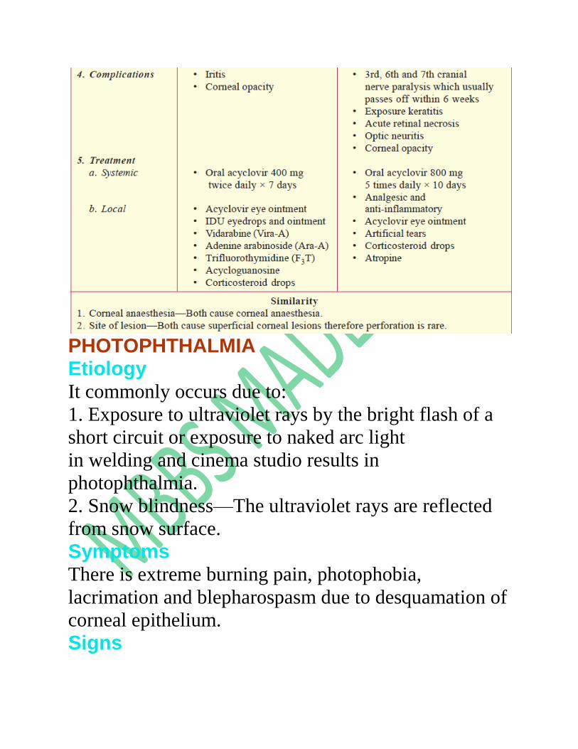
PHOTOPHTHALMIA Etiology It commonly occurs due to:
1. Exposure to ultraviolet rays by the bright flash of a
short circuit or exposure to naked arc light
in welding and cinema studio results in
photophthalmia.
2. Snow blindness—The ultraviolet rays are reflected
from snow surface.
Symptoms There is extreme burning pain, photophobia,
lacrimation and blepharospasm due to desquamation of
corneal epithelium.
Signs

There are multiple epithelial erosions associated with
blepharospasm and swelling of the palpebral
conjunctiva and retrotarsal folds.
Treatment • Cold compresses, astringent lotions and atropine
ointment are effective.
• Bandage both eyes for 24 hours. This helps in
regeneration of the epithelium.
Prophylaxis Wearing of dark glasses (Crooke’s glasses) made of
such materials which cut off practically all the
infrared and ultraviolet rays when such exposure is
anticipated.
DEEP KERATITIS The deep forms of keratitis affect the stroma of the
cornea.
Etiology 1. Congenital syphilis—It is characterised by bilateral
interstitial keratitis, vascularization (Salmon
patches) and uveitis. It affects children between the age
of 5-15 years.
2. Tuberculosis—There is presence of interstitial
keratitis in this condition.
3. Viral infections (disciform keratitis)—A central grey
disc is seen in the stroma. It is unilateral

and seen in adults usually.
4. Sclerosing keratitis—It spreads from scleritis
involving the corneal stroma.
Treatment The basic cause of deep keratitis is treated along with
routine treatment of corneal ulcer.
Megalocornea It is a bilateral condition in which the corneal diameter
is more than 14 mm. The cornea is usually
clear with normal thickness and vision. It is often
associated with Marfan’s syndrome.
Differential diagnosis Megalocornea can be
differentiated from buphthalmos and keratoglobus
i. Buphthalmos—In this condition 10P is raised and
eyeball is enlarged as a whole. Enlarged
cornea is associated with Descemet’s membrane tears.
ii. Keratoglobus—There is congenital bilateral
hemispherical protrusion of the whole cornea.
Keratoglobus In this condition there is thinning and excessive
protrusion of cornea which seems enlarged but its
diameter is usually normal.
Microcornea The corneal diameter is less than 10 mm with
decreased radius of curvature. Hypermetropia and

narrow angle glaucoma may be found in later years.
The condition may occur as an isolated anomaly
or in association with microphthalmos.
Cornea Plana It is a rare anomaly in which cornea is comparatively
flat since birth. It may be associated with
microcornea.
Posterior Embryotoxon There is an unusual prominence of Schwalbe’s line
which is peripheral termination of Descemet’s
membrane. It appears as a ring opacity in deeper layer
of cornea.
Corneal Degenerations These are non hereditary and usually unilateral. They
can be divided into three categories:
i. Primary degeneration
ii. Secondary degeneration
iii. Infiltration associated with metabolic disturbance,
e.g. fatty degeneration, hyaline degeneration,

amyloid degeneration, calcific degeneration (Band
shaped keratopathy) etc.
The basic difference between degenerations and
dystrophies are as under:
Arcus Senilis There is bilateral annular lipoid infiltration of cornea in
old persons with no symptoms. It does not
require any treatment as it does not affect the vision or
vitality of the cornea.
There are concentric grey lines in the upper and lower
part of the cornea.
• The lines join to form a ring 1 mm broad which is
separated from the margin by a rim of clear
cornea about 1.5 mm. It is also known as lucid interval
of Vogt. The outer border of the arcus is
sharp but the inner border appears faint. It is found in
approximately 60% of population between the age of
40 to 60
years and almost in all persons above the age of 80
years. The
arcus is formed by deposition of cholesterol,
cholesterol esters,
phospholipids and triglycerides in the substantial
propria layer.
Arcus Juvenilis

It usually occurs below 40 years of age. It is a rare
condition. A serum lipid profile is indicated to rule
out hereditary anomaly which has a serious prognosis.
White Limbal Girdle of Vogt It is seen mostly in the age group of 40-60 years. It is
seen as chalky line in the nasal and temporal
periphery of inter-palpebral area of cornea. The opacity
is at the level of Bowman’s membrane. It is
due to elastotic degeneration of tissues.
Amyloid Degeneration Amyloid degeneration of cornea is characterized by
deposition of amyloid material underneath its
epithelium. It could be either primary or secondary due
to some disease.
Pigmentary Degeneration Pigment deposition in cornea could be iron, blood
pigment, melanin and other metallic pigments like
cooper, silver, gold etc.
a. Hudson-Stahli-line—It is a horizontal line at the
lower half of the cornea due to deposition of
hemosiderin pigment
b. Fleischer’s ring is seen at the base of keratoconus
c. Stocker-Busaca line—It is seen in front of a
Pterygium.
Band-shaped Keratopathy

• It is common in old, blind, shrunken eyes and in
Still’s
disease of children.
• It is associated with hyperthyroidism, vitamin D
poisoning
or sarcoidosis.
• It could be either primary or secondary to
hypercalcaemia, chronic uveitis, chronic glaucoma,
interstitial keratitis etc.
• A continuous band lies in the interpalpebral area
starting in the inner and outer side.
• It is due to hyaline infiltration in the superficial
stroma followed by calcareous salt deposition

ANTERIOR DYSTROPHIES 1. Reis-Buckler’s Dystrophy It is bilaterally symmetrical dystrophy, which starts in
early childhood as recurrent corneal erosions.
Later there is diffuse scarring of Bowman’s membrane.
Corneal surface becomes rough with
diminished sensation. The opacities have typical ring
like appearance. It is an autosomal dominant
condition. It starts near the Bowman’s membrane.
There are subepithelial grey opacities arranged in
a fish net pattern.
2. Cogan’s Microcystic Dystrophy

It is the commonest epithelial dystrophy with dot, map
or fingerprint opacities characterized by
recurrent attacks of severe pain, watering, photophobia
and blepharospasm. There is increased
hydration of cornea and formation of microcysts under
the epithelium.
3. Messman’s Juvenile Epithelial Dystrophy (Recurrent Corneal Erosion Syndrome) Characterized by appearance of small vesicles between
epithelium and Bowman’s membrane. It is
an autosomal dominant inherited bilateral symmetrical
condition. There are minimum symptoms and
visual loss is very less, hence it does not require any
treatment.
STROMAL DYSTROPHIES 1. Granular Dystrophy • It is autosomal dominant dystrophy.
• There is milky granular hyaline deposits in anterior
stroma.
• There is clear cornea between opacities.
• It develops in first decade of life and vision remains
good until 40 years of life.
2. Macular Dystrophy • It is autosomal recessive dystrophy.
• There is dense opacity in central cornea.
• There is deposition of mycopolysaccharides.

• It starts in first decade and vision lost early in life.
• It requires penetrating keratoplasty.
3. Lattice Dystrophy • There is autosomal dominant inheritance.
• There are amyloid deposits in corneal stroma.
• Spider like opacities are seen in cornea.
• It starts early in life.
• Cornea becomes hazy by the age of 20 years.
• It requires penetrating keratoplasty.
ENDOTHELIAL DYSTROPHIES 1. Fuch’s Endothelial Dystrophy • It was described by Fuch’s in 1910.
• It usually occurs after 50 years of age.
• The female to male ratio is 4:1.
• There is atrophy of the endothelial cells along with
oedema and formation of vesicles. Grey
punctate opacities are seen in the stroma.
• The clinical features are divided into four stages
a. Stage of cornea guttata
b. b. Oedematous stage
c. c. Stage of bullous keratopathy
d. d. Stage of scarring
e. • Treatment
f. a. 5% sodium chloride ointment or solution
(hypertonic saline) is useful.
g. b. Hydrated soft contact lenses may be useful.

h. c. Penetrating keratoplasty can be done.
i. 2. Cornea Guttata j. • It manifests are middle age.
k. • Females are most affected.
l. • It has autosomal dominant inheritance.
m. • There are bilateral symmetric lesions, which
appear as golden hue on the posterior surface of
n. cornea.
• It rarely affects vision.
TARSORRHAPHY The aim is to achieve lid closure so that palpebral
aperture is narrowed. It is not strictly a corneal
operation, but it is performed for corneal conditions.
Indications • Neuroparalytic keratitis as a result of 5th nerve
paralysis.

• Exposure keratitis due to inadequate closure of
palpebral aperture as a result of 7th nerve paralysis
or proptosis.
Method Palpebral aperture is narrowed by placement of
mattress sutures through the small raw areas in
the lid margins and skin. The sutures are tied over
rubber sheet in the skin.
Types 1. Lateral tarsorrhaphy—The suture is placed at the
junction of middle and lateral third of lid
margin.
2. Paramedian tarsorrhaphy—Two sutures are placed
on either side of the middle line as shown
in the diagram.
EYE BANK The primary function of an eye bank is to collect, store
good quality donor’s cornea and make it
available for cornea transplantation for therapeutic use
and research.
Objective of Eye Bank The main objectives of an eye bank can be summarized
as follows,
1. Collection of donor eyes.
2. Preservation of donor cornea.

3. Distribution of highest quality of donor tissue for
cornea transplantation.
4. Promotion, awareness about eye donation from
potential donors.
A person makes a pledge to donate his eyes after death.
No living individual can donate his eye
because the law does not permit it and moreover it is
not practical. The eye cannot be sold or
purchased. Eye bank personnel collect the eyes after
getting information about death and properwritten
consent from close relative. It is important to know the
age of the donor, cause of death and
time of death. Eyes should be removed as early as
possible or atleast within 5-6 hours after death.
Equipments for an Eye Bank The equipments required for any eye bank are listed
below,
Enucleation Eyes should be enucleated soon after death. A
relatively longer interval of 4-6 hours may be allowed

in winter months but in summer, not more than 2-3
hours should elapse between death and enucleation.
The eyes should carry the following information about
the donor:
1. Age and sex.
2. Cause of death.
3. Time and date of death.
4. Time and date of enucleation.
Enucleation should be done aseptically and the
eyeballs should be transported to the eye bank in
a wide mouth sterile glass bottle in an ice box or
thermos flask. The eyeballs are washed with normal
saline, antibiotic drops instilled and the cornea is
examined with good illumination and magnification,
preferably with slit-lamp. Clinical viability is graded
depending upon the degree of stromal oedema
and folds. Usable eyeballs are then transferred to
autoclaved wide mouth bottles containing sterile
cotton gauze pad. Adequate antibiotic solution is
instilled to moisten the pad. The eyeball rests on the
pad with cornea straight up and without touching any
part of the bottle. It is better to have a mouldable
clamp in the bottle to hold the eyeball erect to protect
the cornea. This is particularly important when
these eyes are to be transported to distant corneal
surgeons. The bottles should have tight screw

caps so that even if ice has melted off in the container,
fluid does not enter the bottle. The Eye Bank
Association has designed a thermocol container which
has provision for carrying one or two pairs of
eyeballs with adequate amount of ice for 18 to 24 hours
transport. If Descemet’s membrane folds
and stromal oedema exceed acceptable limits, the
cornea is designated unusable for therapeutic
purpose but can be used for surgical training and
experimental purposes.
One may conclude that when media preservation
facilities are not available, eyeballs enucleated
from a relatively younger person who dies after an
acute episode such as accident, suicide, homicide,
etc. and preserved in moist chamber at 4o C provide
the best donor cornea.
Contraindications for Collection of Donor Eyes There are certain conditions when the donor’s eye are
not suitable for corneal transplant.
1. Systemic causes—These include death due to:
• AIDS (HIV positive)
• Hepatitis B
• Rabies
• Poison
• Severe burn
• Malignancy, leukaemia, lymphoma

• Death from unknown cause.
2. Ocular causes
• Corneal opacities and dystrophy
• Retinoblastoma, malignant melanoma
• Active inflammatory diseases, e.g. conjunctivitis,
iridocyclitis, endophthalmitis
• Congenital abnormalities, e.g. keratoconus,
keratoglobus
• Prior refractive procedures, e.g. radial keratotomy,
laser photoablation
• Anterior segment surgical procedures, e.g. cataract,
glaucoma.
Evaluation of Donor Tissue 1. Gross examination with torch and loupe.
2. Slit-lamp examination.
3. Specular microscopy (for endothelial cell count and
morphology).
A careful slit-lamp examination provides an overall
status of the endothelium. The normal
endothelium shows a pattern of cells of similar size and
shape with no abnormal structures. The
normal cell density is usually between 2000 to 3500
cells per sq. mm.

Preservation of the Donor Eye Traditionally corneal preservation is described under
short-term, intermediate-term and long-term
preservations.
1. Short-term Preservation (up to 96 hours) i. Moist chamber method—Whole globe is preserved in
a moist chamber at 4o C in a refrigerator
for 24 hours.
ii. M.K. (McCarey-Kaufman) medium—It consists of
tissue culture (TC)-199, 5% Dextran-
40, HEPES buffer to adjust pH at 7.4, Gentamicin 0.1
mg/ml, colour-pink.
Corneoscleral button can also be preserved in M-K
medium at 4o C for upto 96 hours.
It is superior than conventional moist-chamber method
and practised widely.
2. Intermediate-term Preservation (upto 2 weeks) K-SOL medium, dexol medium, optisol medium, etc.
are used for preservation.
3. Long-term Preservation (months to years) i. Viable—Organ culture method, cryopreservation.

ii. Non-viable—Glycerine preservation.