tooth morphology basics
TRANSCRIPT

Tooth Morphology Basics
Howard Chi, D.M.D.

Learning Objectives
• Primary and permanent dentition• Tooth identification system• Trait categories• Tissues of the tooth• Dental terminology• Anatomy of tooth structure

Primary and Permanent Dentition

Dentition
Refers to all of the teeth in the maxillae and the mandible
* maxillary arch - maxillary teeth* mandibular arch - mandibular teeth
Maxillary arch
Mandibular arch

Humans Have Two Dentitions Throughout Life
1) Primary Dentition - during childhood2) Permanent Dentition - during adulthood
PrimaryPermanent

Primary Dentition
• There are 20 teeth in the primary dentition• 10 maxillary - 10 mandibular• This dentition is also termed the deciduous
dentition
Permanent 1st Molars

Primary Dentition
The dental formula for one side of the mouth is:I 2 C 1 M 2 = 10 teeth on either side 2 1 2
11 212
11 212

Deciduous Teeth
Those teeth that “fall out” due to a natural process of tooth succession

Permanent Dentition
Also called the succedaneous dentition - that which succeeds the primary dentition
There are 8 teeth in each quadrant
I 2 C 1 PM 2 M 3 = 16 teeth 2 1 2 3 each side

Permanent Dentition
First signs of this dentition appear at age 6

Permanent Dentition
A A A P P P P P
Anterior Teeth - incisors and caninesPosterior teeth - premolars and molars

Tooth Identification Systems

Tooth Identification Systems
There are three main systems used in modern dentistry for the numbering of teeth. They are:
* Universal Numbering System* Palmer Notation System* International Numbering System

Universal Numbering System
1. Suggested by Parreidt in 18822. Adopted by the A.D.A. in 19753. Uses numbers 1 through 32

Universal Numbering System
1 for upper right third molar around to 16upper left third molar
1 16

Dropping down same side to 17 lower leftthird molar and around to 32
Universal Numbering System
1732

For deciduous dentition, letters A through T are used
Universal Numbering System
A B C D E F G H I J
T S R Q P O N M L K

Universal Numbering
System

Palmer Notation System
Utilizes brackets to represent the four quadrants
Upper Right Upper Left
Lower Right Lower Left

• Permanent teeth are labeled 1 to 8 on each side of the midline
• On deciduous teeth same brackets with letters A through E
Palmer Notation System

Palmer System

International Numbering System
• Uses two digits for each tooth• First digit represents dentition, arch and
side• Second number denotes the tooth (1-4
perm. and 5-8 prim.)

International Numbering System
1 = permanent dentition, maxillary, right2 = permanent dentition, maxillary, left3 = permanent dentition, mandibular, left4 = permanent dentition, mandibular, right5 = primary dentition, maxillary, right6 = primary dentition, maxillary, left7 = primary dentition, mandibular, left8 = primary dentition, mandibular, right

International Numbering System

Trait Categories

Trait Categories
Set Traits: (dentition traits) distinguish teeth in the primary from secondary dentition

Trait Categories
Arch Traits: distinguish maxillary
from mandibular

Trait Categories
Class Traits: Distinguish the four categories of teeth ... incisors, canines, premolars, molars

Trait Categories
Type Traits: Distinguish teeth within one class

Tissues of the Tooth

Tissues Of A Tooth
1) Dentin2) Enamel3) Cementum4) Pulp

Enamel
• Makes up the protective outersurface of the anatomic crown
• Mostly inorganic and calcifiedhard, white shiny surface of theanatomic crown

Dentin
• Found in the crown and root, making up bulk of tooth
• Found beneath the enamel and cementum and surrounding the pulp tissue
• Not normally visible• Mostly inorganic and calcified

Cementum
• Makes up the surface of the anatomic root• Very thin next to cervical line• Mostly inorganic calcified• Dull yellow in color

Pulp• Is non calcified found within the pulp chamber• Develops from the dental papilla
(from mesoderm)• Surrounded by dentin except at the
apical foramen

Pulp• Normally not visible except on
dental radiographs• In the coronal portion termed
the pulp chamber• In the root portion termed the
pulp canal(s)

Pulp - Functions
• Formative - dentin producing cells (odontoblasts) produce dentin thoughout the life of a tooth
• Sensory - nerve endings permit the sense of pain

Pulp - Functions
• Nutritive - nutrient transport from the blood stream to extensions of the pulp that reach into dentin
• Defensive/Protective - responds to injury and decay by forming reparative dentin

Junctions Of Tooth Structure
Cementoenamel Junction - also called the cervical line, separates the anatomic crown from the anatomic root.

Junctions Of Tooth Structure
Dentinoenamel Junction - is the inner surface of the enamel cap visible in cross section or in badly worn teeth

Junctions Of Tooth Structure
Cementodentinal Junction - a.k.a. dentinocemental junction is the inner surface of cementum lining the root visible in cross section or badly worn teeth

Anatomic Versus Clinical Crown
Anatomic Crown - that part of the tooth covered in enamel
Clinical Crown - that part of the tooth that is visible in the oral cavity

Dental Terminology

Terminology Used To Distinguish Tooth Surfaces
Facial Surface - the surface next to the face, the outer surface of a tooth resting next to the cheeks or gums. Used in both anterior and posterior teeth

Terminology Used To Distinguish Tooth Surfaces
Buccal Surface - the facial surface of posterior teeth. Meaning next to the cheek

Terminology Used To Distinguish Tooth Surfaces
Labial Surface - the facial surface next to the lips, generally used for anterior teeth

Terminology Used To Distinguish Tooth Surfaces
Proximal Surface - the surface or side of a tooth that is next to an adjacent tooth, not considered self-cleansing

Mesial Surface - is the surface of the tooth nearest to the midline of the dental arch
Terminology Used To Distinguish Tooth Surfaces

Distal Surface - is the surface of the tooth farthest from the midline of the dental arch
Terminology Used To Distinguish Tooth Surfaces

Terminology Used To Distinguish Tooth Surfaces
Lingual Surface - is the surface of maxillary and mandibular teeth nearest the tongue

Terminology Used To Distinguish Tooth Surfaces
Palatal Surface – is the surface of
maxillary teeth nearest the palate

Terminology Used To Distinguish Tooth Surfaces
Occlusal Surface - is the chewing surface of the posterior teeth found within cusp and marginal ridges

Terminology Used To Distinguish Tooth Surfaces
Incisal Edge - is the cutting edge, ridge or surface of anterior teeth

Anatomy of Tooth Structure

Divisions Of The Crown And Root Of A Tooth
Divisions Cervico-occlusally
Cervical 3rdMiddle 3rdIncisal 3rd
Cervical 3rdMiddle 3rdOcclusal 3rd
Anterior Posterior
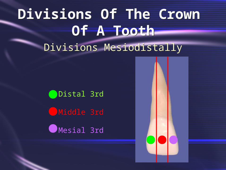
Divisions Of The Crown Of A Tooth
Divisions Mesiodistally
Distal 3rd
Middle 3rd
Mesial 3rd

Divsions Of The Crown Of A Tooth
Divisions Faciolingually
Lingual 3rd
Middle 3rd
Facial 3rd

Divisions Of The Crown And Root Of A Tooth
Divisions Cervico-occlusally
Apical 3rdMiddle 3rdCervical 3rdCervical 3rdMiddle 3rdIncisal 3rd
Apical 3rdMiddle 3rdCervical 3rdCervical 3rdMiddle 3rdOcclusal 3rd
Anterior Posterior

Morphology Of An Anatomic Crown
Cusp - is a point, or peak on the chewing surface of premolar or molar tooth

Morphology Of An Anatomic Crown
Cusp Slopes Or Ridges - are the inclined surfaces that form an angle at the cusp tip

How Many Cusp Ridges Does This Tooth Possess?

Morphology Of An Anatomic Crown
Cingulum - is the enlargement or bulge on the cervical third of the lingual surface of the crown of anterior teeth

Morphology Of An Anatomic Crown
Labial Ridge - is a ridge running cervico-incisally in approximately the center of the labial surface of the canines

Morphology Of An Anatomic Crown
Buccal Ridge - is the ridge running cervico-occlusally in approximately the center of the buccal surface of premolars

Morphology Of An Anatomic Crown
Cervical Ridge - ridge running mesiodistally on the cervical one-third of the buccal surface of the crown, found on all deciduous teeth but only on the permanent molars

Morphology Of An Anatomic Crown
Marginal Ridge - on incisor and canine located on the mesial and distal border of the lingual surface

Morphology Of An Anatomic Crown
Marginal Ridge - on posterior teeth located on the mesial and distal border of the occlusal surface

Morphology Of An Anatomic Crown
Triangular Ridge - on the occlusal surface of posterior teeth, is the ridge from any cusp tip to center of the occlusal surface - ML cusp of upper molars have two

Morphology Of An Anatomic Crown
Oblique Ridge - found only on maxillary molars made of the triangular ridges of the mesiolingual and distobuccal cusps

Transverse Ridge - ridge crossing the occlusal surface of posterior teeth in a B-L direction and made of connecting triangular ridges
Morphology Of An Anatomic Crown

Morphology Of An Anatomic Crown
Mamelon - is one of three tubercules sometimes present on the incisal edge of an incisor tooth that has not been subject to wear

Morphology Of An Anatomic Crown
Sulcus - is a broad depression or valley on the occlusal surface of posterior teeth

Morphology Of An Anatomic Crown
Developmental Groove - is a sharply defined, narrow and linear depression, formed during tooth development separating lobes or a major portion of a tooth - a fissure may be found at the depth of a developmental groove

Morphology Of An Anatomic Crown
Supplemental Groove - small irregularly placed grooves not at the junction of lobes or major portions of the teeth

Morphology Of An Anatomic Crown
Fossa - a depression or hollow found on the lingual surfaces of some anterior teeth and on the occlusal surfaces of posterior teeth

Morphology Of An Anatomic Crown
Pits - often occur at the depths of fossa where two or more grooves join

Morphology Of An Anatomic Crown
Furcation - is the place on multirooted teeth where the root trunk or base divides into separate roots
RootTrunk

Curve Of Spee
Anteroposterior curve of the occlusal plane - curve of the maxillary arch is convex

Curve Of Wilson
Gradual curve of posterior teeth from left to right side viewed from the anterior region - curve of maxillary teeth is convex