tools of the biologist - weeblymarandoscience.weebly.com/uploads/2/3/7/6/23768555/notes...+ viewing...
TRANSCRIPT

+
Tools of the Biologist

+

+Mirror/Light Source
Diaphragm Stage
Ocular/eyepiece Body Tube
Coarse adjustment High power
objective
Fine adjustment Low power
objective
Arm Base
Nosepiece Stage clips

+
The Invention of the
Microscope - YouTube
Eyepiece/Ocular – 10x
magnification usually
Coarse Adjustment – major
changes in focus/ DON’T
use on HIGH power
Fine Adjustment – minor
changes in focus
Arm – for safe
carrying
Stage – place slide
here
Base – for support
Body tube
Nosepiece – rotates
the objective lenses
High Power
Objective (40x mag)
Low Power
Objective (10x mag)
Stage Clips – hold
specimen in place
Diaphragm –
controls the amount
of light on specimen
Light Source
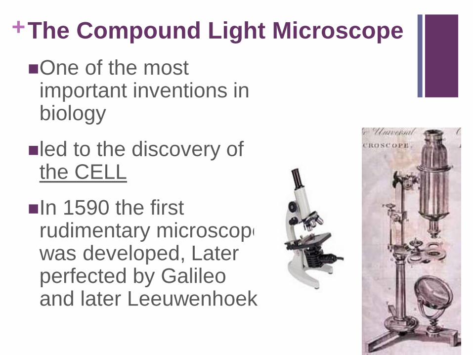
+The Compound Light Microscope
One of the most important inventions in biology
led to the discovery of the CELL
In 1590 the first rudimentary microscope was developed, Later perfected by Galileo and later Leeuwenhoek

+The Compound Light Microscope
Calculating Total Magnification
Multiply
Ocular magnification x objective lens magnification
Ex. If viewing a slide under low power what is the total magnification of the specimen? (assume ocular 10x)
Answer: ocular 10x X low power 10x Total Magnification = 100x

+The Compound Light Microscope
Calculating Total Magnification
Complete the following:
Objective Total Magnification
Scanning objective (4x) ________x
Low Power objective (10x) ________x
High Power objective (40x) ________x
40x
100x
400x

+The Electron MicroscopeUses beams of electrons to magnify the
image.
Living specimens cannot be observed.

+

+The Dissecting/Binocular Microscope
Produces a 3-D image
low magnification
Useful for observing small organisms and
body structures
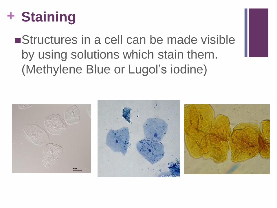
+ Staining
Structures in a cell can be made visible
by using solutions which stain them.
(Methylene Blue or Lugol’s iodine)
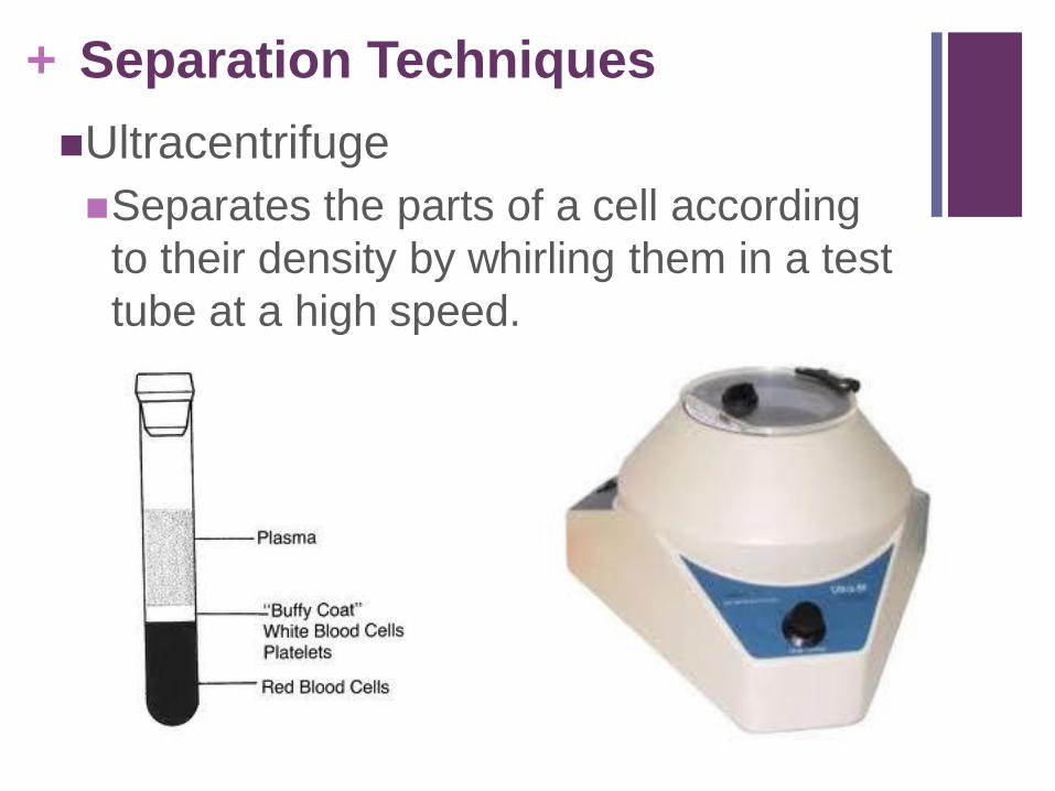
+ Separation Techniques
Ultracentrifuge
Separates the parts of a cell according
to their density by whirling them in a test
tube at a high speed.
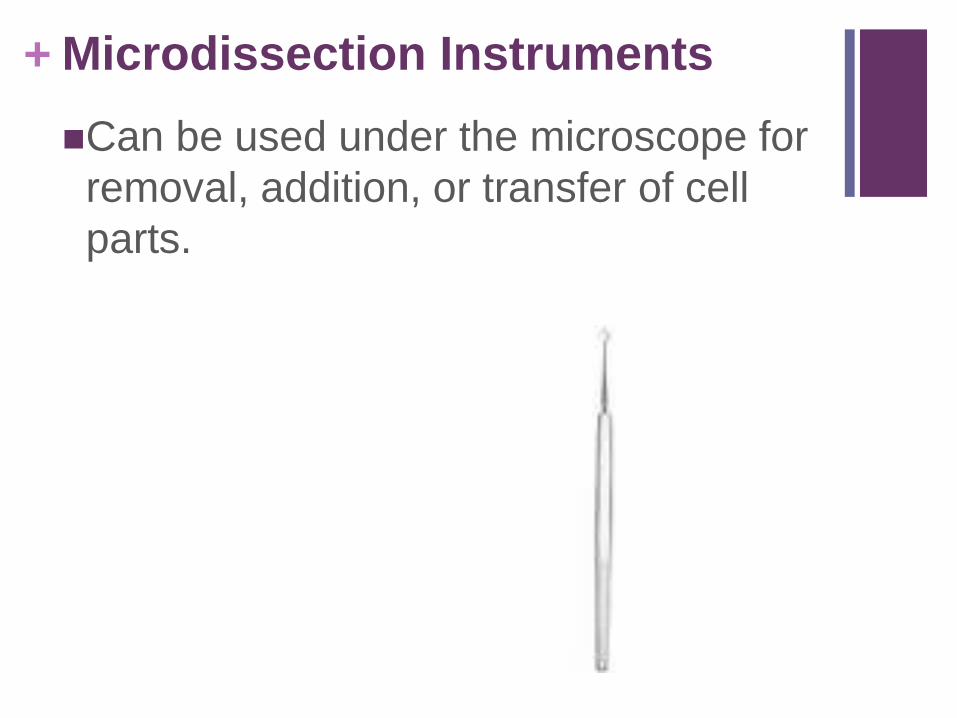
+ Microdissection Instruments
Can be used under the microscope for
removal, addition, or transfer of cell
parts.

+Of the following, which instrument is
most commonly used to observe the
external features of a grasshopper's
abdomen?
1) ultracentrifuge
2) microdissection instrument
3) dissecting microscope
4) electron microscope
+A student observed a Paramecium under the
low power objective of a microscope (100x) and then under high power (400x). The image of the Paramecium under low power, compared to the image of the same Paramecium under high power, would be
1) smaller and in a darker field of view
2) smaller and in a brighter field of view
3) larger and in a darker field of view
4) larger and in a brighter field of view
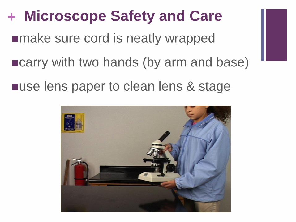
+ Microscope Safety and Care
make sure cord is neatly wrapped
carry with two hands (by arm and base)
use lens paper to clean lens & stage

+ Slide Preparation
Specimen must be THIN for light to
pass through
Stain may be needed to make
specimen more visible
Coverslip should be placed at a 45
degree angle to avoid air bubbles
Making a Wet Mount
for Microscopy - 1:20

+ Viewing a Slide
1. Always begin on the lowest available power (scanning lens)
Has widest and brightest field of view to locate specimen
2. Center specimen in the field of view (up is down, right is left)
3. Focus with coarse adjustment knob
4. Switch to low power and repeat #2 and #3
5. If switching to high power, only use the FINEadjustment to focus!
to prevent losing sight of your specimen & damaging the slide

+ Measurement Conversion
K H D U D C MLarger Smaller

+