title horie, takahiro; nishino, tomohiro; baba,...
TRANSCRIPT

TitleMicroRNA-33b knock-in mice for an intron of sterol regulatoryelement-binding factor 1 (Srebf1) exhibit reduced HDL-C invivo.
Author(s)
Horie, Takahiro; Nishino, Tomohiro; Baba, Osamu; Kuwabara,Yasuhide; Nakao, Tetsushi; Nishiga, Masataka; Usami,Shunsuke; Izuhara, Masayasu; Nakazeki, Fumiko; Ide, Yuya;Koyama, Satoshi; Sowa, Naoya; Yahagi, Naoya; Shimano,Hitoshi; Nakamura, Tomoyuki; Hasegawa, Koji; Kume,Noriaki; Yokode, Masayuki; Kita, Toru; Kimura, Takeshi;Ono, Koh
Citation Scientific reports (2014), 4
Issue Date 2014-06-16
URL http://hdl.handle.net/2433/188354
Right
This work is licensed under a Creative Commons Attribution4.0 International License. The images or other third partymaterial in this article are included in the article's CreativeCommons license, unless indicated otherwise in the credit line;if the material is not included under the Creative Commonslicense, users will need to obtain permission from the licenseholder in order to reproduce the material. To view a copy ofthis license, visit http://creativecommons.org/licenses/by/4.0/
Type Journal Article
Textversion publisher
Kyoto University

MicroRNA-33b knock-in mice for anintron of sterol regulatoryelement-binding factor 1 (Srebf1) exhibitreduced HDL-C in vivoTakahiro Horie1,2*, Tomohiro Nishino1*, Osamu Baba1, Yasuhide Kuwabara1, Tetsushi Nakao1,Masataka Nishiga1, Shunsuke Usami1, Masayasu Izuhara1, Fumiko Nakazeki1, Yuya Ide1,Satoshi Koyama1, Naoya Sowa1, Naoya Yahagi3, Hitoshi Shimano3, Tomoyuki Nakamura4,Koji Hasegawa5, Noriaki Kume6, Masayuki Yokode2, Toru Kita7, Takeshi Kimura1 & Koh Ono1
1Department of Cardiovascular Medicine, Graduate School of Medicine, Kyoto University, Kyoto 606-8507, Japan, 2Department ofClinical Innovative Medicine, Institute for Advancement of Clinical and Translational Science, Graduate School of Medicine, KyotoUniversity, Kyoto 606-8507, Japan, 3Department of Internal Medicine (Endocrinology and Metabolism), Graduate School ofComprehensive Human Sciences, Nutrigenomics Research Group, Faculty of Medicine, and International Institute for IntegrativeSleep Medicine (IIIS), World Premir International Research Center Initiative (WPI), University of Tsukuba, 1-1-1 Tennodai, Tsukuba,Ibaraki 305-8575, Japan, 4Department of Pharmacology, Kansai Medical University, Moriguchi, Osaka 570-8506, Japan,5Division of Translational Research, National Hospital Organization, Kyoto Medical Center, Kyoto 612-8555, Japan, 6Division ofClinical Pharmacy, Faculty of Pharmaceutical Sciences, Kobe Gakuin University, Kobe 650-8586, Japan, 7Department ofCardiovascular Medicine, Kobe City Medical Center General Hospital, Kobe 650-0046, Japan.
MicroRNAs (miRs) are small non-protein-coding RNAs that bind to specific mRNAs and inhibittranslation or promote mRNA degradation. Recent reports, including ours, indicated that miR-33a locatedwithin the intron of sterol regulatory element-binding protein (SREBP) 2 controls cholesterol homeostasisand can be a possible therapeutic target for treating atherosclerosis. Primates, but not rodents, expressmiR-33b from an intron of SREBF1. Therefore, humanized mice, in which a miR-33b transgene is insertedwithin a Srebf1 intron, are required to address its function in vivo. We successfully established miR-33bknock-in (KI) mice and found that protein levels of known miR-33a target genes, such as ABCA1, ABCG1,and SREBP-1, were reduced compared with those in wild-type mice. As a consequence, macrophages fromthe miR-33b KI mice had a reduced cholesterol efflux capacity via apoA-I and HDL-C. Moreover, HDL-Clevels were reduced by almost 35% even in miR-33b KI hetero mice compared with the control mice. Theseresults indicate that miR-33b may account for lower HDL-C levels in humans than those in mice and thatmiR-33b is possibly utilized for a feedback mechanism to regulate its host gene SREBF1. Our mice will alsoaid in elucidating the roles of miR-33a/b in different genetic disease models.
Sterol regulatory element-binding proteins (SREBPs) comprise a subclass of basic helix-loop-helix leucinezipper transcription factors conserved from yeasts to humans and regulate the expression of genes requiredfor maintaining cellular lipid homeostasis1. Mammals possess two SREBP genes, SREBP-1 and SREBP-2
(known as SREBF1 and SREBF2, respectively) that express three major SREBP proteins. Two SREBP-1 isoforms,SREBP-1a and SREBP-1c, primarily regulate fatty acid metabolism, and SREBP-2 is the main regulator ofcholesterol metabolism, although there is some functional overlap among the three SREBP isoforms2–4.
MicroRNAs (miRs) are small non-protein-coding RNAs that bind to specific mRNAs and inhibit translationor promote mRNA degradation5. Recent advances in the understanding of miR biology revealed that the geneticloci encoding for the transcription factors SREBP-1 and SREBP-2 also encode for the miRs miR-33b and miR-33a, respectively. Recent reports, including ours, indicated that miR-33a controls ABCA1 expression and reducesHDL-C levels6–8 and that miR-33a deficiency ameliorates atherosclerosis in mice9–11. However, in rodents, a partof miR-33b is lacking from a Srebf1 intron (Supplementary Fig. 1a), and it is impossible to determine the precisecoordinate mechanisms of miR-33a and miR-33b; the expression of these miR-33s is expected to depend on theircorresponding host genes. Of note, SREBP-1 and SREBP-2 are differentially regulated by hormones, dietary
OPEN
SUBJECT AREAS:EXPERIMENTAL MODELS
OF DISEASE
MIRNAS
Received20 March 2014
Accepted30 May 2014
Published16 June 2014
Correspondence andrequests for materials
should be addressed toK.O. (kohono@kuhp.
kyoto-u.ac.jp)
* These authorscontributed equally to
this work.
SCIENTIFIC REPORTS | 4 : 5312 | DOI: 10.1038/srep05312 1

challenges, or statin treatment, and the amounts and functions ofmiR-33a and miR-33b would be greatly affected under theseconditions.
miR-33a and miR-33b are identical in their seed sequences, andthus have been predicted to repress the same set of genes with similarspecificities. Antisense oligonucleotides against miR-33a are believedto simultaneously target miR-33a and miR-33b. However, thereremains a 2-nucleotide mismatch after the seed sequence betweenmiR-33a and miR-33b (Supplementary Fig. 1a), and whether thisdifference results in differential targeting remains to be established.Moreover, some of the previously established miR-33a target geneswere not dysregulated in our miR-33a-deficient mice. Therefore,humanized mice, in which a miR-33b transgene is inserted withina Srebf1 intron, are required to address its function in vivo.
We successfully established miR-33b knock-in (KI) mice for thesame intron as in humans. The protein levels of known miR-33atarget genes, such as ABCA1, ABCG1, and SREBP-1, were reducedunder basal conditions. An LXR agonist, which induces Srebf1expression, enhanced miR-33b production. In vitro experimentsindicated that macrophages from the miR-33b KI mice had a reducedcholesterol efflux capacity via apoA-I and HDL-C. Moreover, HDL-C levels were reduced by almost 35% even in miR-33b KI hetero micecompared with the control mice.
The feasibility of genetic manipulation is one of the many advan-tages of using mice as a model organism. However, the lack of miR-33b in mouse Srebf1 has raised an important concern regarding thedirect translation of data from rodent models to human physiologyand metabolic disorders. Our mice will aid in answering these ques-tions and will be useful for assessing the risks and benefits of long-term alterations in miR-33s in different disease models. These micemight also be useful for screening of the drugs that alter the levels ofmiR-33a and miR-33b.
ResultsmiR-33b is co-expressed with SREBF1 in the human cell line HepG2.It is assumed that a miR located within an intron of a gene is ex-pressed along with its host gene and exerts its specific function12.Because miR-33b is located in a SREBF1 intron in humans (Supple-mentary Fig. S1a), we stimulated human cell line HepG2 with theLXR agonist T0901317 and determined miR-33b and miR-33aexpression along with the expression of the host genes SREBF1and SREBF2. As shown in Fig. 1a and b, miR-33b expressionseemed to tag along behind SREBF1 expression. In contrast, miR-33a and SREBF2 expression was not affected by LXR stimulation(Fig. 1c and d).
Generation of miR-33b KI mice. Because miR-33b is located inSREBF1 intron 16 in humans and there are high homologies inexons 16 and 17 between human and mouse (82.6% of nucleotides
and 79.7% of amino acids, Supplementary Fig. S1b), we introducedthe human miR-33b sequence into intron 16 of mouse Srebf1. Weisolated and amplified the region that encoded for the complete pre-miR sequence of human miR-33b and adjacent sequence, whichenabled the introduction of miR-33b into intron 16 of mouseSrebf1 (Fig. 2a). Supplementary Figure S2a and Figure 2b show theresults of Southern blotting analysis of genomic DNA from ES cellsand tail genomic DNA from F1 mice that were successfully targetedby a KI vector, respectively. PCR analysis indicated the specificpatterns for wild-type (WT), KI1/2, and KI1/1 mice (Fig. 2c). ThismiR-33b KI strategy did not alter Srebf1 intron 16 splicing, asconfirmed by PCR (Fig. 2d) and sequencing (Fig. 2e). Theexpression levels of miR-33b in miR-33b KI1/2 mice were almosthalf of those in miR-33b KI1/1 mice (Fig. 2f). We also measured thelevels of miR-33b, miR-33a, Srebf1, and Srebf2 in WT and KI mice inboth the liver and the peritoneal macrophages (SupplementaryFigure S2b–d and S3a–d). Srebf1 levels were similar among thesemice (Supplementary Figure S2c and S3c). Although there was nodifference in miR-33a levels in macrophages (Supplementary FigureS3b), miR-33a levels were increased in proportion of the expressionlevels of miR-33b in the liver (Supplementary Figure S2b). The miR-33b KI1/1 mice were born with the expected Mendelian ratios, wereviable, fertile, and did not exhibit any obvious abnormalities in size,shape, or structure up to 8 weeks of age. Relative tissue expressionpattern of miR-33b was similar to that of Srebf1 (Supplementary Fig.S2e and S2f).
miR-33b is upregulated after inducing Srebf1 expression. We nextsought to confirm whether miR-33b expression was affected byendogenous changes in Srebf1 expression by the LXR agonistT090131713. When primary hepatocytes from the miR-33b KI1/1
mice were stimulated with T0901317, Srebf1 and miR-33b mRNAlevels were significantly increased in parallel, although this increasewas faster for Serbf1 than for miR-33b (Fig. 3a and b). To check thiseffect in vivo, T0901317 was suspended in 0.5% carboxymethyl-cellulose and administrated to 8-week-old male miR-33b KI1/1
mice at a dose of 25 mg/kg for 3 days. The average liver weight ofthe T0901317-treated mice was 1.5-fold greater than that of thecontrol mice (Supplementary Fig. S4a). Srebf1 and miR-33bexpression levels in the liver were also significantly increased inparallel (Fig. 3c and d). The average liver weight and Srebf1expression level in the liver of T0901317-treated WT mice wereshown in Supplementary Figure S4b and S4c. These resultsindicate that miR-33b was co-expressed with Srebf1 in the livers ofthe T0901317-treated miR-33b KI mice.
miR-33b KI results in alterations in miR-33a target proteinsABCA1 and SREBP-1. We determined ABCA1, SREBP-1, CPT1a,and AMPKa protein levels in the liver (Fig. 4a and Supplementary
(h)
miR
-33a
(A.U
.)
0 3 6 24 480.0
0.5
1.0
1.5
2.0a
(h)
SREBF1
(A.U
.)
0 3 6 24 480
1
2
3
(h)
miR
-33b
(A.U
.)
0 3 6 24 480
1
2
3
4b c d
(h)
SREBF2
(A.U
.)
0 3 6 24 480.0
0.5
1.0
1.5
******
******
***
*
Figure 1 | miR-33b is co-expressed with SREBF1 in HepG2 cells. HepG2 cells were treated with T0901317 (10 mM) for the indicated time. The relative
expressions of SREBF1 (a), miR-33b (b), SREBF2 (c), and miR-33a (d) are shown (n 5 6–9). Values are mean 6 s.e.m. *p , 0.05, ***p , 0.001
compared with 0 h.
www.nature.com/scientificreports
SCIENTIFIC REPORTS | 4 : 5312 | DOI: 10.1038/srep05312 2
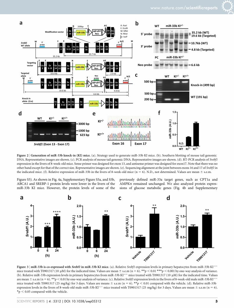
Figure S5). As shown in Fig. 4a, Supplementary Figure S5a, and S5b,ABCA1 and SREBP-1 protein levels were lower in the livers of themiR-33b KI mice. However, the protein levels of some of the
previously defined miR-33a target genes, such as CPT1a andAMPKa remained unchanged. We also analyzed protein expres-sions of glucose metabolic genes (Fig. 4b and Supplementary
a b
c
d e fKI+/-
KI+/+
Exon 16 Exon 17
5’ probe
3’ probe
WT miR-33b KI+/-
35.2 kb (WT)19.6 kb (Targeted)
10.7kb (WT)
4.8 kb (Targeted)
Neo probe 6.6 kb
PC miR-33b KI+/-
WT KI+/- KI+/+
Knock-in (499 bp)
WT (191 bp)
500 bp
500 bp
200 bp
WT KI+/- KI+/+
Srebf1 (Exon 13 - Exon 17)
1000 bp623 bp
3000 bp
miR-33bEx16
pgk-gb2Neo
Ex15
Ex14
Ex19
Ex17
Ex16
Ex3-13
Ex2
209bp
10.0 kb 2.0 kb
Ex15
Ex14
Ex16
Ex3-13
Ex2 miR-33b
Ex15
Ex14
Ex16
Ex3-13
Ex2 miR-33b
miR-33b
10.7 kb
4.8 kb
Ex15
Ex14
Ex16
Ex3-13
Ex2
Ex18
E E
M
A: AseIE: EcoRIM: MfeIN: NheI
E E N/E
35.2 kb
3’ probe
Ex17
Ex18
M
A
pgk-gb2Neo
E EA
pgk-gb2Neo
E EA
Ex19
Ex17
Ex18
Ex19
Ex17
Ex18
N/E
E
5’probe
M
19.6 kb
Neoprobe
E
Ex19
Ex17
Ex18
5’probe
3’ probe
Modifica�on vector
Srebf1WT allele
Targe�ngvector
Targetedallele
Knock-in allele (Cre)
: loxP
N
N
N
6.6 kb
N.D.
miR
-33b
(A.U
.)
WT KI+/- KI+/+0.0
0.5
1.0
1.5
Figure 2 | Generation of miR-33b knock-in (KI) mice. (a). Strategy used to generate miR-33b KI mice. (b). Southern blotting of mouse tail genomic
DNA. Representative images are shown. (c). PCR analysis of mouse tail genomic DNA. Representative images are shown. (d). RT-PCR analysis of Srebf1
expression in the livers of 8-week-old mice. Sense primer was designed for exon 13, and antisense primer was designed for exon17. Note that there was no
other band except for that of the correct size. Representative images are shown. (e). Sequencing alignment at the joint between exons 16 and 17 of Srebf1 in
the indicated mice. (f). Relative expression of miR-33b in the livers of 8-week-old mice (n 5 6). N.D., not determined. Values are mean 6 s.e.m.
a
(h)
miR
-33b
(A.U
.)
0 6 240.0
0.5
1.0
1.5
2.0
b
**** **
***
c d
**
Srebf1
(A.U
.)
Vehicl
e
T0901
317
0
1
2
3
4 **
miR
-33b
(A.U
.)
Vehicl
e
T0901
317
0.0
0.5
1.0
1.5
2.0
(h)
Srebf1
(A.U
.)
0 6 240
1
2
3
4
Figure 3 | miR-33b is co-expressed with Srebf1 in miR-33b KI mice. (a). Relative Srebf1 expression levels in primary hepatocytes from miR-33b KI1/1
mice treated with T0901317 (10 mM) for the indicated time. Values are mean 6 s.e.m (n 5 6). **p , 0.01 ***p , 0.001 by one-way analysis of variance.
(b). Relative miR-33b expression levels in primary hepatocytes from miR-33b KI1/1 mice treated with T0901317 (10 mM) for the indicated time. Values
are mean 6 s.e.m (n 5 6). **p , 0.01 by one-way analysis of variance. (c). Relative Srebf1 expression levels in the livers of 8-week-old male miR-33b KI1/1
mice treated with T0901317 (25 mg/kg) for 3 days. Values are means 6 s.e.m (n 5 6). **p , 0.01 compared with the vehicle. (d). Relative miR-33b
expression levels in the livers of 8-week-old male miR-33b KI1/1 mice treated with T0901317 (25 mg/kg) for 3 days. Values are mean 6 s.e.m (n 5 6).
*p , 0.05 compared with the vehicle.
www.nature.com/scientificreports
SCIENTIFIC REPORTS | 4 : 5312 | DOI: 10.1038/srep05312 3

Figure S5c–f). However, no significant change in protein level wasobserved in PCK1, G6PC, and CREB in the liver of miR-33b KI micecompared with that of control mice. SRC1 was up-regulated in miR-33bKI mice and it was opposite to the results of previous report14.
miR-33b KI reduces cholesterol efflux in macrophages. To inves-tigate a physiological role of miR-33b in mice, we first compared thefunctions of peritoneal macrophages from the WT and miR-33bKI1/1 mice. ABCA1 and ABCG1 protein levels were lower inmacrophages from the miR-33b KI1/1 mice than from the WTmice (Fig. 5a and Supplementary Figure S3e and S3f), which wascompatible with the findings for our miR-33a-deficient mice. Wedetermined apoA-I- and HDL-C-mediated cholesterol efflux fromperitoneal macrophages and found that macrophages from the miR-33b KI1/1 mice had lower apoA-I- and HDL-C-mediated cholesterolefflux than those from the WT mice (Fig. 5b).
A single miR-33b copy reduces serum HDL levels. Hepatic ABCA1overexpression increases HDL-C levels15, and liver-specific deletionof ABCA1 results in a substantial decrease in plasma HDL-C levels(approximately 80%) in chow-fed mice16. Moreover, we previouslyreported that the miR-33a2/2 mice had 22%–39% higher serumHDL-C levels than the WT mice8. Thus, we determined the serumHDL-C levels of the WT, miR-33b KI1/2, and miR-33b KI1/1 mice atthe age of 8 weeks.
Serum HDL-C levels were significantly decreased in the miR-33bKI1/2 and miR-33b KI1/1 mice compared with the WT mice (Table).We also classified and quantified serum lipoproteins using high-performance liquid chromatography (HPLC). Mean plots of theHPLC elution profile of serum from male mice are shown inFig. 5c, and the lipid profiles are summarized in SupplementaryTable S1. These results show that only one copy of miR-33b wassufficient to substantially reduce HDL-C and total cholesterol tothe same levels as those in the miR-33b KI1/1 mice. Moreover, thedecreased HDL levels mainly comprised very large-, large-, and med-ium-sized HDLs (mature HDLs) (Fig. 5c and Supplementary TableS1).
DiscussionIn the present study, we successfully established humanized mice, inwhich a miR-33b transgene was inserted within the same intron asthat in human SREBF1. The LXR agonist T0901317, which is a well-established Srebf1 expression inducer, enhanced miR-33b produc-tion. The protein levels of known miR-33a/b target genes, such asABCA1, ABCG1, and SREBP-1, were reduced under basal condi-tions. In vitro experiments indicated that macrophages from the
miR-33b KI1/1 mice had a reduced cholesterol efflux capacity viaapoA-I and HDL-C. Finally, HDL-C levels were reduced by almost35% even in the miR-33b KI1/2 mice compared with the WT micewithout any changes in triglyceride (TG) levels.
In contrast to humans and other mammals, rodents lack miR-33b and only have miR-33a in Srebf2. This needs to be kept inmind when attempting to directly translate to humans the previousresults that miR-33a inhibition could prevent atherosclerosis inmouse models because of two reasons. First, SREBF1 andSREBF2 are differentially regulated by hormones, dietary chal-lenges, and lipid-lowering agents, including statins17. This indicatesthat both isoforms of miR-33 participate in regulating the primaryrisk factors of metabolic syndrome, which accelerate atheroscler-osis. Second, miR-33b differs from miR-33a by 2-nucleotides andmay have a different target profile, including stronger effects ontargets in the SREBP-1-dependent regulation of fatty acid/TGhomeostasis and insulin signaling. We found increased miR-33bexpression after treatment with LXR agonist in our mice, whichindicated that miR-33b was co-expressed with its Srebf1 host geneand enabled us to study the impact of Srebf1-derived miR-33b oncholesterol/lipid homeostasis.
We have not yet succeeded in identifying miR-33b-specific targetgenes. Even previously reported miR-33b target genes were notreduced in the liver of miR-33b KI mice compared with that ofcontrol mice. One of the reasons of such result may be that theprevious study was conducted in human cell line and potential bind-ing sites of miR-33b are not conserved at least in PCK1 39UTR ofmice. It is also possible that some compensated mechanisms mayhave occurred in miR-33b KI mice. However, the protein levels ofmiR-33a target genes, such as ABCA1, ABCG1, and SREBP-1, werereduced18. Moreover, the protein levels of previously defined miR-33a target genes, which were not dysregulated in miR-33a KO mice,including CPT1a and AMPKa, remained unchanged19,20. Thus, itmay be necessary to assess those conditions when Srebf1 expressionis strongly affected to establish the importance of the functions ofmiR-33b. In any event, the numbers of miR-33b transcripts weregreater than those of miR-33a transcripts, and this underscores theimportance of miR-33b21. Although there were no differences in thelevels of miR-33a in macrophages, it is interesting that the levels ofmiR-33a were increased in proportion to the expression levels ofmiR-33b in the liver. Because Srebf1 level is higher in the liver thanthat in macrophages22, it is possible that miR-33b and miR-33a com-pete for the same target gene binding sites in the liver, and that thedegradation of miR-33a is inhibited by miR-33b expression. In addi-tion, there may be other unknown mechanisms.
ABCA1
β-ac�n
WT KI+/- KI+/+
200
42
AMPKα55
CPT1a97
PrecursorSREBP-1 116
TF2B 36
55MatureSREBP-1
kDa b
β-ac�n
WT KI+/- KI+/+ kD
β-ac�n
G6Pase
WT KI+/- KI+/+kD
4236
200SRC1
55PCK1
36CREB
42
Figure 4 | miR-33b regulates ABCA1 and SREBP-1. (a). Western blotting analysis for ABCA1, SREBP-1, CPT1a, and AMPKa protein levels in the
livers of WT, KI1/2, and KI1/1 mice. Representative images are shown. TF2B and b-actin were used as loading controls. (b). Western blotting analysis for
SRC1, PCK1, CREB and G6Pase protein levels in the livers of WT, KI1/2, and KI1/1 mice. Representative images are shown. b-actin were used as loading
controls.
www.nature.com/scientificreports
SCIENTIFIC REPORTS | 4 : 5312 | DOI: 10.1038/srep05312 4

miRs are known to target long non-coding RNAs whose functionsare largely unknown, and interactions between miRs are also pos-sible23. Thus, miR-33b-specific functions should be determined infuture experiments.
Rayner et al. recently showed that inhibiting miR-33a and miR-33b in healthy male non-human primates increased circulatingHDL-C levels21. More recently, Rottiers et al. reported that miR-33a and miR-33b acted in a redundant manner and that inhibitingboth isoforms by an 8-mer LNA-modified anti-miR enhanced HDL-C levels24. Our data demonstrated that miR-33b indeed functions tocontrol HDL-C levels, which highlights the importance of targetingboth miR-33 family members simultaneously. It is noteworthy thatonly one copy of miR-33b (miR-33b KI1/2 mice) significantlyreduced HDL-C levels to the same levels as those in the miR-33bKI1/1 mice. This explains one of the reasons why human HDL-Clevels are lower than those of mice and indicates that it is importantto considerably reduce miR-33b levels if pharmacological targeting
of miR-33s is used to increase HDL-C levels. In this context, thecurrent LNA-modified anti-miR technique is quite potent for redu-cing the levels of both miR-33 isoforms and may be useful for anti-atherosclerosis therapy.
In addition to the effects on HDL-C, a study by Rayner et al.showed that miR-33 antagonism reduced very low-density lipopro-tein-associated TGs in their cohort of normal male African greenmonkeys21. However, Rottiers et al. did not find any significantchanges in TG levels when using miR-33a/b-targeting LNA-anti-miR treatment24. In our present miR-33b KI study and in previousmiR-33a KO experiments8, we did not observe any changes in TGlevels, indicating that modulation of miR-33s is unlikely to have astrong effect on TG levels, although species differences and differentdietary conditions need to be considered.
In contrast, we found a significant inhibitory effect of miR-33b onSREBP-1. A feedback system of SREBP-2 by cholesterol levels is wellknown, which maintains appropriate levels of cellular cholesterol.
a b c
Cho
lest
erol
effl
ux (%
)
0
5
10
15
(-) ApoA-I HDL-C
WTKI+/+
***
***
Abso
rban
ce U
nits
(mV)
20 22 24 26 28 300
50
100
150 WT
KI+/+
ABCA1
β-ac�n
200
42
WT KI+/+
ABCG1 55
kD
Figure 5 | miR-33b reduces cellular cholesterol efflux and serum HDL-C levels. (a). Western blotting for ABCA1 and ABCG1 proteins in peritoneal
macrophages from WT and KI1/1 mice. Representative images are shown. b-actin was used as the loading control. (b). Cholesterol efflux to apoA-I
and HDL-C in peritoneal macrophages from WT and KI1/1 mice (n 5 6 each). Values are mean 6 s.e.m. ***p , 0.001 (c). Mean plots of HPLC analysis
for serum cholesterol in WT and KI1/1 mice (n 5 4 and 5, respectively).
Table | Serum profiling of WT, KI1/2, and KI1/1 mice
WT (n 5 4) KI1/2 (n 5 4) KI1/1 (n 5 4)
TP (g/dL) 4.375 6 0.1109 4.275 6 0.04787 4.350 6 0.05000ALB (g/dL) 2.950 6 0.1190 2.825 6 0.1109 2.900 6 0.04082BUN (mg/dL) 21.75 6 0.6801 20.58 6 1.248 21.58 6 1.680CRE (mg/dL) 0.1125 6 0.002500 0.0925 6 0.004787 * 0.0975 6 0.006292Na (mEq/L) 152.5 6 0.6455 153.5 6 0.2887 153.8 6 0.4787K (mEq/L) 3.350 6 0.05000 3.325 6 0.0750 3.350 6 0.1041Cl (mEq/L) 110.5 6 0.6455 110.8 6 0.2500 111.0 6 0.5774Ca (mg/dL) 8.500 6 0.1871 8.325 6 0.1109 8.350 6 0.08660IP (mg/dL) 7.775 6 0.4589 7.225 6 0.2955 7.400 6 0.4637T-BIL (mg/dL) 0.0875 6 0.004787 0.0925 6 0.008539 0.0825 6 0.01109AST (IU/L) 39.25 6 1.702 33.50 6 1.658 39.25 6 1.702ALT (IU/L) 26.50 6 3.663 21.00 6 2.415 22.75 6 1.702ALP (IU/L) 505.5 6 48.55 398.5 6 40.01 480.0 6 29.31LDH (IU/L) 278.3 6 77.21 243.5 6 55.30 255.0 6 55.26AMY (IU/L) 2295 6 68.22 2224 6 62.39 2363 6 97.02c-GTP (IU/L) 3. 3. 3.
T-CHO (mg/dL) 98.50 6 5.694 66.25 6 2.287 ** 62.00 6 1.225 ***TG (mg/dL) 34.75 6 2.780 32.25 6 3.065 35.25 6 4.328NEFA (mEq/L) 471.0 6 47.36 474.8 6 71.81 459.5 6 55.01LDL-C (mg/dL) 6.750 6 0.6292 6.750 6 0.6292 6.000 6 0.0HDL-C (mg/dL) 57.75 6 4.171 39.25 6 0.7500 ** 37.25 6 0.6292 **GLU (mg/dL) 216.3 6 22.98 180.5 6 8.930 197.8 6 11.92
Values are mean 6 s.e.m. Blood was obtained from chow-fed 8-wk-old male mice after 4 h fasting.*p , 0.05;**p , 0.01;***p , 0.001 compared with WT mice.
www.nature.com/scientificreports
SCIENTIFIC REPORTS | 4 : 5312 | DOI: 10.1038/srep05312 5

However, a similar mechanism has not been established for SREBP-1. Chronic activation of SREBP-1c in cases of overnutrition can leadto serious obesity-related problems. miR-33b may be utilized for afeedback mechanism to regulate its host gene SREBF1 because insu-lin induces hepatic SREBP-1c expression and promotes lipogenesisand hepatic TG synthesis (Supplementary Fig. S6).
In the present study, we demonstrated the effect of miR-33b onHDL-C levels in vivo. We assume that inhibiting both miR-33a andmiR-33b will have a significant effect on HDL-C levels in clinicalsettings. However, it is known that one miR can have hundreds oftarget genes and unexpected side effects may occur due to long-termtherapeutic modulation of miR-33 to cure metabolic diseases.Careful observations of miR-33b KI and miR-33a-deficient miceand intercrossing of these mice will enable us to detect miR-33a-and miR-33b-specific target genes and to elucidate the overall func-tions of miR-33a and miR-33b in vivo. Moreover, our mice will aid inanalyzing the roles of miR-33a/b in different genetic disease modelsand in screening drug candidates that can modulate miR-33a andmiR-33b levels and activities.
MethodsMaterials. The following antibodies were used: anti-ABCA1 (NB400-105) and anti-ABCG1 (NB400-132) (Novus Biologicals, Littleton, CO, USA); anti-AMPKa (#2532)and anti-CREB (#9197) (Cell Signaling Technology, Beverly, MA, USA); anti-CPT1a(ab128568) and anti-PCK1 (ab70358) (Abcam, Cambridge, UK); anti-b-actin (AC-15; A5441, Sigma-Aldrich, St. Louis, MO, USA); anti-SREBP-1 (sc-13551, sc-8984),anti-SRC1 (sc-8995), anti-G6Pase (sc-27198), and anti-TF2B (sc-225) (Santa Cruz,Biotechnology, California, USA). Anti-mouse, anti-rabbit and anti-goat IgG HRP-linked antibodies were purchased from GE Healthcare (Amersham, UK). HumanapoA-I was purchased from Sigma-Aldrich. Human acetylated LDL (acLDL) andhuman HDL-C were purchased from Biomedical Technologies, Inc. (Stoughton, MA,USA). [1, 2-3H (N)]-Cholesterol was purchased from Perkin Elmer (Boston, MA,USA). T0901317 was purchased from Cayman Chemical (Ann Arbor, MI, USA).
Generation of miR-33b KI mice. A targeting vector was constructed by modifyingbacterial artificial chromosome RP24-310C22 (Invitrogen) using a defectiveprophage l-Red recombination system25,26. As a selection marker, a neomycinresistance cassette flanked by loxP sites (loxP-PGK-gb2-neo-loxP cassette; GeneBridges, Germany) was inserted at the adjacent site of the human pre-miR-33b site.The targeting vector was electroporated into C57BL/6 mouse ES cells (DS PharmaBiomedical) using a Nucleofector system (Lonza). Positive clones were selected byincubating cells with 200 mg/ml geneticin (Invitrogen) for 5 days, and homologousrecombination was confirmed by Southern blotting. Successfully recombined ES cellswere injected into blastocysts from ICR strain mice supplied by Unitech Inc. (Japan),and chimeric mice were bred with C57BL/6 mice to generate F1 mice. F1 micegenotypes were confirmed by Southern blotting. The neomycin resistance cassettewas removed from the mouse germ line by breeding heterozygous mice with Ayu-1Cre KI mice, which expressed Cre recombinase in multiple tissues, including the germline27. Descendant miR-33b knock-in heterozygous mice without the Ayu-1 Cre allelewere crossed with each other to generate the miR-33b KI1/1 mice. All experimentswere performed with male C57BL/6 background mice and wild-type littermates wereused as a control. All of the experimental protocols were approved by the EthicsCommittee for Animal Experiments of Kyoto University and the methods wereperformed in accordance with the guidelines approved by the ethics committee.Primers used for genotyping were as follows: WT/KI sense, ATGGATTTACC-TCAGTTTTAACGAC; WT antisense, CATCACTGAAGCACTGCATCTGC; KIantisense, AAGTGGATCCAGAATTCGTGA; Cre sense, GCTGCCACGACdC-AAGTGACAGCAATG; and Cre antisense, GTAGTTATTCGGATCATCAGC-TACAC.
Southern blotting. Southern blotting was performed using DIG High Prime DNALabeling and Detection Starter Kit II (Roche) according to the manufacturer’sprotocol. Genomic DNA samples were purified and digested with MfeI and AseI for a59 probe, EcoRI for a 39 probe, and NheI for a Neo probe. Primer sequences used toamplify these probes were as follows:
59 probe sense, CACGGTTGTGAGAAGTCAGTATTC; 59 probe antisense,CTTTGCAAGCTCCTTGAGAATAAG; 39 probe sense, AGTAAAATTCTCCTC-AATGAACGTG; 39 probe antisense, CAGTAGGTGACATTGTGATTGATCT; Neoprobe sense, GAACAAGATGGATTGCACGCAGGTTCTCCG; and Neo probeantisense, GTAGCCAACGCTATGTCCTGATAG.
Determination of splicing between exons 16 and 17 in Srebf1. We amplified thefragment between Srebf1 exons 13 and 17 using cDNA from the livers of the indicatedmice by PCR, and these products were then electrophoresed. Extension time wassufficient to expand the fragment when the correct splicing did not occur. There wasno other band except for that of the correct size. Sequencing was performed using a
primer for exon 16 and an ABI 3130 genetic analyzer. Primer sequences used were asfolows:
Exon 13 sense, CCTAGAGCGAGCGTTGAACT; Exon 17 antisense, CTACCT-GGACTGAAGCTGGTG; and Exon 16 sequence primer, AGGGCAGCTCTGTAC-TCCTTC.
Cell culture. HepG2 cells were cultured in Dulbecco’s modified Eagle’s medium(DMEM; Nacalai Tesque, Japan) supplemented with 10% fetal bovine serum (FBS).Mouse primary hepatocytes were obtained from miR-33b KI1/1 mice using a two-step collagenase perfusion method28. In brief, hepatocyte suspensions were obtainedby passing a collagenase type II (Gibco BRL, Life Technologies Inc., Rockville, MD,USA)-digested liver sample through a 70-mm cell strainer, followed by centrifugationto isolate mature hepatocytes. Hepatocytes were then resuspended in DMEMsupplemented with 10% FBS and seeded on collagen type I-coated dishes (IwakiAsahi Glass Co. Ltd., Japan) at a density of 7 3 104 cells/ml. After incubation for 24 h,the cells were used for experiments.
Cholesterol efflux from macrophages. Cellular cholesterol efflux via apoA-I wasdetermined as described previously29. In brief, thioglycollate-elicited mouseperitoneal macrophages were plated in 24-well microplates at a density of 5 3
106 cells/ml. Cells were cultured for 24 h in RPMI 1640 containing 3H-labeled acLDL(1.0 mCi/ml of 3H-cholesterol and 25 mg/ml of acLDL). On the next day, the cellswere washed 3 times with RPMI 1640 and incubated for 6 h in RPMI 1640 with orwithout apoA-I (10 mg/ml) or HDL (100 mg/ml). Cholesterol efflux was expressed asthe percentage of radioactivity released from cells in medium relative to the totalradioactivity in cells plus medium.
Western blotting. Western blotting was performed using standard procedures asdescribed previously30. A lysis buffer was supplemented with a complete miniprotease inhibitor (Roche), ALLN (25 mg/ml), 0.5 mM NaF, and 10 mM Na3VO4 justprior to use. Protein concentrations were determined using a bicinchoninic acidprotein assay kit (Bio-Rad). All samples (20 mg of protein) were suspended in lysisbuffer, fractionated using NuPAGE 4%–12% Bis-Tris (Invitrogen) gels, andtransferred to a Protran nitrocellulose transfer membrane (Whatman). Themembrane was blocked using 13 phosphate-buffered saline (PBS) containing 5%non-fat milk for 1 h and incubated with a primary antibody [anti-ABCA1 (151000),anti-ABCG1 (151000), anti-AMPKa (151000), anti-SREBP-1 (15250), anti-TF2B(151000), anti-b-actin (153000), anti-CREB (151000), anti-PCK1 (151000), anti-SRC1 (15200), anti-G6Pase (15200) or anti-CPT1a (151000)] overnight at 4uC. Afterwashing with PBS–0.05% Tween 20 (0.05% T-PBS), the membrane was incubatedwith a secondary antibody (anti-rabbit, anti-mouse and anti-goat IgG HRP-linked;152000) for 1 h at 4uC. The membrane was then washed with 0.05% T-PBS anddetected with an ECL Western Blotting Detection Reagent (GE Healthcare) using anLAS-1000 system (Fuji Film).
RNA extraction and quantitative RT-PCR (qRT-PCR). Total RNA was isolated andpurified using TriPure Isolation Reagent (Roche). cDNA was synthesized from 1 mgof total RNA using the Transcriptor First Strand cDNA Synthesis Kit (Roche)according to the manufacturer’s instructions. For qRT-PCR, specific genes wereamplified in 40 cycles using SYBRTM Green PCR Master Mix (Applied Biosystems).Expression was normalized to that of the housekeeping gene b-actin. Gene-specificprimers are as follows;
SREBF1 sense, AACAGTCCCACTGGTCGTAGAT; SREBF1 antisense,TGTTGCAGAAAGCGAATGTAGT; SREBF2 sense, AGGAGAACATGGTG-CTGA; SREBF2 antisense, TAAAGGAGAGGCACAGGA; ACTB sense, AGGCA-CTCTTCCAGCCTTCC; ACTB antisense, GCACTGTGTTGGCGTACAGG; Srebf1sense, TAGAGCATATCCCCCAGGTG; Srebf1 antisense, GGTACGGGCCAC-AAGAAGTA; Srebf2 sense, GTGGAGCAGTCTCAACGTCA; Srebf2 antisense,TGGTAGGTCTCACCCAGGAG; Actb sense, GATCTGGCACCACACCTTCT;and Actb antisense, GGGGTGTTGAAGGTCTCAAA.
Quantitative PCR for miRs. Total RNA was isolated using the TriPure IsolationReagent (Roche). miR-33a and miR-33b were measured using TaqMan MicroRNAassay protocols (Applied Biosystems). Products were analyzed using a thermal cycler(ABI PrismH 7900HT sequence detection system). miRs expression of samples werenormalized by U6 snRNA expression.
Serum biochemical analysis. After mice were fasted for 4–6 h, blood was obtainedfrom the inferior vena cava of an anesthetized mouse, and serum was separated bycentrifugation at 4uC and stored at 280uC. Employing standard methods, biochemicalmeasurements were made using a Hitachi 7180 Auto Analyzer (Nagahama LifeScience Laboratory, Nagahama, Japan). Lipoproteins were analyzed by HPLC atSkylight Biotech (Akita, Japan), according to the procedures described previously31.
Statistical analysis. Results are given as mean 6 s.e.m. Statistical comparisons weremade using unpaired two-tailed Student’s t-tests or one-way analysis of variance with theBonferroni post hoc test, as appropriate. A p value of ,0.05 was considered significant.
1. Osborne, T. F. & Espenshade, P. J. Evolutionary conservation and adaptation inthe mechanism that regulates SREBP action: what a long, strange tRIP it’s been.Genes Dev 23, 2578–2591 (2009).
www.nature.com/scientificreports
SCIENTIFIC REPORTS | 4 : 5312 | DOI: 10.1038/srep05312 6

2. Espenshade, P. J. & Hughes, A. L. Regulation of sterol synthesis in eukaryotes.Annu Rev Genet 41, 401–427 (2007).
3. Horton, J. D., Goldstein, J. L. & Brown, M. S. SREBPs: activators of the completeprogram of cholesterol and fatty acid synthesis in the liver. J Clin Invest 109,1125–1131 (2002).
4. Yokoyama, C. et al. SREBP-1, a basic-helix-loop-helix-leucine zipper protein thatcontrols transcription of the low density lipoprotein receptor gene. Cell 75,187–197 (1993).
5. Ono, K., Kuwabara, Y. & Han, J. MicroRNAs and Cardiovascular Diseases. FEBS J(2011).
6. Najafi-Shoushtari, S. H. et al. MicroRNA-33 and the SREBP host genes cooperateto control cholesterol homeostasis. Science 328, 1566–1569 (2010).
7. Rayner, K. J. et al. MiR-33 contributes to the regulation of cholesterol homeostasis.Science 328, 1570–1573 (2010).
8. Horie, T. et al. MicroRNA-33 encoded by an intron of sterol regulatory element-binding protein 2 (Srebp2) regulates HDL in vivo. Proc Natl Acad Sci U S A 107,17321–17326 (2010).
9. Rayner, K. J. et al. Antagonism of miR-33 in mice promotes reverse cholesteroltransport and regression of atherosclerosis. J Clin Invest 121, 2921–2931 (2011).
10. Horie, T. et al. MicroRNA-33 Deficiency Reduces the Progression ofAtherosclerotic Plaque in ApoE(2/2) Mice. J Am Heart Assoc 1, e003376 (2012).
11. Horie, T. et al. MicroRNAs and Lipoprotein Metabolism. J Atheroscler Thromb 21,17–22 (2014).
12. Sowa, N. et al. MicroRNA 26b encoded by the intron of small CTD phosphatase(SCP) 1 has an antagonistic effect on its host gene. J Cell Biochem 113, 3455–3465(2012).
13. DeBose-Boyd, R. A., Ou, J., Goldstein, J. L. & Brown, M. S. Expression of sterolregulatory element-binding protein 1c (SREBP-1c) mRNA in rat hepatoma cellsrequires endogenous LXR ligands. Proc Natl Acad Sci U S A 98, 1477–1482 (2001).
14. Ramirez, C. M. et al. MicroRNA 33 regulates glucose metabolism. Mol Cell Biol 33,2891–2902 (2013).
15. Basso, F. et al. Role of the hepatic ABCA1 transporter in modulating intrahepaticcholesterol and plasma HDL cholesterol concentrations. J Lipid Res 44, 296–302(2003).
16. Timmins, J. M. et al. Targeted inactivation of hepatic Abca1 causes profoundhypoalphalipoproteinemia and kidney hypercatabolism of apoA-I. J Clin Invest115, 1333–1342 (2005).
17. Shimano, H. SREBPs: physiology and pathophysiology of the SREBP family. FEBSJ 276, 616–621 (2009).
18. Horie, T. et al. MicroRNA-33 regulates sterol regulatory element-binding protein1 expression in mice. Nat Commun 4, 2883 (2013).
19. Gerin, I. et al. Expression of miR-33 from an SREBP2 intron inhibits cholesterolexport and fatty acid oxidation. J Biol Chem 285, 33652–33661 (2010).
20. Fernandez-Hernando, C. & Moore, K. J. MicroRNA modulation of cholesterolhomeostasis. Arterioscler Thromb Vasc Biol 31, 2378–2382 (2011).
21. Rayner, K. J. et al. Inhibition of miR-33a/b in non-human primates raises plasmaHDL and lowers VLDL triglycerides. Nature 478, 404–407 (2011).
22. Zhou, X. et al. Genetic deletion of low density lipoprotein receptor impairs sterol-induced mouse macrophage ABCA1 expression. A new SREBP1-dependentmechanism. J Biol Chem 283, 2129–2138 (2008).
23. Memczak, S. et al. Circular RNAs are a large class of animal RNAs with regulatorypotency. Nature 495, 333–338 (2013).
24. Rottiers, V. et al. Pharmacological Inhibition of a MicroRNA Family inNonhuman Primates by a Seed-Targeting 8-Mer AntimiR. Sci Transl Med 5,212ra162 (2013).
25. Copeland, N. G., Jenkins, N. A. & Court, D. L. Recombineering: a powerful newtool for mouse functional genomics. Nat Rev Genet 2, 769–779 (2001).
26. Horiguchi, M. et al. Fibulin-4 conducts proper elastogenesis via interaction withcross-linking enzyme lysyl oxidase. Proc Natl Acad Sci U S A 106, 19029–19034(2009).
27. Morita, M. et al. HLF/HIF-2alpha is a key factor in retinopathy of prematurity inassociation with erythropoietin. EMBO J 22, 1134–1146 (2003).
28. Seglen, P. O. Preparation of isolated rat liver cells. Methods Cell Biol 13, 29–83(1976).
29. Zhang, Z. et al. Expression of cholesteryl ester transfer protein in humanatherosclerotic lesions and its implication in reverse cholesterol transport.Atherosclerosis 159, 67–75 (2001).
30. Horie, T. et al. Acute doxorubicin cardiotoxicity is associated with miR-146a-induced inhibition of the neuregulin-ErbB pathway. Cardiovasc Res 87, 656–664(2010).
31. Usui, S., Kakuuchi, H., Okamoto, M., Mizukami, Y. & Okazaki, M. Differentialreactivity of two homogeneous LDL-cholesterol methods to LDL and VLDLsubfractions, as demonstrated by ultracentrifugation and HPLC. Clin Chem 48,1946–1954 (2002).
AcknowledgmentsWe thank Neal G. Copeland (Institute of Molecular and Cell Biology, Singapore) for thedefective prophage l-Red recombination system; Junji Takeda (Osaka University, Osaka,Japan) and Kosuke Yusa (Osaka University, Osaka, Japan) for the plasmid used for theconstruction of the targeting vector; Ken-ichi Yamamura (Kumamoto University,Kumamoto, Japan) and Kimi Araki (Kumamoto University) for Ayu-1 Cre expressing mice.This work was supported in part by grants from the Japan Society for the Promotion ofScience, by a Grant-in-Aid for Scientific Research from the Ministry of Education, Culture,Sports, Science, and Technology of Japan (to T. Kimura, T. Kita, K.H., T.H., and K.O.), by aGrant-in-Aid for Scientific Research on Innovative Areas ‘‘Crosstalk betweentranscriptional control and energy pathways, mediated by hub metabolites’’(3307) from theMinistry of Education, Culture, Sports, Science and Technology of Japan (to K.O.), byOtsuka Pharmaceutical Co., Ltd. - Kyoto University SRP Program (to T. Kimura, T. Kita,M.Y., T.H., and K.O.), by grants from Banyu Life Science Foundation International, TakedaMemorial Foundation, Suzuken Memorial Foundation, Sakakibara Memorial Foundation,Japan Foundation of Applied Enzymology, SENSHIN Medical Research Foundation, KowaLife Science Foundation, and ONO Medical Research Foundation (to T.H.), and by grantsfrom ONO Medical Research Foundation, the Cell Science Research Foundation,Daiichi-Sankyo Foundation of Life Science, and Takeda Memorial Foundation (to K.O.).
Author contributionsT.H., T.N. and K.O. designed the project; T.H., T.N., O.B., Y.K., T.N., M.N., S.U., M.I., F.N.,Y.I., S.K. and N.S. performed experiments; T.H., T.N., N.Y., H.S., T.N., K.H., N.K., M.Y.,T.K., T.K. and K.O. analyzed and interpreted data; and T.H., T.N. and K.O. prepared themanuscript.
Additional informationSupplementary information accompanies this paper at http://www.nature.com/scientificreports
Competing financial interests: The authors declare no competing financial interests.
How to cite this article: Horie, T. et al. MicroRNA-33b knock-in mice for an intron of sterolregulatory element-binding factor 1 (Srebf1) exhibit reduced HDL-C in vivo. Sci. Rep. 4,5312; DOI:10.1038/srep05312 (2014).
This work is licensed under a Creative Commons Attribution 4.0 InternationalLicense. The images or other third party material in this article are included in thearticle’s Creative Commons license, unless indicated otherwise in the credit line; ifthe material is not included under the Creative Commons license, users will needto obtain permission from the license holder in order to reproduce the material. Toview a copy of this license, visit http://creativecommons.org/licenses/by/4.0/
www.nature.com/scientificreports
SCIENTIFIC REPORTS | 4 : 5312 | DOI: 10.1038/srep05312 7