tissue‐specific study across the stem of taxus media
TRANSCRIPT
Tissue-specific study across the stem of Taxus mediaidentifies a phloem-specific TmMYB3 involved in thetranscriptional regulation of paclitaxel biosynthesis
Chunna Yu1,2, Xiujun Luo1,2, Chengchao Zhang1,2, Xinyun Xu1,2, Jiefang Huang1,2, Yueyue Chen1,2, Shangguo Feng1,2,
Xiaori Zhan1,2, Lei Zhang3, Huwei Yuan4,5, Bingsong Zheng4,5, Huizhong Wang1,2 and Chenjia Shen1,2,*1College of Life and Environmental Sciences, Hangzhou Normal University, Hangzhou 311121, China,2Zhejiang Provincial Key Laboratory for Genetic Improvement and Quality Control of Medicinal Plants, Hangzhou Normal
University, Hangzhou 311121, China,3Department of Plant Pathology, Washington State University, Pullman, WA 99164-6430, USA,4State Key Laboratory of Subtropical Silviculture, Zhejiang A & F University, Hangzhou 311300, China, and5Center for Cultivation of Subtropical Forest Resources (CCSFR), Zhejiang A & F University, Hangzhou 311300, China
Received 10 September 2019; revised 6 January 2020; accepted 22 January 2020.
*For correspondence (e-mail [email protected]).
SUMMARY
Taxus stem barks can be used for extraction of paclitaxel. However, the composition of taxoids across the
whole stem and the stem tissue-specificity of paclitaxel biosynthesis-related enzymes remain largely
unknown. We used cultivated Taxus media trees for analyses of the chemical composition and protein of
major stem tissues by an integrated metabolomic and proteomic approach, and the role of TmMYB3 in
paclitaxel biosynthesis was investigated. The metabolomic landscape analysis showed differences in stem
tissue-specific accumulation of metabolites. Phytochemical analysis revealed that there is high accumula-
tion of paclitaxel in the phloem. Ten key enzymes involved in paclitaxel biosynthesis were identified, most
of which are predominantly produced in the phloem. The full-length sequence of TmMYB3 and partial pro-
moter sequences of five paclitaxel biosynthesis-related genes were isolated. Several MYB recognition ele-
ments were found in the promoters of TBT, DBTNBT and TS. Further in vitro and in vivo investigations
indicated that TmMYB3 is involved in paclitaxel biosynthesis by activating the expression of TBT and TS.
Differences in the taxoid composition of different stem tissues suggest that the whole stem of T. media has
potential for biotechnological applications. Phloem-specific TmMYB3 plays a role in the transcriptional regu-
lation of paclitaxel biosynthesis, and may explain the phloem-specific accumulation of paclitaxel.
Keywords: metabolome, phloem, proteome, R2R3-MYB, paclitaxel biosynthesis, Taxus media.
INTRODUCTION
Paclitaxel is one of the most important chemotherapeutic
agents, and its efficacy against ovarian, breast and head
and neck cancers has been widely documented (Bernabeu
et al., 2017). The importance of Taxus trees as natural
sources of paclitaxel and its derivatives has been high-
lighted in numerous biological, phytochemical and omics
studies (Hao et al., 2012). The commercial value of pacli-
taxel has driven considerable research interest in Taxus
trees in recent decades (Sanchez-Munoz et al., 2018).
The biosynthesis of paclitaxel, a structurally complex
representative of approximately 400 identified taxoids iso-
lated from Taxus trees, can be conceptually divided into 19
steps, from a diterpenoid progenitor to the final product
(Croteau et al., 2006). First, the 2-C-methyl-D-erythritol
4-phosphate (MEP) pathway yields isopentenyl diphos-
phate (IPP) and dimethylallyl diphosphate (DMAPP) to syn-
thesize the universal diterpenoid precursor geranylgeranyl
diphosphate (GGPP) (Eisenreich et al., 1996). Subse-
quently, formation of the taxane skeleton involves cycliza-
tion of GGPP into taxa-4(5),11(12)-diene, which is catalyzed
by a slow-starter enzyme, taxadiene synthase (TS) (Koepp
et al., 1995). To obtain a key intermediate 10-deacetylbac-
catin III (10-DAB), a number of functional groups are added
to the taxane core by a series of cytochrome P450-medi-
ated hydroxylations, CoA-dependent acyl transfers and a
C9 site oxidation (Kaspera and Croteau, 2006). One step of
these reactions is conducted by a taxane 2a-O-benzoyl-
transferase (TBT), which transforms a 2-debenzoyl taxoid-
type intermediate to 10-DAB (Walker and Croteau, 2000).
© 2020 The Authors.The Plant Journal © 2020 John Wiley & Sons Ltd
1
The Plant Journal (2020) doi: 10.1111/tpj.14710
Next, a C13 side chain is appended to baccatin III (BAC),
yielding 30-N-debenzoyl-20-deoxytaxol (Kaspera and Cro-
teau, 2006). Finally, the formation of the functional pacli-
taxel molecule involves an important enzyme, 30-N-
debenzoyl-20-deoxytaxol-N-benzoyltransferase (DBTNBT)
(Onrubia et al., 2011).
Omics studies on Taxus trees have identified a large
number of metabolites and proteins involved in the pacli-
taxel biosynthesis pathway (Yu et al., 2017). For example,
six taxoids were isolated from cultured Taxus seedlings
using a metabolomic approach (Tanaka et al., 2011). Com-
parative metabolomics revealed that most of the interme-
diates of paclitaxel biosynthesis varied hugely in distinct
Taxus species, providing a possible explanation for their
differential accumulation of paclitaxel (Yu et al., 2018).
Comparative proteomic analyses identified four MEP path-
way-related enzymes and three cytochrome P450 taxoid
oxygenases that play important roles in determining inter-
specific differences in taxoid accumulation (Hao et al.,
2017). An integrated metabolomic and proteomic analysis
indicated that 1-deoxy-D-xylulose 5-phosphate reductoi-
somerase (DXR) and 1-hydroxy-2-methyl-2-(E)-butenyl
4-diphosphate reductase (HDR), two key enzymes in the
MEP pathway, are responsible for the stimulation of pacli-
taxel production (Zheng et al., 2016).
Recently, numerous transcription factors (TFs) have
been reported to play crucial roles in the transcriptional
regulation of different paclitaxel biosynthesis-related genes
(Kuang et al., 2019). Among these TFs, MYC family mem-
bers are believed to be involved in the regulation of jas-
monic acid (JA) signal transduction, which is involved in
paclitaxel biosynthesis (Cui et al., 2019). Three basic helix–loop–helix (bHLH) TFs, TcJAMYC1, TcJAMYC2 and TcJA-
MYC4, negatively regulate the expression of paclitaxel
biosynthetic genes in Taxus cuspidata (Lenka et al., 2015).
Another study showed that TcMYC2a may regulate the
expression of TS, TAT, DBTNBT, T13OH and T5OH via
ethylene responsive factor (ERF) regulators that depend on
the JA signaling pathway in Taxus chinensis (Zhang et al.,
2018b). Also in T. chinensis, ERF12 and ERF15, act as
repressor and activator of paclitaxel biosynthesis, respec-
tively, by binding to the GCC-box in the JA-responsive ele-
ment of the TS promoter (Zhang et al., 2015). In
T. chinensis, a WRKY transcription factor, TcWRKY1, par-
ticipates in the transcriptional activation of the promoter of
10-deacetylbaccatin-III-10-b-O-acetyltransferase (DBAT) (Li
et al., 2013). Another two WRKY TFs, TcWRKY8 and
TcWRKY47, significantly increased the expression levels of
several paclitaxel biosynthesis-related genes (Zhang et al.,
2018a).
The plant MYB family consists of four subfamilies, 1R-,
R2R3-, R1R2R3- and 4R-MYB, which have different num-
bers of MYB domain repeats (Dubos et al., 2010). In partic-
ular, R2R3-MYB is a large subfamily, members of which
are involved in secondary metabolism in plants (Stracke
et al., 2001). In poplar, MYB165 and MYB194 are broad
repressors of the flavonoid and phenylpropanoid biosyn-
thetic pathways (Ma et al., 2018). In peach, activator-type
R2R3-MYB genes balance the accumulation anthocyanin
and pro-anthocyanidin by inducing a repressor-type R2R3-
MYB gene PpMYB18 (Zhou et al., 2019a). However, the role
of R2R3-MYB TFs in the regulation of paclitaxel biosynthe-
sis is largely unknown.
Stem bark tissues are major sources of medicinal com-
pounds, such as lignanoids from the bark of Eucommia
ulmoides and taxoids from the bark of Taxus species (Uni-
yal, 2013; Wang et al., 2019). First isolated from the bark of
the Pacific yew Taxus brevifolia, hundreds of taxoids have
since been identified in the stem bark of various Taxus
species (Chan et al., 1994; Shen et al., 2001; Nadeem et al.,
2002). Although the whole Taxus stem is rich in secondary
metabolites, the composition of taxoids across distinct
stem tissues remains poorly understood. Studies of the
chemical composition of the major tissues of the whole
stem will give us a good opportunity to explore the stem
tissue-specificity of taxoid accumulation. Herein, molecular
fingerprints were determined for four major tissue types in
the stem of T. media, namely the cortex, phloem, xylem
and pith. The results reveal the metabolite and protein
profiles of different stem fractions based on integrated
metabolomic and proteomic analyses. Furthermore, a
phloem-specific R2R3-MYB TF, TmMYB3, was identified,
and its function in the regulation of paclitaxel biosynthesis
was investigated. Our findings may help to elucidate the
regulatory mechanism underlying paclitaxel biosynthesis
and accelerate the breeding of species with a high pacli-
taxel yield.
RESULTS
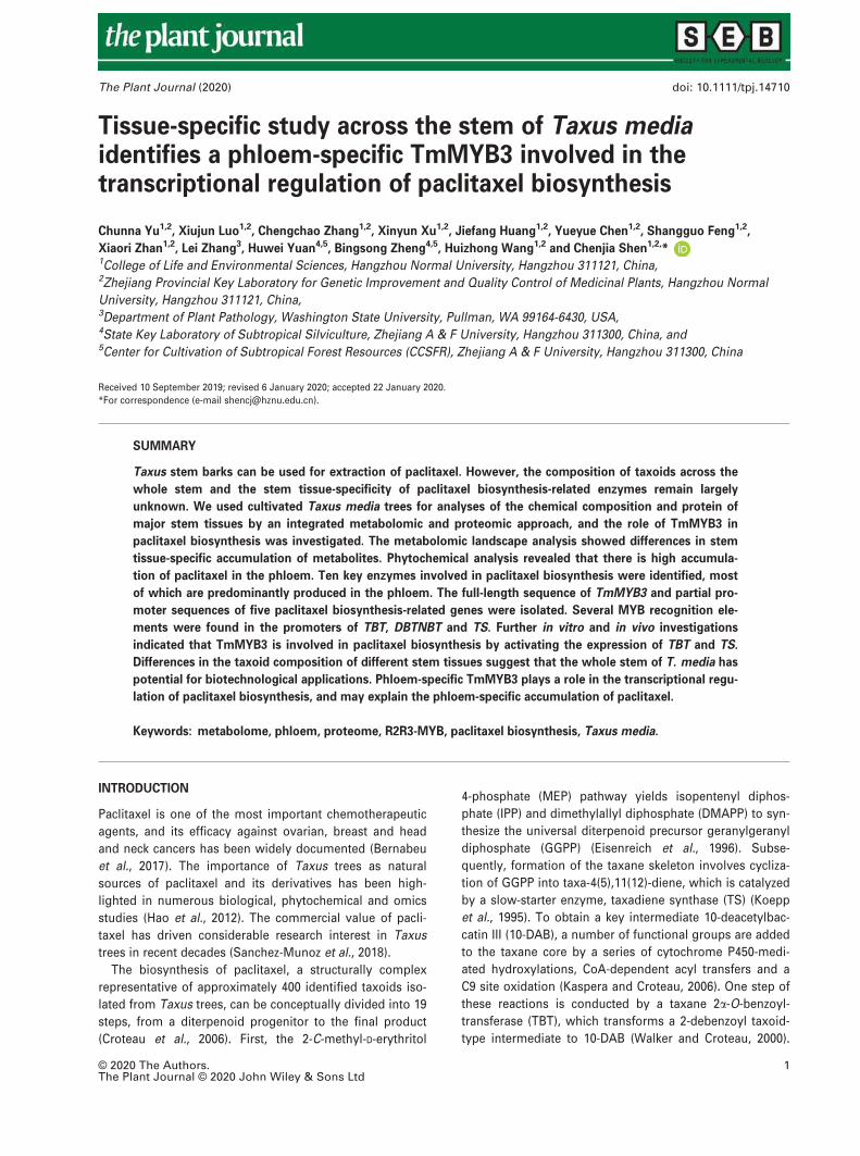
Localization of taxoid accumulation
The stem of Taxus species, especially the stem bark, is
widely used for paclitaxel extraction (Shen et al., 2001;
Nguyen et al., 2003). Four major tissues across the stem of
T. media, the cortex, phloem, xylem and pith, were sepa-
rated by simply peeling. A cross-section of the stem is
shown in Figure 1(a). Quantification analysis was per-
formed to explore the variation of taxoid content among
the four major stem tissues. Four important taxoids,
including paclitaxel, 10-deacetylpaclitaxel (DAP), BAC and
10-DAB, were quantified by ultra-performance liquid chro-
matography – tandem mass spectrometry (UPLC-MS/MS)
and their total ion chromatography (TIC) chromatogram is
shown in Figure 1(b). Most taxoids accumulated to varying
levels in these four stem tissues. Specifically, both pacli-
taxel and 10-DAB accumulated to the highest levels in the
phloem, while BAC predominantly accumulated in the pith
and DAP greatly accumulated in the cortex and phloem
© 2020 The Authors.The Plant Journal © 2020 John Wiley & Sons Ltd, The Plant Journal, (2020), doi: 10.1111/tpj.14710
2 Chunna Yu et al.
tissues. The xylem contained the lowest levels of these
four taxoids (Figure 1c).
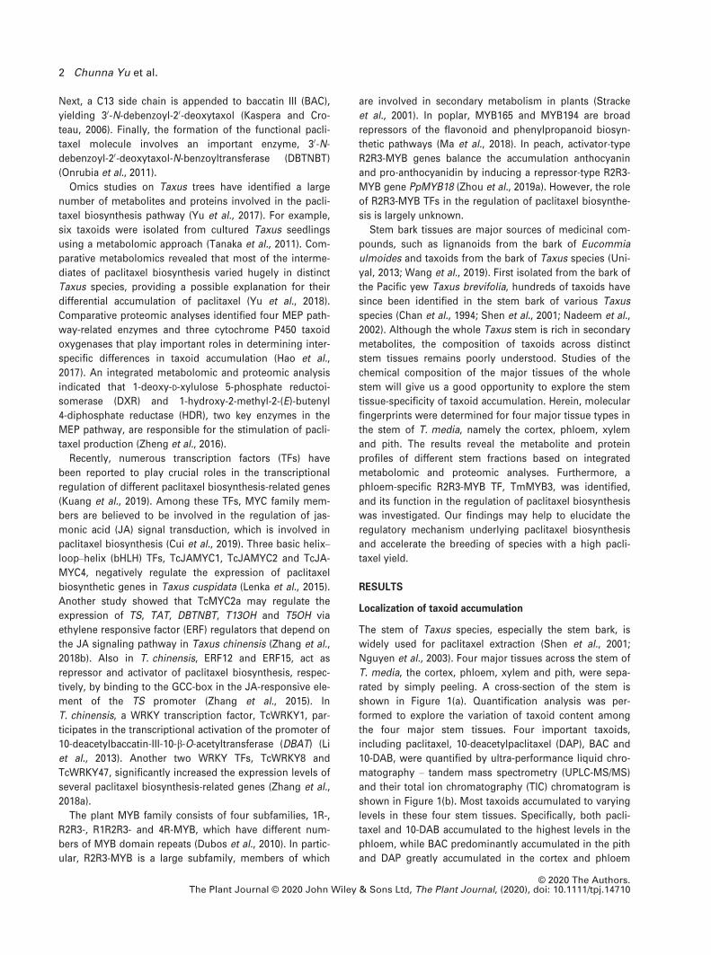
Overview of the metabolomes of different stem tissues
To explore the variation in phytochemical composition of
different stem tissues, an untargeted metabolomic analysis
(n = 10) was performed, and 9512 ion features correspond-
ing to 5162 potential metabolites with annotations were
identified (Table S2 in the online Supporting Information).
Analysis of five checking parameters, including TIC, aver-
age m/z distribution, retention-time width, metabolite
intensity distribution and coefficient of variation distribu-
tion indicated effective sample preparation and high-qual-
ity raw data (Figures 2a and S1). Principal component
analysis (PCA) showed that PC1 and PC2 explained 39.3%
and 15.51% of variation, respectively. Metabolite profiling
of the four stem tissues separated their metabolomes into
three groups (Figure 2b). Metabolomes from the cortex
and phloem were grouped into the same cluster, indicating
high similarity between these tissues (Figure 2c). A total of
5077 annotated metabolites were mapped to different
known metabolic pathways (Table S3). The largest number
of metabolites belonged to the category ‘metabolism of
terpenoids and polyketides’ (275 metabolites), followed by
‘amino acid metabolism’ (239 metabolites) and ‘lipid meta-
bolism’ (224 metabolites).
Analysis of differentially accumulated metabolites (DAMs)
in different stem tissues
To analyze stem tissue-specific accumulated metabolites,
DAMs were clustered into four major clusters (I–IV).Metabolites belonging to Cluster I predominantly accumu-
lated in the cortex; metabolites grouped into Cluster II
were highly accumulated in the phloem; metabolites in
Cluster III were abundant in the pith; and metabolites
belonging to Cluster IV predominantly accumulated in both
the cortex and phloem (Figure 2d). In Cluster I, the major
DAMs were sugars, flavonoids, amino acids and alkaloids;
in Cluster II, the largest number of DAMs belonged to
amino acids, flavonoids and terpenoids; in Cluster III,
Figure 1. Microstructure of the stem and differential accumulation of four representative taxoids across the stem tissues of Taxus media.
(a) Paraffin section of the stem. Four major stem tissues, including cortex, phloem, xylem and pith, were clearly identified.
(b) A representative total ion current chromatogram of four taxoids: peak 1, 10-deacetylbaccatin III (10-DAB); peak 2, baccatin III (BAC); peak 3, 10-deacetylpacli-
taxel (DAP); peak 4, paclitaxel (PTX). cps, counts per second.
(c) Corresponding histograms indicated the differences in the four representative taxoids among different stem tissues. Different letters indicated significant dif-
ferences in the contents of four representative taxoids from different stem tissues (P < 0.05). DW, dry weight.
© 2020 The Authors.The Plant Journal © 2020 John Wiley & Sons Ltd, The Plant Journal, (2020), doi: 10.1111/tpj.14710
TmMYB3 is involved in paclitaxel biosynthesis 3
terpenoids, lipids and alkaloids were the main metabolites;
and in Cluster IV, the predominant metabolites were flavo-
noids, amino acids and alkaloids (Figure 2e).
Overview of the proteomes of different stem tissues
To explore stem tissue-specific proteins, protein samples
(n = 3) from the four stem tissues were isolated for the
quantification of dynamic changes in their proteomes (Fig-
ure S2). The distribution of mass errors and peptide
lengths indicated a high mass accuracy for the mass spec-
trometry data. After searching the database, the number of
effectively matched spectra was 132 730, and the utilization
rate of spectra was 17.42%. Based on the matched spectra,
56 902 peptides were identified, including 47 396 unique
peptides (Figure S3). After filtering, 5604 proteins were
annotated, among which 4461 were quantified (Table S4).
To assess statistical consistency, pairwise Pearson’s
correlation coefficients, PCA and relative standard devia-
tion (RSD) were investigated using all quantified proteins,
suggesting that three samples from each group displayed
good repeatability (Figure S4).
Gene Ontology (GO) annotation, Eukaryotic Orthologous
Groups (KOG) classification and subcellular localization
prediction were performed on all identified proteins. The
most representative GO terms were ‘catalytic activity’
(2273 proteins), ‘metabolic process’ (2194 proteins), ‘bind-
ing’ (2031 proteins), ‘cellular process’ (1433 proteins) and
‘single-organism process’ (1344 proteins); the most abun-
dant KOG terms were ‘general function prediction only’
(600 proteins), ‘post-translational modification’ (599 pro-
teins) and ‘energy production and conversion’ (378 pro-
teins); the dominant subcellular localization categories
were ‘cytoplasm’ (1879 proteins), ‘chloroplast’ (1825 pro-
teins) and ‘nucleus’ (999 proteins) (Table S4).
Figure 2. Untargeted metabolomes identified differentially accumulated metabolites among the four stem tissues.
(a) Quality control parameters, including m/z widths and retention-time widths of the metabolomes.
(b) A principal component (PC) analysis of the metabolomes from four stem tissues of Taxus media.
(c) A heatmap of the abundance of metabolites in the four stem tissues (n = 10). The heatmap scale ranges from �2 to +2 on a log2 scale.
(d) MeV cluster analysis of tissue-specific accumulated metabolites from the untargeted metabolomic profiles. The red ovals indicated stem tissue-specific accu-
mulated metabolites. Cluster I indicates the cortex-specific accumulated metabolites, Cluster II indicates the phloem-specific accumulated metabolites, Cluster III
indicates the pith-specific accumulated metabolites and Cluster IV indicates the metabolites predominantly accumulated in both the cortex and phloem.
(e) Proportions of metabolites in each class.
© 2020 The Authors.The Plant Journal © 2020 John Wiley & Sons Ltd, The Plant Journal, (2020), doi: 10.1111/tpj.14710
4 Chunna Yu et al.

Analysis of differentially produced proteins (DPPs) in
different stem tissues
The numbers of up- and downregulated proteins from dif-
ferent comparisons (cortex/xylem, cortex/phloem, cortex/
pith, xylem/pith, phloem/xylem and phloem/pith) are
shown in Figure S5. Furthermore, proteomic profiling of
the four stem tissues clustered tissue-specific proteins into
four groups (I–IV; Figure 3a). In total, 329 cortex-, 307
phloem-, 94 xylem- and 90 pith-specific proteins were iden-
tified (Figure 3b). Further studies were carried out on
phloem-specific produced proteins (PPPs) to investigate
their relationship with phloem-specific accumulation of
paclitaxel (Table S5). Kyoto Encyclopedia of Genes and
Genomes (KEGG) enrichment analysis classified 307 PPPs
into five significantly enriched KEGG pathways. Interest-
ingly, the ‘terpenoid backbone biosynthesis’ pathway,
which provides precursors for paclitaxel biosynthesis, was
the most significantly enriched KEGG pathway (Figure 3c).
The GO enrichment analysis of DPPs showed that ‘oxidore-
ductase activity’ was the dominant term in the molecular
function category, and ‘isoprenoid biosynthetic process’
was the major biological process subcategory (Figure 3d).
Proteomic analysis identified nine classical TFs, namely
MYB3, NAC25, GATA9-like, TCP2, TFIIB, bHLH82, WRKY31,
Figure 3. Analysis of differentially produced proteins (DPPs) among the four stem tissues.
(a) A heatmap of the abundance of proteins in the four stem tissues (n = 3). The heatmap scale ranges from �2 to +2 on a log2 scale. The black dotted line
frames indicated stem tissue-specific produced proteins.
(b) MeV cluster analysis showing the numbers of tissue-specific produced proteins. The red oval indicates the number of phloem-specific produced proteins
(PPPs).
(c) Kyoto Encyclopedia of Genes and Genomes enrichment analysis of the PPPs.
(d) Gene Ontology enrichment analysis of the PPPs.
© 2020 The Authors.The Plant Journal © 2020 John Wiley & Sons Ltd, The Plant Journal, (2020), doi: 10.1111/tpj.14710
TmMYB3 is involved in paclitaxel biosynthesis 5

DIVARICATA and TFIID (Table S6). Among these TFs,
MYB3, NAC25 and GATA9-like were predominantly pro-
duced in the phloem.
Integrated metabolomic and proteomic analysis of taxoid
metabolic pathways in T. media
Paclitaxel biosynthesis is a sophisticated metabolic path-
way that involves a series of intermediate metabolites and
enzymes (Croteau et al., 2006). In our present study, 12
intermediate metabolites involved in paclitaxel biosyn-
thesis were identified, among which taxa-4(20),11(12)-
dien-5a-yl acetate, 10-DAB, 30-N-debenzoyl-20-deoxytaxol,30-N-debenzoyltaxol and paclitaxel predominantly accumu-
lated in the phloem, while taxa-4(5),11(12)-diene and taxa-4
(20),11(12)-dien-5a,13a-diol accumulated at high levels in
the xylem, and taxa-4(20),11(12)-dien-5a-ol, 10b,14b-dihy-droxytaxa-4(20),11(12)-dien-5a-yl acetate and BAC signifi-
cantly accumulated in the pith (Figure 4a). Additionally, 10
paclitaxel biosynthesis-related enzymes were identified.
The precise positions of the identified intermediate
metabolites and key enzymes involved in paclitaxel biosyn-
thesis are shown in Figure 4(b). Interestingly, most of the
paclitaxel-related enzymes, such as geranylgeranyl diphos-
phate synthase (GGPPS), TS, taxadiene 5a-hydroxylase(T5aOH), taxadien-5a-ol O-acetyltransferase. (TAT), taxane
10b-hydroxylase (T10bOH), TBT, DBAT, baccatin III:3-
amino-3-phenylpropanoyltransferase (BAPT) and DBTNBT,
were predominantly produced in the phloem (Figure 4c).
Figure 4. Integrated metabolomic and proteomic analysis of the paclitaxel biosynthesis pathway.
(a) Differential accumulation of 12 intermediate metabolites involved in paclitaxel biosynthesis from the four major stem tissues.
(b) Overview of the paclitaxel biosynthesis pathway. The orange background indicates the enzymes identified by the proteome. BAPT, baccatin III:3-amino-3-
phenylpropanoyltransferase; DBAT, 10-deacetylbaccatin-III-10-b-O-acetyltransferase; DBTNBT, 3’-N-debenzoyltaxol N-benzoyltransferase; GGPPS, geranylgeranyl
diphosphate synthase; MEP, 2-C-methyl-d-erythritol 4-phosphate; T10bOH, taxane 10b-hydroxylase; T13aOH, taxane 13a-hydroxylase; T5aOH, taxadiene-5a-hy-droxylase; TAT, taxadien-5a-ol O-acetyltransferase; TBT, taxane 2a-O-benzoyltransferase; TS, taxadiene synthase.
(c) Differential production of 10 key enzymes involved in paclitaxel biosynthesis from the four major stem tissues. The heatmap scale ranges from �2 to +2 on a
log2 scale.
© 2020 The Authors.The Plant Journal © 2020 John Wiley & Sons Ltd, The Plant Journal, (2020), doi: 10.1111/tpj.14710
6 Chunna Yu et al.

There are a number of dead-end metabolites and 14-hy-
droxylated taxoids in Taxus trees; however, none of these
intermediate metabolites leads to paclitaxel biosynthesis
(Wang et al., 2011). Herein, four taxusin-like metabolites,
(+)-taxusin, 2a-hydroxytaxusin, 7b-hydroxytaxusin and
2a,7b-dihydroxytaxusin, and one 14-hydroxylated taxoid,
taxuyunnanin C, were identified (Figure 5a). All four taxu-
sin-like metabolites accumulated in the pith, and taxuyun-
nanin C accumulated in both the cortex and phloem
(Figure 5b). Two important enzymes, taxadiene-2a-hydrox-ylase (T2aOH) and taxane-7b-hydroxylase (T7bOH),
involved in the metabolic pathway of taxusin-like metabo-
lites were produced at high levels in the pith. Taxoid 14b-hydroxylase (T14bOH), catalyzing an intermediate step to
synthesize taxuyunnanin C, was produced in both the cor-
tex and phloem (Figure 5c).
Most of the paclitaxel biosynthesis-related enzymes are
found mainly in the phloem, while the intermediate
metabolites involved in paclitaxel biosynthesis are
distributed in different stem tissues. For example,
GGPP, taxa-4(20),11(12)-dien-5a-ol, 10b,14b-dihydroxytaxa-4(20),11(12)-dien-5a-yl acetate and BAC might be produced
in the phloem and then transfer to the pith; taxa-4(5),11
(12)-diene might be produced in the phloem and pith and
transfer to the xylem; taxa-4(20),11(12)-dien-5a,13a-diolmight be produced in the pith and then migrate to the
xylem and phloem; and 10-deacetyl-2-debenzoylbaccatin III
might be produced in the phloem and then transfer to the
cortex and pith (Figure S6). Our data indicated possible
migration routes of the intermediate metabolites involved
in paclitaxel biosynthesis.
Isolation of the promoters of paclitaxel synthesis-related
genes
Due to the unavailability of information on the T. media
genome, promoters of most paclitaxel biosynthesis-related
genes have not yet been isolated. Using the chromosome
walking approach, partial promoter sequences of five pacli-
taxel biosynthesis-related genes, TS, T7OH, T13OH, TBT
and DBTNBT, were successfully cloned in the present work
(Figure S7). To investigate their potential regulators, all five
promoter sequences were scanned for known TF motifs.
The results showed that the promoter sequences of TS,
TBT and DBTNBT contained at least one classical MYB-
binding element (MBE; Figure S7). Interestingly, TmMYB3
was the most significant phloem-specific TF identified by
proteomic analysis, hence we focused on TmMYB3 and its
regulatory role in paclitaxel biosynthesis in subsequent
studies.
Cloning and basic analysis of TmMYB3
Based on the transcriptomes of T. media, the partial
sequence of TmMYB3 was assembled, and using the
assembled fragment as a specific template the full-length
coding sequence (CDS) of TmMYB3 was cloned using 50/30-rapid amplification of cDNA ends (RACE). Sequence
Figure 5. Integrated metabolomic and proteomic analysis of the biosynthetic pathways of other taxoids.
(a) Overview of the metabolic pathway of taxusin-like metabolites and taxuyunnanin C. T2aOH, taxadiene-2a-hydroxylase; T7bOH, taxane-7b-hydroxylase;T14bOH, Taxoid 14b-hydroxylase.(b) Differential accumulation of taxusin-like metabolites and taxuyunnanin C.
(c) Differential production of three key enzymes involved in taxusin-like metabolites and taxuyunnanin C biosynthesis pathway. The heatmap scale ranges from
�2 to +2 on a log2 scale.
© 2020 The Authors.The Plant Journal © 2020 John Wiley & Sons Ltd, The Plant Journal, (2020), doi: 10.1111/tpj.14710
TmMYB3 is involved in paclitaxel biosynthesis 7
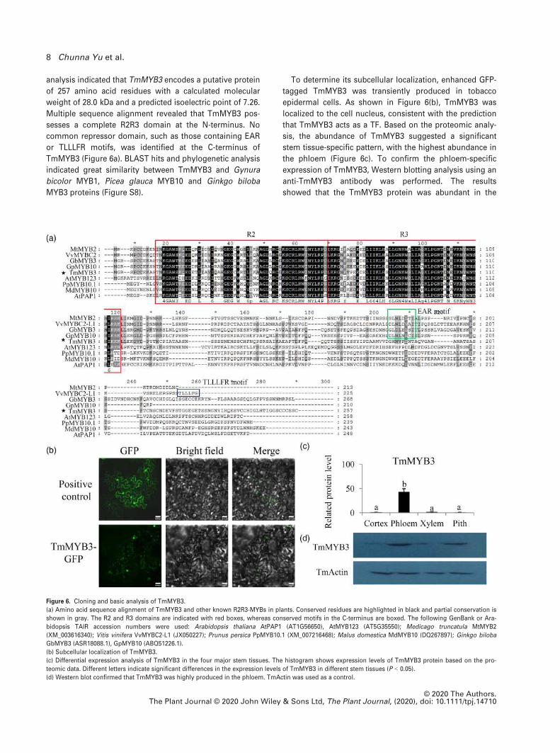
analysis indicated that TmMYB3 encodes a putative protein
of 257 amino acid residues with a calculated molecular
weight of 28.0 kDa and a predicted isoelectric point of 7.26.
Multiple sequence alignment revealed that TmMYB3 pos-
sesses a complete R2R3 domain at the N-terminus. No
common repressor domain, such as those containing EAR
or TLLLFR motifs, was identified at the C-terminus of
TmMYB3 (Figure 6a). BLAST hits and phylogenetic analysis
indicated great similarity between TmMYB3 and Gynura
bicolor MYB1, Picea glauca MYB10 and Ginkgo biloba
MYB3 proteins (Figure S8).
To determine its subcellular localization, enhanced GFP-
tagged TmMYB3 was transiently produced in tobacco
epidermal cells. As shown in Figure 6(b), TmMYB3 was
localized to the cell nucleus, consistent with the prediction
that TmMYB3 acts as a TF. Based on the proteomic analy-
sis, the abundance of TmMYB3 suggested a significant
stem tissue-specific pattern, with the highest abundance in
the phloem (Figure 6c). To confirm the phloem-specific
expression of TmMYB3, Western blotting analysis using an
anti-TmMYB3 antibody was performed. The results
showed that the TmMYB3 protein was abundant in the
Figure 6. Cloning and basic analysis of TmMYB3.
(a) Amino acid sequence alignment of TmMYB3 and other known R2R3-MYBs in plants. Conserved residues are highlighted in black and partial conservation is
shown in gray. The R2 and R3 domains are indicated with red boxes, whereas conserved motifs in the C-terminus are boxed. The following GenBank or Ara-
bidopsis TAIR accession numbers were used: Arabidopsis thaliana AtPAP1 (AT1G56650), AtMYB123 (AT5G35550); Medicago truncatula MtMYB2
(XM_003616340); Vitis vinifera VvMYBC2-L1 (JX050227); Prunus persica PpMYB10.1 (XM_007216468); Malus domestica MdMYB10 (DQ267897); Ginkgo biloba
GbMYB3 (ASR18088.1), GpMYB10 (ABQ51226.1).
(b) Subcellular localization of TmMYB3.
(c) Differential expression analysis of TmMYB3 in the four major stem tissues. The histogram shows expression levels of TmMYB3 protein based on the pro-
teomic data. Different letters indicate significant differences in the expression levels of TmMYB3 in different stem tissues (P < 0.05).
(d) Western blot confirmed that TmMYB3 was highly produced in the phloem. TmActin was used as a control.
© 2020 The Authors.The Plant Journal © 2020 John Wiley & Sons Ltd, The Plant Journal, (2020), doi: 10.1111/tpj.14710
8 Chunna Yu et al.
phloem (Figure 6c,d), which was consistent with the pro-
teomic results.
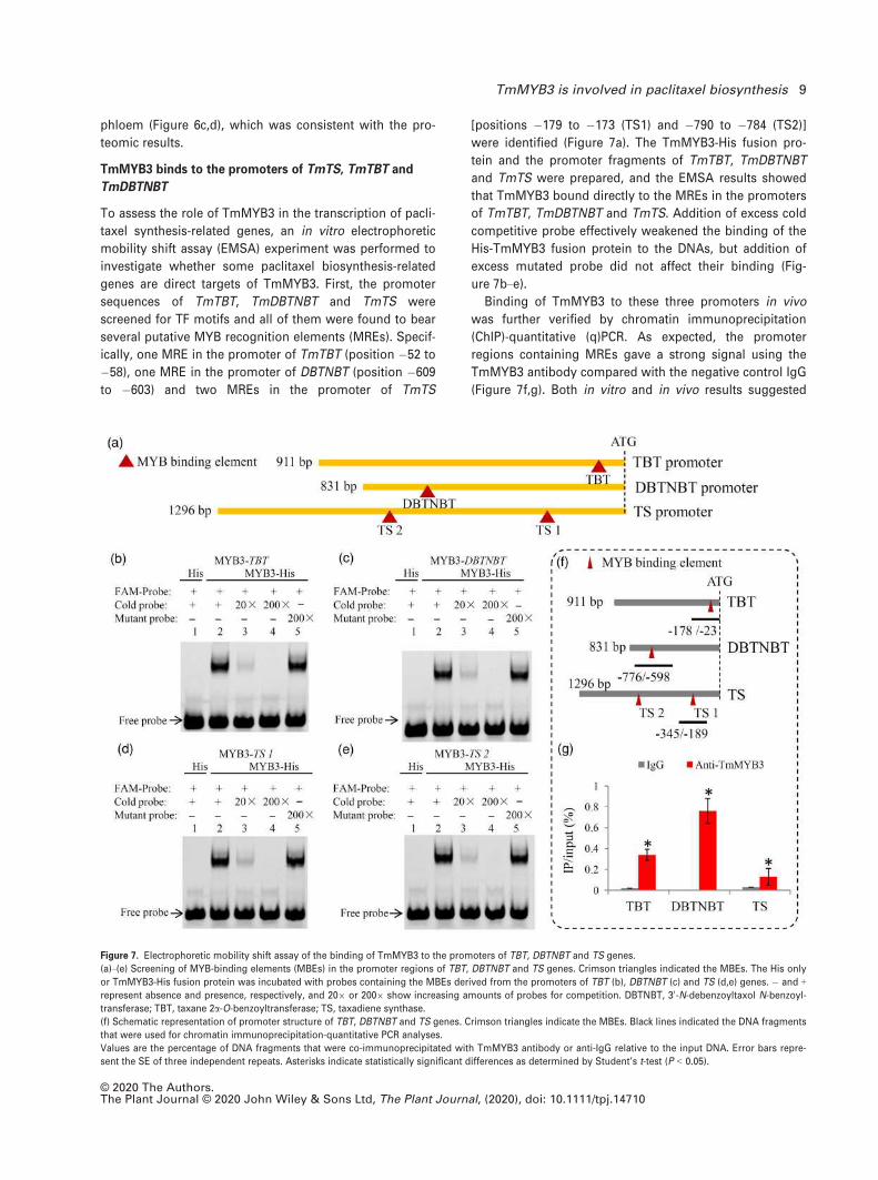
TmMYB3 binds to the promoters of TmTS, TmTBT and
TmDBTNBT
To assess the role of TmMYB3 in the transcription of pacli-
taxel synthesis-related genes, an in vitro electrophoretic
mobility shift assay (EMSA) experiment was performed to
investigate whether some paclitaxel biosynthesis-related
genes are direct targets of TmMYB3. First, the promoter
sequences of TmTBT, TmDBTNBT and TmTS were
screened for TF motifs and all of them were found to bear
several putative MYB recognition elements (MREs). Specif-
ically, one MRE in the promoter of TmTBT (position �52 to
�58), one MRE in the promoter of DBTNBT (position �609
to �603) and two MREs in the promoter of TmTS
[positions �179 to �173 (TS1) and �790 to �784 (TS2)]
were identified (Figure 7a). The TmMYB3-His fusion pro-
tein and the promoter fragments of TmTBT, TmDBTNBT
and TmTS were prepared, and the EMSA results showed
that TmMYB3 bound directly to the MREs in the promoters
of TmTBT, TmDBTNBT and TmTS. Addition of excess cold
competitive probe effectively weakened the binding of the
His-TmMYB3 fusion protein to the DNAs, but addition of
excess mutated probe did not affect their binding (Fig-
ure 7b–e).Binding of TmMYB3 to these three promoters in vivo
was further verified by chromatin immunoprecipitation
(ChIP)-quantitative (q)PCR. As expected, the promoter
regions containing MREs gave a strong signal using the
TmMYB3 antibody compared with the negative control IgG
(Figure 7f,g). Both in vitro and in vivo results suggested
Figure 7. Electrophoretic mobility shift assay of the binding of TmMYB3 to the promoters of TBT, DBTNBT and TS genes.
(a)–(e) Screening of MYB-binding elements (MBEs) in the promoter regions of TBT, DBTNBT and TS genes. Crimson triangles indicated the MBEs. The His only
or TmMYB3-His fusion protein was incubated with probes containing the MBEs derived from the promoters of TBT (b), DBTNBT (c) and TS (d,e) genes. � and +represent absence and presence, respectively, and 209 or 2009 show increasing amounts of probes for competition. DBTNBT, 30-N-debenzoyltaxol N-benzoyl-
transferase; TBT, taxane 2a-O-benzoyltransferase; TS, taxadiene synthase.
(f) Schematic representation of promoter structure of TBT, DBTNBT and TS genes. Crimson triangles indicate the MBEs. Black lines indicated the DNA fragments
that were used for chromatin immunoprecipitation-quantitative PCR analyses.
Values are the percentage of DNA fragments that were co-immunoprecipitated with TmMYB3 antibody or anti-IgG relative to the input DNA. Error bars repre-
sent the SE of three independent repeats. Asterisks indicate statistically significant differences as determined by Student’s t-test (P < 0.05).
© 2020 The Authors.The Plant Journal © 2020 John Wiley & Sons Ltd, The Plant Journal, (2020), doi: 10.1111/tpj.14710
TmMYB3 is involved in paclitaxel biosynthesis 9

that TmTBT, TmDBTNBT and TmTS might be downstream
target genes of TmMYB3.
TmMYB3 enhances the expression of TmTBT and TmTS
To determine the transcriptional activities of TmMYB3
in vivo, dual-luciferase reporter assays were performed in
Nicotiana benthamiana leaves (Figures 8a and S9). Dual-lu-
ciferase analysis showed that TmMYB3 effectively acti-
vated the expression of TmTBT and TmTS pro-LUC
reporter genes compared with the empty construct, indi-
cating that TmMYB3 functions as a transcriptional activator
by binding directly to the promoters of TmTBT and TmTS
genes. However, addition of TmMYB3 did not activate the
expression of the TmDBTNBT gene (Figure 8b,c).
DISCUSSION
The stem bark of Taxus trees is a major source for pacli-
taxel extraction (Shen et al., 2001; Uniyal, 2013). Increasing
market demand for paclitaxel has led to vast amounts of
illegal logging, destroying thousands of rare Taxus trees
(Li et al., 2012b). Despite its importance, current knowledge
about the metabolism and proteome dynamics of Taxus
species is very limited.
Recently, a number of taxoids have been isolated from
Taxus trees, and their distribution and levels vary largely
among different tissues (Ge et al., 2010). A previous study
showed great differences in the content of paclitaxel and
10-DAB among needle, stem and root tissues (Mubeen
et al., 2018). The distribution and amounts of paclitaxel vary
greatly in different shoot parts of T. cuspidata (Fett Neto
and DiCosmo, 1992). However, the composition of taxoids
across the whole stem of T. media has remained poorly
understood due to a lack of stem-specific studies. Herein,
molecular fingerprints of four major stem tissue types (cor-
tex, phloem, xylem and pith) were explored using an untar-
geted metabolomic method. Studies on all major tissues in
the context of the whole stem could reveal the stem tissue
specificity of medicinal compound accumulation.
The phloem is an important stem tissue for the biosyn-
thesis and transport of secondary metabolites (Choudhary
Figure 8. Transcriptional activation ability of TmMYB3.
(a) Schematic view of the constructs used for transient expression analysis and detailed information about plasmid combinations of dual REN/LUC reporters
and effectors. The promoter fragments of TmTS, TmDBTNBT and TmTBT were cloned into pGreenII 0800-LUC vector to generate the reporter constructs. The
effectors were generated by recombining the TmMYB3 gene into the pGreenII 62SK vector. CPMV, cowpea mosaic virus; LUC, firefly luciferase; REN, Renilla
luciferase; UTR, untranslated regions.
(b) The dual luciferase assays in tobacco leaves showed that co-transformation of TmMYB3 activates both TmTS and TmTBT promoters. Representative pictures
were taken: 1, pTS+62SK/GAL4BD; 2, pTS+62SK/TmMYB3; 3, pTBT+62SK/GAL4BD; 4, pTBT+62SK/TmMYB3; 5, pDBTNBT+62SK/GAL4BD; 6, pDBTNBT+62SK/TmMYB3.
(c) The transcriptional activation ability of TmMYB3 is indicated by the LUC/REN ratio. The pBD that produced the GAL4 DNA-binding domain alone was used
as a negative control. Each value is the mean � SE of three biological repeats. The asterisk (*) represents significant differences (P < 0.05).
© 2020 The Authors.The Plant Journal © 2020 John Wiley & Sons Ltd, The Plant Journal, (2020), doi: 10.1111/tpj.14710
10 Chunna Yu et al.
et al., 2016). In Norway spruce, phenolic stilbene gluco-
sides, including astringin, isorhapontin and piceid, are
mainly accumulated in the phloem (Li et al., 2012a; Jyske
et al., 2016). In T. media, paclitaxel, as well as 10-DAB and
DAP, accumulate at high levels in the phloem (Figure 1c),
suggesting that this tissue may be the most significant
source for paclitaxel extraction. Although the stem bark is
a major source for isolation of taxoids, the pith contains
high levels of BAC (Lin et al., 2018). This suggests that the
barkless trunk of Taxus trees might be a valuable material
for extraction of BAC, and could be a cost-effective source
of the key paclitaxel precursor (Lin et al., 2018).
A broad range of taxusin-like metabolites are considered
dead-end metabolites rather than intermediates leading to
paclitaxel formation (Croteau et al., 2006). A synthetic
study demonstrated that a number of putative paclitaxel
biosynthesis metabolites could be prepared by Barton
deoxygenation of the C9 and C10 hydroxyl groups of pro-
tected derivatives of taxusin (Li et al., 2008). For many
years, taxusin-like metabolites have been thought to pre-
dominantly accumulate in yew heartwood (Banskota et al.,
2003; Yu et al., 2018). Our metabolomic results showed
that taxusin-like metabolites predominantly accumulated
in the pith rather than xylem of T. media. Knowledge of
the precise distribution of taxusin and its analogues in the
stem tissues will be useful for exploring the utilization of
prominent side-route metabolites in Taxus trees.
The phloem is a key stem tissue that mediates stress tol-
erance by affecting plant–environment interactions (Sav-
age et al., 2016). A number of secondary metabolites, such
as flavonoids, play essential roles in the responses to abi-
otic and biotic stresses (Brunetti et al., 2018). Flavonoids
accumulate highly in the phloem of T. media, suggesting
that activation of the biosynthesis of flavonoids might play
a role in phloem-mediated resistance to environmental
stresses (Agati et al., 2012). Analysis of the chemical com-
position showed that both the phloem and cortex were
similar in terms of primary and secondary metabolites,
whereas the numbers of pith-specific accumulated lipids
(27 metabolites) and terpenoids (67 metabolites) were lar-
ger than in other stem tissues. Due to the differential
metabolite composition of different stem tissues, the
whole stem has broad potential for biotechnological appli-
cations.
Interestingly, the localizations of enzymes and the inter-
mediate metabolites involved in paclitaxel biosynthesis are
inconsistent in the T. media stem tissues. For example, TS
is produced in the phloem and its downstream product
taxa-4(5),11(12)-diene basically accumulates in the xylem.
Another important intermediate, 10-DAB, accumulates in
the phloem, but its downstream product BAC accumulates
in the pith (Figures 4 and S6). Our data indicate the move-
ments of the intermediates from one tissue to another.
Similar to the taxoids, gibberellins (GAs) belong to another
major class of diterpenoids, and the movements of GAs
have been well studied in recent years (Binenbaum et al.,
2018). Several previous studies have demonstrated the
presence of GAs in the phloem sap and the movement of
GAs through the phloem (Hoad and Bowen, 1968). GA12
moved through the xylem in a root-to-shoot direction but
through the phloem in a shoot-to-root manner (Regnault
et al., 2015). Due to the ability to move out of cells, the
existence of GA efflux transporters is predicted for GA to
effectively move locally, at both tissue and cellular levels
(Kramer, 2006). However, taxoid efflux transporters have
not yet been identified. In addition to active transport, the
ability of taxoids to diffuse through membranes may be
another explanation for the movements of taxoids from
one tissue to another.
Metabolic engineering has the potential to achieve
higher paclitaxel output (Wilson and Roberts, 2014).
Improving the production yield of paclitaxel depends criti-
cally upon a deep understanding of the regulation of the
paclitaxel biosynthesis pathway (Li et al., 2013). Transcrip-
tion factors can increase the accumulation of bioactive
ingredients in medicinal plants by regulating genes in a
specific biosynthetic pathway (Zhou et al., 2019b). As a cul-
tivated Taxus species with a high paclitaxel content, T. me-
dia is a major source for industrial production of paclitaxel
(Shen et al., 2016). Our previous studies identified most
genes in the paclitaxel biosynthesis pathway of T. media,
providing a solid foundation for further studies on the tran-
scriptional regulation of paclitaxel biosynthesis (Hao et al.,
2017; Yu et al., 2017).
In other Taxus species, several TFs, including MYCs in
T. cuspidata and T. chinensis, WRKYs in T. chinensis and
ERFs in T. chinensis, were deemed to be involved in the
paclitaxel biosynthesis pathway (Li et al., 2013; Lenka
et al., 2015; Zhang et al., 2015; 2018a; Cui et al., 2019). To
date, no MYB TFs have been functionally identified in
Taxus species. Herein, a R2R3-MYB TF, TmMYB3, was
identified in T. media. In plants, secondary metabolism is
regulated by a complex regulatory network in which MYBs
are important components (Chezem and Clay, 2016). Previ-
ous studies showed that R2R3-MYB TFs mediate terpenoid
biosynthesis by regulating the expression of MEP pathway
genes such as DXS, DXR and GGPPS (Zhang et al., 2017).
In Stevia rebaudiana Bert, MYB TFs are thought to control
the downstream metabolic flux of MEP and mevalonate
pathways (Singh et al., 2017). Paclitaxel is a bioactive sub-
stance built around a diterpenoid taxane core, and the
expression of key genes involved in paclitaxel biosynthesis
might be regulated by MYB TFs (Croteau et al., 2006).
Sequence-specific DNA-binding sites of R2R3-MYB have
been demonstrated in model plants (Romero et al., 1998).
Due to the unavailability of the Taxus genome, cloning of
promoter sequences is needed to explore the relationships
between TFs and their target genes. Using T. cuspidata
© 2020 The Authors.The Plant Journal © 2020 John Wiley & Sons Ltd, The Plant Journal, (2020), doi: 10.1111/tpj.14710
TmMYB3 is involved in paclitaxel biosynthesis 11
suspension cells, promoters of seven known paclitaxel
pathway genes have been isolated (Lenka et al., 2015). In
T. chinensis, a 550 bp 50-flanking sequence of the TS gene
and a 1740 bp 50-flanking sequence of the DBAT gene have
been reported (Li et al., 2015). Additionally, the role of a
Y-patch promoter region of the BAPT gene has been
revealed using Taxus cell cultures (Sanchez-Munoz et al.,
2018). To verify the target genes of TmMYB3, we isolated
the promoter sequences of five important paclitaxel
biosynthesis-related genes. After scanning the MREs, three
potential target genes of TmMYB3, TS, TBT and DBTNBT,
were identified in T. media (Figure 7a). Taxadiene synthase
catalyzes a committal step in paclitaxel biosynthesis by
producing the taxane core from the intermediate GGPP
(Edgar et al., 2017), TBT catalyzes the conversion of 10-dea-
cetyl-2-debenzoylbaccatin III to 10-DAB (Walker and
Croteau, 2000) and DBTNBT catalyzes the conversion of 30-N-debenzoyltaxol into the target compound, paclitaxel
(Nasiri et al., 2016). All three enzymes play essential roles
in paclitaxel biosynthesis (Croteau et al., 2006). Herein, at
least one potential MBE was identified in the promoter
sequences of TS, TBT and DBTNBT genes, and EMSA
assays confirmed the binding of TmMYB3 to these three
promoters (Figure 7). Although physical binding occurs,
exactly how TmMYB3 regulates downstream target genes
by activation or repression still needs to be addressed. For
example, TcMYC2 in T. chinensis is a positive regulator of
TS, TAT, DBTNBT, T13OH and T5OH genes (Zhang et al.,
2018b), and two WRKYs in T. chinensis, TcWRKY8 and
TcWRKY47, function as transcriptional activators in pacli-
taxel biosynthesis (Zhang et al., 2018a). Two JA-responsive
factors, TcERF12 and TcERF15, act as a repressor and an
activator of the TS gene, respectively (Zhang et al., 2015).
In our present study, dual-luciferase transient expression
analysis demonstrated the transcriptional activation activ-
ity of TmMYB3 against the promoters of the TS and TBT
genes, but not the DBTNBT gene (Figure 8). Although most
of the intermediate metabolites involved in paclitaxel
biosynthesis might be produced in the phloem, several
intermediate metabolites accumulated in different stem tis-
sues. Interestingly, the downstream product of TS, taxa-4
(5),11(12)-diene, was transferred to the xylem, and the
upstream product of TBT, 10-deacetyl-2-debenzoylbaccatin
III, migrated to the pith and cortex. Taken together, our
results revealed that TmMYB3 is a positive regulator
involved in paclitaxel biosynthesis, possibly by activating
the expression of TmTBT and TmTS. Moreover, possible
movements of TS- and TBT-related products among differ-
ent stem tissues are predicted (Figure 9).
In conclusion, the integration of proteomic and metabo-
lomic profiling of Taxus stem tissues provides a molecular
framework for revealing the mechanism underlying tissue-
specific accumulation of medicinal compounds such as
taxoids. The differential metabolite compositions of
different stem tissues highlight the broad potential for
biotechnological applications. Moreover, as a phloem-
specific TF, TmMYB3 has a significant effect on paclitaxel
biosynthesis by activating the expression of the TS and
TBT genes, providing a potential explanation for the
phloem-specific accumulation of paclitaxel.
EXPERIMENTAL PROCEDURES
Plant materials and sampling
Five-year-old T. media (cultivar ‘Zike’) plants were cultivated in anexperimental field within the campus of Hangzhou Normal Univer-sity, Hangzhou, China. In our study, four different stem tissues,including cortex, phloem, xylem and pith, were tested. Indepen-dent samples from the different stem tissues were used for meta-bolomic analysis (25 mg each, n = 10) and proteomic analysis(50 mg each, n = 3). After the isolation of stem tissues, cross sec-tions were observed under a microscope to localize the anatomi-cal position. Young stems of 5-year-old T. media trees werecollected and prepared for microsection observation according toa previously published paper (Chen et al., 2014). All slides weremounted with synthetic resin and observed under a LSM510 laserscanning system (Carl Zeiss, https://www.zeiss.com/).
Quantitative UPLC-MS/MS and untargeted metabolomic
analyses
Fresh stems were harvested from T. media plants (n = 6), and fourmajor tissues of each stem, including cortex, phloem, xylem andpith, were prepared by peeling and scraping. The tissue sampleswere dried at 40°C and ground into a fine powder. Our previouslypublished method was used for the preparation of crude extractsand UPLC-MS/MS quantitative analysis (Yu et al., 2018). For themetabolomic analysis, metabolite extraction, UPLC-MS/MS analy-sis and bioinformatic analysis were performed according to ourprevious work (Yu et al., 2018).
Proteomic analysis
For proteomic analysis, protein extraction, trypsin digestion, Tan-dem Mass Tag (TMT) labeling, peptide fractionation, LC-MS/MSanalysis, protein annotation and functional enrichment were per-formed according to our previous work (Hao et al., 2017). The MS/MS data generated by MaxQuant with integrated Andromedasearch engine v.1.5.2.8 were searched against the transcriptomeof T. media (NCBI accession number GSM3438662).
Promoter isolation by chromosome walking
High-quality T. media DNA was isolated from young twigs usingthe classical cetyl trimethyl ammonium bromide method. Theupstream promoter regions of TBT, TS, DBTNBT, T7OH andT13OH genes were amplified using a Universal Genome WalkerKit (Clontech, https://www.takarabio.com/). In brief, genomic DNAwas digested with four blunt-end restriction enzymes, EcoRV,PvuII, DraI and StuI, and the digested products were ligated withGenome Walker adaptors to produce templates for first-roundnested PCR. The products of first-round PCR were used as tem-plates in secondary PCR with nested primers. The PCR productswere ligated with the pMD18-T vector (TaKaRa, https://www.takarabio.com/) and sequences were analyzed using the PlantCAREprogram (http://bioinformatics.psb.ugent.be/webtools/plantcare/html/). The sequences of primers used for the chromosome walk-ing are listed in Table S1.
© 2020 The Authors.The Plant Journal © 2020 John Wiley & Sons Ltd, The Plant Journal, (2020), doi: 10.1111/tpj.14710
12 Chunna Yu et al.
Isolation of full-length TmMYB3 by 50/30-RACE
The partial TmMYB3 gene sequence was identified from theT. media transcriptome, and full-length TmMYB3 cDNA wasobtained using a SMARTer RACE 50/30 Kit (Clontech) according tothe manufacturer’s manual. In brief, total RNAs were extractedfrom young twigs of T. media using TRIZOL reagent (Qiagen,https://www.qiagen.com/). A mixture containing 1 lg of RNA, 1 llof 50 CDS primer, 1 ll of SMARTer II A oligonucleotide and dou-ble-distilled H2O to the required final volume was served as the 50-RACE-Ready cDNA solution. Master Mix solution was preparedusing 59 First Strand buffer (4 ll), 100 mM dithiothreitol (0.5 ll),20 mM dNTP mix (1 ll), RNase inhibitor (20 U) and SMARTScribeReverse Transcriptase (200 U). The Master Mix was added to the50-RACE-Ready cDNA solution, and the mixture was incubated at42°C for 90 min. The reaction was stopped by heating at 70°C for10 min. Sequences of primers used for 50-RACE are listed inTable S1.
Subcellular localization and phylogenetic analysis of
TmMYB3
The full-length sequence of TmMYB3 was cloned into thepH7FWG2.0 vector with an artificial green fluorescent protein
(GFP) gene fused at the N-terminus. Sequences of primers usedfor the GFP-TmMYB3 construct are listed in Table S1. All vectorswere transiently expressed in N. benthamiana epidermal cells byAgrobacterium tumefaciens (GV3101)-mediated transformation.The GFP fluorescence of the fusion protein was detected using aLSM710 confocal microscope (Carl Zeiss).
The TmMYB3 protein sequence and other tryptic MYB proteinsfrom different plants were used for multiple sequence alignmentsusing CLUSTALW with default parameters. An unrooted phyloge-netic tree was constructed using MEGA 6.1 (http://www.megasoftware.net/) employing the neighbor-joining method.
Preparation of TmMYB3-specific polyclonal antibody and
Western blotting
The full-length TmMYB3 gene was inserted into the pET30a vectorand transformed into Escherichia coli Rosetta (DE3) to producerecombinant TmMYB3 protein with a His tag. After inducing with1 mM isopropyl b-D-1-thiogalactopyranoside at 30°C for 5 h, therecombinant protein was isolated and purified using His60 NiSuperflow Resin (Clontech) according to the manufacturer’sinstructions. His-tagged fusion proteins were separated by SDS-PAGE and bands of interest were used for preparation ofTmMYB3-specific polyclonal antibody. The expression of the
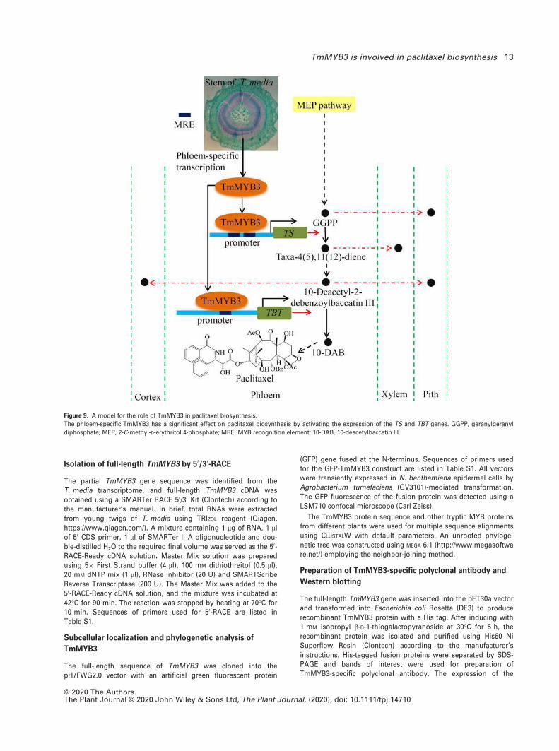
Figure 9. A model for the role of TmMYB3 in paclitaxel biosynthesis.
The phloem-specific TmMYB3 has a significant effect on paclitaxel biosynthesis by activating the expression of the TS and TBT genes. GGPP, geranylgeranyl
diphosphate; MEP, 2-C-methyl-D-erythritol 4-phosphate; MRE, MYB recognition element; 10-DAB, 10-deacetylbaccatin III.
© 2020 The Authors.The Plant Journal © 2020 John Wiley & Sons Ltd, The Plant Journal, (2020), doi: 10.1111/tpj.14710
TmMYB3 is involved in paclitaxel biosynthesis 13

TmMYB3 protein in distinct stem tissues was tested by Westernblotting analysis. Total proteins were extracted from differentstem tissues (100 mg each). About 10 µg of total soluble proteinsfor each tissue were separated on 12% SDS-PAGE gels and trans-ferred to a polyvinyl difluoride membrane. The Western blottinganalysis was performed according to a previous work (Liu et al.,2013).
Electrophoretic mobility shift assay and ChIP-qPCR
analysis
Electrophoretic mobility shift assay was performed as previouslydescribed (Hellman and Fried, 2007). Briefly, probes containingMYB-specific cis-elements (CAGTTA and TGGTTA) derived fromthe promoters of TBT, TS and DBTNBT genes were labeled with506-FAM (FITC) fluorescent dye. Unlabeled probes were used incompetition assays. MYB-specific cis-elements within probes werechanged into CCCGGG and used in mutation assays. The probesequences used for EMSA are listed in Table S1.
Taxus media samples were treated with 1% formic acid (FA)to cross-link genomic DNA and TmMYB3 protein. Chromatin wascut into pieces with an average length of 500 bp by sonicationon ice, and immunoprecipitation of TmMYB3 cross-linked withDNA was carried out using affinity-purified TmMYB3 antibody at4°C overnight. Preimmune IgG was used as a negative control.The cross-linked complex was isolated on Protein A/agarosebeads following a 1 h incubation at 4°C. Beads were washed for10 min at 4°C with low-salt, high-salt, LiCl and TRIS-EDTA wash-ing buffers, and the immunoprecipitated sample was eluted bygentle rotation for 15 min at 65°C. Cross-linking of immunopre-cipitated DNA was reversed by incubation in 0.2 M NaCl at 65°Covernight. The amount of each precipitated DNA fragment wasdetermined by quantitative real-time PCR (qRT-PCR) (Yu et al.,2017).
Dual-luciferase reporter assay
For transcription activity analysis, the full-length cDNA ofTmMYB3 was inserted into the pBD vector under the control ofthe 35S promoter as effector (pBD-TmMYB3). A GAL4-LUC repor-ter and an internal control REN (Renilla luciferase) reporter underthe control of the 35S promoter were constructed as double-re-porter vectors. To determine the binding activity of TmMYB3 tothe promoters of paclitaxel biosynthesis-related genes, the pro-moters of TS, TBT and DBTNBT were cloned into the pGreenII0800-LUC double-reporter vector. The resulting effector andreporter plasmids were co-transformed into tobacco leaves byA. tumefaciens strain GV3101 as described previously (Zhu et al.,2017). The LUC and REN luciferase activities were determinedusing a dual luciferase assay kit (Promega, https://www.promega.com/) according to the manufacturer’s instructions. The bind-ing activities of TmMYB3 to the selected promoters were calcu-lated from the LUC/REN ratio (assured in triplicate). Sequencesof primers used for the dual-luciferase reporter assay are listedin Table S1.
Statistical analysis
For untargeted metabolomic analysis, the Wilcoxon test was per-formed to identify DAMs in each sample comparison, and super-vised partial least squares-discriminant analysis was performed toanalyze the different variables between two sample groups. Fortranscriptomic analysis, P-values, which were used as a thresholdto determine differentially expressed genes in each sample com-parison, were produced by false discovery rate analysis and
adjusted using the Benjamini–Hochberg method. The statisticalanalysis was carried out using SPSS software v.19.0 (https://spss.software.informer.com/). A one-way analysis of variance wascarried out to compare the content differences of taxoids betweentwo sample groups. A P-value < 0.05 was considered statisticallysignificant.
ACCESSION NUMBERS
The metabolomic datasets generated and analyzed during
the current study are available in the ‘Baidu Netdisk’
(https://pan.baidu.com/s/1AlkJRQYcao-oxbx3Q_iO2Q) with
extraction code ytsc. The MS proteomic data have been set
to the ProteomeXchange Consortium by the PRIDE partner
repository program under identifier PXD008871. The tran-
scriptomes of T. media used for proteome annotation have
been set to the NCBI database under accession number
GSM3438662.
ACKNOWLEDGEMENTS
This work was funded by Zhejiang Provincial Natural ScienceFoundation of China under grant no. LY19C160001, the OpenFoundation of State Key Laboratory of Subtropical Silviculture,Zhejiang A & F University (KF201708), Major Increase or DecreaseProgram in the Central Finance Level grant no. 2060302 and Zhe-jiang Provincial Key Research & Development Project grants2017C02011 and 2018C02030. We are also grateful to LC Sciencescompany (Hangzhou, China) for metabolomic analysis and PTMBiolabs Inc. (Hangzhou, China) for proteomic analysis.
CONFLICT OF INTEREST
The authors declare no conflict of interest.
AUTHOR CONTRIBUTIONS
CY, BZ, HW and CS designed the research. CY, XX and HY
performed the metabolite and protein extractions. XL and
SF did the sampling for quantification of four different tax-
oids. XL and CZ performed microsection observation. XL
and XX took care of the plants. CY, XL, CZ, YC and XZ per-
formed targeted UPLC-MS/MS analysis. CY, XL, CZ, XZ,
JX, XX and SF performed proteomic analysis. CY and XX
carried out promoter isolation by chromosome walking
and full-length TmMYB3 isolation by 50/30-RACE. CY, XL
and HY performed subcellular localization and phyloge-
netic analysis. CY, XL, CZ, XX and XZ did TmMYB3-specific
polyclonal antibody production and Western blotting. LZ
performed the EMSA and ChIP-qPCR analysis. HW and CS
wrote the manuscript.
SUPPORTING INFORMATION
Additional Supporting Information may be found in the online ver-sion of this article.
Figure S1. Quality control parameters of the metabolomes.
Figure S2. Protein electrophoresis photograph of isolated proteinsamples.
Figure S3. Quality control parameters of the proteomes.
Figure S4. Checking the statistical consistency.
© 2020 The Authors.The Plant Journal © 2020 John Wiley & Sons Ltd, The Plant Journal, (2020), doi: 10.1111/tpj.14710
14 Chunna Yu et al.

Figure S5. Analysis of differentially produced proteins among thefour stem tissues.
Figure S6. The movements of intermediates among different stemtissues.
Figure S7. The promoter sequences of five important paclitaxelbiosynthesis-related genes.
Figure S8. Phylogenetic analysis of TmMYB3 and several knownMYBs from other plants.
Figure S9. The positive controls of dual-luciferase reporter assays.
Table S1. The primer sequences used in the present study.
Table S2. Detailed information about the data from the untargetedmetabolomes.
Table S3. A total of 5077 annotated metabolites were mapped intodifferent known metabolic pathways.
Table S4. The annotation information for all the identified pro-teins.
Table S5. The detailed information on the phloem-specificexpressed proteins.
Table S6. Proteomic analysis identified nine classical transcriptionfactors.
REFERENCES
Agati, G., Azzarello, E., Pollastri, S. and Tattini, M. (2012) Flavonoids as
antioxidants in plants: Location and functional significance. Plant Sci.
196, 67–76.Banskota, A.H., Tezuka, Y., Nguyen, N.T., Awale, S., Nobukawa, T. and
Kadota, S. (2003) DPPH radical scavenging and nitric oxide inhibitory
activities of the constituents from the wood of Taxus yunnanensis.
Planta Med. 69, 500–505.Bernabeu, E., Cagel, M., Lagomarsino, E., Moretton, M. and Chiappetta,
D.A. (2017) Paclitaxel: What has been done and the challenges remain
ahead. Int. J. Pharm. 526, 474–495.Binenbaum, J., Weinstain, R. and Shani, E. (2018) Gibberellin localization
and transport in plants. Trends Plant Sci. 23, 410–421.Brunetti, C., Fini, A., Sebastiani, F., Gori, A. and Tattini, M. (2018) Modula-
tion of phytohormone Signaling: a primary function of flavonoids in
plant-environment interactions. Front. Plant Sci. 9, 1042.
Chan, K.C., Alvarado, A.B., McGuire, M.T., Muschik, G.M., Issaq, H.J. and
Snader, K.M. (1994) High-performance liquid chromatography and micel-
lar electrokinetic chromatography of taxol and related taxanes from bark
and needle extracts of Taxus species. J. Chromatogr. B, Biomed. Appl.
657, 301–306.Chen, Y.J., Liang, Z.T., Zhu, Y., Xie, G.Y., Tian, M., Zhao, Z.Z. and Qin, J.Q.
(2014) Tissue-specific metabolites profiling and quantitative analyses of
flavonoids in the rhizome of Belamcanda chinensis by combining laser-
microdissection with UHPLC-Q/TOF-MS and UHPLC–QqQ-MS. Talanta,
130, 585–597.Chezem, W.R. and Clay, N.K. (2016) Regulation of plant secondary metabo-
lism and associated specialized cell development by MYBs and bHLHs.
Phytochemistry, 131, 26–43.Choudhary, S.B., Kumar, M., Chowdhury, I., Singh, R.K., Pandey, S.P.,
Sharma, H.K. and Karmakar, P.G. (2016) An efficient and cost effective
method of RNA extraction from mucilage, phenol and secondary
metabolite rich bark tissue of tossa jute (C. olitorius L.) actively develop-
ing phloem fiber. 3 Biotech, 6, 100.
Croteau, R., Ketchum, R.E., Long, R.M., Kaspera, R. and Wildung, M.R. (2006)
Taxol biosynthesis and molecular genetics. Phytochem. Rev. 5, 75–97.Cui, Y., Mao, R., Chen, J. and Guo, Z. (2019) Regulation mechanism of MYC
family transcription factors in jasmonic acid signalling pathway on taxol
biosynthesis. Int. J. Mol. Sci. 20, pii:E1843.
Dubos, C., Stracke, R., Grotewold, E., Weisshaar, B., Martin, C. and Lepi-
niec, L. (2010) MYB transcription factors in Arabidopsis. Trends Plant Sci.
15, 573–581.Edgar, S., Li, F.S., Qiao, K., Weng, J.K. and Stephanopoulos, G. (2017) Engi-
neering of taxadiene synthase for improved selectivity and yield of a key
taxol biosynthetic intermediate. ACS Synth. Biol. 6, 201–205.
Eisenreich, W., Menhard, B., Hylands, P.J., Zenk, M.H. and Bacher, A. (1996)
Studies on the biosynthesis of taxol: the taxane carbon skeleton is not of
mevalonoid origin. Proc. Natl Acad. Sci. USA, 93, 6431–6436.Fett Neto, A.G. and DiCosmo, F. (1992) Distribution and amounts of taxol in
different shoot parts of Taxus cuspidata. Planta Med. 58, 464–466.Ge, G.B., Liang, S.C., Hu, Y., Liu, X.B., Mao, Y.X., Zhang, Y.Y., Luan, H.W.,
Qiu, M.H. and Yang, L. (2010) Rapid qualitative and quantitative determi-
nation of seven valuable taxanes from various Taxus species by UFLC-
ESI-MS and UFLC-DAD. Planta Med. 76, 1773–1777.Hao, D.C., Xiao, P.G., Ge, G.B. and Liu, M. (2012) Biological, chemical, and
omics research of Taxus medicinal resources. Drug Dev. Res. 73, 477–486.
Hao, J., Guo, H., Shi, X., Wang, Y., Wan, Q., Song, Y.B., Zhang, L., Dong, M.
and Shen, C. (2017) Comparative proteomic analyses of two Taxus spe-
cies (Taxus 9 media and Taxus mairei) reveals variations in the metabo-
lisms associated with paclitaxel and other metabolites. Plant Cell
Physiol. 58, 1878–1890.Hellman, L.M. and Fried, M.G. (2007) Electrophoretic mobility shift assay
(EMSA) for detecting protein-nucleic acid interactions. Nat. Protoc. 2,
1849–1861.Hoad, G.V. and Bowen, M.R. (1968) Evidence for gibberellin-like substances
in phloem exudate of higher plants. Planta, 82, 22–32.Jyske, T., Kuroda, K., Suuronen, J.-P., Pranovich, A., Roig-Juan, S., Aoki, D.
and Fukushima, K. (2016) In planta localization of stilbenes within Picea
abies phloem. Plant Physiol. 172, 913–928.Kaspera, R. and Croteau, R. (2006) Cytochrome P450 oxygenases of Taxol
biosynthesis. Phytochem. Rev. 5, 433–444.Koepp, A.E., Hezari, M., Zajicek, J., Vogel, B.S., LaFever, R.E., Lewis, N.G.
and Croteau, R. (1995) Cyclization of geranylgeranyl diphosphate to taxa-
4(5),11(12)-diene is the committed step of taxol biosynthesis in Pacific
yew. J. Biol. Chem. 270, 8686–8690.Kramer, E.M. (2006) How far can a molecule of weak acid travel in the apo-
plast or xylem? Plant Physiol. 141, 1233–1236.Kuang, X., Sun, S., Wei, J., Li, Y. and Sun, C. (2019) Iso-Seq analysis of the
Taxus cuspidata transcriptome reveals the complexity of Taxol biosyn-
thesis. BMC Plant Biol. 19, 210–210.Lenka, S.K., Nims, N.E., Vongpaseuth, K., Boshar, R.A., Roberts, S.C. and
Walker, E.L. (2015) Jasmonate-responsive expression of paclitaxel
biosynthesis genes in Taxus cuspidata cultured cells is negatively regu-
lated by the bHLH transcription factors TcJAMYC1, TcJAMYC2, and
TcJAMYC4. Front. Plant Sci. 6, 115.
Li, H., Horiguchi, T., Croteau, R. and Williams, R.M. (2008) Studies on Taxol
biosynthesis: preparation of taxadiene-diol- and triol-derivatives by
deoxygenation of taxusin. Tetrahedron, 64, 6561–6567.Li, S.H., Nagy, N.E., Hammerbacher, A., Krokene, P., Niu, X.M., Gershenzon,
J. and Schneider, B. (2012a) Localization of phenolics in phloem par-
enchyma cells of Norway spruce (Picea abies). ChemBioChem, 13, 2707–2713.
Li, S.T., Zhang, P., Zhang, M., Fu, C.H., Zhao, C.F., Dong, Y.S., Guo, A.Y. and
Yu, L.J. (2012b) Transcriptional profile of Taxus chinensis cells in
response to methyl jasmonate. BMC Genom. 13, 295.
Li, S., Zhang, P., Zhang, M., Fu, C. and Yu, L. (2013) Functional analysis of a
WRKY transcription factor involved in transcriptional activation of the
DBAT gene in Taxus chinensis. Plant Biol. (Stuttg), 15, 19–26.Lin, S.L., Wei, T., Lin, J.F., Guo, L.Q., Wu, G.P., Wei, J.B., Huang, J.J. and
Ouyang, P.L. (2018) Bio-production of baccatin III, an important precursor
of paclitaxel by a cost-effective approach. Mol. Biotechnol. 60, 492–505.Liu, X., Yang, L., Zhou, X., Zhou, M., Lu, Y., Ma, L., Ma, H. and Zhang, Z.
(2013) Transgenic wheat expressing Thinopyrum intermedium MYB tran-
scription factor TiMYB2R-1 shows enhanced resistance to the take-all dis-
ease. J. Exp. Bot. 64, 2243–2253.Ma, D., Reichelt, M., Yoshida, K., Gershenzon, J. and Constabel, C.P. (2018)
Two R2R3-MYB proteins are broad repressors of flavonoid and phenyl-
propanoid metabolism in poplar. Plant J. 96, 949–965.Mubeen, S., Li, Z.L., Huang, Q.M., He, C.T. and Yang, Z.Y. (2018) Compara-
tive transcriptome analysis revealed the tissue-specific accumulations of
taxanes among three experimental lines of Taxus yunnanensis. J. Agric.
Food Chem. 66, 10410–10420.Nadeem, M., Rikhari, H.C., Kumar, A., Palni, L.M. and Nandi, S.K. (2002)
Taxol content in the bark of Himalayan Yew in relation to tree age and
sex. Phytochemistry, 60, 627–631.
© 2020 The Authors.The Plant Journal © 2020 John Wiley & Sons Ltd, The Plant Journal, (2020), doi: 10.1111/tpj.14710
TmMYB3 is involved in paclitaxel biosynthesis 15

Nasiri, J., Naghavi, M.R., Alizadeh, H. and Moghadam, M.R. (2016) Sea-
sonal-based temporal changes fluctuate expression patterns of TXS,
DBAT, BAPT and DBTNBT genes alongside production of associated tax-
anes in Taxus baccata. Plant Cell Rep. 35, 1103–1119.Nguyen, N.T., Banskota, A.H., Tezuka, Y., Nobukawa, T. and Kadota, S.
(2003) Diterpenes and sesquiterpenes from the bark of Taxus yunnanen-
sis. Phytochemistry, 64, 1141–1147.Onrubia, M., Moyano, E., Bonfill, M., Palazon, J., Goossens, A. and Cusido,
R.M. (2011) The relationship between TXS, DBAT, BAPT and DBTNBT
gene expression and taxane production during the development of
Taxus baccata plantlets. Plant Sci. 181, 282–287.Regnault, T., Daviere, J.M., Wild, M., Sakvarelidze-Achard, L., Heintz, D.,
Carrera Bergua, E., Lopez Diaz, I., Gong, F., Hedden, P. and Achard, P.
(2015) The gibberellin precursor GA12 acts as a long-distance growth
signal in Arabidopsis. Nat. Plants, 1, 15073.
Romero, I., Fuertes, A., Benito, M.J., Malpica, J.M., Leyva, A. and Paz-Ares,
J. (1998) More than 80R2R3-MYB regulatory genes in the genome of Ara-
bidopsis thaliana. Plant J. 14, 273–284.Sanchez-Munoz, R., Bonfill, M., Cusido, R.M., Palazon, J. and Moyano, E.
(2018) Advances in the regulation of in vitro paclitaxel production:
methylation of a Y-patch promoter region alters BAPT gene expression
in Taxus cell cultures. Plant Cell Physiol. 59, 2255–2267.Savage, J.A., Clearwater, M.J., Haines, D.F., Klein, T., Mencuccini, M.,
Sevanto, S., Turgeon, R. and Zhang, C. (2016) Allocation, stress tolerance
and carbon transport in plants: how does phloem physiology affect plant
ecology? Plant Cell Environ. 39, 709–725.Shen, Y.C., Prakash, C.V., Chen, Y.J., Hwang, J.F., Kuo, Y.H. and Chen, C.Y.
(2001) Taxane diterpenoids from the stem bark of Taxus mairei. J. Nat.
Prod. 64, 950–952.Shen, C., Xue, J., Sun, T., Guo, H., Zhang, L., Meng, Y. and Wang, H. (2016)
Succinyl-proteome profiling of a high taxol containing hybrid Taxus spe-
cies (Taxus 9 media) revealed involvement of succinylation in multiple
metabolic pathways. Sci. Rep. 6, 21764.
Singh, G., Singh, G., Singh, P. et al. (2017) Molecular dissection of transcrip-
tional reprogramming of steviol glycosides synthesis in leaf tissue dur-
ing developmental phase transitions in Stevia rebaudiana Bert. Sci. Rep.
7, 11835.
Stracke, R., Werber, M. and Weisshaar, B. (2001) The R2R3-MYB gene fam-
ily in Arabidopsis thaliana. Curr. Opin. Plant Biol. 4, 447–456.Tanaka, K., Li, F., Morikawa, K., Nobukawa, T. and Kadota, S. (2011) Analy-
sis of biosynthetic fluctuations of cultured Taxus seedlings using a meta-
bolomic approach. Phytochemistry, 72, 1760–1766.Uniyal, S.K. (2013) Bark removal and population structure of Taxus wallichi-
ana Zucc. in a temperate mixed conifer forest of western Himalaya. Envi-
ron. Monit. Assess. 185, 2921–2928.Walker, K. and Croteau, R. (2000) Taxol biosynthesis: molecular cloning of a
benzoyl-CoA:taxane 2alpha-O-benzoyltransferase cDNA from Taxus and
functional expression in Escherichia coli. Proc. Natl Acad. Sci. USA, 97,
13591–13596.Wang, Y.F., Shi, Q.W., Dong, M., Kiyota, H., Gu, Y.C. and Cong, B. (2011)
Natural taxanes: developments since 1828. Chem. Rev. 111, 7652–7709.Wang, C.Y., Tang, L., He, J.W., Li, J. and Wang, Y.Z. (2019) Ethnobotany,
phytochemistry and pharmacological properties of Eucommia ulmoides:
a review. Am. J. Chin. Med. 47, 259–300.Wilson, S.A. and Roberts, S.C. (2014) Metabolic engineering approaches for
production of biochemicals in food and medicinal plants. Curr. Opin.
Biotechnol. 26, 174–182.Yu, C., Guo, H., Zhang, Y. et al. (2017) Identification of potential genes that
contributed to the variation in the taxoid contents between two Taxus
species (Taxus media and Taxus mairei). Tree Physiol. 37, 1659–1671.Yu, C., Luo, X., Zhan, X., Hao, J., Zhang, L., Song, Y.B., Shen, C. and Dong,
M. (2018) Comparative metabolomics reveals the metabolic variations
between two endangered Taxus species (T. fuana and T. yunnanensis) in
the Himalayas. BMC Plant Biol. 18, 197.
Zhang, M., Li, S., Nie, L., Chen, Q., Xu, X., Yu, L. and Fu, C. (2015) Two jas-
monate-responsive factors, TcERF12 and TcERF15, respectively act as
repressor and activator of tasy gene of taxol biosynthesis in Taxus chi-
nensis. Plant Mol. Biol. 89, 463–473.Zhang, J., Zhou, L., Zheng, X., Zhang, J., Yang, L., Tan, R. and Zhao, S.
(2017) Overexpression of SmMYB9b enhances tanshinone concentration
in Salvia miltiorrhiza hairy roots. Plant Cell Rep. 36, 1297–1309.Zhang, M., Chen, Y., Nie, L., Jin, X., Liao, W., Zhao, S., Fu, C. and Yu, L.
(2018a) Transcriptome-wide identification and screening of WRKY factors
involved in the regulation of taxol biosynthesis in Taxus chinensis. Sci.
Rep. 8, 5197.
Zhang, M., Jin, X., Chen, Y., Wei, M., Liao, W., Zhao, S., Fu, C. and Yu, L.
(2018b) TcMYC2a, a basic helix-loop-helix transcription factor, trans-
duces JA-signals and regulates Taxol biosynthesis in Taxus chinensis.
Front. Plant Sci. 9, 863.
Zheng, W., Komatsu, S., Zhu, W., Zhang, L., Li, X., Cui, L. and Tian, J. (2016)
Response and defense mechanisms of Taxus chinensis leaves under UV-
A radiation are revealed using comparative proteomics and metabolo-
mics analyses. Plant Cell Physiol. 57, 1839–1853.Zhou, H., Lin-Wang, K., Wang, F. et al. (2019a) Activator-type R2R3-MYB
genes induce a repressor-type R2R3-MYB gene to balance anthocyanin
and proanthocyanidin accumulation. New Phytol. 221, 1919–1934.Zhou, T., Luo, X., Yu, C., Zhang, C., Zhang, L., Song, Y.-B., Dong, M. and
Shen, C. (2019b) Transcriptome analyses provide insights into the
expression pattern and sequence similarity of several taxol biosynthesis-
related genes in three Taxus species. BMC Plant Biol. 19, 33–33.Zhu, F., Luo, T., Liu, C. et al. (2017) An R2R3-MYB transcription factor
represses the transformation of alpha- and beta-branch carotenoids by
negatively regulating expression of CrBCH2 and CrNCED5 in flavedo of
Citrus reticulate. New Phytol. 216, 178–192.
© 2020 The Authors.The Plant Journal © 2020 John Wiley & Sons Ltd, The Plant Journal, (2020), doi: 10.1111/tpj.14710
16 Chunna Yu et al.