tissues cells work together in functionally related groups called tissues how is this done?...
TRANSCRIPT

Tissues Cells work together in functionally
related groups called tissues How is this done?
Attachments Communication
Types of tissues:1. Epithelial – lining and covering2. Connective – support 3. Muscle – movement4. Nervous – control

Epithelial Tissue – General Characteristics & FunctionsCovers a body surface or lines a body cavity
& forms most glandsFunctions of epithelium:
Protection- skinAbsorption, secretion, and ion transport-
pancreatic cellsFiltration- stomach, intestineForms slippery surfaces- lungs

Special Characteristics of EpitheliaCellularity
Mostly cells that are in close contact (tightly packed)… thus they form effective barriers
Specialized contactsSpecialized cell contacts bind adjacent cells
together (helps w/ communication)
Location- body surfaces, lining of hollow organs, forms glandsOutside surface of the bodyLining of digestive, respiratory and urogenital
systemsHeart and blood vesselsLinings of many body cavities

Special Characteristics of Epithelia
SurfacesBasal,
apical and lateral
Supported by connective tissue At the basal
surface, epithelial tissue and connective tissue form the basement membrane
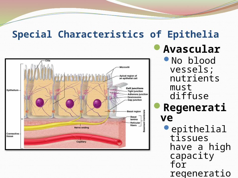
Special Characteristics of EpitheliaAvascular
No blood vessels; nutrients must diffuse
Regenerative epithelial
tissues have a high capacity for regeneration (mitosis!)
Recap.Identify two special characteristics
about epithelial tissue?
Identify two places epithelial tissue can be found.

Bell WorkWhat surface of an epithelial cell
opens up to the outside of the opening of an internal space?
What surface connects to the side of another cell?

Basal SurfaceWhat is it? Where is it?
Non-cellular, non-living supporting sheet two layers (basal lamina & reticular lamina)
Composed of: proteins secreted by the epithelial cells
Function:Selective filter selectively permeable to
molecules from capillaries Point of attachment and support for overlying
epithelial tissues (regenerating cells migrate from this point)

Apical SurfaceWhat is it? Where is it?
Surface that is exposed to the outside or internally to an open space
Located above the Basal LaminaComposed of:
Microvilli – finger-like extensions of plasma membrane Found in the small intestine and kidney Maximize SA across which small molecules enter or leave
Cilia – whip-like, highly motile extensions Found in the lungs Movement is coordinated waves

Lateral Surface FeaturesWhat is it?
sides of epithelial cells that face adjacent cells on either side
Factors holding epithelial cells together:Adhesion proteins link plasma membranes of
adjacent cellsSpecial cell junctions
Tight JunctionsAdherens JunctionsDesmosomesGap Junctions

Tight JunctionsTight junctions– closes off intercellular
space Location: near apical regionPurpose: forms an impermeable junction;
prevents molecules from passing between cells
Formation: transmembrane proteins in the plasma membrane of adjacent cells fuse together
Ie.: epithelial tissue lining the stomach, intestines & urinary bladder prevent contents of these organs from leaking


Adherens JunctionAdherens junctions – anchoring junction
Location- apical lateral bordersPurpose: helps form the tight junction around
apical lateral bordersFormation:
A dense layer of proteins on inside of plasma membrane (plaque) attaches to the cytoskeleton.
Transmembrane linker proteins (cadherins) are anchored into the cell’s plaque and they bind to cadherins of another cell thus joining the two cells.
Ie.: help epithelial surfaces resist separation during contractile activities (food moving through the intestine)

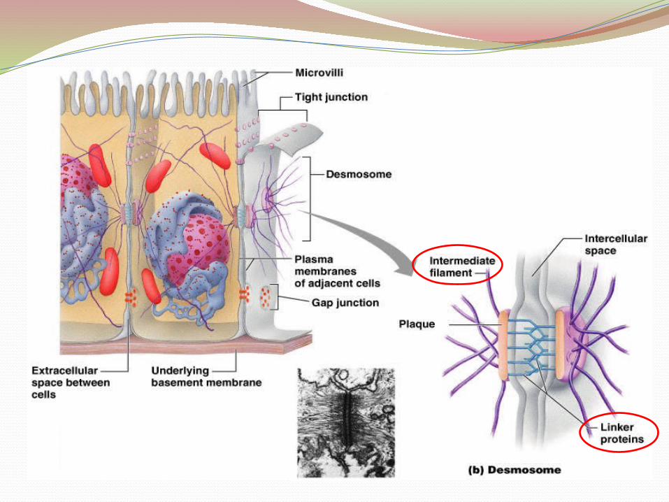
DesmosomesDesmosomes – 2 disc-like plaques
connected across intercellular spaceLocation: found in superficial layers of skinPurpose: reduces tearing, twisting, stretchingFormation:
Plaques of adjoining cells are joined by proteins called cadherins
Desomosomes on one side of the cell are imbedded with intermediate filaments (keratin protein) that extends across the cytosol of a cell to desmosomes on the other side of the same cell

Gap JunctionsGap junctions – passageway between two
adjacent cellsLocation: Present in electrically excitable
tissues (heart, smooth muscle)Purpose: Let small molecules move directly
between neighboring cellsFormation: Cells are connected by a protein
called connexins that form hollow cylinders called connexons
Ie.: lens and the cornea of the eye; enable nerve or muscle impulses to spread rapidly among cells


Bell WorkWhat type of junction is a passage way
between two cells?
What type of junction resists contractile activity?

Create a flip book on the following:Draw & label the surfaces of an epithelial
cell (Basal, apical and lateral)Draw & label each of the following
(include the cell junction’s purpose, where it is found and an analogy to remember it)Tight junction Adherens junction DesmosomeGap junction

Bingo!tissue
EpithelialConnective
Musclenerveapicalbasal
lateral surfacetight junction
adherens junction
desmosomegap junctionConnexonsConnexinsCadherins
plaque microvilli
cilia avascular
impermeaberegenerativeintermediate
filamentlinker proteins

