tissue culture of a medicinal plant-aloe vera l. · tissue culture of a medicinal plant-aloe vera...
TRANSCRIPT
1
TISSUE CULTURE OF A MEDICINAL PLANT-ALOE VERA L.
A
DISSERTATION
By
Mr.Diwakar Aggarwal
Roll No.3010108
Submitted in partial fulfillment of the requirement for the award of the degree of
Masters of Science in Biotechnology
Department of Biotechnology and Environmental Sciences Thapar Institute of Engineering and Technology
(Deemed University) Patiala –147004
2
ABSTRACT
Aloe vera syn barbadensis Mill. is an important medicinal plant and used world
wide in drug and cosmetic industry. Although Aloe propagates vegetatively in its
natural state, but propagation rate is too slow to meet demand of high quality
planting material for commercial cultivation. Micropropagation method for elite
selection of Aloe vera by axillary branching method using shoot tip as explant
was standardized. Shoot cultures were initiated on MS medium containing BA
0.2mg/L with IBA 0.2mg/L. Maximum shoot proliferation was achieved on
medium containing BA 1.0mg/L with IBA 0.2 mg/L within 28 days of culture.
Shoot proliferation was better in liquid medium with same composition. Citric acid
also enhanced shoot proliferation. A maximum of 5-multiplication rate of shoots
was achieved with citric acid (10mg/L) in the medium. Hundred percent rooting of
microshoots was obtained on phytohormone – free MS medium. Regenerated
plants after hardening were transferred to soil and they showed 85% survival.
The regenerated plants were morphologically similar to control plants.
3
CANDIDATE’S DECLARATION
I hereby declare that the work presented in the dissertation entitled,
Tissue culture of a medicinal plant-Aloe vera in partial fulfillment of the
requirement for the award of the degree of Masters in Biotechnology,
Department of Biotechnology and Environmental Sciences, Thapar Institute
of Engineering and Technology, Patiala; is an authentic record of my own
work done during the period of five months from January 2003 to May
2003,under the supervision of Dr. Kuldeep S. Barna, Research scientist,
TIFAC-CORE(Center of Relevance and Excellence) in Agro and
Industrial Biotechnology,Thapar Institute of Engineering and Technology
(TIET),Patiala. I have not submitted the matter embodied in this dissertation
for the award of any other degree or diploma.
Place: Patiala Date: Diwakar Aggarwal This is to certify that the above statement made by the candidate is correct and true to the best of our knowledge. (Dr.Kuldeep S.Barna) (Dr. Sunil Khanna) Supervisor, HEAD, TIFAC-CORE, TIET, Patiala. DBTES, TIET, Patiala.

4
ACKNOWLEDGEMENT
I thank the almighty whose blessings have enabled me to accomplish my
dissertation work successfully.
It is my pride privilege to express my sincere thanks and deep sense of gratitude
to Dr.K.S.Barna, Research scientist, TIFAC-CORE, Thapar Institute of
Engineering and Technology, Patiala, for his valuable advice, splendid
supervision and constant patience through which this work was able to take the
shape in which it has been presented. It was his valuable discussions and
endless endeavors through which I have gained a lot. His constant
encouragement and confidence-imbibing attitude has always been a moral
support for me.
I am grateful to Dr. Sunil Khanna, Coordinator CORE & Head, Department of
Biotechnology and Environmental Sciences, for his immense concern throughout
the project work and for providing me the laboratory facilities.
A special word of thanks to all the faculty members for their constant
encouragement and support throughout this duration.
I take this opportunity as a privilege to thank Sh. Dipal R. Chowdhary for putting
in his best efforts and helping another or me in one way.
Thanks to Dr N.Dass, Assistant Professor, DBTES, TIET, Patiala, for sharing his
DNA isolation kit and stepwise guidance, critical observation and immense help
during my entire period of study at Thapar campus.
5
I am really grateful for the help rendered by all members of the tissue culture lab.
at TIFAC-CORE with special mention of Mr. Rajesh who was always there for
making me understand the minor details of the work.
I feel lacunae of words to express my most heartfelt and cordial thanks to my
friend Sandeep, Rupinder, Anshu, Pankaj and Anita who have always been a
source of inspiration for me, stood by my side at the toughest times.
Finally, I wish to extend a warm thanks to everybody involved directly or indirectly
with my work.
The whole credit of my achievements goes to my parents, who were always
there for me in my difficulties. It was their unshakable faith in me that has always
helped me to proceed further.
(DIWAKAR AGGARWAL)

6
CONTENTS
CHAPTER PAGES
¬ INT R DUCT ION 7 -1 2
¬ R E VIE W OF L IT E R AT UR E 1 3 -1 7
¬ MAT E R IAL S AND ME T HODS 1 8 -2 5
¬ R E S UL T S 2 6 -3 9
¬ DIS CUS S ION 4 0 -4 2
¬ CONCL US ION 4 3 -4 4
¬ B IB L IOGR APHY 4 5 -5 3

7
INTRODUCTION
Aloe vera syn barbadensis Mill. belongs to the family Liliaceae (Anonymous
1985). It is commonly called as 'Burn plant'. It is a xerophyte and can be grown
even in dry lands under rain fed conditions. Aloe is a coarse looking perennial
plant with a short stem, found in the semi-wild state in many parts of the country
(Figure 1). Leaves 30-60 cm long, erect, crowded in a basal rosette, full of juice,
glaucous-green, narrow –lanceolate, long-acuminate, smooth except for the
spiny teeth on the margins. Scape longer than leaves, scaly, branched. Flowers
yellow, in dense racemes terminating the scapes. Commercial Aloes are
obtained from wild as well as cultivated plants. Propagation is primarily by means
of suckers (or) offshoots, which are separated carefully from mature plants and
transplanted. Medium sized suckers are chosen and carefully dugout without
damaging the parent plant at the base and can be directly planted in the field.
Plants will produce a commercial crop in one year (Venkataramaiah 2003).
Leaves exude a bitter liquid, which is dried and known as "bitter Aloes." They
also contain a clear gel, which is a soothing skin remedy. Leaves are broken off
and the clear gel is applied to the skin as a first aid for burns. Aloe contains
cathartic anthra-glycosides and its active principle ranging from 4.5 to 25% of
Aloin. These are extensively used as active ingredients in laxative, anti-obesity
preparation, as a moisturizer, emollient, wound healer, in various cosmetic and
pharmaceutical formulations. It is a drug as well as a cosmetic. There are about
more than 40 Aloe-based formulations being marketed in the global market. Aloin
portion, which forms 'Musambar', is considered as drug and other two portions

8
FIG-1, Elite Plant Material of Aloe vera Growing in the experimental plot.

9
i.e., chips, gel is used in cosmetic industry. The clear gel contained in the leaf is
a remarkably effective healer of wounds and burns, speeding up the rate of
healing and reducing the risk of infection. The yellow sap from the base of the
leaf when dried is known as "bitter Aloes" (Musambar). It is a strong laxative,
useful for short-term constipation.
Key Constituents:
Anthraquinones (aloin, Aloe - emodin), Resins, Tannins, Polysaccharides etc.
The principal constituent of aloin is a water-soluble crystalline glycoside barbaloin
[10(1)-deoxyglucosyl Aloe-emodin anthrone, C21H22O9.H2O, m.p.148-
1490C(anhyd.)].
Key Actions:
It heals wounds, is emollient and stimulates secretions of bile. Aloe is laxative.
Gels of different strengths, oil extracts, leaf concentrates, and powders are
available. South African Aloe gives quality gel. The processed gel is used in
different preparations such as1. Drugs 2. Cosmetics 3. Soft drinks 4.Food
Preparations 5. Moisturizers 6. Healthcare products etc.
Traditional & Current Uses
Beauty treatment: Aloe vera has a long history as a skin lotion. Cleopatra is said
to have attributed her beauty to it.
Western remedy: In the West, Aloe vera first became popular in 1950s when its
ability to heal burns, in particular radiation burns, was discovered.

10
First aid: Aloe vera is an excellent first aid to keep in the home for burns,
scrapes, scalds, and sunburn. A broken off leaf releases soothing gel, which may
be applied to the affected part.
Skin conditioner: The gel is useful for almost any skin condition that needs
soothing and astringing and will help varicose veins to some degree.
Ulcers: The protective and healing effect of Aloe vera also works internally and
the gel can be used for peptic ulcers and irritable bowel syndrome.
Laxative: The bitter yellow liquid in the leaves (bitter Aloes) contains
anthraquinones, which are strongly laxative. They cause the colon to contract,
generally producing a bowel movement 8-12 hours after consumption. At low
doses, the bitter properties of the herb stimulate the digestion. At higher doses,
bitter Aloes is laxative and purgative.
Minor burns & Sunburn: Apply Aloe-vera gel to the affected area as needed.
Stretch marks: Rub Aloe vera gel over the affected area.
Warts: Apply the gel directly to the wart 2-3 times a day for up to 3 months.
Weeping skin: Apply Aloe-vera gel to the affected area 2-4 times a day.
Wounds: Cleanse the wound with the gel and cover with a dressing soaked in
gel.
Out of the 22 amino acids required for the human body, 8 essential amino acids
are found in Aloe vera. Vitamins namely A, B1, B2, B6, B12, C and E, which the
human body cannot prepare by itself, are available in Aloe vera. Daily intake of
Aloe vera Gel free from aloin - a yellow sap, is certainly good for health and
improves natural immunity. The research studies conducted on Aloe vera plant

11
have revealed that through strengthening the T-lymphocyte cells of the blood, it
is able to heal the wounds and improve immunity (Lee & Kim, 2000).
Fresh leaf juice mixed with little turmeric power is best external application for
wounds, burns etc. Tribals and villagers who feed goats and cattle in forest areas
usually apply Aloe vera Juice externally to the scalp (head skin) in extreme
summer to protect the body from the effects of radiation and sunstroke. It is an
ideal, safe, proven anti-oxidant useful to prevent aging process and can be safely
used right from infants to extremely aged groups irrespective of sex, age
differentiation. Aloe vera gel bestowed with very powerful immunomodulatory
effect can be used in Aids, TB, and Cancer patients to overcome the symptoms
and to prolong their life span. It is the natural healer and safe cosmetic for all age
groups of men and women. It protects the skin, and improves complexion. It is a
natural moisturizer.
The technique of tissue and organ culture is used for rapid multiplication of
plants, for genetic improvement of crops, for obtaining disease- free clones and
for preserving valuable germplasm (Bhojwani & Razdan1992). One of the major
applications of plant tissue culture is micropropagation or rapid multiplication.
Compare to conventional propagation, micropropagation has the advantage of
allowing rapid propagation in limited time and space.
Although Aloe barbadenesis propagates vegetatively in its natural state, but
propagation is too slow for commercial plant production (Meyer &Staden1991).
To overcome slow propagation rate, micro propagation will be a very useful
technique for mass multiplication of Aloe.

12
Thus, with all this in view and consideration, the current project was undertaken
to standardize the necessary cultural conditions of Aloe vera by tissue culture
techniques.
Current studies aim at following objectives
Standardize optimum conditions for
• Establishment of axenic shoot culture from elite germplasm
• Multiplication of microshoots,
• Rooting of micro shoots,
• Hardening and transfer of plants to soil.

13
REVIEW OF LITRATURE
Plant tissue culture forms the backbone of plant biotechnology, i.e.
micropropagation, induction of somaclones, somatic hybridization,
cryopreservation and regeneration of transgenic plants. Plant Tissue culture is an
essential component of Plant Biotechnology. Plant Cell and tissue culture has
already contributed significantly to crop improvement and has great potential for
the future (Kumar and Kumar, 1996). Research efforts in plant cell and tissue
culture have increased dramatically worldwide in recent years including efforts in
developing nations. Plant cell and tissue culture is defined as the capability to
regenerate and propagate plants from single cells, tissues and organs under
sterile and controlled environmental conditions (Murashige & Skoog, 1974). In
India, Tissue Culture research began nearly four decades ago with the first report
on production of test tube fertilization (Kanta et al.1962). Tissue culture
techniques are now being widely applied for improvement of field crop, forest,
and horticulture and plantation crop for increased agricultural and forestry
production. Today tissue culture technology is being exploited mainly for large-
scale production or micropropagation of elite planting material with desirable
characteristics. This technology has now been commercialized globally and has
contributed significantly towards the enhanced production of high quality planting
material. Recently, emphasis has been on genetic transformation, especially for
(1) increased production of secondary metabolites, (2) production of alkaloids,
pharmaceutics, nematocidal compounds, and also some novel compounds not
found in the whole plant, () regeneration of plant resistant to herbicides,

14
diseases, and pests, (3) scale up of cultures in bioreactors, (4) plants with
different morphological traits, and (5) transgenic plants for the production of
vaccines etc. These developments have far-reaching implications in the
improvement of medicinal plants as well. (Bajaj, 1998)
Micropropagation has been useful for the rapid initial release of new varieties
prior to multiplication by conventional methods, e.g. pineapple (Drew, 1980) and
strawberry (Smith and Drew, 1990). Micropropagation is also used to promote
germplasm storage for maintenance of disease-free stock in controlled
environmental conditions (Withers, 1980) and in long -term via cryopreservation
(Kartha et al. 1980). The in vitro vegetative propagation has important benefits to
produce stable lines in plants that have no named varieties e.g., Annona spp.
(Bridg, 1993), Australian dioecious papaw genotypes (Drew, 1988; Drew, 1992)
where traditional plant breeding has failed. It has also a great potential for the
propagation of important crops like: Cassava spp., Phaseolus spp., Solanum
spp., (Roca and Mroginski, 1991). The micropropagation of elite or selected
plants showed good results, which benefit the agriculture, horticulture and
forestry (Conger, 1981; Drew, 1997). Worldwide there is much interest to
promote the development of an in vitro technology that permits the propagation
and breeding of commercial valuable woody, semi woody, ornamental, basic
food, industrial and medicinal plants. Which species are in danger of extinction
should receive a priority in terms of germplasm conservation (Conger, 1981;
Deberg and Zimmerman, 1990; Drew, 1997).

15
Aloe vera have been cultured in vitro by various researchers like Sanchez et al.
(1988), Natali et al. (1990), Meyer & Staden (1991), Roy & Sarkar (1991),
Corneanu et al., (1994). Richwine et al. (1995), Abrie & staden (2001), Chaudhuri
& Mukandan (2001).
Sanchez et al. (1988) performed micropropagation from shoot meristems. They
found that in vitro culture of Aloe barbadensis is very difficult for both callus
induction and plant regeneration. A DNA microdensitometric study was
performed on different organs of A.barbadensis and during in vitro culture of
different explants.
Natali et. al. (1990) reported a rapid and highly effective plant micropropagation
from vegetative meristems. Micropropagation was achieved by culturing shoot
apices on medium containing 2,4-D and Kn within 15-30 days. High
morphogenetic ability was maintained by transferring explants on media
containing 2,4-D and 6-benzylaminopurine. (Natali et.al. 1990),
Aloes have been cultured in vitro with 2,4-D and cytokinins in the growth
medium (Groenewald et al. 1975, Natali et al 1990). Axillary bud development
and adventitious bud formation was obtained with decapitated shoot explants of
Aloe barbadensis Mill. Maximal bud growth and rooting of shoots was obtained
on a modified medium of Murashige and Skoog supplemented with IBA. The
optimal temperature for bud growth and development was 250C. Bud growth was
slowed at 100c (Meyer & Staden, 1991).

16
Aloe has also cultured for their high medicinal and ornamental value (Vij et al.,
1980). Little work has been done on callus culture of Aloe species because
establishment of primary cultures is difficult owing to the secretion of the phenolic
substances by explant. There is only one report on callus formation and plant
regeneration from seed calli of Aloe pretoriensis (Groenewald et al., 1975). Roy
and Sarkar (1991) report the rapid propagation by the formation of shoots from
calli of Aloe vera. They induced the callus formation in stem segments from
young axillary shoots grown on the underground rhizomatous stem.
Polyvinylpyrrolidone was used to reduce the secretion of phenolic substances
from the explant. Modified MS media with 2,4-D and Kinetin was used for callus
induction. (Roy & Sarkar, 1991).
Micropropagation of Aloe was carried out by culturing fragments from axillary
shoots on MS medium without growth regulators (the presence of which was
found to inhibit the first stage of development). For the second stage of
development involving the newly formed plantlets, the presence of a magnetic
fluid in the culture medium stimulated secondary shoot production, general plant
development and rhizogenesis (Corneanu et al., 1994).
Richwine et. al. (1995) reported the induction of shoot cultures of Aloe, Gasteria
and Haworthia species from immature inflorescence. Shoots were initiated on a
modified MS medium containing zeatin and later maintained on medium
containing Zeatin and BA.
17
A rapid propagation protocol was established for the highly endangered A.
polyphylla. Seed was germinated in vitro on MS medium with or without sucrose.
Plantlets were cultured on medium containing benzyl adenine (BA) only, or a
combination of BA and NAA. After initial problems with browning, the explants
rapidly formed axillary and adventitious buds (Abrie & staden, 2001).
Chaudhuri and Mukandan (2001), also reported that in Aloe vera formation of
multiple shoots in vitro was a function of cytokinin and auxin concentrations. The
presence of only cytokinin or auxin resulted in formation of callus or roots. Best
multiplication of shoots was obtained on medium containing BA + Adenine
sulphate + IAA.
18
MATERIALS AND METHODS
Plant material:
Elite plants were healthy and free of symptoms of disease, pest problems and
showed good biomass yield. We collected the plant growing in the experimental
plot of TIFAC- CORE building. Shoot with young leaves was collected from the
elite plants. The extra leaves were removed and shoot was trimmed to size of 2-3
cm for further work.
Explant sterilization:
For the surface sterilization, the explants first were washed thoroughly in running
tap water for 30 minutes. After that they were again washed with liquid detergent
(Rankleen, Ranbaxy India) and Tween 20 (Himedia Laboratories, India) for 10
minutes with vigorous shaking. After washing with detergent explants were again
washed with running tap water to remove any traces of detergent for 30 minutes
and kept in 1% w/v solution of Bavistin (BASF India Limited) for one hour. After
that explants was shifted to the 1% v/v solution of savlon (Johnson and Johnson,
USA) for 1-2 minutes. After these treatments explants were taken inside the
laminar hood for further sterilization. Here 2-3 sterile water washings are given.
After these washings, explants were taken out and dipped in 70% ethyl alcohol
for 30 seconds. After alcohol dip, explants were surface sterilized with freshly
prepared 0.1% w/v aqueous solution of Mercuric chloride for 5 minutes. After
Mercuric chloride treatment, explants were thoroughly washed for 3-4 times with
sterile water to remove any traces of Mercuric chloride.

19
Culture media:
The basal medium used for the culture is Murashige and Skoog medium (MS,
1962) with sucrose 3% (Analytical grade, Himedia, India) and 0.8% agar
(Bacteriological grade, Himedia, India). Growth hormones,
Composition of basal MS medium
MACRO SALT (mg/L)
KNO3 1900
NH4NO3 1650
MgSO4.7H2O 370
CaCl2.2H2O 440
KH2PO4 170
MICRO SALTS
MnSO4.H2O 22.3
ZnSO4.7H2O 8.6
H3BO3 6.2
KI 0.83
Na2MoO4.2H2O 0.25
CuSO4.5H2O 0.025
CoCl2.6H20 0.025
Na2Fe-EDTA 37.24

20
ADDITIVES mg/L
Thiamine HCl 0.1
Nicotinic acid 0.5
Pyridoxine HCl 0.5
Glycine 2.0
Myo-inositol 100
Sucrose 30000
6-benzylaminopurine (BA), 3-Indolebutyric acid (IBA), kinetin (Kn), Adenine
sulphate were added to the basal medium either singly or in various
combinations.
The concentrated stock solutions of all the ingredients were prepared and stored
under refrigeration. To prepare stock solution of micro salts, all the micro salts in
required quantities were dissolved in one liter of distilled water and used as stock
solution. Like wise stock solutions of all other ingredients are also prepared and
kept under refrigeration. Similarly stock solutions of growth hormones were also
prepared. Cytokinins were dissolved in few drops of acidic solutions (1N HCl)
and Auxins were dissolved in few drops of basic solutions (1N KOH), after
dissolving final volume is made up with the help of distilled water and kept at 40C.
(Growth regulators are from Sigma, USA).
The medium was prepared by adding required quantities of all the ingredients in
the conical flask. After adding all the ingredients in required amounts, the final

21
volume is made up with the help of distilled water. pH of the medium is adjusted
to 5.8 by using 1N KOH or 1N HCl (Cyberscan 510,Eutech Instruments,
Singapore). After adjusting the pH, agar (Himedia Labs Limited, India) is added
to the medium at the rate of 0.8% w/v for solidification of the medium. For
preparing liquid medium (wherever used) agar was not added to the medium.
After pouring media (50-ml/300ml bottle), bottles are tightly capped and labeled
properly. After that media is autoclaved (Equitron, Medica Instruments. India) at
1210C for 20 minutes at 15psi.
Inoculation of explants:
After sterilization of explants, explants were inoculated in culture bottles
aseptically. For inoculation explants were transferred to large sterile glass
petriplate or glass plate with the help of sterile forceps under strict aseptic
conditions. Here the explants were further trimmed and extra outer leaves were
removed to make them in suitable sizes. Trimming and leaves were removed
with sterile scalpel blade. After cutting explants into suitable size (2-3cm),
explants are transferred to culture bottles containing MS medium with 0.2mg/L
BA and 0.2 mg/L IBA. After vertically inoculating the explants in culture bottle the
mouth of bottle is quick flamed and bottles are tightly capped and mouths of the
bottles was properly sealed with klin film to avoid entry of external air. After
proper labeling clearly mentioning media code, date of inoculation etc. the bottles
was transferred to growth room.
Shoot proliferation:

22
For shoot proliferation, BA (0-1mg/L) and Kn (0-1mg/L) at different
concentrations in combination with IBA (0.2 mg/L), Citric acid (0, 10, 100 mg/L),
adenine sulphate (160 mg/L) and agar (0, 0.8%) used. After 28 days of culture
period of the explants with newly form shoots were taken out under strict aseptic
conditions and were excised from the parent plant with help of sterile scalpel
blade and sterile forceps and inoculated into new bottles containing solid and
liquid MS basal medium with different set of growth hormones as mentioned
earlier. Two shoots per culture bottle were used and 4-6 replicates per treatment
were also used. Data were recorded after 28 days of culture and only shoots
greater than 2cm were considered for taking data. Every possible care has been
taken to prevent any further contamination.
Rooting of microshoots:
Newly formed shoots measuring 3-4cm in length were excised individually from
the parent explant and transferred to rooting media. Three types of rooting
medias were used one MS basal media without hormone and other MS basal
media with hormone (IBA 1mg/L). Here also we used both liquid as well as solid
mediums. Three- five shoots per culture bottle were used and 5 replicates were
used per treatment. Data were recorded after 15 days of culture.
Culture conditions:
All cultures were incubated under 16 hr photoperiod with light intensity of 2000-
2500 lux (Provided by Polylux XL, GE Britain, 36W and temperature of 25± 10C).
Acclimatization:

23
After 15 days of culture on rooting media, the plantlets were shifted to plastic
pots for their hardening prior to final transfer to soil to natural conditions. For
hardening of plants, plants with newly formed roots were taken out from the
culture bottles with the help of forceps with utmost care to prevent any damage to
newly formed roots and dipped in warm water not hot to remove the any traces of
solidified agar media. After removing media, plants were dipped in 1% w/v
solution of Bavistine to prevent any fungal infection to newly developed plants.
After Bavistine treatment the plantlets were carefully planted in plastic pots
containing 1:1 mixture of soil and farmyard manure. After planting the plants
were thoroughly watered and kept under polyhouse having 80% humidity and
31oC temperature for ten days. In-between the ten days plants were thoroughly
watered with the help of sprinkler to maintain required level of humidity. Then the
plants were shifted to shade house with less humidity level and indirect sunlight.
In shade house also plants were watered two times a day i.e. morning and
evening to prevent wilting (if any).
DNA isolation:
For the DNA isolation, we use CTAB plant DNA extraction kit provided by
GENEI, Bangalore. The protocol and solutions used in the kit are as fallow and
as per manufacturer instructions.
Solutions of CTAB DNA isolation kit
Solution A (CTAB extraction buffer)

24
CTAB 2% (w/v)
Tris-HCl 10 mM (pH 8.0)
EDTA 20 mM (pH 8.0)
NaCl 1.4 M
Solution B (Extraction buffer)
Tris-HCl 100 mM (pH 8.0)
EDTA 100 mM (pH 8.0)
NaCl 250 mM
Solution C (CTAB Precipitation solution)
CTAB 1% (w/v)
Tris-HCl 50 mM (pH 8.0)
EDTA 10 mM (pH 8.0)
Solution D (High salt TE buffer)
Tris-HCl 10 mM (pH 8.0)
EDTA 0.1 mM (pH 8.0)
NaCl 1.0 M
Procedure:
The young and soft leaves (~1.2 g) was pulverized to a fine powder in the
presence of liquid nitrogen and was distributed equally to three microfuge tubes
(2.0 ml capacity). Immediately 0.9 ml of solution A + 2% (v/v) â-mercaptoethanol
25
(at 65ºC) was mixed to the tissue to wet it thoroughly. The tubes were incubated
at 65• C for 1 hr. with occas ional mixing, after every 15 min.
Equal volume of chloroform: iso-amyl alcohol (24:1) was added to it. The solution
was mixed well by inversion for 7 min and then centrifuged at 8000 rpm for 5
minutes.
The upper aqueous layer was recovered, to which, 1/10th volume (100 ì l) of 65• C
solution B was added and this solution was mixed well by inversion for 5 min.
This solution was extracted in the same way with equal volume of chloroform:
iso-amyl alcohol (24:1) and the upper aqueous layer were recovered. To this
solution, 1 volume (900 ì l) of solution C was added. T he solution was mixed well
and was incubated at 65• C for ½ hr. This solution was centrifuged for 5 min. at
5000 rpm. T he pellets were resuspended in ~500 ì l of solution D by intermittent
incubation at 65• C. 300 ì l of isopropanol was added to each, mixed well and this
was kept at 4• C for 15 min. T his solution was centrifuged at full speed (rpm
15000) for 15 min., when the pellets were washed with 80% ethanol. The pellets
were air-dried and were finally resuspended in 60 ì l of T E buffer and store at –20
oC.
AGAROSE GEL ELECTROPHORESIS:
Gel Loading Buffer:
Sucrose 35% (w/v)
EDTA 50.0 mM (pH 8.0)
Bromophenol blue 0.2% (w/v)

26
TBE BUFFER (5X):
Tris base 54 g
Boric acid 28 g
EDTA 3.8 g
The pH of the buffer was set at 8.0
Horizontal agarose gel electrophoresis was performed using standard methods.
0.7% agarose gel was made in 0.5X TBE buffer to which ethidium bromide dye
was added and was casted in a gel tray. The DNA samples were loaded after
mixing well with the gel loading buffer containing tracking dye bromophenol blue
and electrophoresis was carried out at 65 V till the tracking dye covered 2/3rd of
the gel length. Finally, the DNA bands were visualized under UV light.
Statistical analysis:
All experiments were repeated once and data presented is mean of two values.
Means and standard deviation were calculated by using software Graph Pad
Prism 2.01.
Photography: Photos were taken with Ashai pentex camera using 400ASA
color film.

27
RESULTS
Shoot culture Initiation:
After surface sterilization of shoot tip explants, the explants were inoculated in
culture bottles aseptically. For inoculation, explants were transferred to large
sterile glass petriplate or glass plate with the help of sterile forceps under strict
aseptic conditions. Here the explants were further trimmed and extra outer
leaves were removed to make them in suitable sizes. Trimming was done with
sterile scalpel blade. After cutting explants into suitable size (2-3cm), explants
are inoculated to culture bottles containing MS medium with 0.2 mg/L BA and 0.2
mg/L IBA. After two weeks of observation, all explants gave axenic cultures.
Plants were free from both fungal as well as bacterial contamination. Explants
starts to show signs of proliferation after two weeks of culturing. New buds starts
to appear from the axil of leaves of shoot explants and buds develop into shoots
by 4 weeks of culture. After successful initiation of the culture (28 day culturing),
newly formed shoots were excised individually from the proliferated explant and
further cultured on the same medium to increase the number of shoots for further
work.
Shoot proliferation: Explants starts to show signs of proliferation after two
weeks of culturing. New buds starts to appear from the axil of leaves of shoot
explants and buds develop into shoots by 4 weeks of culture. Microshoots were
inoculated on MS basal medium with different concentrations and combinations
of BA and Kn (in combination of IBA 0.2 mg/L) for shoot proliferation. Both BA

28
and Kn were found to be give the indications of shoot proliferation after 2 weeks
of incubation.
Table-1. Effect of different combinations of cytokinins* on shoot proliferation in
Aloe vera after four weeks of culture.
Phytohormones
(mg/L)
% Of explants showing
shoot formation
(Mean±SD)
n=5
Number of shoots per
explant
(Mean ±SD)
n=10
Hormone- Free
(Control)
BA 0.2
1.0
Kn 0.2
1.0
nil
100 ± 0
100 ± 0
40 ± 41
90 ± 22
1
3.0 ± 0.8
3.3 ± 0.9
1.4 ± 0.5
3.1 ± 1.2 ∗ In combination with 0.2 mg/L IBA
It was found that BA gave better shoot proliferation than Kn (Table- 1). In
medium containing BA in different concentration, on an average each explant
gave rise to 3.0- 3.3 shoots (Table 1, Figure-2, 3). Hundred percent cultures
showed shoot proliferation on BA containing medium. On medium containing Kn
1mg/L, only 90% cultures showed shoot proliferation. In medium containing
higher concentration of Kn (1.0 mg/L) the average number of shoots per plant
29
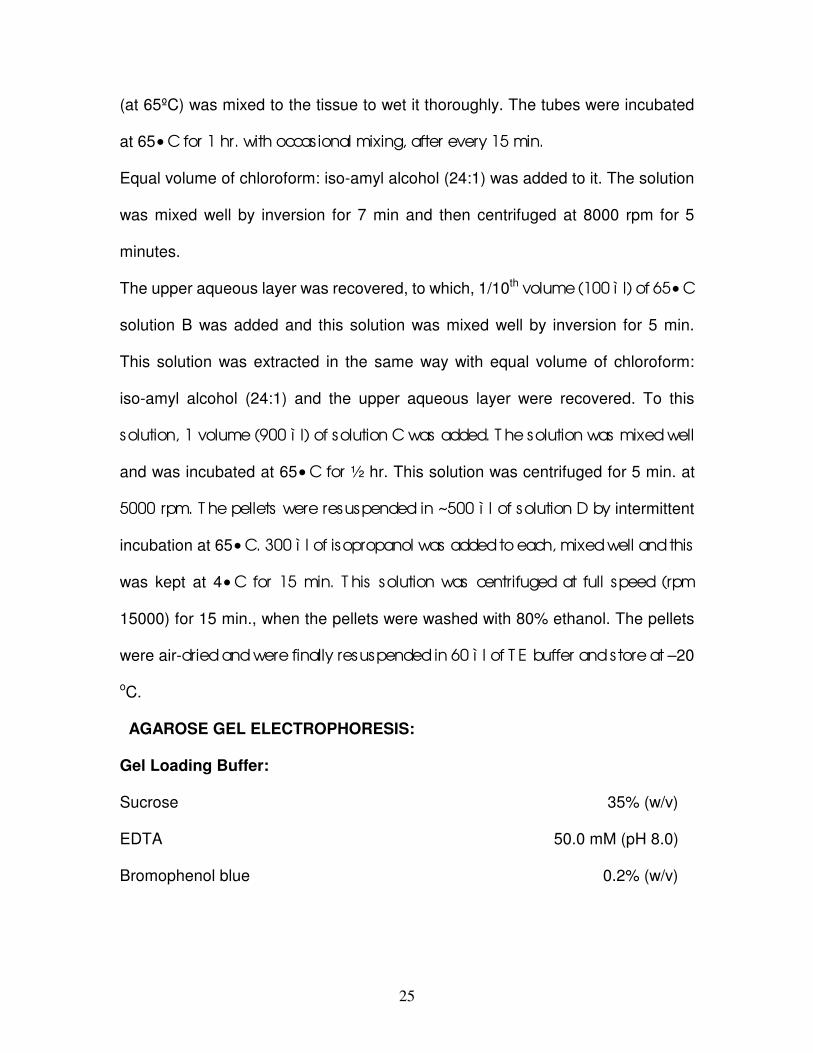
FIG-2, Microshoot inoculated on MS medium with IBA0.2mg/L+BA 1.0mg/L
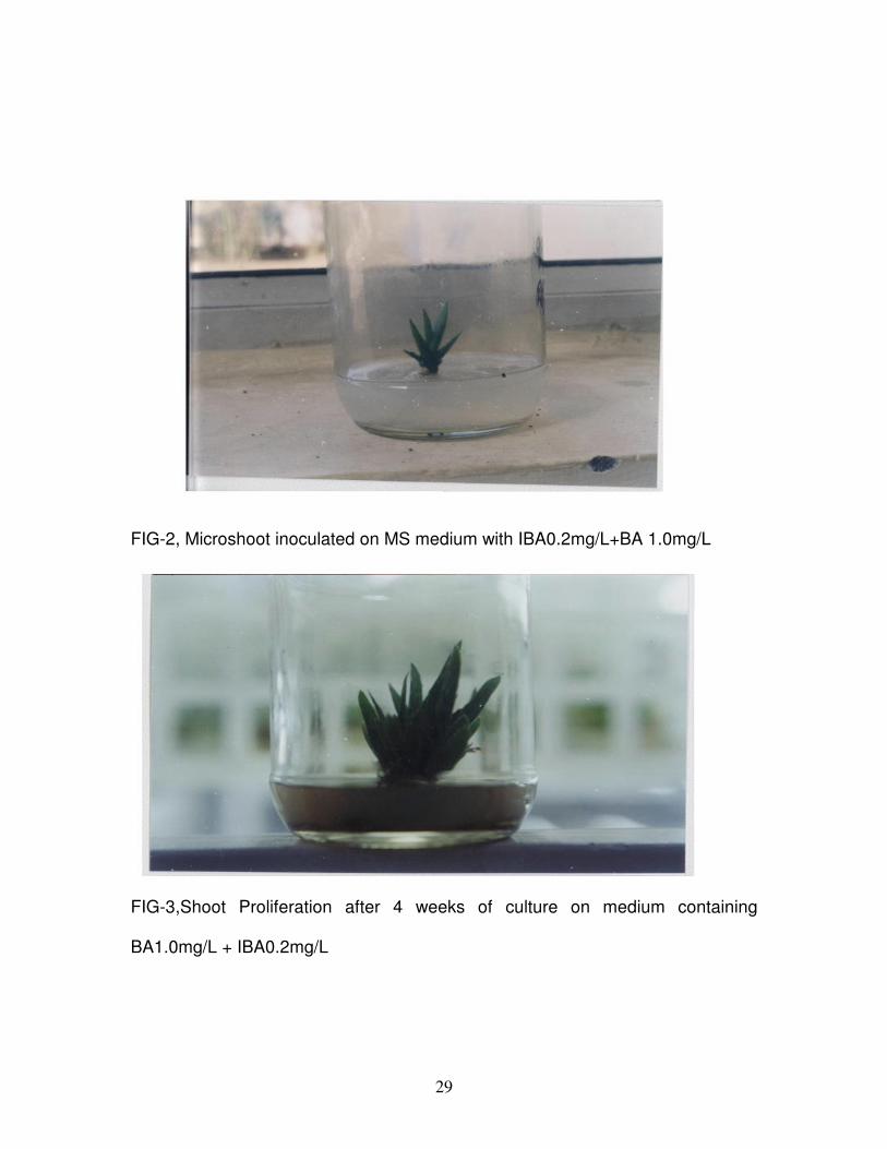
FIG-3,Shoot Proliferation after 4 weeks of culture on medium containing
BA1.0mg/L + IBA0.2mg/L

30
were 3.1±1.2. While on the other hand in medium containing less concentration
of Kn (0.2mg/L) the average number of shoots per plant was 1.4±0.5. The
explants which were cultured on medium without any phytohormone, failed to
produce any new shoots.
Adenine sulphate was also used to check whether it has any effect on shoot
proliferation or not. It was observed that adenine sulphate has no significant
effect on shoot proliferation in Aloe vera.
Table-2. Effect of Adenine sulphate∗ on shoot proliferation in Aloe vera after
Four weeks of culture.
Adenine sulphate
(mg/L)
% of explants
Showing shoot
formation
(Mean ± SD)
n=4
Number of shoots per
explant
(Mean ± SD)
n=8
0
(Control)
160
100 ± 0
75 ± 29
3.1 ± 0.8
3.1 ± 1.8
∗In combination with BA 0.2 mg/L and IBA 0.2 mg/L
In both the cases i.e. with and without adenine sulphate the average number of
shoots per plant was 3.1 (Table-2). The percentage of explant showing shoot
proliferation was also lower than control i.e. 75±29.
31
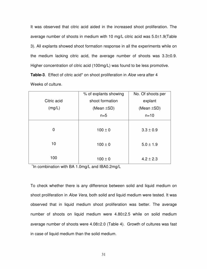
It was observed that citric acid aided in the increased shoot proliferation. The
average number of shoots in medium with 10 mg/L citric acid was 5.0±1.9(Table
3). All explants showed shoot formation response in all the experiments while on
the medium lacking citric acid, the average number of shoots was 3.3±0.9.
Higher concentration of citric acid (100mg/L) was found to be less promotive.
Table-3. Effect of citric acid* on shoot proliferation in Aloe vera after 4
Weeks of culture.
Citric acid
(mg/L)
% of explants showing
shoot formation
(Mean ±SD)
n=5
No. Of shoots per
explant
(Mean ±SD)
n=10
0
10
100
100 ± 0
100 ± 0
100 ± 0
3.3 ± 0.9
5.0 ± 1.9
4.2 ± 2.3
∗In combination with BA 1.0mg/L and IBA0.2mg/L
To check whether there is any difference between solid and liquid medium on
shoot proliferation in Aloe Vera, both solid and liquid medium were tested. It was
observed that in liquid medium shoot proliferation was better. The average
number of shoots on liquid medium were 4.80±2.5 while on solid medium
average number of shoots were 4.08±2.0 (Table 4). Growth of cultures was fast
in case of liquid medium than the solid medium.

32
Table-4, Effect of liquid and solid medium on shoot proliferation in Aloe vera after
4 weeks of culture.
Media*
Percentage of explants
showing shoot proliferation
(Mean±SD)
n=4
No. Of shoots per
explant
(Mean ±SD)
n=8
Solid
Liquid
91±17
91±17
4.08±2.0
4.80±2.5
∗Containing BA 1.0mg/L and IBA 0.2 mg/L
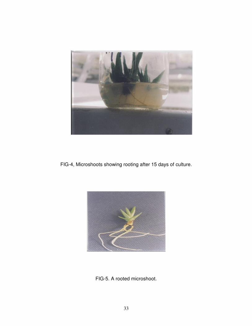
Rooting of Microshoots:
Three to four centimeters long shoots were excised individually from the
proliferated shoot clumps and cultured on rooting medium. The shoots inoculated
on hormone –free (medium lacking IBA) and IBA supplemented medium showed
rooting response within a week of inoculation. However, the response was better
in hormone- free medium. After the 15 days of inoculation, rooting was 100% in
hormone- free medium (Table-5, Figure-4, 5). The number of roots per shoot was
2.8+0.5 on hormone - free medium.
33
FIG-4, Microshoots showing rooting after 15 days of culture.
FIG-5. A rooted microshoot.

34
Table-5. Effect of IBA on root induction in Aloe vera after 15 days of culture.
IBA
(mg/L)
Percentage of
microshoots showing root
formation (Mean±SD)
n=5
No. Of roots per
microshoot
(Mean ±SD)
n=10
0
1
100 ± 0
90 ± 22
2.8 ± 0.5
1.7 ± 1.1
In case of hormone- free medium, roots were more thick and elongated, while the
roots on hormone supplemented medium were thin and less elongated. There
was no difference in colour of roots. In both the cases colour of roots was
creamish yellow. In both the cases roots were without any branches and normal
in appearance. In hormone- free medium average number of roots per plant was
2.8 ±0.5 and on hormone supplemented medium average number of roots per
plant was1.7±1.1.
To check the effect of solid and liquid medium on root induction so that rooting
response can be improved or/and cost of plants produced could be reduced,
Microshoots were inoculated on both the media. The microshoots inoculated on
solid medium showed better rooting response.

35
Table-6. Effect of solid and liquid medium on root induction in Aloe vera after 15
days of culture.
Media*
Percentage of
microshoots showing root
formation (Mean±SD)
n=5
No. Of roots per
microshoot
(Mean ±SD)
n=10
Solid
Liquid
100±0
18±20
2.7±1.2
0.2±0.5
∗MS hormone- free medium.
Hundred percent shoot showed rooting and the mean number of roots per
shoots was 2.7+1.2(Table 6) .On the other hand in liquid medium, only 18%±20
microshoots gave rooting. The shoots inoculated on liquid medium were failed to
give any further rooting response even after 3-4 weeks of inoculation (data not
shown).
Hardening of plantlets:
After 15 days of culture of microshoots on rooting medium, which resulted in the
sufficient rooting of shoots, the plantlets were transplanted to plastic pots
containing garden soil and Farmyard manure (1:1) for their hardening. For first
ten- days the plantlets were kept in polyhouse. To maintain the appropriate
humidity level (80%), plants were thoroughly watered with the help of manual

36
sprinkler every 2 hours The temperature of polyhouse was maintained at 31oC
with humidity level of nearly 80%.
Table-7.Survival rate of plantlets of Aloe vera at different stages of Hardening.
Stage of transplantation Number of plants
transplanted
Percentage of
survival
Poly House
(1st stage)
Shade House∗
(2nd stage)
13
11
85
82
∗Plantlets transferred to shade house after 10 days growth on polyhouse.
Plantlets that were transferred to the plastic pots in polyhouse showed good
percentage of survival of 85% (Table 7,Figure 6). After keeping plantlets for
initial ten days in polyhouse, the plantlets were transferred to shade house under
less humidity and temperature controlled conditions and indirect sunlight. In
shade house, these plants showed percentage of 82% survival (Table 7). In
shade house plants started to elongate and leaves also start to thicken. In shade
house plants were watered two times a day i.e. morning and evening. Among the
survived plants, some plants showed the symptoms of leap tip necrosis during
shade house conditions. But this does not hamper the overall growth of the
plants. Plants with these symptoms were also growing well.

37
FIG-6, Fully Hardened Plants growing in Open Shade.

38
Morphology of the regenerated plants:
All the plants produced were morphologically similar to the mother/control plants.
To find any possible (if any) somaclonal variation had occurred at molecular/DNA
level, genomic DNA of control plants and tissue-cultured plants were isolated. So,
the DNA isolated will be further used to find out any possible somaclonal variation.
Agarose gel electrophoresis showed that all these samples were about 23kb,
when compared with the molecular size marker (Figure-8). Further work with the
isolated is under progress.
Table-8. Quantification of the isolated DNA by UV spectrophotometeric methods
SAMPLE Yield (ì g/ ì l)
1.Normal plant (control)
2.Normal plant (control)
3 Normal plant (control)
4.Regenerated plant (without roots)
5.Regenerated plant (with roots)
6. Regenerated plant (with roots)
0.50
0.65
0.65
1.62
2.15
1.00
.

39
Lane 1: Normal plant genomic DNA; Lane 2: Normal plant genomic DNA; Lane 3: Tissue cultured plant genomic DNA; Lane 4: Tissue cultured plant genomic DNA;
Lane 5: Normal plant genomic DNA; Lane 6:λ-Hind ²²² DNA
FIG-7, Gel Profile of Aloe vera DNA.

40
DISCUSION
For shoot proliferation, growth regulators especially cytokinins (Lane 1979, Stolz
1979, Bhojwani 1980, Garland & Stolz 1981) are one of the most important
factors affecting the response. A range of cytokinins (Kinetin, BA, 2-ip and zeatin)
has been used in micropropagation work (Bhojwani and Razdan 1992).
Murashige (1974) and Hussy (1978) described 2-ip as more effective than either
BA or kinetin. A number such as blueberry (Cohen 1980) and garlic (Bhojwani
1980) were successfully multiplied by using 2-ip. But a wider survey of the
existing literature suggests that BA is the most reliable and useful cytokinin. A
number of plants has been were successfully multiplied on medium containing
BA. In white clover (Bhojwani 1981) and hybrid willow (Bhojwani 1980), chickpea
(Barna & Wakhlu 1994). Nair et al (1979), and Iresine lendenii (Sebastin & Barna
2003) BA is the most effective cytokinin for the shoot tip, meristem and bud
culture. At higher levels cytokinins tends to induce adventitious bud formation
(McComb, 1978; Zimmerman and Broome, 1980) .In the present study also,
shoot proliferation occurred only in the presence of cytokinin. Among the
cytokinins tested, BA proved to me more effective. This is in contrast to earlier
reports in Aloe Vera by Meyer and Staden (1991) and Natali et al (1990) in Aloe
vera. These researchers reported that better proliferation occurred on medium
containing Kn instead of BA in Aloe vera. This difference may be due to
difference in the genotype of plant used. Abrie and Staden (2001) Chaudhuri
and Mukandhan (200) also reported use of BA in shoot proliferation of Aloe
polyphylla and A.vera respectively.

41
Adenine sulphate was also used to check its effect on shoot proliferation. In our
case, adenine sulphate did not improve shoot proliferation in Aloe vera. But
earlier adenine sulphate was used for shoot proliferation in Aloe vera by
Chaudhuri & Mukandan (2001).
Citric acid also helped in the enhanced shoot proliferation in Aloe vera in the
present study. Keeping in mind the cost factor of agar, liquid medium containing
was also used for the shoot proliferation in Aloe vera. In the present study liquid
medium was found to be better for shoot proliferation in Aloe vera. Use of liquid
medium considerably reduces the cost of producing plants for the commercial
purposes.
Rooting response of microshoots is reported to be controlled by growth
regulators in the medium (Bhojwani & Razdan 1992), basal salt composition
(Garland and Stoltz.1981, Zimmerman and Broome.1981, Skirvin & Chu 1979),
genotype (Rines & McCoy 1981) as well as cultural conditions (Murashige 1977).
For most of the species auxin is required to induce rooting. NAA and IBA are
most commonly used for root induction (Bhojwani and Razdan 1992). By the use
of IBA many plants such as Lycoperscicon esculemtum (Sibi 1982), Hedychium
roxburgii (Tripathi & Bitaillion 1985), carnation (Werker & Leshem 1987) gave in-
vitro rooting. For the purpose induction of roots hormone-Free and IBA
supplemented medium were used in the present study. But rooting was observed
better in hormone – free medium. These kinds of observations were also earlier
by Sanchez et.al (1988), Meyer & Staden (1991), and Richwine et al. (1995) in
Aloe vera. Richwine et al. (1995) Also reported induction of roots in hormone-

42
medium for some other plants like Gasteria and Haworthia. Many other plants
such as straw berry (Boxus 1974), Narcissus (Seabrook et al. 1976), Gladiolus
(Hussy 1979)) and Rose (Barna & Wakhlu 1995) was rooted successfully rooted
on hormone-free medium. Decrease in number of roots in IBA supplemented
medium may be due to suparoptimal concentration of IBA in the medium. By
keeping in mind the cost factor, liquid medium was tried for the induction of roots.
But rooting was very poor in liquid medium in present study.
Hardening of tissue culture plants is the most crucial step in micropropagation.
The plants produced are very soft to face ambient environmental conditions.
(Bhojwani & Razdan 1992). These plants are grown under controlled conditions.
Under these conditions the leaves of plants develop cuticle and its photosynthetic
system starts functioning. The most crucial stage is during first 10 days in
polyhouse. During the 2nd hardening stage, mortality is lower as the plants are
comparatively hardened during first hardening stage or during the first 10 days in
polyhouse.
In the present study, rooted plantlets were transferred from culture bottles to
plastic cups in mixture of 1:1 ratio of soil: FYM for their hardening prior to their
final transfer to the soil, showed good percentage of survival (85%) in both
polyhouse and shade house. In shade house also plants showed 82% survival
rate. The growth and elongation of the plants were less in poly house whereas in
shade house growth of the plants was better and they also start to elongate in
shade house. The leaves also start to thicken in shade house.

43
CONCLUSION
Aloe vera syn barbadensis Mill. is a xerophytic medicinal plant of considerable
importance. It is widely used in cosmetic and drug industry and its demand is
increasing day by day. Due to widespread male sterility it propagates only
through vegetative mode of reproduction. But its propagation rate is very slow to
meet commercial demand of high quality planting material for its commercial
cultivation. So keeping this thing in mind, micropropagation work is carried out on
this plant. The objectives of the present study was to standardize optimum
conditions for establishment of axenic culture from elite germplasm, shoot
proliferation, rooting of micro shoots, hardening and transfer of plants to soil. For
the identification of any possible somaclones, in addition to their comparison with
in terms of morphology we planned to do some genetic analysis also. For this
purpose we isolated DNA from both normal as well as plants regenerated
through tissue culture. But due to lack of time we were able to complete only first
portion of the work.
The conclusions Drawn from this study are,
1. Surface sterilization with HgCl2 (0.1% for 5-minutes) with 70% alcohol dip
was best for the surface sterilization of the explants.
2. For the initiation of the culture,MS medium with BA 0.2 mg/L with IBA 0.2
mg//L was used.
3. Best shoot proliferation was achieved on MS medium containing
BA1.0mg/L with IBA 0.2mg/L.

44
4. Liquid medium with same composition was found to be better than solid
medium for shoot proliferation.
5. Adenine sulphate did not promoted shoot proliferation in the present
study.
6. Addition of 10mg/Lcitric acid in the medium aided in the enhanced shoot
proliferation. Citric acid in higher concentration (100mg/L) was found to be
less effective.
7. Hundred percent shoot showed rooting response on hormone -free
medium.
8. In liquid medium rooting response was found to very poor.
9. Regenerated plantlets, 85% survival during polyhouse conditions and
82% during shade house stage of hardening.
10. Regenerated plants were found to be morphologically similar to the
mother/control plant.

45
BIBLIOGRAPHY
Abrie, A. and Staden, J.V. (2001) Micropropagation of endangered Aloe
polyphylla, Plant Growth Regulation, 33(1): 19-23.
Anonymous (1976) The wealth of India- Raw materials, CSIR, New Delhi Vol.-
1A: 191-193.
Atherton, P. (1998) Aloe Vera: Revisited, British Journal of Physiotherapy, 4(4):
176-183.
Balasubramaniun, D., Bryce, C.F.A., Dharmarlinghum, K. and Jayaram, K.
(1998) Concepts in Biotechnology, university press, Hyderabad.
Barna, K.S and Wakhlu, A.K. (1994) Whole plant regeration of Cicer arietium
from callus culture via organogenesis, Plant Cell Reports,13:510-513.
Barna, K.S. and Wakhlu, A.K. (1995) Effects of Thiadizuron on the
micropropagation of rose, In Vitro Cellular and Developmental Biology-
Plant,33:44-46.
Bhojwani S.S. and Razdan, M.K. (1992) Plant tissue culture: Theory and
practice, Elsevier, Amsterdam, London, New York, Tokyo.
Bhojwani S.S. (1981) A tissue culture method for propagation and low
temperature storage of Trifolium repens genotypes, Physiol. Plant, 52:187-
190.

46
Bhojwani S.S. (1980 (1980) Micropropagation method for hybrid willow (Salix
matsudana x alba NZ-10002), N.Z.J. Bot.,18:209-21.
Bhojwani S.S. (1980) In vitro propagation of garlic by shoot proliferation,
Sci.Hortic.,13:47-52.
Boxus, P. (1974) The production of strawberry plants by in vitro
micropropagation, J.Hortic. Sci., 49:209-211.
Cavallini, A., Natali, L., Cionini, C., Sessoli, O. and Sanchez, I.C. (1993) In-
vitro culture of Aloe Barbadensis Mill.: Quantitative DNA variation in
regenerated plants, Plant Science , 91: 223-224.
Chaudhuri, S. and Mukundan, U. (2001) Aloe Vera L.—Micropropagation and
Characterization of its gel, Phytomorphology, 51(2): 155-157.
Cohen, D. (1980) Application of micropropagation methods for blueberries and
tamarillos, Proc.Int.Plant Prop.Soc.,30:144-146.
Corneanu,M., Corneanu,G.,Vekas,M., and Minea, R. (1994) In vitro
organogenesis of Aloe arborescencs.(Liliaceae),Revue Roumaine de
Biologie.39(1):45-52.
Davis, R.H. and Leiter, M.G. (1988) Aloe Vera: A natural approach for treating
wounds, edema, and pain diabetes, Journal of The American Podiatric
Medical Association, 78(2): 60-68.
Drew, R.K.L. (1979) effect of activated charcoal on embryo genesis and
regeneration of plantlets from suspension culture of carrot (Daucus carota
L.), Ann. Bot., 44:387:389.

47
Farooqi, A.A., Kathiresan, C., Pasha, K.N. and Anuradha, M.N. (2001) Scope
of Biotechnology in Medicinal plants, Science Tech Entrepreneur, 45-55.
Gamborg, O.L. and Phillips, G.C. (1998) Plant cell, Tissue and Organ Culture:
Fundamental methods, Narosa publishing house New Delhi.
Garland, P. and Stoltz, L.P. (1980) In vitro propagation of tarragon,
HortScience, 15:739.
Garland, P. and Stoltz, L.P. (1981) Micropropagation of Pissrdi plum, Ann. Bot.,
48:387-389.
Groenewald, E.G., Koeleman, A. and Wessels, D.C.J. (1975) Callus formation
and plant regeneration from seed tissue of Aloe pretoriensis,
Z.Pflanzenphysiol. 75:270-272.
Guha, S. and Maheshwari, S.C. (1966) Cell division and differentiation of
embryos from anthers of Datura in vitro, Nature (London), 204:497.
Gui, Y.L. and Xu, T.Y. (1990) Studies on stem tissue culture and organogenesis
of Aloe vera,Acta Botanica Sinica,32(8): 606-610.
Hussy, G. (1978) The application of tissue culture to the vegetative propagation
of plants, Sci.Prog.,65:185-208.
Hussy, G. (1981) In vitro propagation .In: D.S. Ingram and J.P.Hegelson
(Editors), Tissue culture methods for plant pathologists, Blackwill Scientific
Publishers, Oxford, 51-61.
48
Jasso, C.D. and Rodriguez, G.R. (1996) An overview of tissue culture of
outstanding species from semiarid and arid lands in Mexico, Biotechnologia
Aplicada, 13(4): 294.
John, H.D. and Roberts, L.W. (1987) Experiments in Plant Tissue Culture, 2nd
edition, Cambridge University Press, New York.
Kanta,K. and Maheshwari, P. (1963) Test tube fertilization in some
angiosperms,Phytomorphology,13:230-237
Kartha, K.K., Leung, N.L. and Pahl, K. (1980) Cryopreservation of strawberry
meristems and mass propagation of plantlets, J.Am. Soc.
Hortic.Sci.,105:481-484.
Keijzer, C.J. and Cresti, M. (1987) A comparison of anther tissue development
in male sterile Aloe vera, and male fertile Aloe ciliaris, Ann. Bot, 59: 533-
542.
Khan, I.A. and Khanum, A. (1999) Role of Biotechnology in Medicinal and
Aromatic plants, vol-2, Ukaaz publications Hyderabad.
Kumar, A. and Kumar, V.A. (1996) Plant Biotechnology and Tissue Culture
Principles and Perpectives, International Book Distributing Co, Lucknow.
Lane, W.D. (1979) In vitro propagation of Spirea bumalda and Prunus cistena
from shoot apics, Can.J. Plant Sci., 59:1025-1029.
Larkin, P.J. and Scowcroft, W.R. (1981) Somaclonal variation novel source of
variability from cell cultures for plant improvement, Theor Appl Genet,
60:197-214.
49
Lee, K.H. and Kim, J.H. (2000) Anti-lukaemic and anti -mutagenic effects of di
(2-ethylhexyl) phthalate isolated from Aloe vera Linne.,Journal of Pharmacy
and Pharmacology,52(5):593-598.
Martin, K.P., Beena, M.M. and Joseph, D. (2003) High frequency axillary bud
multiplication and ex-vitro rooting of Wedelia chinensis (osbek) Merr. – A
medicinal plant, Indian Journal of Experimental Biology, 41: 262-266.
McComb, J.A. (1978) Clonal propagation of woody plant species with special
reference to apples, Proc.Int.Plant Prop.Soc.,28:413-426.
Meyer, H.J. and Staden, J.V. (1991) Rapid in vitro propagation of Aloe
barbadensis Mill., Plant cell, Tissue and Organ Culture, 26:167-171.
Murashige, T. (1977) Clonal crops through tissue culture. In: W.Barz et al.
(Editors), Plant Tissue Culture and its in Biotechnological Application,
Springer-Verlag, Berlin, 392-403.
Murashige, T. and Skoog, F. (1962) A revised medium for rapid growth and
bioassays with tabacco tissue cultures. Physiol. Plant, 115: 493-497.
Murashige, T. and Skoog, F. (1974) Plant propagation through tissue cultures,
Ann.Rev.,Plant Phisiol.,25:135-165.
Natali, L., Sanchez, I.C. and Cavallini, A. (1990) In vitro culture of Aloe
barbadensis Mill.:Micropropagation from vegetative meristems, Plant Cell,
Tissue and Organ Culture, 20 :71-74.
Panda, H. (2002) Medicinal plants cultivation and their uses, Asia Pacific
Business Press Inc, New Delhi. 163-164 and 167-168.

50
Richwine, A.M., Tipton, J.L. and Thompson, A. (1995) Establishment of Aloe,
Gasteria, and Haworthia shoot cultures from inflorescence explants, Hort
Science, 30(7): 1443-1444.
Rines, H.W and McCoy, T.J. (1981) Tissue culture initiation and plant
regeneration in hexaploid species of oats, Crop Science,21:837-842.
Roy, S.C. and Sarkar, A. (1991) In vitro regeneration and micro propagation of
Aloe vera, Scientia Horticulturae, 47(1-2): 107-114.
Sanchez, I.C., Natali, L. and Cavallini, A. (1988) In vitro culture of Aloe
barbadensis Mill. Morphogenetic ability and nuclear DNA content, Plant
Science, 55:53-59.
Seabrook, J.E.A and Cumming, B.G. (1977) The in vitro propagation of
Amaryllis (Hippeastrum spp. Hybrids). In Vitro, 13:831-836.
Sebastin, J. and Barna, K.S. (2003) Plant regeneration through callus culture of
Iresine lindenii, In Vitro Cellular and Developmental Biology,
Communicated.
Sibi, M. (1982) Heritable epigenetic variation from in vitro tissue culture of
Lycopersian esculentum (Var. Monalbo). In: E.D. Earle and Y. Demarly
(Editors), Variability in plants regenerated from tissue culture, Proc.NSF-
CNRS Congr.Osray.Prager, New York, 228-244.
Skirvin, R.M. and Chu, M.C. (1979) In vitro propagation of ‘‘Forever Yours’’
rose, HortScience, 14:608-610.

51
Stoltz, L.P. (1979) In vitro propagation of Acalypha wilkesiana, HortScience,
14:702-703.
Tripathi, B.K. and Bitallion, C. (1995) In vitro plant regeration of Hedychium
roxburgii blume through rhizome meristem culture, HortScince,4:11-17.
Vasil, I.K. and Vasil,V.(1980) Clonal propagation,Int.Rev.Cytol.(Suppl),11A:145-
173.
Venkataramaiah, V. (2003) Aloe: A key medicinal herb for drug and cosmetic
industry,sapthgiri.http://www.tirumala.org/sapthagiri/012003/Aloe.htm.,Acce
sed on 08-05-2003.
Vij, S.P., Sharma, M. and Toor, I.S. (1980) Cytogenetical investigations into
some garden ornamentals 2,the genus Aloe, Cytologia, 45:515-532.
Werker, E. and Leshem, B. (1987) Structural changes during vitrification of
carnation plantlets, Ann.Bot. (London),59:377-385.
Withers, L.A. (1980) Preservation of germplasm. In: I. K.Vasil (Editor),
Perspectives in Plant Cell and Tissue Culture, Int. Rev.Cytol.Suppl.,11b:
101-136.
Zimmerman, R.H. and Broome, O.C. (1980) Blueberry micropropagation. In:
Proceedings of the Conference on Nursery production of fruit plants through
Tissue culture-Application and Feasibility. Agric. Res. Sci. Educ. Admin.,
U.S.D.A., Beltsville, 44-47.

52
Zimmerman, R.H. and Broome, O.C. (1981) Phloroglucinol and in vitro rooting
of apple cultivar cuttings, J.Am.Soc.Hortic.Sci.,106:648-652.