tissue clearing pro-organoid - tocris bioscience
TRANSCRIPT
TISSUE CLEARING PRO-ORGANOIDGUIDEBOOK V1.1
2
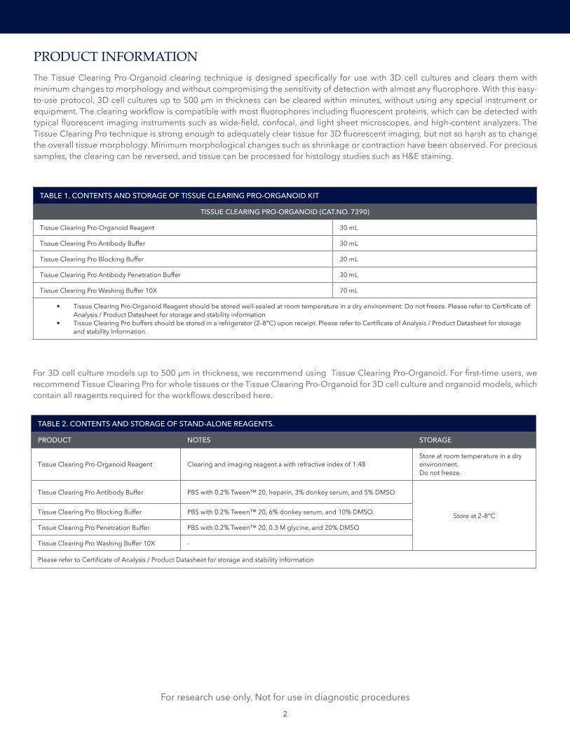
PRODUCT INFORMATIONThe Tissue Clearing Pro-Organoid clearing technique is designed specifically for use with 3D cell cultures and clears them with minimum changes to morphology and without compromising the sensitivity of detection with almost any fluorophore. With this easy-to-use protocol, 3D cell cultures up to 500 µm in thickness can be cleared within minutes, without using any special instrument or equipment. The clearing workflow is compatible with most fluorophores including fluorescent proteins, which can be detected with typical fluorescent imaging instruments such as wide-field, confocal, and light sheet microscopes, and high-content analyzers. The Tissue Clearing Pro technique is strong enough to adequately clear tissue for 3D fluorescent imaging, but not so harsh as to change the overall tissue morphology. Minimum morphological changes such as shrinkage or contraction have been observed. For precious samples, the clearing can be reversed, and tissue can be processed for histology studies such as H&E staining.
TABLE 1. CONTENTS AND STORAGE OF TISSUE CLEARING PRO-ORGANOID KIT
TISSUE CLEARING PRO-ORGANOID (CAT.NO. 7390)
Tissue Clearing Pro-Organoid Reagent 30 mL
Tissue Clearing Pro Antibody Buffer 30 mL
Tissue Clearing Pro Blocking Buffer 30 mL
Tissue Clearing Pro Antibody Penetration Buffer 30 mL
Tissue Clearing Pro Washing Buffer 10X 70 mL
• Tissue Clearing Pro-Organoid Reagent should be stored well-sealed at room temperature in a dry environment. Do not freeze. Please refer to Certificate of Analysis / Product Datasheet for storage and stability information
• Tissue Clearing Pro buffers should be stored in a refrigerator (2–8°C) upon receipt. Please refer to Certificate of Analysis / Product Datasheet for storage and stability information.
TABLE 2. CONTENTS AND STORAGE OF STAND-ALONE REAGENTS.
PRODUCT NOTES STORAGE
Tissue Clearing Pro-Organoid Reagent Clearing and imaging reagent a with refractive index of 1.48 Store at room temperature in a dry environment.Do not freeze.
Tissue Clearing Pro Antibody Buffer PBS with 0.2% Tween™ 20, heparin, 3% donkey serum, and 5% DMSO
Store at 2–8°CTissue Clearing Pro Blocking Buffer PBS with 0.2% Tween™ 20, 6% donkey serum, and 10% DMSO.
Tissue Clearing Pro Penetration Buffer PBS with 0.2% Tween™ 20, 0.3 M glycine, and 20% DMSO
Tissue Clearing Pro Washing Buffer 10X -
Please refer to Certificate of Analysis / Product Datasheet for storage and stability information
For research use only. Not for use in diagnostic procedures
For 3D cell culture models up to 500 μm in thickness, we recommend using Tissue Clearing Pro-Organoid. For first-time users, we recommend Tissue Clearing Pro for whole tissues or the Tissue Clearing Pro-Organoid for 3D cell culture and organoid models, which contain all reagents required for the workflows described here.
3
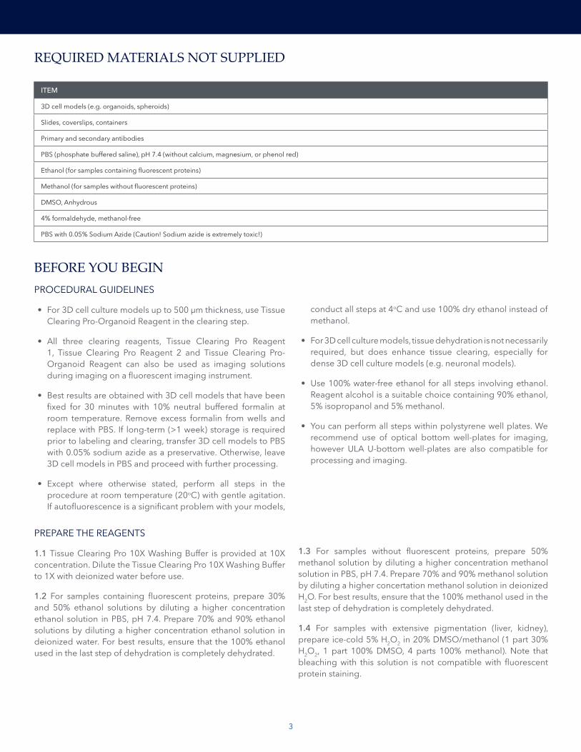
REQUIRED MATERIALS NOT SUPPLIED
BEFORE YOU BEGINPROCEDURAL GUIDELINES
• For 3D cell culture models up to 500 μm thickness, use Tissue Clearing Pro-Organoid Reagent in the clearing step.
• All three clearing reagents, Tissue Clearing Pro Reagent 1, Tissue Clearing Pro Reagent 2 and Tissue Clearing Pro-Organoid Reagent can also be used as imaging solutions during imaging on a fluorescent imaging instrument.
• Best results are obtained with 3D cell models that have been fixed for 30 minutes with 10% neutral buffered formalin at room temperature. Remove excess formalin from wells and replace with PBS. If long-term (>1 week) storage is required prior to labeling and clearing, transfer 3D cell models to PBS with 0.05% sodium azide as a preservative. Otherwise, leave 3D cell models in PBS and proceed with further processing.
• Except where otherwise stated, perform all steps in the procedure at room temperature (20oC) with gentle agitation. If autofluorescence is a significant problem with your models,
ITEM
3D cell models (e.g. organoids, spheroids)
Slides, coverslips, containers
Primary and secondary antibodies
PBS (phosphate buffered saline), pH 7.4 (without calcium, magnesium, or phenol red)
Ethanol (for samples containing fluorescent proteins)
Methanol (for samples without fluorescent proteins)
DMSO, Anhydrous
4% formaldehyde, methanol-free
PBS with 0.05% Sodium Azide (Caution! Sodium azide is extremely toxic!)
conduct all steps at 4oC and use 100% dry ethanol instead of methanol.
• For 3D cell culture models, tissue dehydration is not necessarily required, but does enhance tissue clearing, especially for dense 3D cell culture models (e.g. neuronal models).
• Use 100% water-free ethanol for all steps involving ethanol. Reagent alcohol is a suitable choice containing 90% ethanol, 5% isopropanol and 5% methanol.
• You can perform all steps within polystyrene well plates. We recommend use of optical bottom well-plates for imaging, however ULA U-bottom well-plates are also compatible for processing and imaging.
PREPARE THE REAGENTS
1.1 Tissue Clearing Pro 10X Washing Buffer is provided at 10X concentration. Dilute the Tissue Clearing Pro 10X Washing Buffer to 1X with deionized water before use.
1.2 For samples containing fluorescent proteins, prepare 30% and 50% ethanol solutions by diluting a higher concentration ethanol solution in PBS, pH 7.4. Prepare 70% and 90% ethanol solutions by diluting a higher concentration ethanol solution in deionized water. For best results, ensure that the 100% ethanol used in the last step of dehydration is completely dehydrated.
1.3 For samples without fluorescent proteins, prepare 50% methanol solution by diluting a higher concentration methanol solution in PBS, pH 7.4. Prepare 70% and 90% methanol solution by diluting a higher concertation methanol solution in deionized H2O. For best results, ensure that the 100% methanol used in the last step of dehydration is completely dehydrated.
1.4 For samples with extensive pigmentation (liver, kidney), prepare ice-cold 5% H2O2 in 20% DMSO/methanol (1 part 30% H2O2, 1 part 100% DMSO, 4 parts 100% methanol). Note that bleaching with this solution is not compatible with fluorescent protein staining.
4
Except where otherwise stated, perform all steps in the procedure at room temperature with gentle agitation.
2.1 Obtain 3D cell models of interest. See “Procedural guidelines” on page 3 for guidelines on fixation.
2.2 Wash 3D cell models twice in PBS, pH 7.4 (without calcium, magnesium, or phenol red) for at least 15 minutes. STOPPING POINT. (Optional) You can store the 3D cell models at 4°C in the dark for up to 3 days without detrimental effects.
2.3 Dehydrate the 3D cell models with increasing concentrations of ethanol at 4°C. See Table 3 for required volumes and incubation times. Using an excess volume in the dehydration steps ensures proper clearing.
Note: For 3D cell culture models, tissue dehydration is not required, but it can enhance tissue clearing speed for dense or especially large 3D cell culture models (e.g. neuronal models).
2.3.1 Treat 3D cell models with 30% ethanol in PBS with gentle shaking.
2.3.2 Treat 3D cell models with 50% ethanol in PBS with gentle shaking.
2.3.3 Treat 3D cell models with 70% ethanol in deionized water with gentle shaking.
2.3.4 Treat 3D cell models with 90% ethanol in deionized water with gentle shaking.
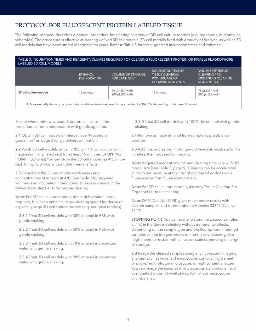
PROTOCOL FOR FLUORESCENT PROTEIN LABELED TISSUEThe following protocol describes a general procedure for clearing a variety of 3D cell culture models (e.g. organoids, microtissues, spheroids). The procedure is effective at clearing unfixed 3D cell models, 3D cell models fixed with a variety of fixatives, as well as 3D cell models that have been stored in formalin for years. Refer to Table 3 for the suggested incubation times and volumes.
TABLE 3. INCUBATION TIMES AND REAGENT VOLUMES REQUIRED FOR CLEARING FLUORESCENT PROTEIN OR FIXABLE FLUOROPHORE-LABELED 3D CELL MODELS
ETHANOL DEHYDRATION
VOLUME OF ETHANOL FOR EACH STEP
INCUBATION TIME IN TISSUE CLEARING PRO-ORGANOID CLEARING REAGENTS
VOLUME OF TISSUE CLEARING PRO-ORGANOID CLEARING REAGENTS [1]
3D cell culture models 15 minutes 75 µL (384-well) 200 µL (96-well)
15 minutes 75 µL (384-well)200 µL (96-well)
[1] For especially dense or large models, incubation time may need to be extended by 30–50%, depending on degree of fixation.
2.3.5 Treat 3D cell models with 100% dry ethanol with gentle shaking.
2.4 Remove as much ethanol from sample as possible via pipetter.
2.5 Add Tissue Clearing Pro-Organoid Reagent, incubate for 15 minutes, then proceed to imaging.
Note: Required reagent volume and clearing time vary with 3D model size (see Table 3, page 5). Clearing can be accelerated at room temperature at the cost of decreased endogenous fluorescence from fluorescent protein.
Note: For 3D cell culture models, use only Tissue Clearing Pro-Organoid for tissue clearing.
Note: DAPI (Cat. No. 5748) gives much better results with cleared samples and is preferable to Hoechst 33342 (Cat. No 5117).
STOPPING POINT. You can seal and store the cleared samples at 4°C in the dark indefinitely without detrimental effects. Depending on the sample type and the fluorophore, mounted samples can be imaged weeks to months after clearing. You might need to re-stain with a nuclear stain depending on length of storage.
2.8 Image the cleared samples using any fluorescent imaging analyzer such as widefield microscope, confocal, light sheet or single/multi-photon microscope, or high content analyzer. You can image the samples in any appropriate container, such as mounted slides, 96-well plates, light sheet microscope chambers, etc
5
3.1 Obtain 3D cell models of interest and fix them, if needed. See “Procedural guidelines” on page 3 for guidelines on fixation.
3.2 Wash 3D cell models twice in PBS, pH 7.4 (without calcium, magnesium, or phenol red) for at least 15 minutes each.
Note: For 3D cell culture models that are particularly difficult to immunolabel due to the spheroids being highly dense in nature or the presence of significant ECM, Tissue Clearing Pro Permeabilization Buffer can be used (See Table 2). Incubate at room temperature for 15-30 minutes with gentle shaking and wash several times with PBS to remove as much buffer as possible before proceeding with next steps. Please note that Tissue Clearing Pro Permeabilization Buffer should not normally be needed for most 3D cell culture models.
Note: Tissue Clearing Pro Permeabilization Buffer cannot be used if immunolabeling 3D cell models that contain fluorescent protein.
3.4 Permeabilize 3D cell models by washing them through a gradient of methanol (samples without fluorescent protein) at room temperature, or ethanol (samples with fluorescent protein) at 4oC with gentle agitation. See Tables 4 and 5 for required volumes and incubation times.
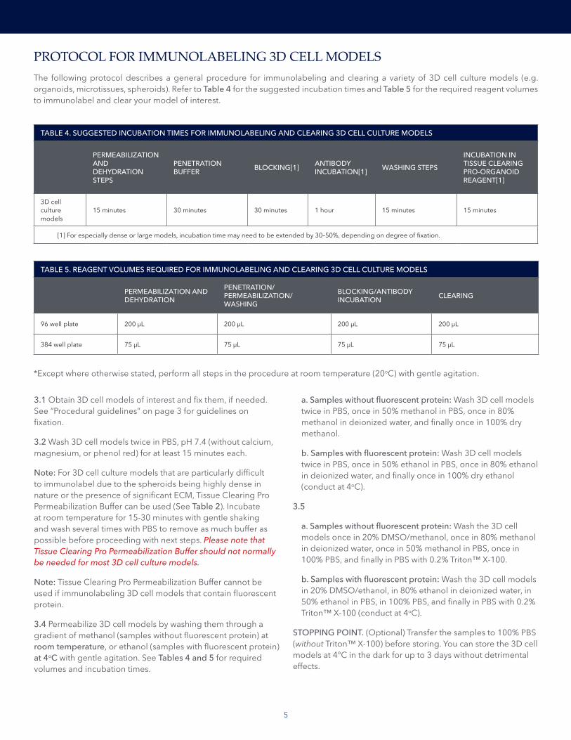
PROTOCOL FOR IMMUNOLABELING 3D CELL MODELSThe following protocol describes a general procedure for immunolabeling and clearing a variety of 3D cell culture models (e.g. organoids, microtissues, spheroids). Refer to Table 4 for the suggested incubation times and Table 5 for the required reagent volumes to immunolabel and clear your model of interest.
TABLE 4. SUGGESTED INCUBATION TIMES FOR IMMUNOLABELING AND CLEARING 3D CELL CULTURE MODELS
PERMEABILIZATION AND DEHYDRATION STEPS
PENETRATION BUFFER BLOCKING[1] ANTIBODY
INCUBATION[1] WASHING STEPS
INCUBATION IN TISSUE CLEARING PRO-ORGANOID REAGENT[1]
3D cell culture models
15 minutes 30 minutes 30 minutes 1 hour 15 minutes 15 minutes
[1] For especially dense or large models, incubation time may need to be extended by 30–50%, depending on degree of fixation.
TABLE 5. REAGENT VOLUMES REQUIRED FOR IMMUNOLABELING AND CLEARING 3D CELL CULTURE MODELS
PERMEABILIZATION AND DEHYDRATION
PENETRATION/ PERMEABILIZATION/WASHING
BLOCKING/ANTIBODY INCUBATION CLEARING
96 well plate 200 µL 200 µL 200 µL 200 µL
384 well plate 75 µL 75 µL 75 µL 75 µL
a. Samples without fluorescent protein: Wash 3D cell models twice in PBS, once in 50% methanol in PBS, once in 80% methanol in deionized water, and finally once in 100% dry methanol.
b. Samples with fluorescent protein: Wash 3D cell models twice in PBS, once in 50% ethanol in PBS, once in 80% ethanol in deionized water, and finally once in 100% dry ethanol (conduct at 4oC).
3.5
a. Samples without fluorescent protein: Wash the 3D cell models once in 20% DMSO/methanol, once in 80% methanol in deionized water, once in 50% methanol in PBS, once in 100% PBS, and finally in PBS with 0.2% Triton™ X-100.
b. Samples with fluorescent protein: Wash the 3D cell models in 20% DMSO/ethanol, in 80% ethanol in deionized water, in 50% ethanol in PBS, in 100% PBS, and finally in PBS with 0.2% Triton™ X-100 (conduct at 4oC).
STOPPING POINT. (Optional) Transfer the samples to 100% PBS (without Triton™ X-100) before storing. You can store the 3D cell models at 4°C in the dark for up to 3 days without detrimental effects.
*Except where otherwise stated, perform all steps in the procedure at room temperature (20oC) with gentle agitation.
6
3.6 Incubate the samples in Tissue Clearing Penetration Buffer with gentle shaking.
3.7 Block the samples in Tissue Clearing Blocking Buffer with gentle shaking at 37°C. For samples containing fluorescent protein, incubate at 4oC.
STOPPING POINT. (Optional) Transfer the samples to 100% PBS before storing. You can store the 3D cell models at 4°C for up to 1 month without detrimental effects.
3.8 Transfer the samples to primary antibody dilutions prepared in Tissue Clearing Antibody Buffer and incubate at 37°C with gentle shaking. For samples containing fluorescent protein, incubate at 4oC.
Note: For most broadly expressing epitopes, a dilution of 1:50 to 1:500 is typically required, but antibody concentration should be optimized for 3D cell models according to the guidelines described on page 13.
3.9 Wash the samples 5 times in Tissue Clearing Pro Washing Buffer (diluted to 1X with DI H2O; see Step 1.1, page 4) with gentle shaking.
STOPPING POINT. (Optional) Transfer the samples to 100% PBS before storing. You can store the 3D cell models at 4°C for up to 2 weeks without detrimental effects.
3.10 If using secondary antibody detection, incubate the samples in secondary antibody dilutions (1:50 to 1:500, depending on the dilution of the primary antibody) in Tissue Clearing Pro Antibody Buffer at 37oC with gentle shaking.
STOPPING POINT. (Optional) Transfer the samples to 100% PBS before storing. You can store the 3D cell models at 4°C for up to 2 weeks without detrimental effects.
3.11 (Optional) Add nuclear stain (e.g. DAPI) to a dilution of 1:1000 to 1:5000 (depending on the stain). You can perform this step concurrently with antibody labeling steps, or separately in Tissue Clearing Pro Washing Buffer. DAPI (Cat. No. 5748) gives better results and should be used as a nuclear stain instead of Hoechst 33342 (Cat. No 5117).
3.12 Wash the samples 5 times in Tissue Clearing Pro Wash Buffer with gentle shaking. You can keep the samples in Tissue Clearing Pro Wash Buffer indefinitely before proceeding with the subsequent steps.
Note: Samples which have not been stained with antibodies normally require only 3 washes. In 384-well plates, due to the difficulty in removal of all liquid within the wells, an increased number of washes should be performed (e.g. 7-10 washes) If excess background staining still occurs, increase the number of washes.
STOPPING POINT. (Optional) You can store the 3D cell models at 4°C in the dark for up to 3 days without detrimental effects.
Note: For 3D cell culture models, tissue dehydration is not required, but it can enhance tissue clearing speed for dense 3D cell culture models (e.g. neuronal models).
3.13 (Optional) Dehydrate the 3D cell models with increasing concentrations of methanol samples without fluorescent protein) or ethanol (samples with fluorescent protein) at 4°C with gentle shaking. See Tables 4 and 5 (page 7/8) for required volumes and incubation times. Using an excess volume in the dehydration steps ensures proper clearing.
a. Samples without fluorescent protein: Treat 3D cell models with 50% methanol in PBS, then with 80% methanol in deionized water, and finally in 100% methanol with gentle shaking.
b. Samples with fluorescent protein: Treat 3D cell models with 50% ethanol in PBS, then with 80% ethanol in deionized water, and finally in 100% ethanol with gentle shaking at 4°C.
STOPPING POINT. (Optional) You can store the 3D cell models at 4°C for up to 3 days without detrimental effects.
3.15 Remove as much methanol / ethanol as possible from sample.
3.16 Add Tissue Clearing Pro-Organoid Reagent, incubate for 15 minutes (may require longer incubation time for thicker 3D cell models), then proceed to imaging (conduct at 4°C for samples with fluorescent protein).
Note: Required reagent volume and clearing time vary with 3D cell model sample size (see Table 3). However, tissue clearing can be accelerated substantially at 37°C with gentle shaking without damage to tissue, at the compromise of increased autofluorescence. Do not use higher temperature incubation with samples containing fluorescent protein.
STOPPING POINT. You can seal and store the cleared samples at 4°C in the dark indefinitely without detrimental effects. Depending on the sample type and the fluorophore, mounted samples can be imaged weeks to months after mounting.
3.19 Image the cleared samples using confocal, light sheet, or single or multi-photon microscopy.
7
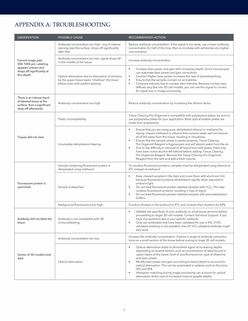
OBSERVATION POSSIBLE CAUSE RECOMMENDED ACTION
Cannot image past 500–1000 μm. Labeling appears uneven and drops off significantly at this depth.
Antibody concentration too high: ring of intense staining near the surface, drops off significantly after that.
Reduce antibody concentration. If the signal is too weak, use a lower antibody concentration for half of the time, then re-incubate with antibodies at a higher concentration.
Antibody concentration too low: signal drops off in the middle of the tissue.
Increase antibody concentration
Optical attenuation due to absorption of photons by the upper tissue layers “shadows” the tissue below, even with perfect staining.
• Increase laser power and gain with increasing depth. Some microscopes can automate laser power and gain corrections.
• Caution! Higher laser power increases the rate of photobleaching. • Ensure that the samples contain no air bubbles. • Compare intensity loss to nuclear stain intensity. Because nuclear stain
diffuses very fast into 3D cell models, you can use this signal to correct for signal loss in image processing.
There is an intense band of labeled tissue at the surface, then a significant drop-off afterwards.
Antibody concentration too high Reduce antibody concentration by increasing the dilution factor.
Tissues did not clear
Plastic incompatibilityTissue Clearing Pro-Organoid is compatible with polystyrene plates, be sure to use polystyrene plates for your application. Most optical bottom plates are made from polystyrene.
Incomplete dehydration/clearing
• Ensure that you are using pure, dehydrated ethanol or methanol for drying. Impure methanol or ethanol that contains water will not remove all of the water from the tissue, resulting in cloudiness.
• Ensure that the sample vessel is sealed properly. Tissue Clearing Pro-Organoid Reagent is hygroscopic and will absorb water from the air.
• Due to the difficulty in removal of all liquid from well plates, there may have been some alcohol left behind before adding Tissue Clearing Pro-Organoid Reagent. Remove the Tissue Clearing Pro-Organoid Reagent from the well and add a fresh volume.
Fluorescent protein is quenched
Sample containing fluorescent protein is dehydrated using methanol.
To visualize fluorescent proteins, samples must be dehydrated using ethanol at 4°C instead of methanol
Sample is bleached.
• Keep cleared samples in the dark and cover them with aluminum foil, because fluorescent proteins photobleach rapidly when exposed to ambient light.
• Do not treat fluorescent protein-labeled samples with H2O2. This step oxidizes fluorescent proteins, resulting in loss of signal.
• Do not treat fluorescent protein-labeled samples with permeabilization buffers.
Background fluorescence too high. Conduct all steps in the protocol to 4°C and increase their duration by 50%.
Antibody did not label the tissue
Antibody is not compatible with 3D immunolabeling.
• Validate the specificity of your antibody on small tissue sections before proceeding to larger 3D cell models. Contact Technical Support, if you have any questions about your specific antibody.
• Only use antibodies that have been validated for use in IHC. If IHC validated antibody is not available, then IF/ ICC validated antibody might also work.
Center of 3D models look dark
Antibody concentration too low.Increase the antibody concentration. Explore a range of antibody concentra-tions on a small section of the tissue before scaling to large 3D cell models.
Optical attenuation
• Optical attenuation leads to diminished signal at increasing depths depending on several factors, such as concentration of label bound in upper layers of the tissue, level of autofluorescence, type of objective, and laser power.
• Modify laser power and gain according to tissue depth to account for optical attenuation. This can be automated in systems such as the Leica SP5 and SP8.
• Histogram matching during image processing can account for optical attenuation at the cost of increased noise at greater depths
APPENDIX A: TROUBLESHOOTING
8
APPENDIX B: GUIDELINES FOR VALIDATING ANTIBODIES AND OPTIMIZING ANTIBODY CONCENTRATION If you are using an antibody for the first time, we recommend that you validate the anti-body and optimize its concentration.
• Fix the 3D cell models with 10% neutral buffered formalin at room temperature for 30 minutes. Do not over-fix the 3D cell models.
• Label 3D cell models using various concentrations of the primary antibody, ranging from 1:50 to 1:500 (e.g. 1:50, 1:100, 1:200, 1:300, 1:500), diluted in Tissue Clearing Pro Antibody Buffer.
• Typically, 1:100 dilution of the secondary antibody works well. However, you might have to optimize the secondary antibody concentration if you observe low signal or high background.
• To evaluate the evenness of staining, image the 3D cell models using a confocal microscope. Obtain a z-stack image spanning the entire thickness of the tissue section using two color channels: the channel corresponding to the fluorescent conjugate for antibody staining, and the channel used for nuclear stain. Because nuclear stains penetrate 3D cell models rapidly and homogenously, the nuclear stain channel serves as a control for optical attenuation.
• Optical attenuation will naturally cause deeper layers of 3D cell models to appear dimmer than the outer layers, this can be corrected by normalizing the intensity of each slice in the Z stack.
• Examine the z-stacks in ImageJ program (or other image processing software). Observe the XZ and YZ planes by viewing “Orthogonal Views” and examine the evenness of staining.
• If the staining is even, you should see relatively consistent intensity (with respect to nuclear stain) across the tissue (Figure 1). Some dimming in the inner layers is expected, but signal should be visible across tissue.
• If the concentration of the immunolabel is too high, you will see a bright ring of staining at the surface layers, with uneven staining at a lower intensity deeper into the tissue.
• If the concentration of the immunolabel is too low, you will see slight staining at the surface layer, a dark interior, and uneven spots of stain.
Tissue Clearing Pro reagents were developed with Visikol Inc, who developed the Visikol® HISTO™ tissue clearing technology.
9
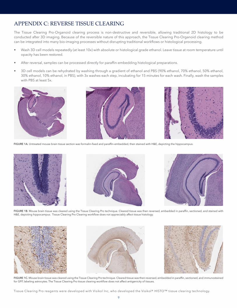
FIGURE 1A. Untreated mouse brain tissue section was formalin-fixed and paraffin-embedded, then stained with H&E, depicting the hippocampus.
FIGURE 1B. Mouse brain tissue was cleared using the Tissue Clearing Pro technique. Cleared tissue was then reversed, embedded in paraffin, sectioned, and stained with H&E, depicting hippocampus. Tissue Clearing Pro Clearing workflow does not appreciably affect tissue histology.
FIGURE 1C. Mouse brain tissue was cleared using the Tissue Clearing Pro technique. Cleared tissue was then reversed, embedded in paraffin, sectioned, and immunostained for GFP, labeling astrocytes. The Tissue Clearing Pro tissue clearing workflow does not affect antigenicity of tissues.
APPENDIX C: REVERSE TISSUE CLEARINGThe Tissue Clearing Pro-Organoid clearing process is non-destructive and reversible, allowing traditional 2D histology to be conducted after 3D imaging. Because of the reversible nature of this approach, the Tissue Clearing Pro-Organoid clearing method can be integrated into many bio-imaging processes without disrupting traditional workflows or histological processing.
• Wash 3D cell models repeatedly (at least 10x) with absolute or histological grade ethanol. Leave tissue at room temperature until opacity has been restored.
• After reversal, samples can be processed directly for paraffin-embedding histological preparations.
• 3D cell models can be rehydrated by washing through a gradient of ethanol and PBS (90% ethanol, 70% ethanol, 50% ethanol, 30% ethanol, 10% ethanol, in PBS), with 3x washes each step, incubating for 15 minutes for each wash. Finally, wash the samples with PBS at least 5x.

WHERE SCIENCE INTERSECTS INNOVATION™
bio-techne.com
Global [email protected] bio-techne.com/find-us/distributors TEL +1 612 379 2956 North America TEL 800 343 7475 Europe | Middle East | Africa TEL +44 (0)1235 529449 China [email protected] TEL +86 (21) 52380373
For research use or manufacturing purposes only. Trademarks and registered trademarks are the property of their respective owners.
BR_Tissue Clearing Pro-Organoid Guidebook_STRY0097515