time-dependent activation of myocardial and peripheral oxidative stress is dissociated after...
TRANSCRIPT
Results: At 4 weeks of MI, body weight, fasting blood glucose and plasma insulin
level were increased in HFDþMI compared to NDþMI. Oxidative stress measured
by LV thiobarbituric acid reactive substances was also increased in HFDþMI. LV
cavity dilatation and dysfunction were significantly exacerbated in HFDþMI com-
pared to NDþMI, which was accompanied by a increase in interstitial fibrosis of
the non-infarcted LV. Treatment of MI mice with apocynin significantly attenuated
LV cavity dilatation and dysfunction. It also decreased LVEDP and lung weight.
Conclusion: Exacerbated post-MI LV remodeling and failure in DM were amelio-
rated by NADPH oxidase inhibitor. Oxidative stress derived from NAD(P)H oxidase
plays a critical role on the progression of heart failure after MI associated with DM.
1035
Enhanced Oxidant Stress Under Acute Decompensated Congestive Heart
Failure Exacerbates Left Ventricular Remodeling at its Stable PhaseMEGUMI KUNISHIGE, YOSHIYUKI KIJIMA, TAKU SAKAI, OSAMUAKUTAGAWA, AKIKO MATSUO, AKIRA NISHIBE, YUSUKE NAKAGAWA,TAKESHI HATADepartment of Cardiology, Higashi-osaka City General Hospital, Osaka, Japan
Progression of left ventricular remodeling (LVR) is crucial for prognosis of conges-
tive heart failure (CHF). We recently reported the enhancement of oxidant stress (OS)
and collagen turnover in patients with acute decompensated CHF (AD-CHF). The
aim of this study was to clarify the association between enhanced OS and LVR in
such patients. Thirty-two patients were admitted into our hospital due to AD-CHF.
After successful treatment, they were followed up with conventional medicines.
LVR was monitored with echocardiography at admission, 2 weeks later, and 6
months later. Immunoreactive 8-iso-prostaglandin F2alpha (8-iso-PGF2alpha), a reliable
maker for OS in vivo, was measured in urine at admission. The patients were divided
into two groups according to the median of urinary 8-iso-PGF2alpha (235pg/mg cre-
atinine), the lower OS (n 5 15) and the higher OS group (n 5 17). In both groups,
LV ejection fraction, mitral valve deceleration time, and left atrial diameter were im-
proved from acute phase to 6 months. Parameters for LVR, however, were improved
only in the lower OS but not in the higher OS group; i.e., LV mass index (188.9653.8
to 145.9630.2 vs. 163.2634.2 to 163.1639.2 g/m2, p!0.05) and LV end-diastolic
diameter (59.8610.3 to 52.465.2 vs. 57.367.8 to 55.767.7 mm, p!0.05). In con-
clusion, enhanced OS under AD-CHF appears to antagonize the beneficial effect of
conventional medical therapy on LVR at its stable phase.
1036
Vagal Nerve Stimulation Acutely Decreases the Generation of Free Radicals in
Failing MyocardiumTAKAKI TSUTSUMI1, TOMOMI IDE1, MAYUMI YAMATO2, ATSUNORIKAMIYA3, HIDEO UTSUMI4, YOSHITAKA HIROOKA1, KENJI SUNAGAWA1
1Department of Cardiovascular Medicine, Graduate School of Medical Sciences,Kyushu University, Fukuoka, Japan, 2Department of REDOX Medicinal Science,Graduate School of Pharmaceutical Sciences, Kyushu University, 3Department ofCardiovascular Dynamics, National Cardiovascular Center, Research Institute,Osaka, Japan, 4Department of Bio-functional Science, Graduate School ofPharmaceutical Sciences, Kyushu University
Background: Vagal nerve stimulation attenuates cardiac remodeling and improves
survival after myocardial infarction. However, its precise mechanisms responsible
for cardiac remodeling are unknown. Recently we have shown the role of reactive
oxygen species (ROS) in the pathogenesis of cardiac remodeling. Herein, we hypoth-
esized that acute vagal nerve stimulation attenuates ROS production in failing heart.
Methods and Results: Mice that survived 28 days after MI were sham-stimulated
(SS) or vagal-stimulated (VS) for 15 min. Five minutes after stimulation we directly
measured the cardiac ROS by in vivo electron spin resonance (ESR) spectroscopy. In
comparison with Non-MI Shams, the rate of ESR signal decay of methoxycarbonyl-
PROXYL was increased in the MI-SS group (0.1360.01 vs 0.1660.01 min-1,
p!0.05), whereas significantly attenuated in the MI-VS group (0.1360.01 min-1,
p!0.05). Furthermore, this effect was completely abolished by the administration
of atropine sulfate (0.1760.02 min-1, p!0.05). In neonatal rat cardiomyocytes, nor-
epinephrine (NE) increased the extracellular oxidants, assessed by the DCF-DA
fluorescence, in a dose-dependent manner. Co-incubation with acetylcholine (Ach)
reduced the elevated DCF-DA, which was antagonized by atropine.
Conclusions: Vagal nerve stimulation decreases ROS generation in failing myocar-
dium via muscarinic Ach receptors. This vagally mediated anti-oxidative mechanism
might be responsible for the beneficial effect of chronic vagal stimulation on cardiac
remodeling.
1037
Identification of Signaling Pathways that Mediate the Oxidative Stress-induced
Translocation of Myocardial GLUT4 using a Novel Lenti-viral-mediated
GLUT4 Reporter GeneTAKAHIRO HORIE1, KOH ONO1, YUKIKO ABE2, TERUHISA KAWAMURA2,AKIRA SHIMATSU2, TORU KITA1, KOJI HASEGAWA2
1Department of Cardiovascular Medicine, Kyoto University, 2Division of TraslationalResearch, Kyoto Medical Center, National Hospital Organization, Kyoto, Japan
GLUT4 is a glucose transporter protein expressed in the heart and translocates from
cytoplasm to sarcolemma under ischemia. Hearts from mice with cardiac-selective
GLUT4 deficiency develop profound and irreversible dysfunction after ischemia.
Therefore, GLUT4 translocation represents protection against ischemic injury. To
identify the signaling pathways of myocardial GLUT4 translocation, we established
a novel method for quantifying the relative proportion of sarcolemmal GLUT4 to to-
tal GLUT4. We constructed a GLUT4 reporter containing a c-Myc epitope tag in the
first exofacial loop and fused GFP to the C-terminus in the lenti-virus vector. Cell-
surface GLUT4 reporter was detected using an anti-Myc primary antibody and
a PE-conjugated secondary antibody by flowcytometry. The ratio of cell-surface
GLUT4 was represented as PE/FITC. We transduced this reporter into primary car-
diomyocytes and found that 90% of these cells expressed GFP. H2O2 stimulation re-
sulted in a concentration-dependent increase in GLUT4 translocation, peaked at 15
min. The PI3K-inhibitor (LY294002), dominant-negative PI3K and AMPK almost
completely inhibited the H2O2-induced GLUT4 translocation as well as Akt phos-
phorylation. Recently, two AMPKKs, CaMKK and LKB1 have been identified. Dom-
inant-negative CaMKK and the CaMKK-inhibitor (STO-609) inhibited translocation
in an earlier phase, whereas dominant-negative LKB1 inhibited translocation in a later
phase. These results suggest that oxidative stress causes GLUT4 translocation
through the serial activation of CaMKK and LKB1, which leads to activation of an
AMPK-PI3K/Akt pathway.
1038
Time-dependent Activation of Myocardial and Peripheral Oxidative Stress is
Dissociated after Myocardial InfarctionTAKAHIRO INOUE1, TOMOMI IDE1, MAYUMI YAMATO2, MASAYOSHIYOSHIDA1, NAOKO SERI2, TAKAKI TSUTSUMI1, HIROYUKI TSUTSUI3,KENJI SUNAGAWA1
1Graduate School of Medical Science, Kyushu University, Fukuoka, Japan,2Department of REDOX Medical Science, Graduate School of PharmaceuticalScience, Kyushu University, Fukuoka, Japan, 3Cardiovascular Medicine, HokkaidoUniversity Graduate School of Medicine, Sapporo, Japan
Purpose: The generation of reactive oxygen species (ROS) increases in the heart af-
ter MI. However, the time-course of oxidative stress in the post-MI heart and periph-
eral blood remains unknown. We investigated the redox profiles with biomarkers of
oxidative stress during cardiac remodeling.
Method: MI was created in mice by ligating the left coronary artery. These mice
were followed by echocardiography, plasma and tissue levels of TBARS, antioxi-
dants, and immunohistochemical studies.
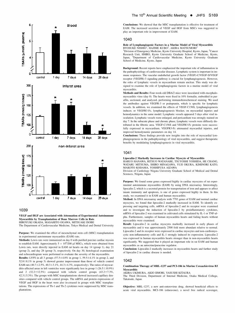
Result: The generation of ROS increased in non-infarcted LV myocardium in the late
remodeling. However, the activation of ROS in myocardium in the late remodeling
did not affect TBARS or antioxidants in peripheral blood.
Conclusion: Oxidative stress is localized in the post-MI heart. Hence, the anti-oxida-
tive stress strategy should focus on the survived myocardium in the late-remodeling.
S168 Journal of Cardiac Failure Vol. 12 No. 8 Suppl. 2006
1039
VEGF and HGF are Associated with Attenuation of Experimental Autoimmune
Myocarditis by Transplantation of Bone Marrow Cells in RatsHIROYUKI OKADA, MASAHITO OGAWA, MITSUAKI ISOBEThe Department of Cardiovascular Medicine, Tokyo Medical and Dental University
Purpose: We examined the effect of mesenchymal stem cell (MSC) transplantation
in experimental autoimmune myocarditis (EAM) rats.
Methods: Lewis rats were immunized on day 0 with purified porcine cardiac myosin
to establish EAM. Approximately 5 � 106/200 ml MSCs, which were obtained from
Lewis rats, were directly injected in EAM rat hearts on day 14 (group 1), day 21
(group 2), and day 28 (group 3), respectively. On day 30, histological examination
and echocardiogram were performed to evaluate the severity of the myocarditis.
Results: LVFS in all 3 groups (57.564.0% in group 1, 59.564.1% in group 2, and
52.868.1% in group 3) showed greater improvement than those of vehicle control
EAM rats (40.762.5%, 40.363.1%, 44.364.3%, respectively). The ratios of myocar-
ditis-affected areas in left ventricles were significantly less in group 1 (26.5610.9%)
and 2 (32.2612.5%) compared with vehicle control groups (43.267.3%,
42.365.3%). The groups with MSC transplantations showed increased capillary den-
sities compared with vehicle control groups. The mRNA and protein expressions of
VEGF and HGF in the heart were also in-creased in groups with MSC transplan-
tations. The expressions of Th-1 and Th-2 cytokines were suppressed by MSC trans-
plantations.
Conclusions: We showed that the MSC transplantation is effective for treatment of
EAM. The increased secretion of VEGF and HGF from MSCs was suggested to
play an important role in improvement of EAM.
1040
Role of Lymphangiogenic Factors in a Murine Model of Viral MyocarditisRYOSUKE NISHIO1, HAJIME KUBO2, AKIRA MATSUMORI3
1Division of Emergency Medicine, Kyoto University Hospital, Kyoto, Japan, 2CancerResearch Unit, HMRO, Kyoto University Graduate School of Medicine, Kyoto,Japan, 3Department of Cardiovascular Medicine, Kyoto University GraduateSchool of Medicine, Kyoto, Japan
Background: Recent reports have emphasized the important role of inflammation in
the pathophysiology of cardiovascular diseases. Lymphatic system is important in im-
mune responses. The vascular endothelial growth factor (VEGF)-C/VEGF-D/VEGF
receptor (VEGFR)-3 signaling pathway is crucial for lymphangiogenesis. However,
the roles of lymphatic vessels in myocardium remain unclear. This study was de-
signed to examine the role of lymphangiogenic factors in a murine model of viral
myocarditis.
Methods and Results: Four-week-old DBA/2 mice were inoculated with encephalo-
myocarditis virus (day 0). The hearts were fixed in 10% formalin, embedded in par-
affin, sectioned, and analyzed, performing immunohistochemical staining. We used
the antibodies against VEGFR-3 or podopranin, which is specific for lymphatic
vessels. In addition, we examined the effects of VEGF-C156S, lymphangiogenesis
inducer, or VEGFR3-Fc, lymphangiogenesis blocker, on myocardial injuries and
hemodynamics in the same model. Lymphatic vessels appeared 3 days after viral in-
oculation. Lymphatic vessels were enlarged, and pericardium was strongly stained on
day 7. In the subacute phase and chronic phase, lymphatic vessels were diffusely dis-
tributed in the fibrotic area. VEGF-C156S and VEGFR3-Fc proteins were success-
fully expressed in myocardium. VEGFR3-Fc attenuated myocardial injuries, and
improved hemodynamic parameters on day 14.
Conclusions: These findings provide new insights into the role of myocardial lym-
phangiogenesis in the pathophysiology of viral myocarditis, and suggest therapeutic
benefits by modulating lymphangiogenesis in viral myocarditis.
1041
Lipocalin-2 Markedly Increases in Cardiac Myocyte of MyocarditisHARUO HANAWA, RITSUO WATANABE, TSUYOSHI YOSHIDA, HE CHANG,WATARU MITSUMA, SHIRO MINAGAWA, YUJI OKURA, KIMINORI KATO,MAKOTO KODAMA, YOSHIFUSA AIZAWADivision of Cardiology Niigata University Graduate School of Medical and DentalSciences, Niigata, Japan
Purpose: We found some genes expressed highly in cardiac myocytes of rat exper-
imental autoimmune myocarditis (EAM) by using DNA microarray. Interestingly,
lipocalin-2, which is a secreted protein for transportation of iron and appears to affect
innate immunity and apoptosis, is one of genes expressed highly in them. In this
study, we examined it in EAM and human myocarditis.
Method: In DNA microarray analysis with 7793 genes of EAM and normal cardiac
myocytes, we found that lipocalin-2 markedly increased in EAM. To identify ex-
pressing and targeting cells, mRNA of lipocalin-2 and its receptor were examined
and to investigate the induction of lipocalin-2 by proinflammatory cytokines,
mRNA of lipocalin-2 was examined in cultivated cells stimulated by IL-1 or TNF-al-
pha. Furthermore, samples of human myocarditis hearts and failing hearts without
myocarditis were examined.
Result: Lipocalin-2 in cardiac myocytes markedly increased in early phases of
myocarditis and it was approximately 2500 fold more abundant relative to normal.
Lipocalin-2 and its receptor were expressed in cardiac myocytes and non-cardiomyo-
cytic non-inflammatory cells and IL-1 strongly induced its expression. Lipocalin-2
was expressed in human myocarditis hearts stronger than in non-myocarditis hearts
significantly. We suggested that it played an important role in rat EAM and human
myocarditis as an autocrine/paracrine regulator.
Conclusion: Lipocalin-2 markedly increases in myocarditis hearts and further study
of lipocalin-2 in cardiac disease is needed.
1042
Combination Therapy of MRL-1237 and PCI-186 in Murine Coxsackievirus B3
MyocarditisAKIRA UKIMURA, AKIO IIMORI, YASUSHI KITAURAThe Third Division, Department of Internal Medicine, Osaka Medical College,Takatsuki, Japan
Objective: MRL-1237, a new anti-enterovirus drug, showed beneficial effects to
acute viral myocarditis. MCI-186 (edaravone), a novel free radical scavenger,
The 10th Annual Scientific Meeting � JHFS S169