tiger stripes: neurally-informed benchwork that helps ... · tiger stripes: neurally-informed...
TRANSCRIPT
• 6 • 2014 IASI Yearbook of Structural Integration
Tiger Stripes: Neurally-Informed Benchwork That Helps Achieve Third Hour or Ninth Hour Goals
Kirstin SchumakerKirstin Schumaker, BCSICM, graduated from the KMI program in 2005. Since then she has studied with four independent neural and visceral manipulation teachers, each of whom has shaped her palpatory understanding of the nerves. She is grateful for their significant influence on the development of her own style of “neurovascularly-informed” structural integration (SI) work. Since 2012 she has been teaching nerve mobilization to her colleagues, helping them to differentiate myofascial layers while keeping the nerve and artery pathways in mind, thus giving structural ease to the neurovascular system while working on achieving the greater goals of SI.
AbstractThe author describes how her understanding of the intercostal nerve pathways (and the parallel lumbar nerves) informed her development of a benchwork technique that she calls tiger stripes. Through demonstration and discussion the author illuminates how to use this technique to open up the lateral body for improved torso rotation and lateral flexion. Photo documentation supports the idea that this technique by itself is very helpful for achieving the SI goals we have for the Lateral Line - Third Hour session. This technique is also very useful for upper body integration during the Ninth Hour (KMI Session Ten) because it can unlock remaining restrictions to torso rotation while the client is upright, for promotion of a more fluid contra-lateral movement through the torso.
One purpose of this article is to show you how useful it can be to keep the peripheral nerves in
mind while working with myofascia. I also wish to show those of you who have studied neural work from an osteopathic-visceral perspective that it is possible to mobilize nerves while the client is upright and moving in gravity, and while you are “making the skin pink” (a “no-no” according to one of my nerve teachers). Ultimately though, I simply want to pass on a great discovery that I made in my own private laboratory (office), so that you too can use the technique which I have dubbed tiger stripes. (Tiger stripes are distinct from the familiar pink, skunk-like stripes that run parallel to the spine after performing classic backstripes benchwork.)
Before I describe this benchwork technique, I want to remind you that the upper portion of the Lateral Line (side of torso from iliac crest to neck) is often depicted as a series of Xs moving up the side of the body, Xs which represent the basket weave of fascial fibers governing rotational and spiral movement through the torso, in addition to regulating stability. On a micro level, fiber directions within the intercostals and obliques form a multilayered criss-cross pattern that controls the ribs’ relationships to one another and the rib cage’s relationship to the pelvis. This criss-cross pattern helps drive the spinal
engine of oscillating left and right torso rotation that is a vital part of integrated cross-crawl movement. (See Myers’ discussion of the Lateral Line in Anatomy Trains (2009), Dalton’s discussion of spiraling spring systems in his Core Stability and Back Pain article (2011), which includes a description of Gracovetsky’s spinal engine, Gracovetsky’s book (1989) and video (1988) on spinal assessment, and Earls’ soon-to-be-released book, Born to Walk (2014).
We know from our SI training and from clinical experience that working broadly in a diagonal direction up or down the side of the torso with some sort of client movement (breathing or reaching) can help open up the torso for greater side-bending capacity, and we know in principle that differentiation between myofascial layers is good in the grand scheme of things. So, we differentiate the lats from the underlying layers, working diagonally with or against their fibers, we work diagonally across the ribs to help accordion them open with the breath, or we work into the intercostal spaces, because we want more space between the ribs and we want release for the intercostal muscles that are part of the fascial X-pattern. We’d all agree that a combination of these techniques is quite helpful in opening up the side body.
2014 IASI Yearbook of Structural Integration • 7 •
When nerves are stretched, or when they have been historically, chronically
inflamed . . . , they feel like strings within the myofascial fabric, which
can function rather like the reinforcing strings in packing tape.
Next, I want to describe the diagonal pattern that the peripheral nerves take as they weave through the myofascial layers of the side body, so that you may gain new perspective on how torso rotation can be limited by restricted nerves. You will then have a new rationale for working diagonally when opening up the Lateral Line. After a discussion of the neural anatomy, I will demonstrate how rotational benchwork can quickly help you meet the goals of improved lateral flexion, rib cage expansion, and torso rotation (KMI Session 3 and traditional Third Hour), balance through the Upper Spiral Line (KMI Session 4), and integrated contralateral movement through the torso (KMI Session 10 and traditional Ninth Hour). My presentation is in cookbook fashion, with photo illustrations of the procedures and results, and explanatory text. You will have to work with the technique to discover for yourself the nuances of its application. It is my hope that the before and after photos will make you want to explore.
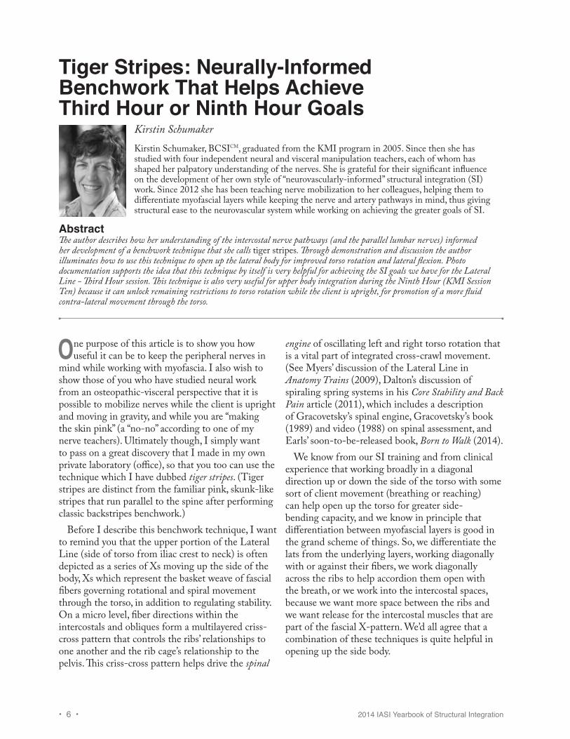
Neural Anatomy of the Rib Cage and WaistI would like to draw your attention to the intercostal nerves that ride around the body beneath each rib, embedded within the myofascia of the intercostals, but with cutaneous branches sprouting to the surface, and the lumbosacral nerves that travel in a similar pattern. These nerves spiral around the torso within the intercostal spaces, reinforcing one “leg” of the fascial X-pattern, traveling from high posterior to low anterior, in a similar direction to that of the external obliques (see Figure 1). These nerves are accompanied along their pathways by small arteries and veins that together with the nerves form neurovascular tracts.
In general, smaller nerves (such as the intercostal nerves) have a higher percentage of fibrous connective tissue than larger nerves. Yet
fibrous connective tissue makes up 50-90% of the composition of nerves in the arm, with the axillary nerve having the highest percentage. Mechanical strain is likely the reason for high connective tissue content in larger nerves. (See Barral’s (2007, p. 22) discussion of connective tissue ratio in peripheral nerves).
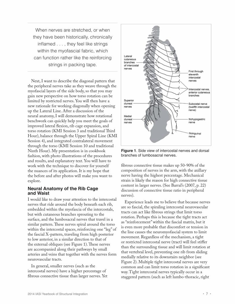
Experience leads me to believe that because nerves are so fascial, the spiraling intercostal neurovascular tracts can act like fibrous strings that limit torso rotation. Perhaps this is because the tight tracts act as “reinforcement” within the fascial matrix, but it is even more probable that discomfort or tension in the line causes the neuromyofascial system to limit movement. Regardless of the mechanism, a tight or restricted intercostal nerve (tract) will feel stiffer than the surrounding tissue and will limit rotation at that vertebral level, preventing one rib from sliding medially relative to its downstairs neighbor (see Figure 2). Multiple tight intercostal nerves are very common and can limit torso rotation in a significant way. Tight intercostal nerves typically occur in a staggered pattern (such as left lumbo-thoracic, right
Figure 1. Side view of intercostal nerves and dorsal branches of lumbosacral nerves.
• 8 • 2014 IASI Yearbook of Structural Integration
low to mid-thoracic, left mid- to upper-thoracic) in cases of mild scoliosis (see Figure 3). (These statements are based on my clinical experience.)
In the side view shown in Figure 1, notice the intercostal nerves spiraling down along the same angle as the ribs, like the stripes on a barbershop pole. This pathway will be surprisingly closer to vertical amongst the lower ribs when the rib cage is posteriorly tilted (relative to the floor, in standing position), because the ribs in this case will be fascially held down in a chronic exhale position. As the rib cartilage starts to swoop back upward (at the costal margin on the front of the torso), the intercostal nerves continue instead along their spiraling path, leveling out as they approach the edge of rectus abdominis. These nerves are the motor supply for the abdominal muscles, but they also have branches that sprout to the surface to supply the skin.
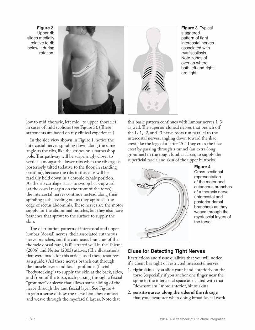
The distribution pattern of intercostal and upper lumbar (dorsal) nerves, their associated cutaneous nerve branches, and the cutaneous branches of the thoracic dorsal rami, is illustrated well in the Thieme (2006) and Netter (2003) atlases. (The illustrations that were made for this article used these resources as a guide.) All these nerves branch out through the muscle layers and fascia profundis (fascial “bodystocking”) to supply the skin at the back, sides, and front of the torso, each passing through a fascial “grommet” or sleeve that allows some sliding of the nerve through the taut fascial layer. See Figure 4 to gain a sense of how the nerve branches connect and weave through the myofascial layers. Note that
this basic pattern continues with lumbar nerves 1-3 as well. The superior cluneal nerves that branch off the L-1, -2, and -3 nerve roots run parallel to the intercostal nerves, angling down toward the iliac crest like the legs of a letter “A.” They cross the iliac crest by passing through a tunnel (an extra-long grommet) in the tough lumbar fascia, to supply the superficial fascia and skin of the upper buttocks.
Figure 2. Upper rib
slides medially relative to rib
below it during rotation.
Figure 3. Typical staggered pattern of tight intercostal nerves associated with mild scoliosis. Note zones of overlap where both left and right are tight.
Figure 4. Cross-sectional representation of the motor and cutaneous branches of a thoracic nerve (intercostal and posterior dorsal branches) as they weave through the myofascial layers of the torso.
Clues for Detecting Tight Nerves Restrictions and tissue qualities that you will notice if a client has tight or restricted intercostal nerves: 1. tight skin as you slide your hand anteriorly on the
torso (especially if you anchor one finger near the spine in the intercostal space associated with that “downstream,” more anterior, bit of skin)
2. sensitive areas along the sides of the rib cage that you encounter when doing broad fascial work
2014 IASI Yearbook of Structural Integration • 9 •
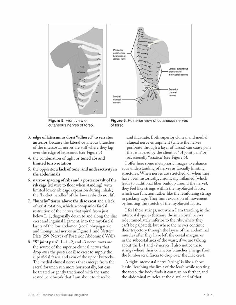
3. edge of latissumus dorsi “adhered” to serratus anterior, because the lateral cutaneous branches of the intercostal nerves are stiff where they lap over the edge of latissimus (see Figure 5)
4. the combination of tight or toned abs and limited torso rotation
5. the opposite: a lack of tone, and underactivity in the abdominals
6. narrow spacing of ribs and a posterior tilt of the rib cage (relative to floor when standing), with limited lower rib cage expansion during inhale; the “bucket handles” of the lower ribs do not lift
7. “bunchy” tissue above the iliac crest and a lack of waist rotation, which accompanies fascial restriction of the nerves that spiral from just below L-1, diagonally down to and along the iliac crest and inguinal ligament, into the myofascial layers of the low abdomen (see iliohypogastric and ilioinguinal nerves in Figure 1, and Netter: Plate 259, Nerves of Posterior Abdominal Wall)
8. “SI joint pain”: L-1, -2, and -3 nerve roots are the source of the superior cluneal nerves that drop over the posterior iliac crest to supply the superficial fascia and skin of the upper buttocks. The medial cluneal nerves that emerge from the sacral foramen run more horizontally, but can be treated or gently tractioned with the same seated benchwork that I am about to describe
and illustrate. Both superior cluneal and medial cluneal nerve entrapment (where the nerves perforate through a layer of fascia) can cause pain that is labeled by the client as “SI joint pain” or occasionally “sciatica” (see Figure 6).
I offer here some metaphoric images to enhance your understanding of nerves as fascially limiting structures. When nerves are stretched, or when they have been historically, chronically inflamed (which leads to additional fiber buildup around the nerve), they feel like strings within the myofascial fabric, which can function rather like the reinforcing strings in packing tape. They limit excursion of movement by limiting the stretch of the myofascial fabric.
I feel these strings, not when I am traveling in the intercostal spaces (because the intercostal nerves ride immediately inferior to the ribs, where they can’t be palpated), but where the nerves continue their trajectory through the layers of the abdominal muscles after they have left the costal margin, or in the subcostal area of the waist, if we are talking about the L-1 and -2 nerves. I also notice these strings where their cutaneous branches emerge from the lumbosacral fascia to drop over the iliac crest.
A tight intercostal nerve “string” is like a short leash: Reaching the limit of the leash while rotating the torso, the body finds it can turn no further, and the abdominal muscles at the distal end of that
Figure 5. Front view of cutaneous nerves of torso.
Figure 6. Posterior view of cutaneous nerves of torso.
• 10 • 2014 IASI Yearbook of Structural Integration
nerve may be situationally (or chronically) hyper- or hypotonic (less functional) due to shearing stress on the nerve.
However, a tight nerve “leash” also causes problems at the proximal end, near the central nervous system: A nerve that cannot stretch or glide acts like a tether on the associated vertebra, as the person attempts torso rotation, which prevents the vertebra from rotating in the appropriate ipsilateral direction. This can contribute to a local rotational conflict within the vertebral column (two adjacent vertabrae are counter-rotated), or it can act like a tether on the spinal cord itself (neural-dural restriction). When a person attempts to flex the spine with head hanging (as when seated on the bench for traditional benchwork), the spinal cord must be free to slide upward within the vertebral column. If multiple peripheral nerves are anchored in myofascia, the spinal cord can’t slide upward in the vertebral column, spinal flexion will be limited, and attempts to force spinal flexion may even put tractional stress on the brain stem. (These statements are my own conclusions based on clinical experience. Have you witnessed a client in your practice or in your clinical training “sweating bullets” while the practitioner attempts backstripes, with little gains in ability to flex forward? Adrenergic sweating combined with a mental aversion or negative emotional reaction to doing backstripes could indicate there is tractional stress on the brain stem.)
One additional comment about restricted neurovascular tracts: In “treating” or addressing the tracts with a mind to freeing the nerves, the arteries often benefit. However, there are ways to target the vascular network more specifically (Barral, 2011), if that is what is needed. (Tiger stripes were developed with the nerves in mind, before I began working with arteries. To keep this discussion simple, I won’t discuss the arteries further, just know that the intercostal arteries [and veins], in addition to the intercostal nerves, are receiving alternating tension and slack during the tiger stripe procedure.)
DemonstrationI chose an 18-year-old model to receive this work, who had received a short series from me when he was fifteen. Since then he has had some “triage” sessions intended to help him recover from injuries. At the time of the demo session, he recently had
been experiencing lower thoracic back pain. He agreed to be my model for this technique demo, after which we would do a session more specifically targeted toward his body’s needs. My goal with this demo was to simply show how I can work the myofascia in the intercostal spaces, with the intent of freeing up the torso for rotation, and that this has a fabulous effect also on side-bending capacity and overall posture.
In this demo session, we did mainly tiger stripe benchwork, with my fingers tracing pathways through the intercostal spaces while the client turned to the left and the right. This work was preceded by a little side-lying work with the cutaneous nerves (which might appear to an observer like I am working with the skin or the superficial fascia), and was followed by a traditional backstripe and some neckwork. That comprised the entire session. The postural after pictures (four views) and the after views of seated rotation were taken before I did the final traditional backstripe and the neckwork, so they really do represent what was achieved with the tiger stripe technique by itself (preceded by some gentle superficial work with the cutaneous nerves).
The forward flexion and extension after pictures were taken after the integrative, traditional backstripe and neckwork, at the very end of the session. Lateral flexion photos were taken both before and after the final integrative work. You can see how much smoother the lateral flexion curve is after just one backstripe and some integrative neckwork, testament that much was accomplished with the seated rotational work that was made more evident after some integration. I have found in my practice that conventional backstripes are very easy (the client’s body does the work with less effort) after I have done tiger stripes with them. Rotational work in the intercostal spaces seems to open the door to easier forward flexion as well as lateral flexion.
(Note: This client has a history of skateboarding and snowboarding with his right foot leading, favoring right torso rotation and probably right hip flexion combined with adduction. He has played soccer as a goalkeeper for years, leaping out to fall with arms outstretched, which may explain why he carries his shoulders up to his ears—repeated strain on the brachial plexus nerve pathways connecting neck to arms.)
2014 IASI Yearbook of Structural Integration • 11 •
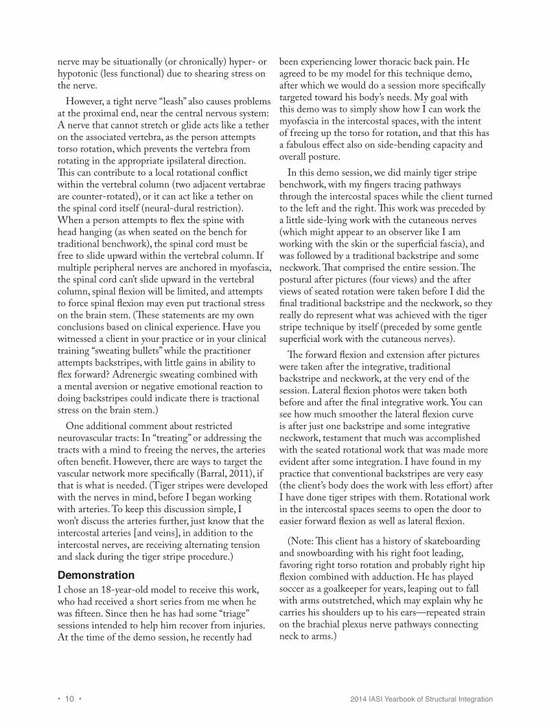
Testing RotationBeforeIn the pretest for rotation, I look for sections of the spine where the spinous processes do not shift laterally very much. These are the vertebral levels from which the intercostal spaces need to be worked. In rotation to the right, you see how the skin (with its ink marks) has shifted left, but the lowest thoracic vertebrae have not shifted, they remain stacked vertically (see Figure 7).
We should not expect much lateral translation of the lumbar vertebrae however, because little rotation happens in the lumbar spine due to orientation of the facet joints. The ink marks in the after pictures represent intercostal spaces that received work. I did more work on the left side of the body, because
that is where I saw the work was needed. It also felt tighter and more “interesting” to work in these intercostal spaces.
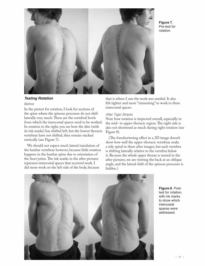
After Tiger StripesNote how rotation is improved overall, especially in the mid- to upper-thoracic region. The right side is also not shortened as much during right rotation (see Figure 8).
(The foreshortening effect in a 2D image doesn’t show how well the upper-thoracic vertebrae make a tidy spiral in these after images, but each vertebra is shifting laterally relative to the vertebra below it. Because the whole upper thorax is turned in the after pictures, we are viewing the back at an oblique angle, and the lateral shift of the spinous processes is hidden.)
Figure 7. Pre-test for rotation.
Figure 8. Post-test for rotation, with ink marks to show which intercostal spaces were addressed.
• 12 • 2014 IASI Yearbook of Structural Integration
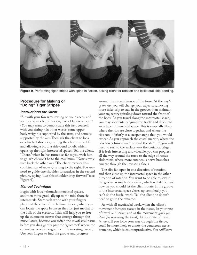
Procedure for Making or “Doing” Tiger StripesInstructions for Client“Sit with your forearms resting on your knees, and your spine in a bit of flexion, like a Halloween cat.” (You may want to demonstrate this first yourself with you sitting.) In other words, some upper body weight is supported by the arms, and some is supported by the core. Then ask the client to look over his left shoulder, turning the chest to the left and allowing a bit of a side-bend to left, which opens up the right intercostal spaces. Tell the client, “There,” when he has turned as far as you wish him to go, which won’t be to the maximum. “Now slowly turn back the other way.” The client reverses this combination of moves, turning to the right. You may need to guide one shoulder forward, as in the second picture, saying, “Let this shoulder drop forward” (see Figure 9).
Manual TechniqueBegin with lower-thoracic intercostal spaces, and then move gradually up to the mid-thoracic intercostals. Start each stripe with your fingers placed at the edge of the laminar groove, where you can locate the space between the ribs, just medial to the bulk of the erectors. (This will help you to free up the cutaneous nerves that emerge through the musculature, because you soften the myofascial tissue before you drag gently past the “grommet” where the cutaneous nerve emerges from the investing fascia.) Use your fingers to find the groove and progress
around the circumference of the torso. At the angle of the ribs you will change your trajectory, moving more inferiorly to stay in the groove; then maintain your trajectory spiraling down toward the front of the body. As you travel along the intercostal space, you may accidentally “jump the track” and drop into an adjacent intercostal space. This is especially likely where the ribs are close together, and where the ribs run inferiorly at a steeper angle than you would expect. As you approach the costal margin, where the ribs take a turn upward toward the sternum, you will need to surf to the surface over the costal cartilage. If it feels interesting and valuable, you can progress all the way around the torso to the edge of rectus abdominis, where more cutaneous nerve branches emerge through the investing fascia.
The ribs fan open in one direction of rotation, and then close up the intercostal space in the other direction of rotation. You want to be able to stay in the groove as much as possible, which will determine how far you should let the client rotate. If the groove of the intercostal space closes up completely, you can’t do the fascial work. Tell the client he doesn’t need to go to the extreme.
As with all myofascial work, when the client’s movement increases tension in the tissue, let your rate of travel slow down; and as the movement gives you slack (by reversing the twist), let your rate of travel increase. If you force your way through the tissue, you’ll be more likely to annoy the cutaneous nerve branches, which is counterproductive. You will leave
Figure 9. Performing tiger stripes with spine in flexion, asking client for rotation and ipsilateral side-bending.
2014 IASI Yearbook of Structural Integration • 13 •
a nice pink stripe, however, which helps you keep track of where you have already worked.
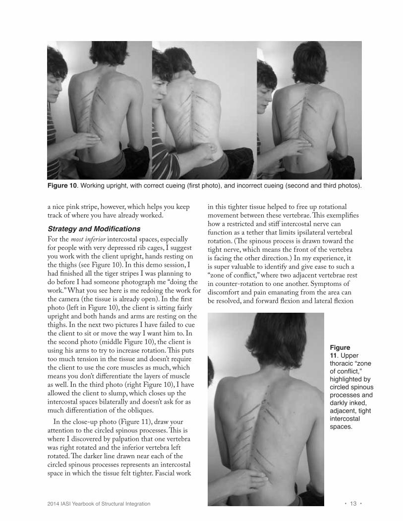
Strategy and ModificationsFor the most inferior intercostal spaces, especially for people with very depressed rib cages, I suggest you work with the client upright, hands resting on the thighs (see Figure 10). In this demo session, I had finished all the tiger stripes I was planning to do before I had someone photograph me “doing the work.” What you see here is me redoing the work for the camera (the tissue is already open). In the first photo (left in Figure 10), the client is sitting fairly upright and both hands and arms are resting on the thighs. In the next two pictures I have failed to cue the client to sit or move the way I want him to. In the second photo (middle Figure 10), the client is using his arms to try to increase rotation. This puts too much tension in the tissue and doesn’t require the client to use the core muscles as much, which means you don’t differentiate the layers of muscle as well. In the third photo (right Figure 10), I have allowed the client to slump, which closes up the intercostal spaces bilaterally and doesn’t ask for as much differentiation of the obliques.
In the close-up photo (Figure 11), draw your attention to the circled spinous processes. This is where I discovered by palpation that one vertebra was right rotated and the inferior vertebra left rotated. The darker line drawn near each of the circled spinous processes represents an intercostal space in which the tissue felt tighter. Fascial work
in this tighter tissue helped to free up rotational movement between these vertebrae. This exemplifies how a restricted and stiff intercostal nerve can function as a tether that limits ipsilateral vertebral rotation. (The spinous process is drawn toward the tight nerve, which means the front of the vertebra is facing the other direction.) In my experience, it is super valuable to identify and give ease to such a “zone of conflict,” where two adjacent vertebrae rest in counter-rotation to one another. Symptoms of discomfort and pain emanating from the area can be resolved, and forward flexion and lateral flexion
Figure 10. Working upright, with correct cueing (first photo), and incorrect cueing (second and third photos).
Figure 11. Upper thoracic “zone of conflict,” highlighted by circled spinous processes and darkly inked, adjacent, tight intercostal spaces.
• 14 • 2014 IASI Yearbook of Structural Integration
are typically improved, when you trace through the specifically restricted intercostal spaces to liberate the conflicted vertebrae.
In the lumbar region, I usually continue making short tiger stripes at an angle that approximates the legs of a letter “A” even though we have run out of intercostal spaces. This is the direction that the upper cluneal nerves run, through the deeper layers of muscle on their way to the iliac crest, where they emerge from the lumbar fascia to supply the superficial fascia and skin lying over the upper gluteal area. Working inferiorly at an angle as you work toward the iliac crest, with the client seated upright and rotating, will prep each of these upper cluneal nerves for the gentle stretch they will receive as you drop past the crest with the client upright and rotating. (Your finger pads may even be aware of a string-like cluneal nerve as you drop over the iliac crest. Tune into its tension and be gentle about “asking” for length.)
Client-reported pain “at the SI joint” after a traditional backstripe is often simply a cranky upper cluneal nerve “talking” that got stretched too tight as you finished the stroke. In the flexed position, when engaged in a traditional backstripe, with client actively descending by engaging his spinal extensors eccentrically, you may not be able to work deeply in the erectors before dropping past the iliac crest. Whereas working in an upright position on the lumbosacral area with client rotation, before doing traditional backstripes, has the effect of: a) flossing the cluneal nerves out of the place where
they emerge from the spinal column, then b) prepping their short length of passage through
the spinal erectors, before c) you floss them gently out of the “grommets”
where they emerge through the lumbar fascia at the iliac crest.
It is very beneficial to do this before you ask for greater length of the upper cluneal nerves during combined spinal and hip flexion.
Even more significantly, here is another reason to attend to the lumbar nerves with tiger stripes, even though the lumbar vertebrae don’t appreciably rotate much due to architectural design. The nerve root that emerges between L-1 and L-2 splits into two nerves that are very important posturally. These are called the iliohypogastric and ilioinguinal nerves. As their name suggests, they run along the iliac crest
toward the inguinal ligament and the low belly wall. They start deep to transversus abdominis (TVA) and gradually perforate one layer of muscle, and then the next, and then the next, running for a while within each intermuscular septum (Netter, Plate 259, Nerves of Posterior Abdominal Wall). When the obliques and TVA are not well-differentiated near the iliac crest, these nerves are trapped. Torso rotation puts a shearing strain on these nerves, which causes pain or low back ache for some people. Discomfort initially inhibits rotational movement, but this leads to more adhesion between the muscle layers. When these nerves have been chronically irritated, fascially reinforced, and shortened over time, they function as tethers that bring the thoracolumbar junction anteriorly, toward the front of the iliac crest, which increases lumbar lordosis. Alternatively, in the case of a relatively flat lumbar curve, or reduced lumbar lordosis, and a posteriorly tilted pelvis, the pubis is brought superiorly so that it is closer to the upper lumbar vertebrae, shortening the path of nerves arcing through the myofascia toward the low belly.
In the case of an asymmetrical spine, when there is a left shift of the rib cage and upper lumbar vertebrae, with the waist muscles posturally maintaining a right rotation of the lower rib cage relative to the pelvis, you can expect the shortened left iliohypogastric and ilioinguinal nerves to be buried in stiffer myofascia than that which is found on the right, which limits left rotation of the ribcage relative to the pelvis. (Try this postural position out in your own body and notice that you have brought L-2 closer to your left iliac crest, shortening these nerve pathways.)
In all these examples, balancing the anterior and posterior bends of the spine or reducing a left or right shift of the rib cage will depend upon release and differentiation of the muscle layers above the iliac crest, the layers which confine the iliohypogastric and ilioinguinal nerves. Upright tiger stripes are just the ticket for release, but you may need to prepare for such seated work with side-lying work superior to the iliac crest (work that hugs the contour of the bones then progresses posteriorly or anteriorly along the same diagonal). For side-lying differentiation of these tissues, I like to ask the client to do exaggerated belly breathing: “let your belly expand as you breathe in, and then draw it in as you exhale.” This is a way of getting them
2014 IASI Yearbook of Structural Integration • 15 •
to lengthen and shorten transversus abdominis as you work through the obliques, establishing layer differentiation.
Analysis of ChangesPostural Views
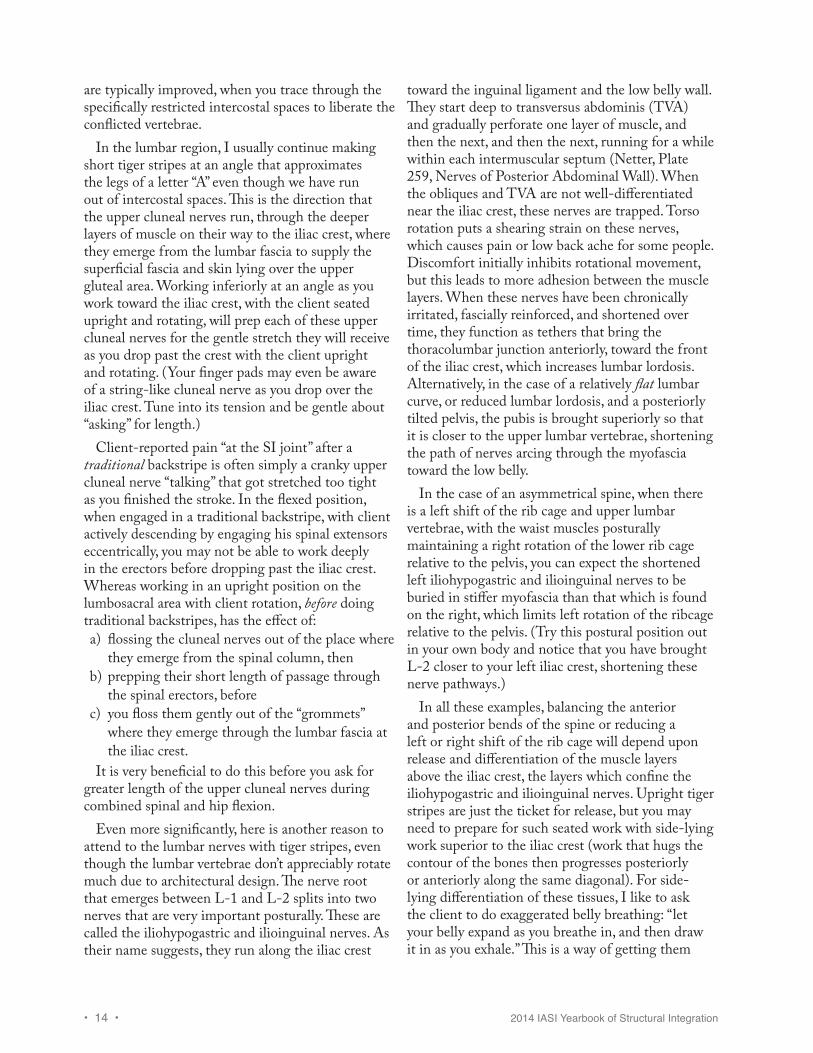
Before WorkRight side-bend pronounced, head shifted anteriorly and neck right tilted, posterior tilt of ribcage, left shiftof ribcage on pelvis. Right rotation of thorax relative to hips and hips relative to feet (see Figure 12).
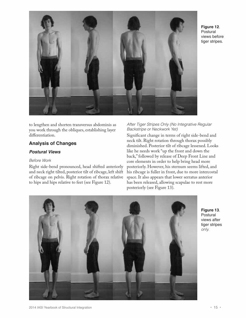
After Tiger Stripes Only (No Integrative Regular Backstripe or Neckwork Yet)Significant change in terms of right side-bend and neck tilt. Right rotation through thorax possibly diminished. Posterior tilt of ribcage lessened. Looks like he needs work “up the front and down the back,” followed by release of Deep Front Line and core elements in order to help bring head more posteriorly. However, his sternum seems lifted, and his ribcage is fuller in front, due to more intercostal space. It also appears that lower serratus anterior has been released, allowing scapulae to rest more posteriorly (see Figure 13).
Figure 12. Postural views before tiger stripes.
Figure 13. Postural views after tiger stripes only.
• 16 • 2014 IASI Yearbook of Structural Integration
Side-Bending
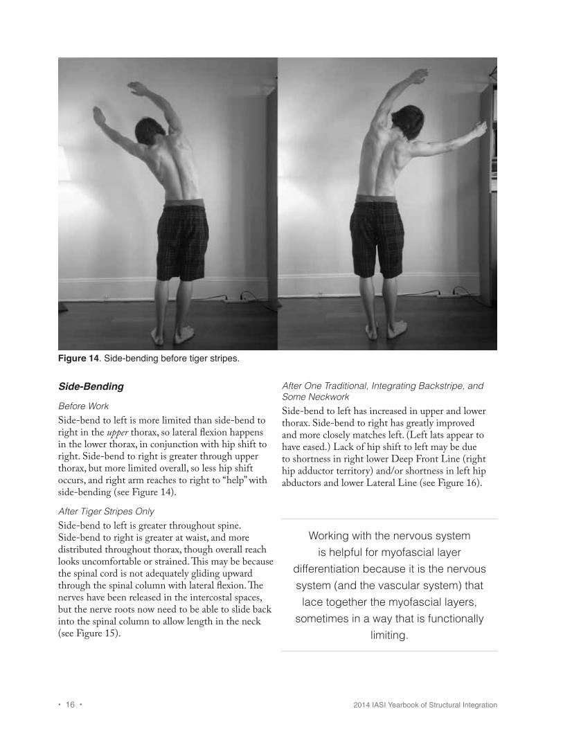
Before WorkSide-bend to left is more limited than side-bend to right in the upper thorax, so lateral flexion happens in the lower thorax, in conjunction with hip shift to right. Side-bend to right is greater through upper thorax, but more limited overall, so less hip shift occurs, and right arm reaches to right to “help” with side-bending (see Figure 14).
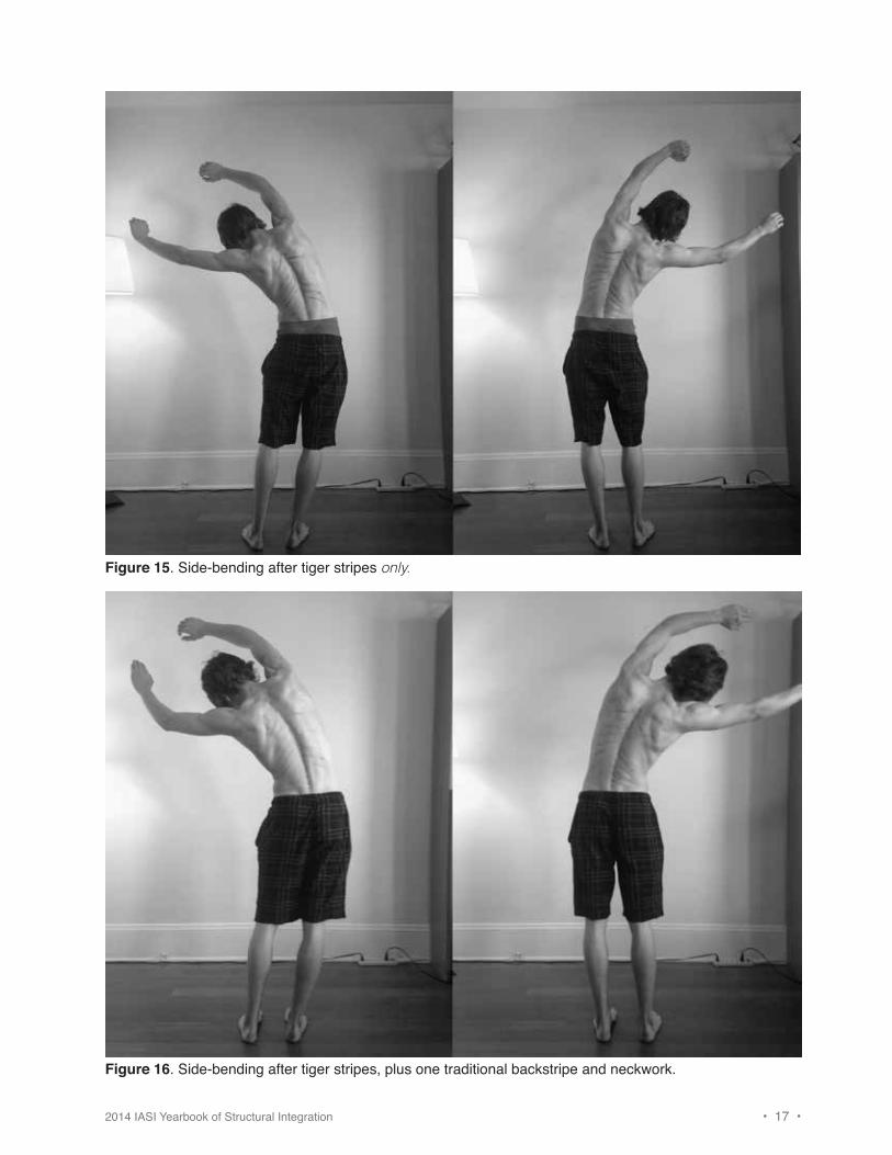
After Tiger Stripes OnlySide-bend to left is greater throughout spine. Side-bend to right is greater at waist, and more distributed throughout thorax, though overall reach looks uncomfortable or strained. This may be because the spinal cord is not adequately gliding upward through the spinal column with lateral flexion. The nerves have been released in the intercostal spaces, but the nerve roots now need to be able to slide back into the spinal column to allow length in the neck (see Figure 15).
After One Traditional, Integrating Backstripe, and Some NeckworkSide-bend to left has increased in upper and lower thorax. Side-bend to right has greatly improved and more closely matches left. (Left lats appear to have eased.) Lack of hip shift to left may be due to shortness in right lower Deep Front Line (right hip adductor territory) and/or shortness in left hip abductors and lower Lateral Line (see Figure 16).
Figure 14. Side-bending before tiger stripes.
Working with the nervous system is helpful for myofascial layer
differentiation because it is the nervous system (and the vascular system) that lace together the myofascial layers,
sometimes in a way that is functionally limiting.
2014 IASI Yearbook of Structural Integration • 17 •
Figure 15. Side-bending after tiger stripes only.
Figure 16. Side-bending after tiger stripes, plus one traditional backstripe and neckwork.
• 18 • 2014 IASI Yearbook of Structural Integration
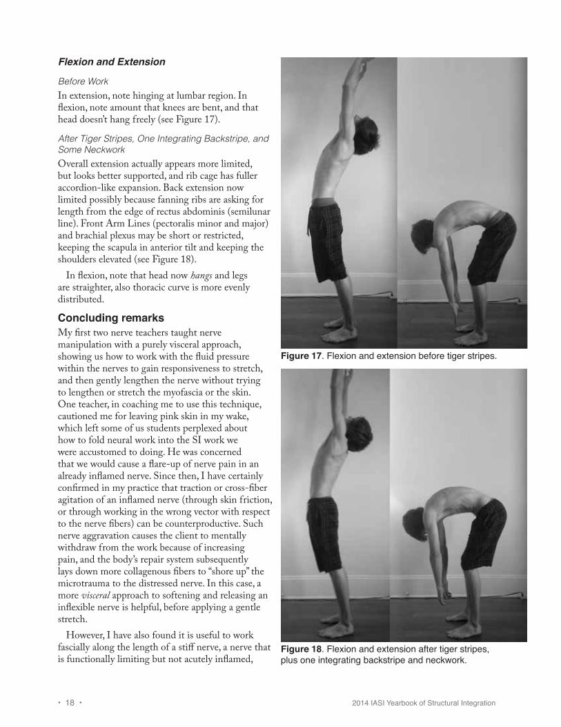
Flexion and Extension
Before WorkIn extension, note hinging at lumbar region. In flexion, note amount that knees are bent, and that head doesn’t hang freely (see Figure 17).
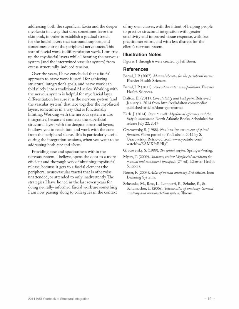
After Tiger Stripes, One Integrating Backstripe, and Some NeckworkOverall extension actually appears more limited, but looks better supported, and rib cage has fuller accordion-like expansion. Back extension now limited possibly because fanning ribs are asking for length from the edge of rectus abdominis (semilunar line). Front Arm Lines (pectoralis minor and major) and brachial plexus may be short or restricted, keeping the scapula in anterior tilt and keeping the shoulders elevated (see Figure 18).
In flexion, note that head now hangs and legs are straighter, also thoracic curve is more evenly distributed.
Concluding remarksMy first two nerve teachers taught nerve manipulation with a purely visceral approach, showing us how to work with the fluid pressure within the nerves to gain responsiveness to stretch, and then gently lengthen the nerve without trying to lengthen or stretch the myofascia or the skin. One teacher, in coaching me to use this technique, cautioned me for leaving pink skin in my wake, which left some of us students perplexed about how to fold neural work into the SI work we were accustomed to doing. He was concerned that we would cause a flare-up of nerve pain in an already inflamed nerve. Since then, I have certainly confirmed in my practice that traction or cross-fiber agitation of an inflamed nerve (through skin friction, or through working in the wrong vector with respect to the nerve fibers) can be counterproductive. Such nerve aggravation causes the client to mentally withdraw from the work because of increasing pain, and the body’s repair system subsequently lays down more collagenous fibers to “shore up” the microtrauma to the distressed nerve. In this case, a more visceral approach to softening and releasing an inflexible nerve is helpful, before applying a gentle stretch.
However, I have also found it is useful to work fascially along the length of a stiff nerve, a nerve that is functionally limiting but not acutely inflamed,
Figure 17. Flexion and extension before tiger stripes.
Figure 18. Flexion and extension after tiger stripes, plus one integrating backstripe and neckwork.
2014 IASI Yearbook of Structural Integration • 19 •
addressing both the superficial fascia and the deeper myofascia in a way that does sometimes leave the skin pink, in order to establish a gradual stretch for the fascial layers that surround, support, and sometimes entrap the peripheral nerve tracts. This sort of fascial work is differentiation work. I can free up the myofascial layers while liberating the nervous system (and the intertwined vascular system) from excess structurally-induced tension.
Over the years, I have concluded that a fascial approach to nerve work is useful for achieving structural integration’s goals, and nerve work can fold nicely into a traditional SI series. Working with the nervous system is helpful for myofascial layer differentiation because it is the nervous system (and the vascular system) that lace together the myofascial layers, sometimes in a way that is functionally limiting. Working with the nervous system is also integrative, because it connects the superficial structural layers with the deepest structural layers; it allows you to reach into and work with the core from the peripheral sleeve. This is particularly useful during the integration sessions, when you want to be addressing both core and sleeve.
Providing ease and spaciousness within the nervous system, I believe, opens the door to a more efficient and thorough way of obtaining myofascial release, because it gets to a fascial element (the peripheral neurovascular tracts) that is otherwise unattended, or attended to only inadvertently. The strategies I have honed in the last seven years for doing neurally-informed fascial work are something I am now passing along to colleagues in the context
of my own classes, with the intent of helping people to practice structural integration with greater sensitivity and improved tissue response, with less practitioner effort, and with less distress for the client’s nervous system.
Illustration NotesFigures 1 through 6 were created by Jeff Boser.
ReferencesBarral, J. P. (2007). Manual therapy for the peripheral nerves.
Elsevier Health Sciences.Barral, J. P. (2011). Visceral vascular manipulations. Elsevier
Health Sciences.Dalton, E. (2011). Core stability and back pain. Retrieved
January 4, 2014 from http://erikdalton.com/media/published-articles/dont-get-married
Earls, J. (2014). Born to walk: Myofascial efficiency and the body in movement. North Atlantic Books. Scheduled for release July 22, 2014.
Gracovetsky, S. (1988). Noninvasive assessment of spinal function. Video posted to YouTube in 2012 by S. Gracovetsky. Retrieved from www.youtube.com/watch?v=EAMK7yR9RgI
Gracovetsky, S. (1989). The spinal engine. Springer-Verlag. Myers, T. (2009). Anatomy trains: Myofascial meridians for
manual and movement therapists (2nd ed). Elsevier Health Sciences.
Netter, F. (2003). Atlas of human anatomy, 3rd edition. Icon Learning Systems.
Scheunke, M., Ross, L., Lamperti, E., Schulte, E., & Schumacher, U. (2006). Thieme atlas of anatomy: General anatomy and musculoskeletal system. Thieme.