tidwell-2011
TRANSCRIPT

Author's personal copy
Molecular Immunology 49 (2011) 260– 272
Contents lists available at SciVerse ScienceDirect
Molecular Immunology
j ourna l ho me pag e: www.elsev ier .com/ locate /mol imm
Characterization of a new ARID family transcription factor (Brightlike/ARID3C)that co-activates Bright/ARID3A-mediated immunoglobulin gene transcription
Josephine A. Tidwell a, Christian Schmidta, Phillip Heatonb, Van Wilsonb, Philip W. Tuckera,∗
a Institute for Cellular and Molecular Biology, Section of Molecular Genetics and Microbiology, The University of Texas at Austin, 1 University Station A5000, Austin, TX 78712, USAb Department of Microbial and Molecular Pathogenesis, Texas A&M Health Science Center, College Station, TX 77843-1114, USA
a r t i c l e i n f o
Article history:Received 18 August 2011Accepted 27 August 2011Available online 28 September 2011
Keywords:Transcriptional regulationImmunoglobulinsB cell developmentGene discovery
a b s t r a c t
Two members, Bright/ARID3A and Bdp/ARID3B, of the ARID (AT-Rich Interaction Domain) transcriptionfamily are distinguished by their ability to specifically bind to DNA and to self-associate via a seconddomain, REKLES. Bright and Bdp positively regulate immunoglobulin heavy chain gene (IgH) transcriptionby binding to AT-rich motifs within Matrix Associating Regions (MARs) residing within a subset of VH
promoters and the E� intronic enhancer. In addition, REKLES provides Bright nuclear export function, anda small pool of Bright is directed to plasma membrane sub-domains/lipid rafts where it associates with andmodulates signaling of the B cell antigen receptor (BCR). Here, we characterize a third, highly conserved,physically condensed ARID3 locus, Brightlike/ARID3C. Brightlike encodes two alternatively spliced, SUMO-I-modified isoforms that include or exclude (�6) the REKLES-encoding exon 6. Brightlike transcriptsand proteins are expressed preferentially within B lineage lymphocytes and coordinate with highestBright expression in activated follicular B cells. Brightlike, but not Brightlike�6, undergoes nuclear-cytoplasmic shuttling with a fraction localizing within lipid rafts following BCR stimulation. Brightlike,but not Brightlike�6, associates with Bright in solution, at common DNA binding sites in vitro, andis enriched at Bright binding sites in chromatin. Although possessing little transactivation capacity ofits own, Brightlike significantly co-activates Bright-dependent IgH transcription with maximal activitymediated by the unsumoylated form. In sum, this report introduces Brightlike as an additional functionalmember of the family of ARID proteins, which should be considered in regulatory circuits, previouslyascribed to be mediated by Bright.
© 2011 Elsevier Ltd. All rights reserved.
1. Introduction
ARID (AT-Rich Interaction Domain)-containing proteins are dis-tinguished by a highly conserved, helix-turn-helix DNA bindingdomain (reviewed in Kortschak et al., 2000; Wilsker et al., 2005)that binds to the major grove of DNA (Herrscher et al., 1995;Iwahara et al., 2002; Kim et al., 2004). Members of the ARID-superfamily of proteins are found in a broad array of organismsfrom fungi to mammals and are involved in functions ranging fromchromatin remodeling to cell cycle control and are sub-groupedinto 7 families (Wilsker et al., 2005). Bright (B cell regulator of IgHtranscription) and Bdp (Bright-Dri-like protein) are members of theARID3 subgroup (Herrscher et al., 1995; Numata et al., 1999).
Bright/ARID3A and Bdp/ARID3B share extended (e) identitybeyond their ARID DNA binding domain (Fig. 1), including a domainrequired for self-association, termed REKLES (named for a hexapep-tide within the motif that is conserved among orthologues (Kim
∗ Corresponding author. Tel.: +1 512 475 7705; fax: +1 512 475 7707.E-mail address: [email protected] (P.W. Tucker).
et al., 2007; Kortschak et al., 2000]). Sequence alignments allowthe REKLES domain to be divided into two sub-domains: A mod-estly conserved N-terminal REKLES-� and a highly conserved 20amino acid C-terminal REKLES-� (Fig. 1; Kortschak et al., 2000).REKLES-� and REKLES-� are required for the nuclear import (NLS)and nuclear export (NES) of Bright, respectively (Kim and Tucker,2006). REKLES-� has also been identified as a region necessary forboth homo- and heteromeric eARID interactions (Kim et al., 2007).REKLES-mediated interactions with SUMO-1 E2 and E3 conjuga-tion enzymes (Ubc9 and PIAS-1) led to the finding that a fraction ofSUMO-I-modified Bright partitions into PML-nuclear bodies (Zonget al., 2000) and within plasma membrane lipid rafts (Schmidt et al.,2009). The small pool of lipid rafts-localized Bright associates withthe BCR to dampen signaling, independent of its nuclear functionas a transcription factor (Schmidt et al., 2009).
Bright expression is tightly regulated during B-cell differentia-tion in normal murine and human lymphocytes (Nixon et al., 2004;Schmidt et al., 2009; Webb et al., 1998, 2011). In association withTFII-I and Bruton’s tyrosine kinase, Bright transactivates the IgHlocus by binding to specific ATC-rich motifs within the nuclearmatrix attachment regions (MARs) flanking the intronic enhancer
0161-5890/$ – see front matter © 2011 Elsevier Ltd. All rights reserved.doi:10.1016/j.molimm.2011.08.025

Author's personal copy
J.A. Tidwell et al. / Molecular Immunology 49 (2011) 260– 272 261
(E�) and upstream of a number of VH-promoters, including theVH1 member of the VS107 family (Herrscher et al., 1995; Rajaiyaet al., 2005, 2006; Webb et al., 2000). Mutation of the single SUMO-Iconjugation site in Bright had no effect on IgH enhancer/promoterDNA binding or transcriptional activities (Schmidt et al., 2009). TheSUMO-I-deficient human orthologue of Bright also was shown tobind DNA indistinguishably from Bright (Prieur et al., 2009).
In contrast with Bright, Bdp is ubiquitously expressed exclu-sively within the nucleus as it lacks the NES encoded in Bright’sREKLES-� domain (Kim et al., 2007; Kim and Tucker, 2006; Webbet al., 2011). Initially identified as a putative tumor suppressor thatinteracts with retinoblastoma protein (Rb) (Numata et al., 1999),Bdp binds to the same IgH MARs as Bright and can form het-eromeric complexes with Bright via their REKLES domains (Kim andTucker, 2006). No significant transactivation alterations have beenobserved for Bright/Bdp complexes when compared with Brightalone in vitro (Kim and Tucker, 2006; Lin et al., 2007). Nor doesBdp compensate for embryonic lethality and hematopoietic defi-ciencies resulting from the targeted elimination of Bright in mice(Webb et al., 2011).
Here, we identify and characterize a compressed genomic locus,which is, in all vertebrate genomes examined, predicted to encodea third ARID3 gene, Brightlike/ARID3C. We show that Brightliketranscripts and proteins are expressed coordinately with Brightwith highest levels accumulating in activated B cells. Alternative
pre-mRNA splicing generates two Brightlike isoforms, which aredifferentially expressed in lymphocyte populations as well assub-cellularly, depending on inclusion or exclusion of a REKLES-�-encoding exon 6. Brightlike and Bright are shown to interactthrough their REKLES domains but appear to traffic independentlyof one another under identical BCR activation conditions into andout of lipid rafts. Furthermore, Brightlike binds non-competitivelyto the same Bright DNA target sequences in vitro and in vivo, andcollaborates with Bright in coactivating VH1-S107-MAR-drivenIgH transcription. This function of Brightlike is enhanced by loss ofsumoylation at a single SUMO-I conjugation motif, conserved withBright.
2. Results
2.1. The Brightlike/ARID3C genomic locus
A genomic search led to the identification of a region thatappears to encode a gene paralogous to Bright/ARID3A andBdp/ARID3B, which we named Brightlike/ARID3C (Wilsker et al.,2005). Clustal-W sequence alignments identified an orthologousgene in all vertebrates examined (data not shown, but furtheraddressed in Section 3). The genomic size of Brightlike is com-pressed when compared to Bdp and Bright (6 kb, as compared to 40and 50 kb, respectively; Fig. 1A). Large deletions within Brightlike
Fig. 1. Comparison of ARID3 family genes and domains. (A) Schematic presentation of mouse ARID3 genomic loci drawn to scale. Brightlike/ARID3C is compressed relativeto Bright/ARID3A and Bdp/ARID3B. (B) Alignment (not to scale) of mouse ARID3 exons indicating DNA binding (ARID), SUMO-I conjugation (SUMO) and multifunctionalhomomerization/nuclear export (REKLES) domains. (C) Amino-acid conservation among ARID3 proteins within essential domains and motifs. Upper panels, ARID domain;lower panel (left), SUMO-I consensus motif with conjugated K (�); right, REKLES domain, subdivided into NLS-containing (alpha) and NES-containing (beta) regions. Aminoacids identities or similarities among paralogues are colored identically. (For interpretation of the references to color in this figure legend, the reader is referred to the webversion of the article.)
Author's personal copy
262 J.A. Tidwell et al. / Molecular Immunology 49 (2011) 260– 272
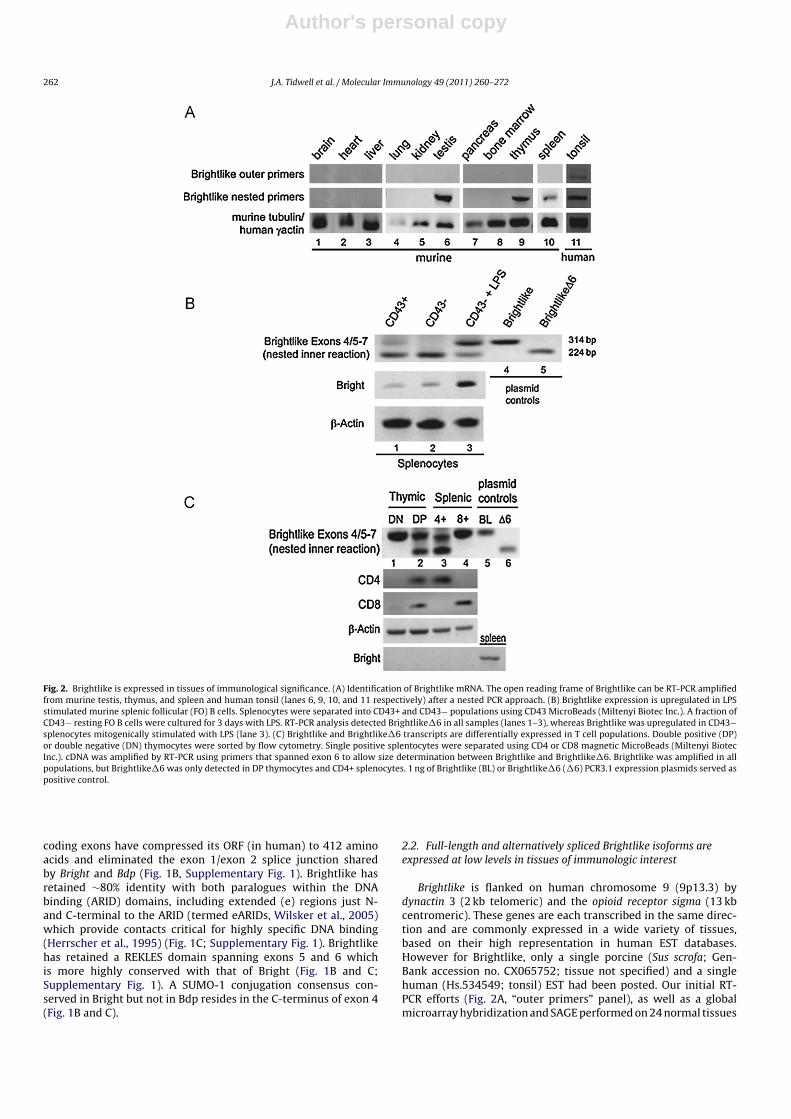
Fig. 2. Brightlike is expressed in tissues of immunological significance. (A) Identification of Brightlike mRNA. The open reading frame of Brightlike can be RT-PCR amplifiedfrom murine testis, thymus, and spleen and human tonsil (lanes 6, 9, 10, and 11 respectively) after a nested PCR approach. (B) Brightlike expression is upregulated in LPSstimulated murine splenic follicular (FO) B cells. Splenocytes were separated into CD43+ and CD43− populations using CD43 MicroBeads (Miltenyi Biotec Inc.). A fraction ofCD43− resting FO B cells were cultured for 3 days with LPS. RT-PCR analysis detected Brightlike�6 in all samples (lanes 1–3), whereas Brightlike was upregulated in CD43−splenocytes mitogenically stimulated with LPS (lane 3). (C) Brightlike and Brightlike�6 transcripts are differentially expressed in T cell populations. Double positive (DP)or double negative (DN) thymocytes were sorted by flow cytometry. Single positive splentocytes were separated using CD4 or CD8 magnetic MicroBeads (Miltenyi BiotecInc.). cDNA was amplified by RT-PCR using primers that spanned exon 6 to allow size determination between Brightlike and Brightlike�6. Brightlike was amplified in allpopulations, but Brightlike�6 was only detected in DP thymocytes and CD4+ splenocytes. 1 ng of Brightlike (BL) or Brightlike�6 (�6) PCR3.1 expression plasmids served aspositive control.
coding exons have compressed its ORF (in human) to 412 aminoacids and eliminated the exon 1/exon 2 splice junction sharedby Bright and Bdp (Fig. 1B, Supplementary Fig. 1). Brightlike hasretained ∼80% identity with both paralogues within the DNAbinding (ARID) domains, including extended (e) regions just N-and C-terminal to the ARID (termed eARIDs, Wilsker et al., 2005)which provide contacts critical for highly specific DNA binding(Herrscher et al., 1995) (Fig. 1C; Supplementary Fig. 1). Brightlikehas retained a REKLES domain spanning exons 5 and 6 whichis more highly conserved with that of Bright (Fig. 1B and C;Supplementary Fig. 1). A SUMO-1 conjugation consensus con-served in Bright but not in Bdp resides in the C-terminus of exon 4(Fig. 1B and C).
2.2. Full-length and alternatively spliced Brightlike isoforms areexpressed at low levels in tissues of immunologic interest
Brightlike is flanked on human chromosome 9 (9p13.3) bydynactin 3 (2 kb telomeric) and the opioid receptor sigma (13 kbcentromeric). These genes are each transcribed in the same direc-tion and are commonly expressed in a wide variety of tissues,based on their high representation in human EST databases.However for Brightlike, only a single porcine (Sus scrofa; Gen-Bank accession no. CX065752; tissue not specified) and a singlehuman (Hs.534549; tonsil) EST had been posted. Our initial RT-PCR efforts (Fig. 2A, “outer primers” panel), as well as a globalmicroarray hybridization and SAGE performed on 24 normal tissues

Author's personal copy
J.A. Tidwell et al. / Molecular Immunology 49 (2011) 260– 272 263
Fig. 3. Detection of endogenous Brightlike protein in normal and transformed lymphocytes. Nuclear extracts were prepared from the indicated cell lines (lanes 1–4), andwestern analysis was performed using polyclonal anti-Brightlike, anti-Bright, or anti �-actin rabbit antisera as described in Section 4. CD43− follicular (FO) B cells werecultured without (lanes 5) or with (lane 6) anti-IgM (�-�) + anti-CD19 (CD19) or without (lane 7) or with (lane 8) LPS under conditions described in Section 4 to activateproliferation and differentiation. Bright and Brightlike are co-expressed in some but not all cell lines, whereas mitogen but not anti-BCR stimulation led to upregulation ofboth proteins within nuclei of FO B cells.
(www.ncbi.nlm.nih.gov/geo) failed to detect Brightlike expressionwith the exception of the human tonsil (Fig. 2A, lane 11). Thus,Brightlike transcripts are exceedingly rare, suggesting they areinduced under rare environmental conditions or in a narrow subsetof tissues.
Using a nested RT-PCR amplification scheme with 5′ and 3′ UTRprimers flanking the predicted translational start and stop codons,amplicons of both the predicted size and slightly smaller were iden-tified only in murine testes, thymus, and spleen (Fig. 2A “nestedprimers” panel and data not shown). Cloning and sequencing iden-tified full-length Brightlike as well as a splice variant (termedBrightlike�6), which lacked the C-terminal portion of the highlyconserved protein–protein interaction (REKLES-�; exon 6) domain(see Kim et al., 2007 for details on the REKLES-� domain). In confir-mation, Southern blotting of the spleen RT-PCR reaction hybridizedwith an exon 6-specific DNA probe revealed species that differedfrom the full-length DNA in size by ∼90 nt (data not shown). Theintensity of bands from this experiment along with the sizableenrichment of cDNA for Brightlike�6 suggested that this REKLES-deficient isoform is more abundant, although formal confirmationremains to be obtained.
2.3. Brightlike and Bright are co-expressed in LPS stimulatedsplenic B cells
Bright expression is highly B lineage restricted in adult mice,with optimal levels observed in transitional/immature and in acti-vated follicular (FO) stages (Nixon et al., 2004). Reasoning thatBrightlike might be similarly restricted, we purified murine splenicFO B cells and assayed for changes seen after LPS-induced activa-tion. As shown in Fig. 2B, we detected full length Brightlike onlyin LPS stimulated FO (CD43−) blasts, where Bright expression ismaximal (lanes 3). Brightlike�6 was more abundantly expressedin unstimulated FO as well as in the non-FO (CD43+) splenic frac-tions (lanes 1 and 2), but was down-regulated in LPS stimulatedblasts (lane 3).
These results indicated that Bright and full length Brightlike areco-expressed, at least in a subset of mitogenically stimulated FOB cells, where both Bright levels (Nixon et al., 2004) and BrightDNA binding (Webb et al., 1991; Herrscher et al., 1995) are max-imally induced. However, Bright expression is not requisite forBrightlike expression. As shown in Fig. 2C, Brightlike isoforms aredifferentially expressed within the Bright-negative TcR�� lineage.Both forms accumulate in DP (lane 2) and CD4 T cells (lane 3),
whereas DN only express full length (lane 1) and CD8 only expressBrightlike�6 (lane 4).
To detect Brightlike protein, antisera were raised against N- orC-terminal regions, not conserved with either Bright or Bdp, andemployed in Western analyses. Brightlike isoforms could not be dis-tinguished because of their modest (30 amino acids) size difference.Consistent with their exceedingly low mRNA levels, Brightlike wasonly marginally detectable in whole cell lysates (data not shown).However, enrichment by nuclear fractionation allowed detectionof an endogenous species of predicted size (∼50 kD) in some (lanes2 and 3) but not all human mature B and T cell lines (Fig. 3 anddata not shown). Consistent with the RT-PCR data of Fig. 2B, lev-els of Brightlike in nuclei of 3 day mitogen-activated splenic FO Bcell blasts (lane 8) were significantly elevated above untreated con-trols (lane 7). Stimulation via the BCR ligation following short termculture with anti-� + anti-CD19 as previously described (Schmidtet al., 2009) had only modest effects on nuclear levels of Bright andBrightlike (compare lanes 5 and 6).
2.4. Exon 6-encoded REKLES- ̌ is required for nuclear export ofBrightlike
We have reported elsewhere that Bright shuttles between thenucleus and the cytoplasm in a CRM1-dependent manner (Kim andTucker, 2006; Kim et al., 2007). The NES of Bright has been mappedto the REKLES-�, the domain excluded in Brightlike�6. Based onBright’s REKLES-� dependent nuclear export, we expected Bright-like to be localized in the nucleus and cytoplasm and Brightlike�6to reside preferentially in the nucleus.
An N-terminally fused GFP-Brightlike was constructed andtransfected into NIH 3T3 fibroblasts. As predicted, Brightlikelocalized within both the cytoplasm and nucleus and the exon 6-deficient form was retained in the nucleus (Fig. 4). These resultswere confirmed by anti-Bright antibody histochemistry (Fig. 4). Thedata suggest that Brightlike and Bright are subject to equivalentnuclear import and export cues, as opposed to the totally nuclearresidence of Bdp/ARID3B.
2.5. Brightlike associates with Bright through its REKLES-ˇdomain
Amino acid residues 521–541 of the REKLES-� subdomainare required for homo- and heteromeric-interactions of Brightwith itself or with Bdp (Kim et al., 2007). This region is highly

Author's personal copy
264 J.A. Tidwell et al. / Molecular Immunology 49 (2011) 260– 272
Fig. 4. REKLES-� is required for nuclear export of Brightlike. NIH 3T3 cells were transfected with GFP-tagged forms of Brightlike or the REKLES-�-lacking (�6) splice variant.48 h-post-transfection, fixed cells were analyzed by fluorescence microscopy or by anti-Bright staining. Brightlike�6 was primarily nuclear, consistent with REKLES-�-dependent nuclear export. Brightlike localized to the cytoplasm and nucleus.
conserved within exon 6 of Brightlike. This suggested that Bright-like would interact with Bright and Bdp as well as homomerizewith itself, whereas REKLES-�-deficient Brightlike�6 would not.The hypothesis was tested by co-immunoprecipitation (CoIP) of thetwo proteins following their ectopic expression in non-lymphoidcells (HEK-293T, abbreviated as 293T) which do not express theirendogenous forms. 293T cells were transfected with combinationsof HA-tagged Brightlike, HA-Brightlike�6, GFP-Bright and emptyvectors under conditions previously established in non-lymphoidcell lines to detect Bright–Bright or Bright–Bdp interactions (Kimet al., 2007). Anti-Brightlike and pre-immune sera were used for Co-IP, followed by anti-HA or anti-GFP Western blotting. As shown inFig. 5A, although inputs of both isoforms of Brightlike were precip-itated, co-IP of Brightlike�6 and Bright was not observed (lane 6).
Similar Co-IP experiments to determine whether theBright–Brightlike interaction demonstrated by over-expressioncould be observed for the endogenous proteins were unsuccess-ful, owing at least in part to the low abundance of Brightlikewithin soluble fractions (data not shown). We reasoned thatBright–Brightlike association might be enriched in the chromatinfraction, where Bright was reported to be enriched (Lin et al., 2007;Schmidt et al., 2009). Cross-linked chromatin, prepared from theAB1.2 murine plasmacytoma line, which expresses Brightlike atreasonable levels (Fig. 3), was subjected to IP with antibodiesagainst Bright, Brightlike, and pre-immune controls. As shown inFig. 5B, Western blot analysis (Fig. 4B) developed with anti-Brightindicated that anti-Bright (lane 3) and anti-Brightlike (lane 4)precipitated Bright containing chromatin under conditions inwhich pre-immune sera (lanes 2) did not.
We conclude from these experiments that Bright and Brightlikeundergo stable association when over-expressed in nonlymphoidcells and in lymphoid cells following enrichment by chromatincross-linking. Our data support the notion that the REKLES-� sub-domain mediates the formation of Bright/Brightlike complexes.
2.6. Brightlike is sumoylated at a consensus motif conserved inBright
It is reported that Bright is SUMO-I conjugated at a single site,which may be equivalent to K284 of Brightlike (Fig. 1C; Schmidtet al., 2009; Prieur et al., 2009). K284 of Brightlike falls into a strongsumoylation consensus (http://www.abgent.com/doc/sumoplot),prompting point mutation and further analysis. In vitro
Fig. 5. Brightlike, but not Brightlike�6, associates with Bright. (A) 293T cells weretransiently co-transfected with GFP-Bright and HA-Brightlike or HA-Brightlike�6.Whole cell lysates were subjected to co-immunoprecipitation (CoIP) with Brightlikeantisera or pre-immune serum (P.I.) as a negative control. Western blots were devel-oped with the antibodies indicated at the left. As predicted, Brightlike precipitatesBright (lane 5) and Brightlike�6 does not (lane 6). (B). Chromatin immunoprecipi-tation of Bright–Brightlike complexes. Chromatin prepared from AB1.2 hybridomacells, which express high levels of Bright, was cross-linked with formaldehyde andassociated proteins were subjected to IP with antibodies (�) against Bright antis-era (lane 3), Bright-preimmune antisera (pre-immune, lane 2), or against Brightlike(lane 4). Western blot analysis developed with anti-Bright indicated that anti-Bright(lane 3) and anti-Brightlike (lane 4) precipitated Bright containing chromatin andpre-immune serum (lane 2) did not.

Author's personal copy
J.A. Tidwell et al. / Molecular Immunology 49 (2011) 260– 272 265
Fig. 6. Brightlike is sumoylated within a consensus motif conserved with Brightin vitro and in vivo. (A) In vitro sumoylation assays were performed using in vitrotranslated, full length and Brightlike�6 in the presence (+) or absence (−) of SUMOE1 (Sae1/2), SUMO E2 (Ubc9) and Sumo-I as described previously (Schmidt et al.,2009) and detailed in Section 4. Substitution of K284R within the Sumo-I consensusmotif (283-�KxE/D-286) eliminates the SUMO-I conjugated form (*, lanes 4 and 8).(B) Jurkat cells were transfected with GFP-Brightlike or GFP-BrightlikeK284R ± GFP-SUMO. Whole cell lysates were analyzed by anti-GFP and anti-Brightlike antibodies.Mutation of the SUMO-I consensus motif (K284R; lane 5) eliminated the SUMO-Iconjugated form (*, lane 3).
sumoylation assays were performed using in vitro tran-scribed/translated Brightlike and Brightlike�6 in the presence orabsence of purified SUMO E1 (Sae1/2) and SUMO E2 (Ubc9), asdescribed previously (Rangasamy and Wilson, 2000; Rosas-Acostaet al., 2005a,b; Schmidt et al., 2009). In the presence of sumoylationenzymes E1 and E2, slower migrating species, equivalent in sizeto SUMO-I mono-conjugated forms of Brightlike (lane 2) andBrightlike�6 (lane 6) were observed (denoted by asterisks inFig. 6A). Substitution of K284R within the SUMO-I consensus motif(283-�KxE/D-286) eliminated the SUMO-I conjugated forms ofboth Brightlike isoforms (lanes 4 and 8).
To verify these observations, the properties of GFP-tagged mam-malian expression versions of the same mutants and SUMO-I wereanalyzed by forced over-expression in Jurkat T cells. As shown in
Fig. 6B, lysates from the wild-type (lane 3), but not the K284Rmutant forms of Brightlike (lane 5), contains species correspondingin size and composition to GFP-SUMO-I-mono-conjugated Bright-like. These observations indicate that Brightlike is modified in vivowith SUMO-1 at K284.
2.7. Brightlike is recruited to plasma membrane lipid raftsfollowing B cell antigen receptor stimulation
We have established (Schmidt et al., 2009) that Bright, whenlocalized within plasma membrane lipid rafts, increases the sig-naling threshold of the BCR in response to BCR ligation in normalB cells and in transformed lymphoblastoid B cell lines. These Bcell lines included one Burkett’s lymphoma, RAJI, which expresseddetectable levels of endogenous Brightlike (Fig. 3C) and which isresponsive to strong BCR (anti-IgM + anti-CD19) ligation (Schmidtet al., 2009). Schmidt et al. (2009) further reported that dischargeof Bright from lipid rafts occurred shortly following BCR ligation.Discharge, at least in part, required Sumo-I modification andcorrelated with restoration of BCR signaling. While Bdp/ARID3Bis undetectable outside of the nucleus (Kim and Tucker, 2006), wereasoned that the nuclear export and sumoylation properties ofBrightlike might engender a Bright phenotype.
RAJI and RAMOS B cells were activated using anti-IgM + anti-CD19 (Methods and Materials and Schmidt et al., 2009), asjudged by total phosphotyrosine incorporation (data not shown).Lipid rafts were isolated and confirmed for purity as described(Nagamatsu et al., 1992; Schmidt et al., 2009; Section 4). Proteinswere extracted and assayed by Western blotting for the presence ofBright and Brightlike. A lipid raft-restricted protein, Raftlin, whoselevels are insensitive to BCR ligation (Saeki et al., 2003; Schmidtet al., 2009), served as a loading control. As shown in Fig. 7A andconsistent with previous findings, BCR ligation resulted in Brightdischarge from lipid rafts of RAJI (middle panel, compare lanes 3and 4) and RAMOS (lanes 7 and 8). Brightlike was also detectedin lipid rafts, but in contrast to Bright, trafficked in the oppositedirection; i.e., accumulating within rafts following BCR ligation (toppanel, lanes 3 vs 4 and 7 vs 8). This was a particularly significantenrichment in RAMOS, as Brightlike was hardly detectable in nucleiunder relatively similar input concentrations (Fig. 3, lane 4).
Since Bright and Brightlike interact (Fig. 5), raft-localized Bright-like might exist as homomeric Brightlike or as Bright–Brightlikeheteromers. We over-expressed Bright by transient transfectionin RAJI and RAMOS cells (compare lanes 1–4 and 5–7 of the mid-dle panel of Fig. 7A) and repeated the above-described analysis.Under these conditions, Brightlike appeared to be titrated from lipidrafts of both B cell lines following BCR stimulation (Fig. 7A, lanes
Fig. 7. Brightlike accumulates within plasma membrane lipid rafts. (A) B cell antigen receptor ligation drives Brightlike into lipid rafts and Bright out of lipid rafts. RAJIor RAMOS B cells were cultured alone or with anti-IgM (�-�) + anti-CD19 (�-CD19) with (+ lanes) or without (− lanes) transfection of Bright or with empty vector underconditions previously shown (Schmidt et al., 2009) to stimulate early events of BCR signaling. Lipid rafts were prepared, proteins were fractionated by SDS/PAGE and themembrane was western blotted for the proteins indicated on the right. Loading was normalized to the raflin control. (B) HA-tagged Brightlike, but not HA-Brightlike�6 ormutants deficient in sumoylation (HA-BrightlikeK284R or HA-Brightlike�6K384R) accumulate in lipid rafts of transiently transfected BTR fibroblasts. Endogenous H-Raswas employed as a loading control. Relatively equal levels of transfected proteins were confirmed by Western blots of whole cell lysates (data not shown).

Author's personal copy
266 J.A. Tidwell et al. / Molecular Immunology 49 (2011) 260– 272
Fig. 8. In vitro and in vivo recruitment of Brightlike to heavy chain variable region-associated MARs. (A) Schematic presentation of the rearranged VH1-IgH S107 locus(adapted from Herrscher et al., 1995) denotes MARs positioned upstream of the proximal promoter (Tx125 and Bf150) previously shown to bind specifically to Bright andBdp (Webb et al., 1991; Herrscher et al., 1995; Kim and Tucker, 2006). (B) Full length Brightlike, but not mutants lacking a conserved ARID domain residue (Y197A) or deletionmutants lacking a REKLES-� domain, binds to a VH1-associated MAR in vitro. In vitro translated Bright, full length, HA-tagged Brightlike, Brightlike�6 or mutants carryingsubstitutions in an essential DNA binding residue (Y197A) were incubated with Bf150 and subjected to an electrophoretic mobility shift analysis (EMSA). Bright and Brightlikeformed protein/DNA complexes that could be super-shifted with anti-Bright (lane 3) or anti-Brightlike (lane 6) or anti-HA (lane 7) antibody, whereas Brightlike�6 (lane8) and Y197A mutants (lanes 9, 10) did not. (C) Chromatin immunoprecipitation detects recruitment of Bright and Brightlike to VH1-associated MARs in vivo. Cross-linkedchromatin, prepared from AB1.2 hybridoma cells, which co-express Bright and Brightlike (Fig. 3), was immunoprecipitated with either anti-Bright (red), anti-Brightlike(anti-BL, green), or the corresponding pre-immune sera (blue). Cross links were reversed, the precipitated DNA was sheared to ∼1 kbp, and then subjected to quantitativePCR analysis using primers flanking the Bf150-Tx125 promoter region or the C� open reading frame (ORF). Highly significant enrichment (P < .01 for 3 independent replicas)was observed for both Brightlike and Bright across the Tx125 and Bf150 MAR region but not within the ORF. (For interpretation of the references to color in this figure legend,the reader is referred to the web version of the article.)
4 and 8). The most straightforward interpretation of these results(albeit more complicated interpretations cannot be excluded) isthat raft-localized Brightlike exists primarily as homomers, whichwhen forced into Bright heteromers by mass action, are dis-charged from lipid rafts in a manner indistinguishable from Brightmonomers.
To address this hypothesis further, we transfected HA-tagged-Brighlike wild type and mutants into BTR fibroblasts, which expressneither endogenous Bright nor Brightlike (Schmidt et al., 2009, anddata not shown). Ras was used as an endogenous loading controlfor isolated raft inputs. As shown in Fig. 7B, both Bright (lane 2) andBrighlike (lane 3) accumulated in lipid rafts, suggesting as in pre-vious studies (Schmidt et al., 2009) that B cell-specific factors arenot required for initial entry. Consistent with our model, Brightlikemutants which fail to form homomeric complexes (Brightlike�6and Brightlike�6-K234R, lanes 6 and 7) fail to enter rafts. Unex-pectedly, based on our previous results with Bright (Schmidt et al.,2009), Sumo-I deficient but homomeric-competent full lengthBrightlike-K234R also was excluded from lipid rafts (lane 5). Takenwith the reverse trafficking response to BCR ligation observed inB cells (Fig. 7A), these data suggest that, whereas Bright requiressumoylation for rafts exit, sumoylation of Brightlike is required forrafts entry.
2.8. Brightlike, but not Brightlike�6, binds to IgH S107 variableregion-associated MARs in vitro and in vivo
Bright is a MAR binding protein (Herrscher et al., 1995; Webbet al., 1991) and may regulate transcription by altering chromatin
structure (Kaplan et al., 2001; Lin et al., 2007). Bright and Bdphave been shown to bind MARs that both flank the intronic(E�) IgH enhancer and to MARs (termed Tx125 and Bf150)positioned upstream of the VH1 member of the S107 variableregion family (Herrscher et al., 1995; Fig. 8A). The presence ofthe highly conserved eARID domain in Brightlike suggested thatBrightlike might recognize and bind to the same IgH-associatedMARs.
Electrophoretic mobility shift assays (EMSAs) were performedto examine Brightlike’s MAR binding ability in vitro (Fig. 8B). SinceREKLES-� (within exon 6) is required for self-association, andthus for Bright to bind to the IgH promoter region (Kim et al.,2007), Brightlike�6 was predicted to not bind to these probes.Radio-labeled DNA fragments containing the Bf150 (or Tx125; seeHerrscher et al., 1995 for details) binding site with and withoutan excess of nonspecific cold competitor DNA were incubated within vitro translated (IVT) Brightlike or with nuclear extracts preparedfrom Brightlike-transfected cells. Protein–DNA complexes wereresolved by non-denaturing gel electrophoresis. Antibody super-shifts were used to confirm specificity of gel retarded complexes.Western blot analysis confirmed that Bright and Brightlike�6 IVTprotein inputs were equivalent (data not shown).
Consistent with our prediction, Brightlike but not Brightlike�6bound to the Bf150 (and Tx125) MARs with relative high affinity(Fig. 8B, lanes 4 and 8 and data not shown). Binding was elimi-nated (lane 9) by point mutation of a conserved tyrosine in theARID domain of Brightlike (Y197A), whose paralogous residue inBright is required for DNA binding (Nixon et al., 2004). Consistentwith previous results on Bright (Schmidt et al., 2009) and DRIL1

Author's personal copy
J.A. Tidwell et al. / Molecular Immunology 49 (2011) 260– 272 267
Fig. 9. Brightlike enhances Bright transactivation driven by the S107VH1 heavy chain promoter-associated MARs. (A) Schematic of luciferase reporter (Kim and Tucker, 2006)containing MARs previously shown (Herrscher et al., 1995) to contain specific Bright binding sites upstream of the S107 IgH variable region. (B) Loss of SUMO-I modificationincreases Brightlike enhancement of Bright-mediated IgH promoter activity. NIH3T3 cells stably transfected with the luciferase reporter construct of (A) were transientlyco-transfected with Renilla luciferase and with increasing DNA concentrations (indicated by triangles and detailed in Section 4) of empty expression vector (Control, backbars), Brightlike (BL, gray bars), Brightlike�6 (BL-�6, blue bars), the Sumo-I-deficient mutant, Brightlike-K284R (BL-K284R, green bars), Bright (B, red bars) or a DNA-bindingdeficient Bright mutant (B-Y257A, yellow bars). To test the potential combined effect of Bright and Brightlike, the above transfections were repeated with the addition ineach case of a Bright at the lowest DNA concentration (indicated by the straight lines + the triangles). Western blotting confirmed that increasing transfected DNA increasedBright and Brightlike protein levels accordingly (data not shown). Firefly luciferase activities were measured and normalized to Renilla luciferase activities. Values are plottedrelative to 100% for the empty vector control at lowest DNA concentration. Corresponding bar colors indicate enhancements. Results and error bars are representative of 3independent experiments for each DNA concentration. Bright consistently activated reporter expression at levels previously documented for this system (Kim and Tucker,2006). Neither Brightlike, Brightlike�6, Brightlike-K284R, nor Brightlike-Y257A showed statistically significant luciferase activity above control alone, but Brightlike (P < .01)and Brightlike-K284R (P < .001) significantly co-activated Bright levels. (For interpretation of the references to color in this figure legend, the reader is referred to the webversion of the article.)
(Prieur et al., 2009), mutation of the sumoylation conjugation sitehad no effect on DNA binding (lane 11).
Chomatin immunoprecipitation (ChIP) was carried out to assessin vivo recruitment of Brightlike to these same VH-associatedMARs. Cross-linked chromatin immunoprecipitated from the AB1.2murine plasmacytoma line (Fig. 5B) was reverse cross-linked, andthe deproteineized DNA was PCR-amplified with primers previ-ously shown (Rajaiya et al., 2006) to be specific for Bf150 orTx125. As shown in Fig. 8C, both anti-Bright and anti-Brightlikeprecipitates showed significant enrichment relative to preimmunecontrols for both MARs. Indeed, Brightlike recruitment to themore promoter-proximal, TX-125 MAR, was particularly impres-sive given its modest protein expression levels relative to Bright(Fig. 3).
2.9. Brightlike co-activates Bright-mediated IgH transactivation
Given that Brightlike binds to the S107VH1 promoter MARs andinteracts with Bright in chromatin, we reasoned that Brightlikewould regulate IgH transcription on its own or in combinationwith Bright. Bright and other nuclear matrix proteins have beenshown to most robustly transactivate reporters that are chromatinintegrated (Kaplan et al., 2001; Kim and Tucker, 2006). Thus,we utilized NIH3T3 cells that stably maintain a VH1-S107 distal(Bf150 + Tx125) heavy chain promoter region fused upstream of
SV40 promoter-driven firefly luciferase (Fig. 9A; Kim and Tucker,2006; Schmidt et al., 2009). These cells were transiently transfectedeither with increasing combinations of Bright and Brightlike wild-types and mutants or with constant levels of Bright plus increasinglevels of Brightlike wild-types and mutants. Luciferase activitieswere measured relative to co-transfected Renilla luciferase controland equivalent protein inputs were confirmed by Western blotting(data not shown).
As shown in Fig. 9B, neither full-length Brightlike (BL),Brightlike�6 (BL-�6), nor a DNA binding-deficient mutant (BL-Y197A) activated reporter expression. Sumo-I-deficient Brightlike-K284R consistently showed modest (average of ∼1.5-fold) trend(P < .1) activation. Consistent with previous results (Kim and Tucker,2006; Schmidt et al., 2009), Bright activated reporter activity 4.5–7-fold. This activity was further stimulated in a dose dependent andstatistically significant manner by co-transfection with Brightlike(P < .01), with maximal stimulation afforded by the sumoylationdeficient form (BL-K284R; P < .001). Co-transfections of Bright witheither RECKLES-�-deficient (BL-�6) or DNA binding-deficient (BL-Y197A) Brightlike achieved no further enhancement over Brightalone.
The results indicate that Brightlike has little transactivationactivity on its own but can collaborate with Bright as a co-activatorof IgH transcription. Mutant data further indicated that the non-sumoylated Brightlike form is a considerably stronger co-activator

Author's personal copy
268 J.A. Tidwell et al. / Molecular Immunology 49 (2011) 260– 272
and that both interaction with Bright through REKLES-� and withDNA are required.
3. Discussion
Sequence alignments indicate that ARID3c is an ancient gene(Wilsker et al., 2005). Conservation of the 5′ UTR of Brightlikeextends to amphibians and fish. The 3′ UTR is less conserved, but it isstill highly conserved within mammals. These comparisons supportthe view that ARID3c was created by duplication before the diver-gence of fish (400 million years ago). Comparison of paralogoussequences flanking the ARID DNA binding domain domains alongwith the retention by Brightlike of REKLES-mediated nuclear exportfunction (Fig. 4) support a more recent divergence of Brightlike andBright/ARID3A.
Brightlike transcript amplification required a nested approach,indicative of exceedingly low abundance (Fig. 2). Its absence fromEST databases further suggested it was a rare transcript, per-haps only expressed at low levels, in a small subset of cells orafter an environmental cue. We identified Brightlike transcriptsin murine spleen, thymus, testis, and in human tonsil; i.e., tissuesof immunological significance. The majority of cloned amplificonsencoded an alternatively spliced form of Brightlike (Brightlike�6)that lacks a highly conserved (RELKES-�) domain, which in addi-tion to NES function endows all ARID3 paralogues the ability forhomo/heteromerization and sequence-specific DNA binding (Kimand Tucker, 2006). It cannot be excluded that the apparent dom-inance of the shorter (by 90 b) Brightlike�6 transcript resultsfrom PCR amplification artifact. That caveat notwithstanding, weobserved virtually all-or-none accumulation of a single Bright-like isoform in different stages of T cell and B cell differentiation(Fig. 2). Most notable in the context of Bright co-activation, weobserved upregulation of the full length form at the near fullexpense of the RELKES-�-deficient form following activation of fol-licular B cells by LPS treatment (Fig. 2). It was previously shownthat Bright mRNA, protein and DNA binding activity is maximallyupregulated in mitogenically stimulated B cells (Webb et al., 1991;Herrscher et al., 1995). However, Brightlike�6 is not predicted toact as a conventional dominant negative, since it neither inter-acts with Bright (Fig. 5) nor binds DNA (Fig. 8). Choice of �m/�s
heavy chains and CD45 isoforms are well documented examples ofpre-mRNA splicing events that play crucial roles in B lineage dif-ferentiation events (Hathcock et al., 1992; Bruce et al., 2003). Inthe Brightlike context, a splicing decision that favors exon 6 exclu-sion would favor B cell differentiation by increasing the relativelevels (Fig. 2) of a Bright transcriptional co-activator—full lengthBrightlike (Fig. 9). This assumes that mitogenic or antigenic/T cellactivation of FO B cells is sufficient to elevate Brightlike to lev-els required to form functional Brightlike–Bright heteromers. Incontrast, the quite dramatic change in Brightlike isoform ratio dur-ing T lineage development (Fig. 2) suggests that Brightlike�6 canalso function independently of a B cell-specific collaboration withBright.
MARs in general (Glazko et al., 2003) and IgH-associated MARsin specific (Fernandez et al., 1998) have been linked to chro-mosomal organization by allowing enhancers to act over largedistances via association with the nuclear matrix. Both in vitroand in vivo, Brightlike binds upstream of the basal IgH promoterat MARs previously shown to bind Bright (Fig. 8; Webb et al.,1991; Herrscher et al., 1995; Rajaiya et al., 2006). Brightlike�6,which lacks critical REKLES-� subdomain functions, fails to bindthese MARs. A mutation in a single parologous tyrosine withinthe eARID domain of Bright (Y330A) or Brightlike (Y197A) dis-rupts DNA binding function (Nixon et al., 2004; Fig. 8). BecauseBright and Brightlike can associate in solution or in chromatin
(Fig. 5) and can bind to the same DNA motifs (Fig. 8), it is prob-able that heterologous Bright/Brightlike/DNA complexes occupythe VH1 promoter-distal Bf150 and Tx125 MARs. However, co-incubation in vitro of various ratios of Bright and Brightlike failed toresult in distinct heteromeric complexes in EMSA/antibody super-shift experiments (data not shown). There are several explanations,including, but not limited to, a possible requirement for specificchromatin context, need for additional transacting factors (absentin IVT lysates) or significantly differing affinities, which may pre-clude formation of heteromeric complexes in the presence of excessprobe.
The strongest support for functional Brightlike–Bright promoterinteractions came from reporter assays of Fig. 9. We observed thatBrightlike, while incapable of transacting expression of VH1 MARs-driven luciferase on its own, synergized with Bright to significantlyincrease transcription in NIH3T3 fibroblasts. Brightlike enhance-ment required both an ability to interact with Bright and an abilityto interact with DNA, as mutants in either of these functions wereinactive (Fig. 9). While these results provide strong support for aco-activator role for Brightlike via Bright–Brightlike interaction, thesituation in B cells is more complicated. A ternary complex of Brightwith Bruton’s tyrosine kinase (Btk), the defective gene product in X-linked immunodeficiency disease, and with the transcription factorTFII-I is required for maximal Bright-dependent transcription of IgH(Webb et al., 2000; Rajaiya et al., 2006). Our studies in fibroblasts,while demonstrating co-activation in the presence of only two Bcell-restricted components, raise fundamental questions as to howBrightlike fits into the ternary complex model in B cells.
Brightlike and Brightlike�6 are sumoylated in vitro and in vivo(Fig. 6) at a consensus motif conserved in Bright, but not inBdp/ARID3B (Fig. 1). This indicates that SUMO-I modification dif-ferentially diversifies the function of the paralogous members ofthe ARID3 subfamily. Consistent with what was recently demon-strated for Bright (Prieur et al., 2009; Schmidt et al., 2009), mutationof the sumoylation motif in Brightlike had no effect on nuclear-cytoplasmic localization (data not shown), nor on DNA binding(Fig. 8). However, SUMO-I-deficient Brightlike-K284R was a signifi-cantly stronger Bright transcriptional co-activator (Fig. 9). This is inaccord with the prevailing model (Gill, 2005) in which sumoylationinhibits transcription factors by relocalizing them to heterochro-matin enriched domains.
A second way in which ARID3 function is diversified was uncov-ered in our analysis of the lipid rafts localization of Brightlike.Antigen receptor stimulation of two responsive human lymphoidlines resulted in discharge of Bright, accumulation of but Brightlikein lipid rafts (Fig. 7). Since Bright discharge is directly correlatedwith reduced BCR signaling threshold (Schmidt et al., 2009), it istempting to speculate that the opposite phenotype—an enhancedsignaling threshold—results from Brightlike inclusion. We furtherobserved that (1) overexpression of Bright in these same B celllines eliminated Brightlike inclusion into rafts; (2) that Brightlikewas capable of entering lipid rafts in non-B cells in the absenceof Bright; (3) but that Brightlike�6 was not. An interpretationof these results that we favor is that the two ARID3 membersoccupy lipid rafts as homomers. Notably, the SUMO-1-deficientform of Brightlike also is blocked from lipid raft entry (Fig. 7),suggesting that the consequence of Brightlike sumoylation withrespect to lipid rafts trafficking is opposite from that of Bright; i.e.,SUMO-I additional to Bright signals exit and to Brightlike signalsentry.
We identify the transcription factor Brightlike as an unsus-pected component of the network of ARID transcription factors.Our studies have implicated Brightlike in functions as diverse as atranscriptional coactivator and a potential regulator of early eventsin BCR signaling. We submit that our findings provide rationale forfurther investigation of Brightlike localization and function.

Author's personal copy
J.A. Tidwell et al. / Molecular Immunology 49 (2011) 260– 272 269
4. Materials and methods
4.1. Computational analysis
ARID3C was identified as an uncharacterized region on mousechromosome 4 and human chromosome 9 by comparison withBright and Bdp coding sequences. Genomic sequences wereobtained from Ensembl.org. The National Center for BiotechnologyInformation (NCBI) web site (http://www.ncbi.nlm.nih.gov/) wasused to compare (BLAST) sequences against the protein and nucleicacid databases. Alignments were generated using ClustalW.
4.2. RT-PCR
Tissues or cells were either stored in RNA Later or directly addedto Trizol reagent (GibcoBRL). Tissues were homogenized by a poly-tron homogenizer. Cells were lyzed by repetitive pipetting using aP100. RNA was isolated following Invitrogen’s Trizol protocol. RNAwas resuspended in DEPC water. After removal of DNA contamina-tion with DNase I for 1 h at 37 ◦C, the reaction was terminated byadding EDTA (final concentration 2.5 mM) at 70 ◦C for 10 min. RNAconcentration was determined by using a nanodrop spectropho-tometer. SuperScriptTM First-Strand Synthesis System for RT-PCR(Invitrogen) was used to make all cDNAs according to manufac-turer’s protocol. 2–5 �g of total RNA was used as template in a 20ultotal reaction. Oligo (dT) was used to synthesis all poly-A mRNAtranscripts. 2 �l of cDNA was used for Brightlike PCR reactions and1ul was used for tubulin reactions. For full length amplification anouter PCR reaction was performed using 5′ and 3′ UTR primers ∼50nucleotides upstream of the start site and stop codon. The UTRreaction was used as a template for the full length open readingframe amplification. Full length primers amplified the open read-ing frame. Taq polymerase (NEB or Gene Choice) was used with theprovided reagents following manufacturer’s protocol. Tubulin wasused as a positive control in every PCR reaction using cDNA. The PCRproducts were cloned into Topo 2.1 with the Topo-TA cloning kit(Invitrogen) and the insert was sequenced by the dye terminationmethod (UT sequencing core).
4.3. Cell lines
Cell lines were incubated at 37 ◦C and maintained in anatmosphere of 5% CO2. Adherent cells were cultured in DMEM(GibcoBRL/Invitrogen) supplemented with 10% fetal calf serum(PAA Laboratories), 1 mM l-glutamine, 1% non-essential aminoacids (GibcoBRL/Invitrogen). Non-adherent cells were cultured inRPMI-1640 (GibcoBRL/Invitrogen) supplemented with, 2 mM l-glutamine, and 10% fetal calf serum. Cells were generally split every3–4 days.
4.4. B cell stimulations
Treatment with LPS (20 �g/ml) was performed for 3 days incomplete growth medium as previously described (Herrscher et al.,1995). B cell receptor ligation was achieved following 5 min treat-ment of ∼5 × 10⁸ cells using 500 ng of F(ab′)2 fragments of anti-IgM(clone JDC-15; Dako) and anti-CD19 (clone HD37; Dako) at 37 ◦C.
4.5. Anti-Brightlike antibody production
Anti-Brightlike polyclonal antisera was raised in New Zealandwhite rabbits against N-terminal (amino acids 4–100) or C-terminal(214–380) portions of Brightlike�6, selected because they lack sig-nificant homology to Bright or Bdp. The N- and C-term sequencesof Brightlike were cloned into the EcoR1 site of the GST vec-tor pGEX6p1. The fusion proteins were expressed and purified
from Escherichia coli, and following 3 immunizations, rabbits weresacrificed and sera collected. Specificity and optimization of anti-Brightlike sera was confirmed by positive Western signals ofNIH-3T3 fibroblasts, transfected with Brightlike and by negativesignals when transfected with Bright.
4.6. Western blot analysis
Cell lysates were made using RIPA buffer (150 mM NaCl, 50 mMTris pH 7.4, 0.1% SDS, 1% Triton X-100, 1% Deoxycholate, 1 mMEDTA) supplemented with protease inhibitors (Roche), incubatedon ice for 30 min then cleared by centrifugation at 3000 RPM. Brad-ford (Bio-rad) reagent was used to determine protein concentrationof cell lysates. 10 or 12% SDS-PAGE gels were used to separateproteins from cell lysates. 6× SDS loading buffer (300 mM Tris,10% glycerol, 6% SDS, 0.03% bromophenol blue, pH adjusted to 6.8,30% �-mercapthoethanol) was added to samples and then boiledfor 5 min before loading proteins into wells. Proteins were trans-ferred to a nitrocellulose membrane (PROTRAN®) using a standardsemi-dry transfer apparatus. Membranes were blocked with 5%milk in PBS-T (150 mM NaCl, 10 mM Tris pH 8, 0.1% Tween-20)at room temperature for 2 h or overnight at 4 ◦C with agitation.Membranes were incubated with primary antibody for 3 h at roomtemperature or overnight at 4 ◦C with agitation. Membranes werewashed twice for 5 min and once for 15 min with PBS-T (150 mMNaCl, 10 mM Tris pH 8, 0.05% Tween-20). Membranes were incu-bated with secondary antibody for 1 h at room temperature andwashed with PBS-T as described above. Blots were developed usingECL Western blotting detection reagent (Amersham PharmaciaBiotech) according to the manufacturer’s instructions. The follow-ing dilutions were used for each antibody: HA-1:1000, Brightlike-1:1000, Bright 1:5000, anti-GFP 1:5000, anti-mouse or anti-rabbit1:8000).
4.7. Expression constructs
Brightlike, Brightlike�6, their putative sumoylation or DNAbinding mutants were subcloned into PCR3.1 (Invitrogen) and/orpEGFP-C1 (GenBank). Brightlike was PCR amplified with Bgl II flank-ing primers and cloned into pGEM-t Easy (Promega). The fragmentwas digested and gel-purified and ligated into the BamH I site ofpE-GFP-C1, disrupting both Bgl II and BamH I sites. Digestion withKpn I was used to determine proper orientation of the insert. 5′
Sal I and 3′ Not I primers were used to amplify Brightlike frompEGFP-C1 and ligated into PCR3.1-HA cut with Sal I and Not I. DNAsequencing was used to verify all constructs using the dye ter-mination method. Plasmid constructs were grown in appropriateE. coli strains and isolated using either Qiagen or Invitrogen min-prep kits, according to manufacturer’s protocols. DNA was elutedusing ddH2O, warmed to 70 ◦C. Concentrations were obtained usinga nanodrop spectrophotometer.
4.8. Transient transfections
FuGENE6 (Roche) or Mirus (Mirus Bio Corporation) were usedto transfect constructs into fibroblast diploid cell lines (NIH-3T3and BTR) following manufacturer’s protocol. Exponentially grow-ing Jurkat T cells or RAJI and RAMOS B cells (∼1.5 × 10⁷) weretransfected by electroporation (320 V; 975 �F) with 20 �g of DNA.
4.9. GFP fusion protein subcellular localization studies
pEGFP-C1 Brightlike constructs were transiently transfectedinto NIH3T3 cells grown in either 100 mm plates containing cov-erslips or in 4 well chamber slides. Cells were fed 24 h posttransfection. Images were acquired 48 h post transfection. Nuclei

Author's personal copy
270 J.A. Tidwell et al. / Molecular Immunology 49 (2011) 260– 272
were visualized by staining with 1 ng/ml DAPI (4′,6′-diamidino-2-phenylindole) for 5 min at 25 ◦C. Stained sells were mounted withVectashield mounting media (Vector) containing Hoechst 33342(final concentration 1 �g/ml). Images were acquired using an Olym-pus IX-70 inverted microscope, equipped with a 100 W HBO Hgilluminator and a Diagnostic Instruments Spot RT-KE monochromecooled CCD camera.
For indirect immunofluorescence analysis, cells (1 × 10⁶) werecollected, washed twice with PBS, and then fixed with 500 �lof 4% paraformaldehyde (PFA) for 20 min at 25 ◦C and perme-abilized with 0.1% of Triton X-100 in PBS for 15 min at 25 ◦C.After 3 PBS washes, the cells were attached onto slides (Poly-tech) coated with 1% poly-l-lysine (Sigma), and the slides wereimmersed first in ice-cold methanol for 5 min and then in ice-cold acetone for 30 s. The cells were blocked with 20% FBS inPBS for 15 min at 25 ◦C and incubated with rabbit anti-Brightlikeantiserum (diluted 1:2000). After a washing, the cells werestained with donkey fluorescein isothiocyanate-conjugated anti-rabbit antibody (diluted 1:500; Santa Cruz) for 1 h at 25 ◦C in thedark. Slides were air dried, mounted, and analyzed as describedabove.
4.10. Cell sorting
Double positive T cells were sorted BD FACSAria using FITC-conjugated anti-CD4 and PE-conjugated anti-CD8 (BD Pharmin-gen). All remaining thymocytes were labeled as double negative,although other populations of cells remained. Thymuses weretaken from 5 to 6 weeks old C57BL/6 mice. Only sorted cellswith higher than 99% purity were used in the experiment. Tocollect CD4 and CD8 single positive splenocytes, single cell sus-pensions were generated from pooled spleens from C57B/6 mice,followed by a subsequent magnetic separation of cell populationsusing anti-mouse CD4 (L3T4) or anti-mouse-CD8a (Ly-2) coatedmagnetic MicroBeads, according to the manufacturer’s instruc-tions (Miltenyi Biotec Inc. Auburn, CA). The resulting CD4 andCD8 cell populations were pelleted and stored at −80 ◦C untilused for RNA isolation. CD43 positive cells were isolated frommurine spleen using anti-CD43 MicroBeads, according to manu-facturer’s protocol (Miltenyi Biotec Inc. Auburn, CA). Unlableledcells were collected as CD43 negative cells. FACS analysis con-firmed this fraction to be >95% B220 positive B cells (data notshown).
4.11. Co-immunoprecipitation
300 mg protein-A beads (Sigma, P3391) were washed in 10 mlof low IPB buffer (25 mM Tris pH 7.5, 150 mM NaCl, 2 mM EDTA,0.5% NP-40) with 1 mg/ml BSA for at least an hour while rotatingat room temperature or overnight at 4 ◦C. Beads were washed 2times in 10 ml low IPB and resuspended in an equal volume oflow IPB. Lysates of transfected cells, as indicated in the figures,were pre-cleared with 50% Sepharose A bead slurry. Fresh proteaseinhibitors (Roche) were added to the 50% slurry before each use.An aliquot of pre-cleared lysate was saved for input lanes. Lysateswere precipitated with �-Bright, �-Brightlike, or pre-immuneserum over-night at 4 ◦C with the addition of protease inhibitors(Roche) and 12 mM PMSF. Beads were spun down at 3000 RPMand washed with lysis buffer (50 mM HEPES-KOH, pH 7.4, 200 mMKCL, 10% glycerol, 1% NP-40, 1 mM EDTA, 1 mM DTT) 3× 5 min.The first 2 washes were supplemented with 1% Trition-X. Beadswere resuspended in 30 �l of 2× SDS sample buffer (100 mM Tris,25% glycerol, 2% SDS, 0.01% bromophenol blue, pH adjusted to 6.8,10% �-mercapthoethanol), 10 �l extract was used for loading of alane.
4.12. In vitro sumoylation assay
35S-Methionine HA-Brightlike IVT reaction was incubated withor without purified SUMO-1, Ubc9 and SAE1/SAE2 as previouslydescribed (Rangasamy and Wilson, 2000; Rosas-Acosta et al.,2005a,b).
4.13. Site directed mutagenesis
Stratagene’s mutation protocol for the QuikChange II Site-Directed Mutagenesis kit was used to introduce putativesumoylation or DNA binding mutations into pGEM T-easy vec-tors (Invitrogen) containing Brightlike or Brightlike�6. Howeverwe used our own reagents for this standard PCR-based muta-genesis. Phusion DNA polymerase was used with an extensiontemperature of 72 ◦C for 1 min/kb. Pfu HF buffer was used (Strata-gene). 10 ng of template was used in the reaction and 125 ng ofeach Y197A mutant oligonucleotide or 200 ng of SUMOK1mutb.Oligonucleotide sequences for the sumoylation mutant (K284R)are: Forward:GCCCGAGCCCAGTAAGGAAAGAGGAGAG; Reverse:CTCTCCTCTTTCCT TACTGGGCTCGGGC and for the DNA bindingdeficient mutant (Y197A) are: Forward: GAAGTATTTGTACCCA-GACGAGTGCGAGACACGGG and Reverse: CCCGTGTCTCGCACTCGTCTGGGTACAAATACTTC. Primers were annealed at 55 ◦C for 30 s.16 cycles were used for the DNA binding mutation reaction and18 cycles were used for the sumoylation mutation reaction. DNAsequencing by dye termination method (UT sequencing core) wasused to confirm the mutations.
4.14. In vitro translation
T7 or T3 RNA polymerase was used to synthesize PCR3.1 HABrightlike or Bright protein, respectively, using Promega’s TNT®
Quick Coupled Transcription/Translation System according to man-ufacturer’s protocol. The translation reaction was always incubatedat 32 ◦C for 2 h instead of the recommended 30 ◦C. 250 ng of PCR3.1-HA Brightlike or Bright constructs were used as a template for thein vitro translation reactions with a total volume of 12.5 �l (1/4 ofrecommended amount) or 1 �g in a total volume of 50 �l.
4.15. Isolation and purity of lipid rafts
500 mg of wet cell pellets of RAJI, RAMOS and BTR cells werewashed twice in ice-cold PBS and homogenized in 5 ml of 10 mMTris/Cl (pH 7.4), 1 mM EDTA, 250 mM sucrose, 1 mM phenylmethyl-sulfonyl fluoride and 1 �g/ml leupeptin (all from Sigma, St. Louis,Montana) in a tightly fitted Dounce homogenizer using five strokes.The resulting homogenate was centrifuged at 900 × g for 10 minat 4 ◦C, the resulting supernatant was then subjected to centrifu-gation at 110,000 × g for 90 min at 4 ◦C (Nagamatsu et al., 1992).The membane pellet was resuspended in ice-cold 500 �l TNE buffer(10 mM Tris/Cl [pH 7.4], 150 mM NaCl, 5 mM EDTA, 1% Triton X-100[Sigma], 10× protease inhibitors [Complete tablets, Roche, Indi-anapolis, Indiana]). Sucrose gradients for the preparation of lipidrafts were assembled exactly as described in an earlier publication(Fuentes-Pananá et al., 2005).
Lipid rafts were isolated by flotation on discontinuous sucrosegradients. Membrane pellets were extracted for 30 min on ice inTNE buffer. For the discontinuous sucrose gradient, 1 ml of clearedsupernatant was mixed with 1 ml of 85% sucrose in TNE and trans-ferred to the bottom of an ultracentrifugation tube, followed byoverlay with 6 ml of 35% sucrose in TNE and 3.5 ml of 5% sucrosein TNE. Samples were spun at 200,000 × g for 30 h at 4◦ C; frac-tions were collected from the top of the gradient and analyzedusing for SDS-PAGE/western blotting. B cell preparations were nor-malized by re-probing the filter with antibody directed against the

Author's personal copy
J.A. Tidwell et al. / Molecular Immunology 49 (2011) 260– 272 271
lipid rafts-specific marker Raflin. Fibrobast internal normalizationsutilized the lipid rafts marker, Ras.
4.16. Electrophoretic mobility shift assays
Bf150 or Tx125 probes were digested from 20 to 25 �g of the fol-lowing plasmids constructed in our lab: PUC-PCRIL5 or 251-125Rby Hind III or EcoR I, respectively. The Erag enhancer used as anegative control was generated by PCR. The PCR fragment (199 bp)corresponds with Region A of the RAG enhancer (Hu et al., 2006).Digested or PCR products were labeled with �-ATP32 using polynu-cleotide kinase (New England Biolabs) and gel purified from a 7%polyacrylamide gel. The running buffer for the gel is 1× TBE (90 mMTris–Borate, 2 mM EDTA). In vitro translated Bright, Brightlike orputative DNA binding mutants were incubated with probes in abinding buffer for 30 min at room temperature (20 mM HEPES pH7.9, 40 mM KCl, 6 mM MgCl2, 1 mM DTT, 0.1% NP40, 3 mg/ml BSA,10% glycerol, 2% Ficoll, 50 �g/ml of sonicated salmon sperm DNAand protease inhibitor cocktail (Roche)). For super shift reactionsanti-HA, anti-Bright or anti-Brightlike was added and incubated onice for 30 min. Pre-immune serum was used as a control. Bindingreactions were loaded onto a 4% polyacrylamide non-denaturinggel (6% glycerol in 0.5× TBE) and run overnight at 4 ◦C in 0.5× TBErunning buffer. Gels were dried and analyzed by a phosphorimager(Molecular Dynamics).
4.17. Chromatin immunoprecipitation (ChIP)
A 20 ml high-density AB1.2 hybridoma culture containing0.5–0.6 × 10⁷ cells was used per IP. Formaldehyde was addeddirectly to culture medium (540 �l added dropwise using a com-mercial stock solution of 37% HCHO/10% MetOH) and cellularchromatin was crosslinked at RT for 20 min with periodic rockingby hand. The reaction was terminated by immediately diluting witha maximal volume of ice-cold PBS supplemented with proteaseinhibitors. After cell lysis and preparation of nuclei, the chromatinwas sonicated with a Branson 250 Sonifier sonicator (90% dutycycle, power setting = 3) using 5 pulses of 15 s each; the chromatinwas incubated 1 min on ice between pulses. An aliquot was reversecrosslinked at 65 ◦C in the presence of 5 M NaCl overnight and ana-lyzed by gel electrophoresis to determine that the chromatin hadbeen successfully sheared to an average size of 200–1000 bp.
Chromatin supernatants were collected and spun at 13,000 rpmat 4 ◦C for 10 min, and the clarified supernatant was transferred toa new tube and then diluted 1:10 in IP buffer (0.1% SDS, 1% TritonX-100, 1.5 mM EDTA, 150 mM NaCl, 15 mM Tris–HCl pH 8.0, 1× pro-tease inhibitor cocktail, 10 mM NaF, 5 mM Na Butyrate) and werepre-cleared by rocking on a platform with 2 �g sheared salmonDNA and protein A-sepharose (50% slurry in PBS + NaN3) overnightat 4 ◦C. The chromatin was immunoprecipitated using 6 �g per IPof affinity-purified rabbit anti-Bright or anti-Brightlike polyclonalantibody or with 6 �g preimmune rabbit IgG obtained from eachrabbit as control. Immunoprecipitation was performed overnightat 4 ◦C.
After washes with sequential buffer solutions, one-third ofthe beads were removed, spun down, resuspended and boiled inLaemmli loading solution, and run on a 7.5% Western gel to con-firm the successful pulldown of Bright or Brightlike. To elute theremaining two-thirds, 250 �l of freshly prepared elution buffer (1%SDS, 0.1 M NaHCO3) was added for 15 min at RT on a platformrocker; the elution was repeated once more. Eluates were heatedat 65 ◦C (in the presence of 5 M NaCl) for at least 6 h to reverse theformaldehyde cross-linking. Chromatin was extracted once withphenol-chloroform, then once more with chloroform only.
A 1.5 �l aliquot (from a total ∼400 �l) was used for real-time quantitative PCR assays using the following TaqMan
primers and probes (Integrated DNA Technologies), previ-ously optimized by Rajaiya et al. (2005), that flanked the∼500 bp region spanning the VH1-associated Tx125 and Bf150MARs: forward: 5′-CTAGATCCACATGTATGATTT-3′; reverse, 5′-GTCTTTCAGACAATAGATTGG-3′); The C� ORF was employed asa negative control: forward: 5′-GTATCCAGTGTGAGGTGAAGC-3′;reverse: 5′-GAGCTTCCCATCCTTTAGCCA-3′. Reactions were per-formed with 96-well plates using the following conditions: 50 ◦Cfor 2 min and 95 ◦C for 10 min, followed by 40 cycles at 95 ◦C for 15 sand 55 ◦C. Standards were run with every experiment for consis-tency and quantification of the amplified DNA. Data from triplicatesamples were averaged and expressed as fold enrichment usingthe standardized curve and analyzed using ABI Prism 7700 SDSsoftware (Applied Biosystems).
4.18. Luciferase assays
NIH 3T3 cells, stably transfected with Bf150/Tx125-sv40-luciferase (Kim and Tucker, 2006), were plated at 1 × 10⁵ cellsper well in 6 well plates. 24 h later, cells were transiently co-transfected with increasing concentrations (100, 200, and 400 ng)of construct DNAs, with final concentration of 1.5 �g/well achievedby addition of varying amounts of empty vector along with 5 ng ofpRL luciferase construct (Promega). Validation of resulting proteinconcentrations were confirmed by anti-Bright and anti-Brightlikeprobed western blots employing �-actin as a loading control. Theluciferase activity was measured 48 h post-transfection by usingDual-luciferase Reporter Assay System (Promega) according tothe manufacturer’s instructions. Values were expressed relative toarbitrary 1.00 assigned to the lowest concentration of empty vec-tor after normalization of transfection efficiency, as measured byRenilla luciferase. Values achieved at each DNA concentration werederived from at least 3 independent transfections.
4.19. Statistics
For each pair of treatments, a 3-sample t-test was carried out,followed by a multiple testing correction to determine errors. P-values of <0.05 were considered statistically significant.
Acknowledgements
The authors thank Paul Das for administrative assistance. Wethank Chhaya Das, Maya Ghosh, June V. Harriss and MelissaPopowski for expert technical assistance. We thank members ofour labs for critically reading the manuscript. The work is sup-ported by the Marie Betzner Morrow Endowment and N. I. H. grantCACA31534 (PWT).
Appendix A. Supplementary data
Supplementary data associated with this article can be found, inthe online version, at doi:10.1016/j.molimm.2011.08.025.
References
Bruce, S.R., Dingle, R.W.C., Peterson, M.L., 2003. B-cell and plasma-cell splicing dif-ferences: a potential role in regulated immunoglobulin RNA processing. RNA 9,1264–1273.
Fernandez, L.A., Winkler, M., Forrester, W., Jenuwein, T., Grosschedl, R., 1998. Nuclearmatrix attachment regions confer long-range function upon the immunoglob-ulin mu enhancer. Cold Spring Harb. Symp. Quant. Biol. 63, 515–524.
Fuentes-Pananá, E.M., Bannish, G., van der Voort, D., King, L.B., Monroe, J.G., 2005.Ig�/Ig� complexes generate signals for B cell development independent of selec-tive plasma membrane compartmentalization. J. Immunol. 174, 1245–1252.
Gill, G., 2005. Something about SUMO inhibits transcription. Curr. Opin. Genet. Dev.15, 536–541.

Author's personal copy
272 J.A. Tidwell et al. / Molecular Immunology 49 (2011) 260– 272
Glazko, G.V., Koonin, E.V., Rogozin, I.B., Shabalina, S.A., 2003. A significant fractionof conserved noncoding DNA in human and mouse consists of predicted matrixattachment regions. Trends Genet. 19, 119–124.
Hathcock, K.S., Hiran, H., Murakami, S., Hodes, R.J., 1992. CD45 expression by B cells.Expression of different CD45 isoforms by subpopulations of activated B cells. J.Immunol. 149, 2286–2294.
Herrscher, R.F., Kaplan, M.H., Lelsz, D.L., Das, C., Scheuermann, R., Tucker, P.W.,1995. The immunoglobulin heavy-chain matrix-associating regions are boundby Bright: a B cell-specific trans-activator that describes a new DNA-bindingprotein family. Genes Dev. 9, 3067–3082.
Hu, H., Wang, B., Borde, M., Nardone, J., Maika, S., Allred, L., Tucker, P.W., Rao, A.,2006. Foxp1 is an essential transcriptional regulator of B cell development. Nat.Immunol. 7, 819–826.
Iwahara, J., Iwahara, M., Daughdrill, G.W., Ford, J., Clubb, R.T., 2002. The structure ofthe Dead ringer-DNA complex reveals how AT-rich interaction domains (ARIDs)recognize DNA. EMBO J. 21, 1197–1209.
Kaplan, M.H., Zong, R.T., Herrscher, R.F., Scheuermann, R.H., Tucker, P.W., 2001.Transcriptional activation by a matrix associating region-binding protein.contextual requirements for the function of bright. J. Biol. Chem. 276,21325–21330.
Kim, D., Probst, L., Das, C., Tucker, P.W., 2007. REKLES is an ARID3-restricted multi-functional domain. J. Biol. Chem. 282, 15768–15777.
Kim, D., Tucker, P.W., 2006. A regulated nucleocytoplasmic shuttle contributes toBright’s function as a transcriptional activator of immunoglobulin genes. Mol.Cell. Biol. 26, 2187–2201.
Kim, S., Zhang, Z., Upchurch, S., Isern, N., Chen, Y., 2004. Structure and DNA-bindingsites of the SWI1 AT-rich interaction domain (ARID) suggest determinants forsequence-specific DNA recognition. J. Biol. Chem. 279, 16670–16676.
Kortschak, R.D., Tucker, P.W., Saint, R., 2000. ARID proteins come in from the desert.Trends Biochem. Sci. 25, 294–299.
Lin, D., Ippolito, G.C., Zong, R.T., Bryant, J., Koslovsky, J., Tucker, P., 2007.Bright/ARID3A contributes to chromatin accessibility of the immunoglobulinheavy chain enhancer. Mol. Cancer 6, 23.
Nagamatsu, S., Kornhauser, J.M., Burant, C.F., Seino, S., Mayo, K.E., Bell, G.I., 1992. Glu-cose transporter expression in brain. cDNA sequence of mouse GLUT3, the brainfacilitative glucose transporter isoform, and identification of sites of expressionby in situ hybridization. J. Biol. Chem. 26, 467–472.
Nixon, J.C., Rajaiya, J., Webb, C.F., 2004. Mutations in the DNA-binding domain of thetranscription factor Bright act as dominant negative proteins and interfere withimmunoglobulin transactivation. J. Biol. Chem. 279, 52465–52472.
Numata, S., Claudio, P.P., Dean, C., Giordano, A., Croce, C.M., 1999. Bdp, a new memberof a family of DNA-binding proteins, associates with the retinoblastoma geneproduct. Cancer Res. 59, 3741–3747.
Prieur, A., Nacerddine, K., van Lohuizen, M., Peeper, D.S., 2009. SUMOylation of DRIL1directs its transcriptional activity towards leukocyte lineage-specific genes. PLoSONE 4, e5542.
Rajaiya, J., Hatfield, M., Nixon, J.C., Rawlings, D.J., Webb, C.F., 2005. Bruton’s tyrosinekinase regulates immunoglobulin promoter activation in association with thetranscription factor Bright. Mol. Cell. Biol. 25, 2073–2084.
Rajaiya, J., Nixon, J.C., Ayers, N., Desgranges, Z.P., Roy, A.L., Webb, C.F., 2006. Inductionof immunoglobulin heavy-chain transcription through the transcription factorBright requires TFII-I. Mol. Cell. Biol. 26, 4758–4768.
Rangasamy, D., Wilson, V.G., 2000. Bovine papillomavirus E1 protein is sumoylatedby the host cell Ubc9 protein. J. Biol. Chem. 275, 30487–30495.
Rosas-Acosta, G., Langereis, M.A., Deyrieux, A., Wilson, V.G., 2005a. Proteins of thePIAS family enhance the sumoylation of the papillomavirus E1 protein. Virology331, 190–203.
Rosas-Acosta, G., Russell, W.K., Deyrieux, A., Russell, D.H., Wilson, V.G., 2005b. Auniversal strategy for proteomic studies of SUMO and other ubiquitin-like mod-ifiers. Mol. Cell. Proteomics 4, 56–72.
Saeki, K., Miura, Y., Aki, D., Kurosaki, T., Yoshimura, A., 2003. The B cell-specificmajor raft protein, Raftlin, is necessary for the integrity of lipid raft and BCRsignal transduction. EMBO J. 22, 3015–3026.
Schmidt, C., Kim, D., Ippolito, G.C., Naqvi, H.R., Probst, L., Mathur, S., Rosas-Acosta,G., Wilson, V.G., Oldham, A.L., Poenie, M., et al., 2009. Signalling of the BCRis regulated by a lipid rafts-localised transcription factor, Bright. EMBO J. 28,711–724.
Webb, C.F., Das, C., Eaton, S., Calame, K., Tucker, P.W., 1991. Novel protein-DNA inter-actions associated with increased immunoglobulin transcription in response toantigen plus interleukin-5. Mol. Cell. Biol. 11, 5197–5205.
Webb, C.F., Smith, E.A., Medina, K.L., Buchanan, K.L., Smithson, G., Dou, S., 1998.Expression of bright at two distinct stages of B lymphocyte development. J.Immunol. 160, 4747–4754.
Webb, C.F., Yamashita, Y., Ayers, N., Evetts, S., Paulin, Y., Conley, M.E., Smith, E.A.,2000. The transcription factor Bright associates with Bruton’s tyrosine kinase,the defective protein in immunodeficiency disease. J. Immunol. 165, 6956–6965.
Webb, C.F., Bryant, J., Popowski, M., Allred, L., Kim, D., Harriss, J., Schmidt, C., Miner,C.A., Rose, K., Cheng, H.L., Griffin, C., Tucker, P.W., 2011. The ARID family tran-scription factor Bright is required for both hematopoietic stem cell and B lineagedevelopment. Mol. Cell. Biol. 31, 1041–1053.
Wilsker, D., Probst, L., Wain, H.M., Maltais, L., Tucker, P.W., Moran, E., 2005. Nomen-clature of the ARID family of DNA-binding proteins. Genomics 86, 242–251.
Zong, R.T., Das, C., Tucker, P.W., 2000. Regulation of matrix attachmentregion-dependent, lymphocyte-restricted transcription through differentiallocalization within promyelocytic leukemia nuclear bodies. EMBO J. 19,4123–4133.