thyroiditis (3, (5).dm5migu4zj3pb.cloudfront.net/manuscripts/104000/104579/jci62104579.pdfone...
TRANSCRIPT

Journal of Clinical InvestigationVol. 41, No. 5, 1962
THE THYROID CYTOTOXICAUTOANTIBODY
By I. J. FORBES,* I. M. ROITT, DEBORAHDONIACHAND I. L. SOLOMONt
(From the Middlesex Hospital Medical School, London, England)
(Submitted for publication November 27, 1961; accepted January 18, 1962)
The presence of circulating autoantibodies inpatients with Hashimoto's disease suggests thatautoimmunity is implicated in the disease process,but the ultimate proof of an autoimmune patho-genesis of thyroiditis and other human diseasesmust be obtained by demonstrating the autoag-gressive action of antibodies or immunologicallycompetent cells on the living tissue in its normalenvironment.
The demonstration by Pulvertaft, Doniach, Roittand Hudson (1, 2) of a serum factor capable ofdestroying human thyroid cells in tissue cultureis an advance in this direction. This finding hasbeen confirmed and amplified by Irvine (3, 4) andby Goudie and McCallum (5). Further investi-gations of this phenomenon are reported in thepresent paper. The cytotoxic factor is shown tobe an antibody reacting with the thyroid micro-somal antigen; its mode of action has been furtherstudied and its incidence determined in a varietyof thyroid conditions and in persons withoutovert thyroid disease.
METHODSAND MATERIALS
Tissue culture. Primary monolayer cultures of trypsin-dispersed thyroid epithelium were set up in chambersprepared on microscope slides by the method of Pulver-taft and co-workers (1). The chambers consisted of aplastic ring attached to a histology slide with siliconegrease, and sealed with a coverslip. Fresh thyroid tis-sue was dispersed for 1 hour in 0.25 per cent trypsin(Difco, 1: 250), and after centrifugation a suitableinoculum was prepared by adding a concentrated sus-pension of cells to Parker 199 medium until a density of5 epithelial clumps per field was seen when a drop wasviewed through a 1oX objective. The ideal cell con-centration resulted in the growth of 30 to 50 cell clumpsin a control chamber. Each chamber contained 0.1 mlof test serum, 0.1 ml fresh normal serum, and 0.6 ml ofcell inoculum. The fresh normal serum used routinelyhad a complement titer of between 160 and 320 minimalhemolytic doses (MHD) per ml, as determined by the
* Present address: University of Michigan MedicalCenter, Ann Arbor, Mich.
t Present address: Children's Hospital, Columbus, Ohio.
micromethod of Donnelley (6), and was known to sup-port good cell growth. Fresh normal serum was alwaysused in tests with serum fractions or absorbed sera.Guinea pig serum could be used as a source of comple-ment but was occasionally cytotoxic. The cultures wereincubated for 18 to 24 hours at 370C.
The sensitivity of each gland was established by in-cluding as controls known weakly cytotoxic sera andpotent Hashimoto sera, sometimes set up in dilutions.With sensitive glands, all the cells were killed by astrong standard serum; the weaker sera usually alloweda few clumps of cells to be established. Where cellssurvived in final dilutions of less than 1: 120 of thestandard strong serum, the gland was too insensitive topermit evaluation of unknown sera. Provided the glandwas sensitive, a serum was judged to be cytotoxic wheneither no clumps of healthy cells were visible or therewas a gross reduction of surviving cells relative to con-trol cultures. All positive sera were retested in at leastone subsequent culture.
Zone electrophoresis on "Pevikon" blocks. This wascarried out by a method similar to that of Muller-Eberhard (7). Approximately 50 g of polyvinyl-chlo-ride powder (Pevikon, Fosfatbolaget, Stockviksverkin,Sweden) washed three times in barbiturate buffer, pH8.6, was made into a thick slurry and poured into aPerspex trough, 7 X 2 X 1/2 inches. The serum wasdialyzed overnight at 40C against the buffer and a traceof bromophenol blue added. After equilibration of theblock in the electrode chamber at 4VC for 30 minutes, 1.5ml of serum was added to a depression in the block, 1inch from the cathodal end. Electrophoresis was con-tinued for approximately 18 hours in the cold, using acurrent of 7 ma until the blue-stained albumin waswithin 1 inch of the anodal end of the block. At the endof electrophoresis the block was cut transversely intosix equal portions. The fractions were stirred with 2ml of Parker's 199 medium in tubes and the supernatantrecovered after centrifugation. Each fraction was dia-lyzed against 250 ml of phosphate-buffered saline at 4°C(8) overnight, then against 10 ml of Parker 199 for8 hours. The fractions were stored at - 20°C untiltested.
The protein fractions were identified by immunoelec-trophoresis, with Scheidegger's (9) micromethod. Fourdrops of each fraction in serial fivefold dilutions wereset up for tissue culture; 0.2 ml of fresh normal humanserum was added to each dilution. A duplicate undilutedtube from each fraction was inactivated at 56°C for 30minutes before culture to ensure that cytotoxicity wascomplement dependent.
996

THYROID CYTOTOXICAUTO-ANTIBODY
Sodium sulfate precipitation of 'y-globulins f rom cy-totoxic sera. Two vol of 27 per cent sodium sulfate inphosphate buffer, pH 7.8, was added to 1 vol of a cyto-toxic serum, over a period of 10 minutes, with continu-ous stirring. After centrifugation at 10,000 rpm for 10minutes at room temperature, the supernatant was de-canted and saved. The precipitate was washed in 18per cent sodium sulfate and after centrifugation was dis-solved in a volume of distilled water equal to the volumeof the supernatant. The solutions were dialyzed againstphosphate-buffered saline and Parker's 199 medium foruse in the tissue culture test and for the detection of cy-toplasmic antibodies by Coons' technique.
Chromatography of -y-globulin obtained by zone elec-trophoresis on Pezikon blocks. The first three fractionscontaining y-globulin were used from two Pevikon blocksprepared according to the method described above.Chromatography was carried out on a diethylaminoethyl(DEAE)-cellulose column by the method of Fahey andHorbett (10).
The Pevikon fractions were eluted in Parker's 199medium; the final volume of solution after elution was9.5 ml. This solution was dialyzed against a 0.01 Mphosphate buffer, pH 8.1, overnight.
The column was prepared with DEAE-cellulose fromServa Entwicklungslabor, Heidelberg, and washedthrough overnight with 0.01 M phosphate buffer. Thedialyzed 'y-globulin solution was added to the column andwashed on with 1 ml of buffer. A further 48 ml ofbuffer was passed through the column before gradientelution was begun. The gradient was developed by run-ning a 0.10 M phosphate buffer, pH 8.1, containing 0.35M sodium chloride into a constant volume mixing cham-ber (volume, 280 ml) containing the original buffer. Theeluate was collected in 4-ml aliquots. The absorbance at280 mju in a 0.5 cm cell was measured in a Hilger spectro-photometer. The solutions were pooled into 4 fractionscontaining 32, 48, 40, and 130 ml, respectively. The frac-tions were concentrated approximately 20-fold overnightby applying a vacuum outside the solutions contained indialysis tubing. The four concentrated fractions weredialyzed against phosphate-buffered saline and then Par-ker's 199 medium. All operations were carried out at4°C. The fractions and a heat-inactivated control ofeach were tested for cytotoxicity in the tissue culturetest.
Sucrose gradient ultracentrifugation. Five-tenths ml ofa 1: 5 dilution of serum was layered above 4.5 ml of a12 to 36 per cent sucrose density gradient and the tubesspun at 35,000 rpm in the SW39 head of the Spincomodel L ultracentrifuge for 15 hours. After centrifu-gation the bottom of the tube was pierced, and separate19S and 7S fractions were collected. The proteins wereidentified by immunoelectrophoresis and tested for cyto-plasmic antibody by Coons' technique. Dialyzed frac-tions were tested for cytotoxicity in one experiment.
Other tests for thyroid antibodies. Coons' fluorescentantibody technique was used for the determination ofcytoplasmic staining of unfixed frozen sections of toxic
thyroid gland, as described by Holborow, Brown, Roittand Doniach (11). Antibodies to the colloid were dem-onstrated by alcohol-fixed thyroid sections (12). Thetanned cell hemagglutination (TRC) test for thyroglobu-lin antibodies was carried out with a formalinized sheepcell preparation (13). The complement fixation test(CFT) was done with thyrotoxic gland homogenate using2 MHD(14) or 11/4 MHDof complement.
Enzymes. The following were used for cell dispersal:trypsin (1: 250 Difco Lab., Detroit, Mich.); collagenase(Worthington Biochemicals Corp., Freehold, N. J.);papain (British Drug Houses, Poole, England); and ficin(crude, Light & Co., Colnbrook, England).
Case material. Patients with primary myxedema andthyrotoxicosis were accepted if the clinical evidence wasunequivocal or was confirmed by laboratory studies.Cases of nontoxic nodular goiter may have included somein which focal thyroiditis was present but was notdisclosed by laboratory studies. The patients with Hashi-moto's disease were accepted on the criteria of myxedemawith a firm smooth goiter which became smaller on thy-roid therapy, or in euthyroid patients with diffuse goiterwho had confirmatory evidence from 131 studies andserum protein abnormalities. The diagnosis of Hashi-moto's disease was proved histologically in 29 of the 48cases tested; of the remainder, 18 gave a positive pre-cipitin test. This series was separate from that re-ported by Pulvertaft and colleagues (2). Sera wereobtained from 118 relatives (72 siblings, 30 offspring,and 16 parents) of 54 propositi with thyroid disease, 46with Hashimoto's disease, 2 with biopsy-proved focalthyroiditis, 4 with thyrotoxicosis, and 2 with suspectedHashimoto's disease.
Ninety-one control sera from persons without overtthyroid disease were obtained from the antenatal clinic(21 sera) and from the routine pathology laboratory.Diagnoses were obtained from information in the casenotes. Sera of 27 blood donors, 8 healthy young adultmedical workers, 4 relatives of an 11 year old girl withsystemic lupus erythematosus, and 31 hospital patientswere also tested. The diagnoses of the latter were: or-thopedic conditions (6 cases), gingivitis, hepatic cir-rhosis with aplastic anemia, dysmenorrhea, migraine, al-lergic rhinitis, ulcerative colitis, Addison's disease, paro-titis, idiopathic thrombocytopenic purpura, anxiety states(3 cases), hypopituitarism, obesity (2 cases), lupuserythematosus (5 cases), diabetes mellitus, rheumatoidarthritis, ischemic heart disease, hypotension of unknownetiology, and idiopathic hemolytic anemia.
RESULTS
Requirements for complement. Heating cyto-toxic serum to 56°C for 30 minutes abolished itscytotoxic effect, and addition of fresh normal hu-man or guinea pig serum to the heated serum re-stored its potency. The addition of increasingamounts of fresh human serum to a heated cyto-
997

FORBES, ROITT, DONIACHAND SOLOMON
TABLE I
Zone electrophoresis of cytotoxic serum *
Cytotoxic factorFinal dilutions Titer of
cytoplasmicFraction Analysis 6 24 120 600 3,000 stainingt
1 -y-Globulin + + 4 - - 102 -y-Globulin + + 1: - 503 y + ,2A-Globulin + 1 - - 104 as + #-Globulins - - - - 15 a, + a2-Globulins - - - - - Neg.6 a,-Globulin + - - - - - Neg.
albumin
* All cells in culture killed, +; few cells surviving, A; good growth of cells,-.t Titer given as reciprocal of highest dilution of fraction giving positive staining in relation to control section treated
with negative serum.
toxic serum in the standard system showed thatapproximately 5 MHDof complement were re-
quired to restore potency; thus 0.1 ml of freshserum was routinely used to provide an adequateexcess.
Serum fractionation studies. The cytotoxic fac-tor was completely precipitated by 18 per centsodium sulfate in one experiment, while cytoplas-
mic staining was simultaneously removed. Tracesof activity remained in the supernatant in a fur-ther precipitation experiment.
In electrophoretic separations the cytotoxic fac-tor was predominantly localized to the y-globulinfractions of serum. The results of a typical sepa-
ration are shown in Table I. The slow f8-globulinfraction always had low cytotoxic activity, al-
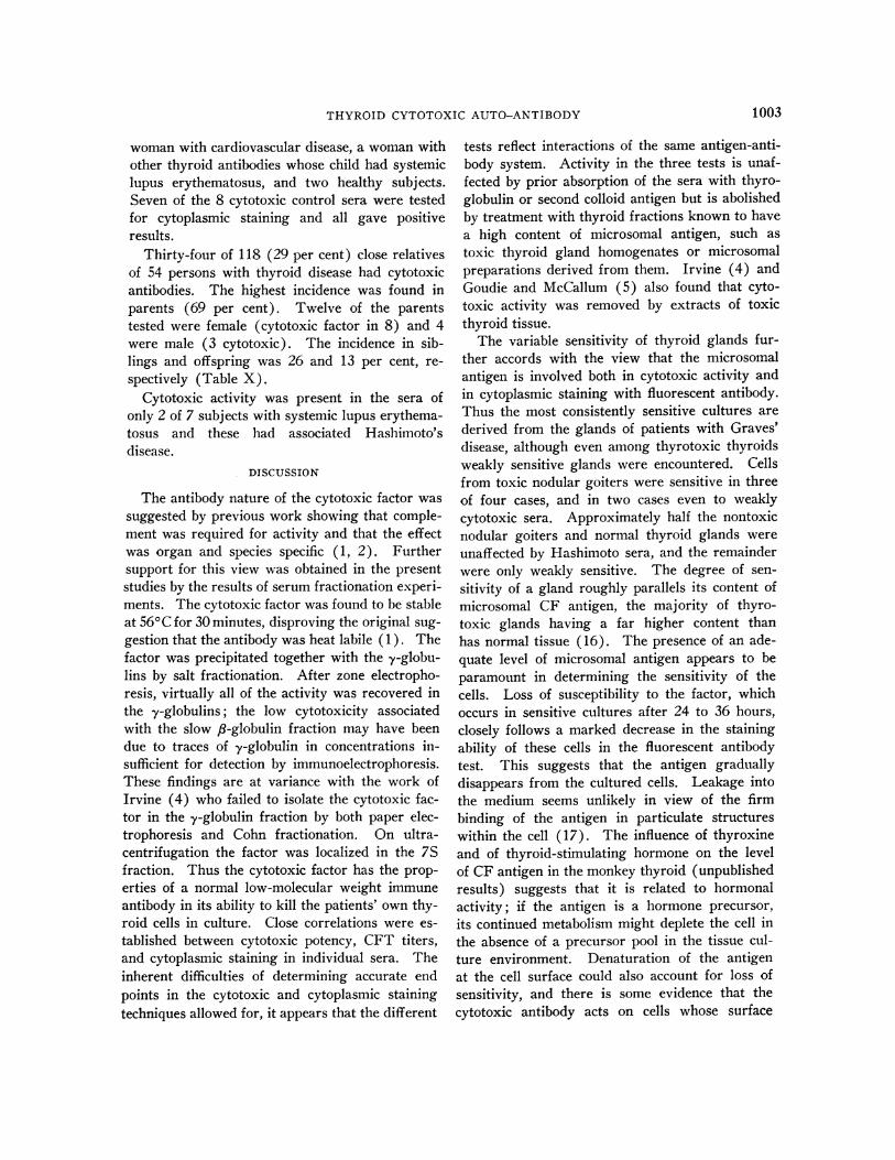
I I -2 1 3 1 4. 1FIG. 1. CHROMATOGRAPHYON DEAE-CELLULOSECOLUMNSOF -y-GLOBULIN FROM CYTOTOXIC
SERUMOBTAINED BY ZONE ELECTROPHORESISON "PEVIKON" BLOCKS. Pools of eluate used intesting for cytotoxic factor in tissue culture: 1, 2, 3, 4.
998
THYROID CYTOTOXICAUTO-ANTIBODY
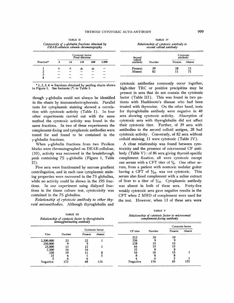
TABLE II
Cytotoxicity of -y-globulin fractions obtained byDEAE-cellulose column chromatography
Cytotoxic factorFinal dilutions
Fraction* 6 24 120 600 3,000
1 + + 4 _2 - - - - _3 - - - _ -
4 - - - _ -
* 1, 2, 3,4 = fractions obtained by pooling eluate shownin Figure 1. See footnote (*) to Table I.
though y-globulin could not always be identifiedin the eluate by immunoelectrophoresis. Paralleltests for cytoplasmic staining showed a correla-tion with cytotoxic activity (Table I). In fourother experiments carried out with the samemethod the cytotoxic activity was found in thesame fractions. In two of these experiments thecomplement-fixing and cytoplasmic antibodies weretested for and found to be contained in they-globulin fractions.
When y-globulin fractions from two Pevikonblocks were chromatographed on DEAE-cellulose,(10), activity was recovered in the breakthroughpeak containing 7S y-globulin (Figure 1, Table
Five sera were fractionated by sucrose gradientcentrifugation, and in each case cytoplasmic stain-ing properties were recovered in the 7S globulins,while no activity could be shown in the 19S frac-tions. In one experiment using dialyzed frac-tions in the tissue culture test, cytotoxicity wascontained in the 7S globulins.
Relationship of cytotoxic antibody to other thy-roid autoantibodies. Although thyroglobulin and
TABLE III
Relationship of cytotoxic factor to thyroglobulinhemagglutinating antibody
Cytotoxic factor
Titer Number Present Absent
2,500,000 23 22 1250,000 9 9
25,000 13 12 12,500 16 16
250 19 1 1 825 6 1 5
5 6 6Negative 172 48 124
TABLE IV
Relationship of cytotoxic antibody tosecond colloid antibody
CytotoxicSecond antibodycolloid
antibody Number Present Absent
Present 39 28 11Absent 82 11 71
cytotoxic antibodies commonly occur together,high-titer TRC or positive precipitins may bepresent in sera that do not contain the cytotoxicfactor (Table III). This was found in two pa-tients with Hashimoto's disease who had beentreated with thyroxine. On the other hand, testsfor thyroglobulin antibody were negative in 48sera showing cytotoxic activity. Absorption ofcytotoxic sera with thyroglobulin did not affecttheir cytotoxic titer. Further, of 39 sera withantibodies to the second colloid antigen, 28 hadcytotoxic activity. Conversely, of 82 sera withoutcolloid staining, 11 were cytotoxic (Table IV).
A close relationship was found between cyto-toxicity and the presence of microsomal CF anti-body (Table V): of 86 sera giving thyroid-specificcomplement fixation, all were cytotoxic exceptone serum with a CFT titer of ¼. One other se-rum, from a patient with nontoxic nodular goiterhaving a CFT of '/'6, was not cytotoxic. Thisserum also fixed complement with a saline extractof liver to a titer of ¼6 Cytoplasmic antibodywas absent in both of these sera. Forty-fiveweakly cytotoxic sera gave negative results in theCFT when 2 MHDof complement were used forthe test. However, when 13 of these sera were
TABLE V
Relationship of cytotoxic factor to microsomalcomplement-fixing antibody
Cytotoxic factor
CF titer Number Present Absent
512 10 10256 9 9128 15 15
64 15 1532 8 816 16 15 1
8 8 84 6 5
Negative 176 45 131
999

FORBES, ROITT, DONIACHAND SOLOMON
4.Or correlation coeff.= 0248
0 0 0
4.Or
2 3.04._
x°0 2.0
0
o4 1.00
0
c5D 0
01.0 2.0 3.0
log CFT titre
correlation coeff. = 0.895
0 00
I-0
0
086p0
1.0 2.0 3.0
log O cytoplasmic staining titre
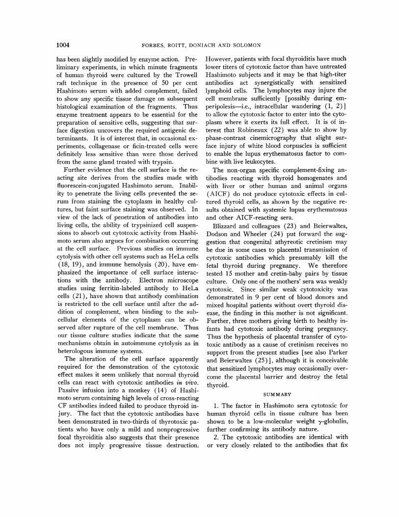
FIG. 2. LEFT: CORRELATIONOF CYTOTOXICITY OF HASHIMOTOSERA WITH COMPLEMENT-FIXING ABILITY. The log10 of the reciprocal of the highest serum dilution showing cyto-toxicity was plotted against log,0 of the titer in the CFT test, with 11/4 minimal hemolyticdoses of complement.
RIGHT: CORRELATIONOF CYTOTOXICITY OF HASHIMOTOSERA WITH CYTOPLASMIC STAIN-
ING ABILITY. The log10 of the titer in the cytotoxic test was plotted against log,0 of thereciprocal of the highest dilution giving positive fluorescent staining of thyroid cyto-plasm in Coons' sandwich technique.
retested with 1¼ MHDof complement and ahigher antigen concentration, 9 were positive and4 had become anticomplementary. In view of thedifficulties inherent in determining the titer of cy-totoxic activity, the correlation coefficient of 0.848obtained between log (CFT titer) and log (cyto-toxic titer) is good (Figure 2, left).
The fluorescent test appears to be the mostsensitive and specific for microsomal antibodies,and results of cytoplasmic staining agreed wellwith cytotoxic tests (r = 0.895; Figure 2, right;Table VI). Of 156 sera tested by both methods,67 were positive in both, 83 were negative by thetwo tests, while weak cytoplasmic staining wasobtained in 6 sera in which cytotoxicity could notbe demonstrated. This apparent greater sensi-tivity of the fluorescent antibody test is undoubt-edly due to the fact that undiluted serum is used,
TABLE VI
Relationship of cytotoxic factor tocytoplasmic antibody
Cytotoxicfactor
Cytoplasmicantibody Number Present Absent
Present 73 67 6Absent 83 83
whereas the minimal serum dilution used in thecytotoxic test is 1: 8.
Sensitivity of various types of thyroid tissue.The results obtained with different types of thy-roid gland are summarized in Table VII. Al-though small proportions of colloid goiter and"normal" tissues were susceptible, they were notsatisfactory for testing unknown sera, since cellswere not uniformly affected, and a variable num-ber survived in the presence of potent sera. Inthree cases normal tissues were from the healthy
TABLE VII
Sensitivity of various types of thyroid tissue tocytotoxic factor; analysis of 49 cultures
No. of Sensitivity*glands
Type of thyroid tissue cultured - + + +
Graves' disease 25 5 20Toxic nodular goiter 4 1 1 2Nontoxic nodular goiter 11 7 4Papillary. carcinoma 2 2Metastasis of papillary
carcinoma 1 1Hashimoto's disease 1 1Normal thyroid tissue 4 2 2Monkey thyroid 1 1
* Good growth of cells in potent cytotoxic-sera, -; cellsaffected by potent sera only, +; cells killed by low levelsof cytotoxic antibody, + +.
3.0-f
2.01-
.-
x
-u
0
01.01-
0
1000
0 61

THYROID CYTOTOXICAUTO-ANTIBODY
lobe of glands with single adenomata; two wereinsensitive and the other partially sensitive. Un-equivocally normal thyroid tissue taken duringsurgical exploration of the neck yielded cells thatwere killed by the high-titer sera but not by weaklyactive sera. Glands having a high content of mi-crosomal antigen measured by the technique ofRoitt, Doniach and Couchman (15) yielded sen-sitive cells while, in general, insensitive glandshad a low antigen content. One gland with sub-stantial lymphoid infiltration yielded highly sen-sitive cells, although the antigen content per gramof wet tissue was in the low range. Attempts toincrease the sensitivity of cells from nontoxic nodu-lar goiters by incubation with thyrotrophin, iodide,iodine, carbimazole, and thyrotrophin plus iodidein high concentration were unsuccessful. In theseexperiments primary cultures were set up in thepresence of potent cytotoxic sera with added com-plement in the usual concentrations, and the fol-lowing concentrations of these substances wereadded: thyrotrophin (Armour), 1.25 U per ml;potassium iodide, 6.25 mg per ml; carbimazole,0.25 mg per ml; Lugol's solution to give iodine33.3 ug per ml; Lugol's solution as above plusthyrotrophin, 1.25 U per ml. In each case therewas no reduction in the number of surviving cellsas compared with control chambers containingcytotoxic sera only.
Sensitivity of autologous thyroid cells to auto-antibodies. Serum containing cytotoxic factorkilled cell cultures obtained from the patient's owngland provided this was sensitive with knownpositive sera. Cytotoxicity was demonstrated toautologous cells in 12 experiments, and in 4 otherexperiments the patient's serum having no cyto-toxic factor allowed good growth of autologouscells.
The use of enzymes for cell dispersal. The pos-sibility that the enzymes used for dispersal mightaffect the sensitivity of thyroid cells was investi-gated; 0.25 per cent trypsin, 1 per cent papain,0.5 per cent ficin, and 0.1 per cent collagenase inParker 199 medium released cells readily. Phase-contrast microscopy showed that cells were rap-idly damaged by papain unless harvested andwashed within 20 minutes. They were not read-ily released when papain was diluted in phosphate-buffered saline; with ficin, cells survived well for1 hour.
The sensitivity of cells released by each enzymewas compared with that of trypsin-dispersed cells.While the cells obtained by all the enzymes weresensitive to cytotoxic factor, a small percentage ofcollagenase-treated cells survived in chamberscontaining cytotoxic sera, indicating a reduced sen-sitivity of cells dispersed by this enzyme. Thebest results were obtained when collagenase wasmixed with trypsin. Ficin-treated cells were com-pared with trypsinized cells in four experiments.Concordant results were obtained on three oc-casions, but in one experiment the ficin-treatedcells were insensitive, although the trypsinizedcells from the same gland were fully susceptible tothe cytotoxic antibody.
Absorption studies. Activity was absorbedfrom a strongly cytotoxic serum (titer 1/3,000)by mixing 0.3 ml of a 1: 10 dilution of the serumwith 0.2 ml of packed trypsinized cells from a thy-rotoxic gland. Cytotoxicity was similarly ab-sorbed out when 0.3 ml of a serum having a cy-totoxic titer of 1/600 was allowed to react atroom temperature for 3 hours with 2.7 ml of a20 per cent toxic thyroid gland homogenate, thesupernatant obtained being tested after centri-fugation at 105,000 G for 40 minutes. Cytotoxiceffects were also abolished in two of three Hashi-moto sera when 0.2 ml of serum was absorbed withmicrosomes derived from 10 g of thyrotoxic tis-sue. Mitochondrial fractions from the same ho-mogenate did not absorb out the factor, nor didcomparable treatment with cells or microsomalfractions from colloid goiters.
Fluorescent tests on thyroid cells in culture.Fluorescein-conjugated Hashimoto serum was leftin contact with living susceptible monolayers at370C for 45 minutes. There was no observablechange in the cells, and only a faint halo of fluores-cence was seen when the culture was viewed byultraviolet light. Addition of complement pro-duced cytotoxic changes showing that conjugatedserum was still active. When monolayers fromtoxic and nontoxic glands were washed brieflyand allowed to dry, the cytoplasm was weaklystained by conjugated globulins from a Hashi-moto serum, while no fluorescence was observedwith a conjugated normal serum. When conju-gated antihuman-y-globulin serum was applied af-ter untreated Hashimoto serum (sandwich tech-nique), the dried cells showed bright fluorescence.
1001

FORBES, ROITT, DONIACHAND SOLOMON
Far weaker staining of the cell cytoplasm wasseen with normal controls, although some non-specific uptake of y-globulin occurred.
Cultures grown in normal serum were testeddaily for susceptibility to cytotoxic factor, andthe cytoplasmic staining of dried monolayers wascompared (Table VIII). Cytotoxic serum andcomplement were applied to living cells and incu-bated for 30 minutes. Characteristic cytolyticchanges were seen in 24-hour old monolayers-i.e.,condensation and vesiculation of mitochondria, in-creased density of the nuclear membrane, loss ofnuclear structure with final swelling, and ruptureof the cells. A few cells remained sensitive in 48-hour old monolayers, but no effects were obtainedin older cultures.
TABLE VIII
Comparison of sensitivity of thyrotoxic cells in culture withcytoplasmic staining of dried monolayers at 24-hour
intervals *
Age of culture (hours)
24 48 72 96
Sensitivity + 1 - -Direct Hashimoto + i: 4i
conjugateDirect normal - - -
conjugateSandwich technique, + + + + + +
Hashimoto serumSandwich technique, 4 : + not
normal serum doneRabbit antiglobulin ±i i ±
conjugate
* Strong cytoplasmic fluorescence, + +; weak cyto-plasmic fluorescence, +; barely perceptible fluorescence,4; no cyctoplasmic fluorescence, -.
Fluorescent staining was more specific with di-rect Hashimoto conjugates, and fluorescence wasmaximal at 24 hours, decreasing thereafter. Thesandwich technique gave bright staining up to 48hours and then decreased, but some staining wasalso seen with normal serum in controls.
Clinical incidence of cytotoxic factor. The in-cidence in various conditions is presented in TableIX. Cytotoxic factor was demonstrable in all but2 of 48 Hashimoto subjects. A high incidence wasalso found in patients with focal thyroiditis, myx-edema, and thyrotoxicosis. Weaker cytotoxic ac-tivity was demonstrable in a small proportion ofpatients with colloid goiters and thyroid carci-noma. Only 1 of 8 patients with histologically
TABLE IX
Incidence of cytotoxic factor
No. of Cytotoxicsera factor
Condition tested present
Hashimoto's disease 48 46 96Focal thyroiditis 6 6Myxedema 9 6Thyrotoxicosis 51 34 67Nontoxic nodular goiter 31 5 16Carcinoma of thyroid 11 2Subacute thyroiditis 8 1Mothers of cretins 15 1Relatives of persons
having thyroid disease 118 34 29Lupus erythematosus
without thyroid disease 4 0Lupus erythematosus
with thyroid disease 3 2Control series 91 8 9
proven de Quervain's disease had cytotoxic fac-tor. Very weak cytotoxic antibody was found in1 of 15 mothers of athyreotic cretins and the cy-toplasmic staining given by her serum was alsoweak.
Eight of 91 sera (9 per cent) tested from per-sons without overt thyroid disease had cytotoxicantibody. In 7 of these the antibodies were weakas judged by their inability to kill all cells in aprimary culture. Stronger cytotoxic antibodieswere found in a case of acute hemolytic anemia.This serum was titered on a moderately sensitivegland and was completely cytotoxic at % but didnot affect the cells significantly at 24-. Serumtaken after the hemolytic process had remitted un-der steroid therapy was barely cytotoxic even tohighly susceptible cells. Three of these positivecontrols were found in a group of 21 pregnantwomen, and they gave birth to healthy babies.The other positive controls were a 75 year old
TABLE X
Incidence of cytotoxic factor in relativesof 54 persons having thyroid disease
Cytotoxic Any thyroidNumber factor antibody*
Siblings 72 19 26 34 47Offspring 30 4 13 7 23Parents 16 11 69 12 75Total 118 34 29 53 45
* One or more tests positive for thyroid antibodies (anti-bodies to thyroglobulin, second colloid antigen, or cyto-toxic factor).
1002
THYROID CYTOTOXICAUTO-ANTIBODY
woman with cardiovascular disease, a woman withother thyroid antibodies whose child had systemiclupus erythematosus, and two healthy subjects.Seven of the 8 cytotoxic control sera were testedfor cytoplasmic staining and all gave positiveresults.
Thirty-four of 118 (29 per cent) close relativesof 54 persons with thyroid disease had cytotoxicantibodies. The highest incidence was found inparents (69 per cent). Twelve of the parents
tested were female (cytotoxic factor in 8) and 4were male (3 cytotoxic). The incidence in sib-lings and offspring was 26 and 13 per cent, re-
spectively (Table X).Cytotoxic activity was present in the sera of
only 2 of 7 subjects with systemic lupus erythema-tosus and these had associated Hashimoto'sdisease.
DISCUSSION
The antibody nature of the cytotoxic factor was
suggested by previous work showing that comple-ment was required for activity and that the effectwas organ and species specific (1, 2). Furthersupport for this view was obtained in the presentstudies by the results of serum fractionation experi-ments. The cytotoxic factor was found to be stableat 560C for 30 minutes, disproving the original sug-
gestion that the antibody was heat labile (1). Thefactor was precipitated together with the y-globu-lins by salt fractionation. After zone electropho-resis, virtually all of the activity was recovered inthe y-globulins; the low cytotoxicity associatedwith the slow fl-globulin fraction may have beendue to traces of y-globulin in concentrations in-sufficient for detection by immunoelectrophoresis.These findings are at variance with the work ofIrvine (4) who failed to isolate the cytotoxic fac-tor in the y-globulin fraction by both paper elec-trophoresis and Cohn fractionation. On ultra-centrifugation the factor was localized in the 7Sfraction. Thus the cytotoxic factor has the prop-
erties of a normal low-molecular weight immuneantibody in its ability to kill the patients' own thy-roid cells in culture. Close correlations were es-
tablished between cytotoxic potency, CFT titers,and cytoplasmic staining in individual sera. Theinherent difficulties of determining accurate endpoints in the cytotoxic and cytoplasmic stainingtechniques allowed for, it appears that the different
tests reflect interactions of the same antigen-anti-body system. Activity in the three tests is unaf-fected by prior absorption of the sera with thyro-globulin or second colloid antigen but is abolishedby treatment with thyroid fractions known to havea high content of microsomal antigen, such astoxic thyroid gland homogenates or microsomalpreparations derived from them. Irvine (4) andGoudie and McCallum (5) also found that cyto-toxic activity was removed by extracts of toxicthyroid tissue.
The variable sensitivity of thyroid glands fur-ther accords with the view that the microsomalantigen is involved both in cytotoxic activity andin cytoplasmic staining with fluorescent antibody.Thus the most consistently sensitive cultures arederived from the glands of patients with Graves'disease, although even among thyrotoxic thyroidsweakly sensitive glands were encountered. Cellsfrom toxic nodular goiters were sensitive in threeof four cases, and in two cases even to weaklycytotoxic sera. Approximately half the nontoxicnodular goiters and normal thyroid glands wereunaffected by Hashimoto sera, and the remainderwere only weakly sensitive. The degree of sen-sitivity of a gland roughly parallels its content ofmicrosomal CF antigen, the majority of thyro-toxic glands having a far higher content thanhas normal tissue (16). The presence of an ade-quate level of microsomal antigen appears to beparamount in determining the sensitivity of thecells. Loss of susceptibility to the factor, whichoccurs in sensitive cultures after 24 to 36 hours,closely follows a marked decrease in the stainingability of these cells in the fluorescent antibodytest. This suggests that the antigen graduallydisappears from the cultured cells. Leakage intothe medium seems unlikely in view of the firmbinding of the antigen in particulate structureswithin the cell (17). The influence of thyroxineand of thyroid-stimulating hormone on the levelof CF antigen in the monkey thyroid (unpublishedresults) suggests that it is related to hormonalactivity; if the antigen is a hormone precursor,its continued metabolism might deplete the cell inthe absence of a precursor pool in the tissue cul-ture environment. Denaturation of the antigenat the cell surface could also account for loss ofsensitivity, and there is some evidence that thecytotoxic antibody acts on cells whose surface
1003
FORBES, ROITT, DONIACHAND SOLOMON
has been slightly modified by enzyme action. Pre-liminary experiments, in which minute fragmentsof human thyroid were cultured by the Trowellraft technique in the presence of 50 per centHashimoto serum with added complement, failedto show any specific tissue damage on subsequenthistological examination of the fragments. Thusenzyme treatment appears to be essential for thepreparation of sensitive cells, suggesting that sur-
face digestion uncovers the required antigenic de-terminants. It is of interest that, in occasional ex-
periments, collagenase or ficin-treated cells were
definitely less sensitive than were those derivedfrom the same gland treated with trypsin.
Further evidence that the cell surface is the re-
acting site derives from the studies made withfluorescein-conjugated Hashimoto serum. Inabil-ity to penetrate the living cells prevented the se-
rum from staining the cytoplasm in healthy cul-tures, but faint surface staining was observed. Inview of the lack of penetration of antibodies intoliving cells, the ability of trypsinized cell suspen-
sions to absorb out cytotoxic activity from Hashi-moto serum also argues for combination occurringat the cell surface. Previous studies on immunecytolysis with other cell systems such as HeLa cells(18, 19), and immune hemolysis (20), have em-
phasized the importance of cell surface interac-tions with the antibody. Electron microscopestudies using ferritin-labeled antibody to HeLacells (21), have shown that antibody combinationis restricted to the cell surface until after the ad-dition of complement, when binding to the sub-cellular elements of the cytoplasm can be ob-served after rupture of the cell membrane. Thusour tissue culture studies indicate that the same
mechanisms obtain in autoimmune cytolysis as inheterologous immune systems.
The alteration of the cell surface apparentlyrequired for the demonstration of the cytotoxiceffect makes it seem unlikely that normal thyroidcells can react with cytotoxic antibodies in vivo.Passive infusion into a monkey (14) of Hashi-moto serum containing high levels of cross-reactingCF antibodies indeed failed to produce thyroid in-jury. The fact that the cytotoxic antibodies havebeen demonstrated in two-thirds of thyrotoxic pa-
tients who have only a mild and nonprogressivefocal thyroiditis also suggests that their presence
does not imply progressive tissue destruction.
However, patients with focal thyroiditis have muchlower titers of cytotoxic factor than have untreatedHashimoto subjects and it may be that high-titerantibodies act synergistically with sensitizedlymphoid cells. The lymphocytes may injure thecell membrane sufficiently [possibly during em-peripolesis-i.e., intracellular wandering (1, 2)]to allow the cytotoxic factor to enter into the cyto-plasm where it exerts its full effect. It is of in-terest that Robineaux (22) was able to show byphase-contrast cinemicrography that slight sur-face injury of white blood corpuscles is sufficientto enable the lupus erythematosus factor to com-bine with live leukocytes.
The non-organ specific complement-fixing an-tibodies reacting with thyroid homogenates andwith liver or other human and animal organs(AICF) do not produce cytotoxic effects in cul-tured thyroid cells, as shown by the negative re-sults obtained with systemic lupus erythematosusand other AICF-reacting sera.
Blizzard and colleagues (23) and Beierwaltes,Dodson and Wheeler (24) put forward the sug-gestion that congenital athyreotic cretinism maybe due in some cases to placental transmission ofcytotoxic antibodies which presumably kill thefetal thyroid during pregnancy. We thereforetested 15 mother and cretin-baby pairs by tissueculture. Only one of the mothers' sera was weaklycytotoxic. Since similar weak cytotoxicity wasdemonstrated in 9 per cent of blood donors andmixed hospital patients without overt thyroid dis-ease, the finding in this mother is not significant.Further, three mothers giving birth to healthy in-fants had cytotoxic antibody during pregnancy.Thus the hypothesis of placental transfer of cyto-toxic antibody as a cause of cretinism receives nosupport from the present studies [see also Parkerand Beierwaltes (25)], although it is conceivablethat sensitized lymphocytes may occasionally over-come the placental barrier and destroy the fetalthyroid.
SUMMARY
1. The factor in Hashimoto sera cytotoxic forhuman thyroid cells in tissue culture has beenshown to be a low-molecular weight y-globulin,further confirming its antibody nature.
2. The cytotoxic antibodies are identical withor very closely related to the antibodies that fix
1004

THYROID CYTOTOXICAUTO-ANTIBODY
complement in the presence of toxic thyroid mi-crosomes, and are demonstrable by Coons' indi-rect fluorescent antibody technique in unfixedfrozen sections of toxic thyroid tissues.
3. Thyroid glands having a high content of mi-crosomal antigen yielded cells sensitive to thecytotoxic antibody, while insensitive glands hada low antigen content.
4. The cytotoxic antibodies were active againstautologous thyroid cells in culture.
5. The loss, with time, in sensitivity of cul-tures to the cytotoxic antibodies parallels the lossin ability of the cells to stain with specific "anti-microsomal" sera in the Coons' fluorescent anti-body technique.
6. Cytotoxic antibodies were found in 29 percent of close relatives of patients with thyroiddisease. Serum from patients with systemic lupuserythematosus was not cytotoxic except in two in-stances of an associated thyroiditis.
7. No evidence was obtained to support the viewthat athyreotic cretinism results from transpla-cental transfer of these cytotoxic antibodies.
ACKNOWLEDGMENTS
We wish to thank Professor A. Kekwick and theClinical Research Committee for facilities afforded inthe Institute of Clinical Research and are grateful toProfessor Sir Charles Dodds and Professor F. Dickensfor their unfailing support. We are greatly indebted toProfessor R. J. V. Pulvertaft for help and advice withtissue culture techniques, and to Mr. K. G. Couchmanand Mr. C. Shapland for skilled assistance. The work issupported in part by a grant to the Middlesex HospitalMedical School from the British Empire Cancer Cam-paign. Some equipment was generously donated by theMary Kinross Charity Fund. Thanks are due to theVaughan Hudson Clinical Research Trust, the WellcomeTrust (I.J.F.), and the Children's Hospital, Columbus,Ohio (I.L.S.), for financial support.
REFERENCES
1. Pulvertaft, R. J. V., Doniach, D., Roitt, I. M., andHudson, R. V. Cytotoxic effects of Hashimotoserum on human thyroid cells in tissue culture.Lancet 1959, 2, 214.
2. Pulvertaft, R. J. V., Doniach, D., and Roitt, I. M.The cytotoxic factor in Hashimoto's disease andits incidence in other thyroid diseases. Brit. J.exp. Path. 1961, 42, 496.
3. Irvine, W. J. An investigation of the pathogenesisof Hashimoto's disease by thyroid tissue culture.J. Endocr. 1960, 20, 83.
4. Irvine, W. J. The cytotoxic factor in thyroid dis-ease. Scot. med. J. 1960, 5, 511.
5. Goudie, R. B., and McCallum, H. M. Loss of tissue-specific autoantigens in thyroid tumors. Lancet1962, 1, 348.
6. Donnelley, M. Studies in experimental immunologyof influenza. VII. An improved complement-fixa-tion technique. Aust. J. exp. Biol. med. Sci. 1951,29, 137.
7. Mfiller-Eberhard, H. J. A new supporting mediumfor preparative electrophoresis. Scand. J. clin.Lab. Invest. 1960, 12, 33.
8. Dulbecco, R., and Vogt, M. Plaque formation andisolation of pure lines with poliomyelitis viruses.J. exp. Med. 1954, 99, 167.
9. Scheidegger, J. J. Une micro-methode de l'immuno-electrophorese. Int. Arch. Allergy 1955, 7, 103.
10. Fahey, J. L., and Horbett, A. P. Human gammaglobulin fractionation on anion exchange cellulosecolumns. J. biol. Chem. 1959, 234, 2645.
11. Holborow, E. J., Brown, P. C., Roitt, I. M., andDoniach, D. Cytoplasmic localization of "com-plement-fixing" auto-antigen in human thyroidepithelium. Brit. J. exp. Path. 1959, 40, 583.
12. Balfour, B. M., Doniach, D., Roitt, I. M., andCouchman, K. G. Fluorescent antibody studiesin human thyroiditis: Auto-antibodies to an antigenof the thyroid colloid distinct from thyroglobulin.Brit. J. exp. Path. 1961, 42, 307.
13. Fulthorpe, A. J., Roitt, I. M., Doniach, D., andCouchman, K. G. J. clin. Path. In press.
14. Roitt, I. M., and Doniach, D. Human auto-immunethyroiditis: Serological studies. Lancet 1958, 2,1027.
15. Roitt, I. M., Doniach, D., and Couchman, K. Stud-ies on an intracellular thyroid auto-antigen inMechanisms of Antibody Formation. New York,Academic Press, 1960, p. 70.
16. Belyavin, G., and Trotter, W. R. Investigationsof thyroid antigens reacting with Hashimoto sera:Evidence for an antigen other than thyroglobulin.Lancet 1959, 1, 648.
17. Roitt, I. M., Doniach, D., Wilson, E. G., and Couch-man, K. Biochemical studies of thyroid auto-antigens. Bull. Soc. Chim. biol. (Paris) 1960, 42,1165.
18. Goldberg, B., and Green, H. Immune cytolysis. I.The release of ribonucleoprotein particles. II.Membrane-bounded structures arising during cellfragmentation. J. biophys. biochem. Cytol. 1960,7, 645.
19. Hiramoto, R., Goldstein, M. N., and Pressman, D.Limited fixation of antibody by viable cells. J.nat. Cancer Inst. 1960, 24, 255.
20. Lepow, I. H., and Ross, A. Studies on immune cel-lular injury. II. Functional role of C'1 esterasein immune cytotoxicity. J. exp. Med. 1960, 112,1107.
1005

FORBES, ROITT, DONIACHAND SOLOMON
21. Easton, J., Goldberg, B., and Green, H. Electronmicroscopic localization of ferritin-labeled anti-body in ascites tumor cells. Fed. Proc. 1961, 20,16.
22. Robineaux, R. Study of L. E. cell formation by phasecontrast microcinematography in Henry Ford Hos-pital International Symposium on Mechanisms ofHypersensitivity, J. H. Shaffer, G. A. LoGrippo,and M. W. Chase, Eds. Boston, Little, Brown,1959, p. 371.
23. Blizzard, R. M., Chandler, R. W., Landing, B. H.,Pettit, M.D., and West, C. D. Maternal autoim-munization to thyroid as a probable cause ofathyrotic cretinism. New Engl. J. Med. 1960,263, 327.
24. Beierwaltes, W. H., Dodson, V. N., and Wheeler,A. H. Thyroid autoantibodies in the families ofcretins. J. clin. Endocr. 1959, 19, 179.
25. Parker, R. H., and Beierwaltes, W. H. Thyroidantibodies during pregnancy and in the newborn.J. clin. Endocr. 1961, 21, 792.
1006