thyphoid fever.ppt
TRANSCRIPT

8/13/2019 thyphoid fever.ppt
http://slidepdf.com/reader/full/thyphoid-feverppt 1/66
TYPHOID FEVER
ND
P R TYPHOID FEVERGuoli Lin
Department of Infectious Diseases
The Third Affiliated Hospital of SYSU

8/13/2019 thyphoid fever.ppt
http://slidepdf.com/reader/full/thyphoid-feverppt 2/66
Typhoid and Paratyphoid
Definition
Etiology
Pathogenesis
Epidemiology
Clinicalmanifestations
The laboratory andother examinations
Complications
Diagnosis anddifferential
diagnosis Prognosis
Treatment
Preventions
Paratyphoid Fever

8/13/2019 thyphoid fever.ppt
http://slidepdf.com/reader/full/thyphoid-feverppt 3/66
Definition of Typhoid fever
Acute enteric infectious disease
caused by Salmonella typhi (S.Typhi). prolonged fever, Relative bradycardia,
apathetic facial expressions, roseola,
splenomegaly, hepatomegaly, leukopenia.

8/13/2019 thyphoid fever.ppt
http://slidepdf.com/reader/full/thyphoid-feverppt 4/66
Etiology
Serotype: D group of Salmonella
Gram-negativenon-spore
flagella
aerob/fakultatif anaerob

8/13/2019 thyphoid fever.ppt
http://slidepdf.com/reader/full/thyphoid-feverppt 5/66
Antigens: located in the cell capsule
H (flagellar antigen) Protein,
termolabil
O (Somatic or cell wall antigen)
lipopolisakarida, termostabil
Vi (polysaccharide virulence)
S.Typhi, S. Paratyphi C
8/13/2019 thyphoid fever.ppt
http://slidepdf.com/reader/full/thyphoid-feverppt 6/66
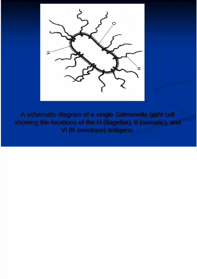
A schematic diagram of a single Salmonella typhi cell
showing the locations of the H (flagellar), 0 (somatic), and
Vi (K envelope) antigens.
8/13/2019 thyphoid fever.ppt
http://slidepdf.com/reader/full/thyphoid-feverppt 7/66
Endotoxin
A variety of plasmids
Resistance: Live 2-3 weeks in water.
1-2 months in stool. Die out quickly in
summer
Resistance to drying and cooling

8/13/2019 thyphoid fever.ppt
http://slidepdf.com/reader/full/thyphoid-feverppt 8/66
Epidemiology
continues to be a global health problem
areas with a high incidence include Asia,
Africa and Latin America
affects about 6000000 people with more
than 600000 deaths a year. 80% in Asia .
sporadic occur usually, sometimes have
epidemic outbreaks.

8/13/2019 thyphoid fever.ppt
http://slidepdf.com/reader/full/thyphoid-feverppt 9/66

8/13/2019 thyphoid fever.ppt
http://slidepdf.com/reader/full/thyphoid-feverppt 10/66
• InfeksiSalmonella peroral
Masa Inkubasi10-14 hari
•
GejalaProdromal
Minggu I• Bradi relatif•
Typhoid tounge• Organomegali
Minggu II danselanjutnya
Gang. KesadaranRoseole spot
Demam,anoreksia, mual,
muntah, obstipasi,cefalgia, myalgia
8/13/2019 thyphoid fever.ppt
http://slidepdf.com/reader/full/thyphoid-feverppt 11/66
Transmissionfecal-oral route
close contact with patients or
carriers
contaminated water and food
flies and cockroaches.
8/13/2019 thyphoid fever.ppt
http://slidepdf.com/reader/full/thyphoid-feverppt 12/66
Susceptibility and immunity
all people equally susceptible to
infection
acquired immunity can keep longer,
reinfection are rare
immunity is not associated with
antibody level of “H”, “O”and “VI”.
No cross immunity between typhoid
and paratyphoid.
8/13/2019 thyphoid fever.ppt
http://slidepdf.com/reader/full/thyphoid-feverppt 13/66
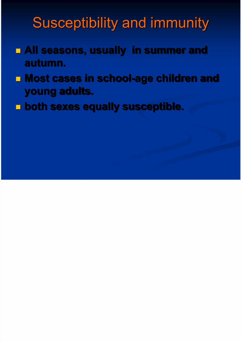
Susceptibility and immunity
All seasons, usually in summer and
autumn.
Most cases in school-age children and
young adults.
both sexes equally susceptible.

8/13/2019 thyphoid fever.ppt
http://slidepdf.com/reader/full/thyphoid-feverppt 14/66
Pathogenesis
gastrointestinal tract host-
pathogen interactions
The amount of bacilli infection(>105baeteria).

8/13/2019 thyphoid fever.ppt
http://slidepdf.com/reader/full/thyphoid-feverppt 15/66
ingested orally
Stomach barrier (some Eliminated)
enters the small intestine
Penetrate the mucus layer
enter mononuclear phagocytes of ileal peyer's
patches and mesenteric lymph nodes
proliferate in mononuclear phagocytes
spread to blood. initial bacteremia (Incubation
period).
Pathogenesis
8/13/2019 thyphoid fever.ppt
http://slidepdf.com/reader/full/thyphoid-feverppt 16/66
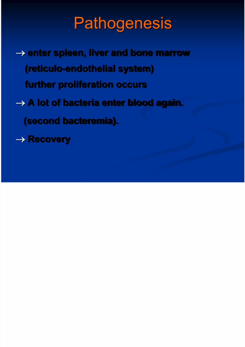
Pathogenesis
enter spleen, liver and bone marrow
(reticulo-endothelial system)
further proliferation occurs
A lot of bacteria enter blood again.
(second bacteremia).
Recovery
8/13/2019 thyphoid fever.ppt
http://slidepdf.com/reader/full/thyphoid-feverppt 17/66
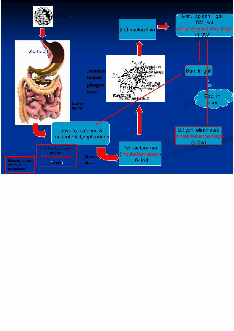
S.Typhi.
stomach
Lower
ileum
peyer's patches &
mesenteric lymph nodes
thoracic
duct
1st bacteremia
(Incubation stage)
10-14d
(monon
uclear
phagoc
ytes )
2nd bacteremia
liver 、spleen、gall、
BM ,ect
early stage&acme stage
(1-3W)
LN Proliferate,swell
necrosis
defervescence stage
3-4w)
Bac. In gall
Bac. In
feces
S.Typhi eliminated
convalvescence stage
(4-5w)
Enterorrhagia,i
ntestinal
perforation

8/13/2019 thyphoid fever.ppt
http://slidepdf.com/reader/full/thyphoid-feverppt 18/66
Pathology
essential lesion:proliferation of RES (reticuloendothelial
system )
specific changes in lymphoid tissuesand mesenteric lymph nodes.
"typhoid nodules“
Most characteristic lesion:
ulceration of mucous in the region of the
Peyer’s patches of the small intestine

8/13/2019 thyphoid fever.ppt
http://slidepdf.com/reader/full/thyphoid-feverppt 19/66
回肠:
集 淋巴结(PEYER’SPATC
HES)增生

8/13/2019 thyphoid fever.ppt
http://slidepdf.com/reader/full/thyphoid-feverppt 20/66

8/13/2019 thyphoid fever.ppt
http://slidepdf.com/reader/full/thyphoid-feverppt 21/66
Major findings in lower ileum
Hyperplasia stage(1st week):swelling lymphoid tissue and
proliferation of macrophages.
Necrosis stage(2nd week):
necrosis of swelling lymph nodes
or solitary follicles.
8/13/2019 thyphoid fever.ppt
http://slidepdf.com/reader/full/thyphoid-feverppt 22/66
Major findings in lower ileum
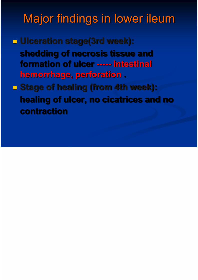
Ulceration stage(3rd week):
shedding of necrosis tissue and
formation of ulcer ----- intestinal
hemorrhage, perforation .
Stage of healing (from 4th week):
healing of ulcer, no cicatrices and nocontraction
8/13/2019 thyphoid fever.ppt
http://slidepdf.com/reader/full/thyphoid-feverppt 23/66
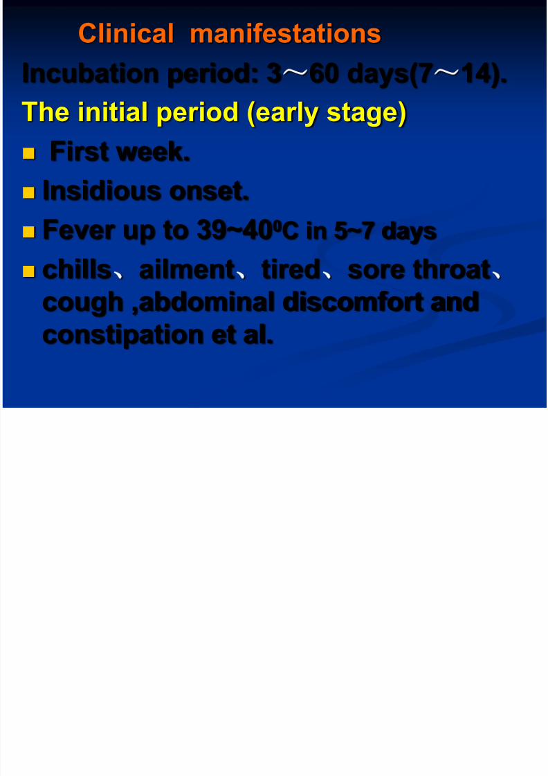
Clinical manifestations
Incubation period: 3~60 days(7~14).
The initial period (early stage)
First week.
Insidious onset. Fever up to 39~400C in 5~7 days
chills ailment tired sore throat
cough ,abdominal discomfort and
constipation et al.
8/13/2019 thyphoid fever.ppt
http://slidepdf.com/reader/full/thyphoid-feverppt 24/66
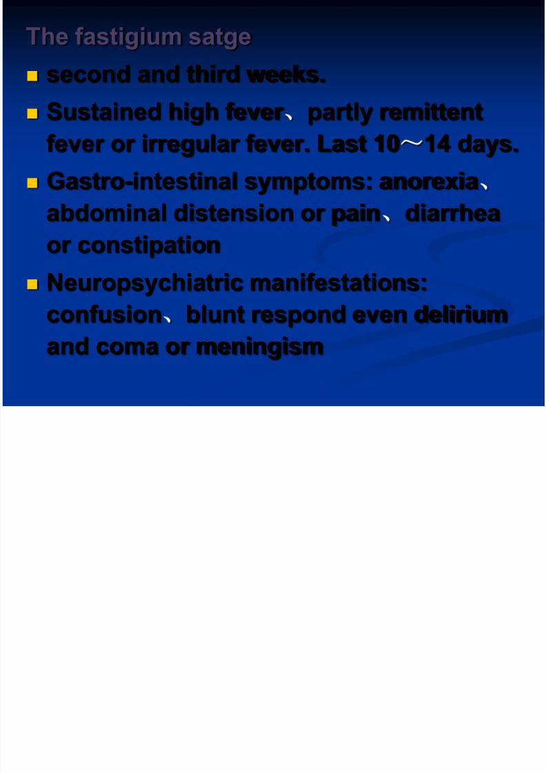
The fastigium satge
second and third weeks.
Sustained high fever partly remittent
fever or irregular fever. Last 10~14 days.
Gastro-intestinal symptoms: anorexia
abdominal distension or pain diarrhea
or constipation
Neuropsychiatric manifestations:confusion blunt respond even delirium
and coma or meningism
8/13/2019 thyphoid fever.ppt
http://slidepdf.com/reader/full/thyphoid-feverppt 25/66
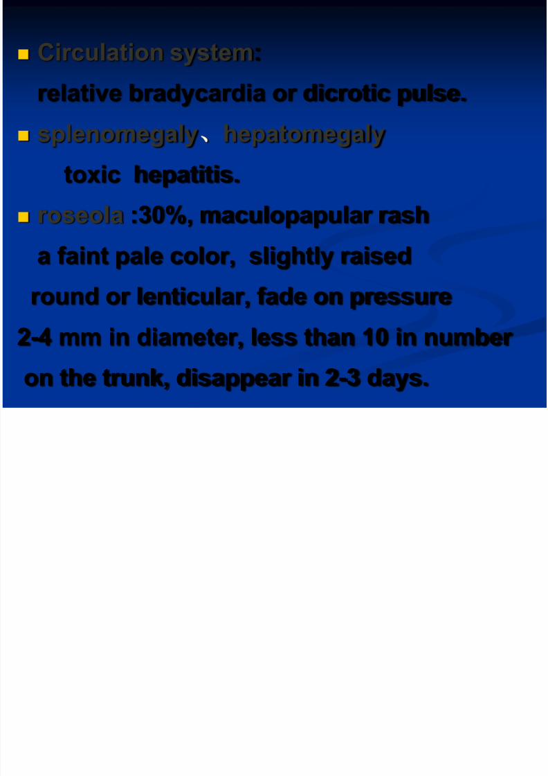
Circulation system:
relative bradycardia or dicrotic pulse. splenomegaly hepatomegaly
toxic hepatitis.
roseola :30%, maculopapular rash
a faint pale color, slightly raised
round or lenticular, fade on pressure
2-4 mm in diameter, less than 10 in number
on the trunk, disappear in 2-3 days.

8/13/2019 thyphoid fever.ppt
http://slidepdf.com/reader/full/thyphoid-feverppt 26/66
8/13/2019 thyphoid fever.ppt
http://slidepdf.com/reader/full/thyphoid-feverppt 27/66
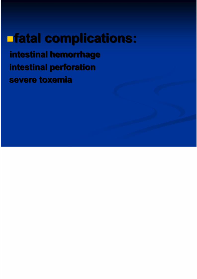
fatal complications:intestinal hemorrhage
intestinal perforationsevere toxemia

8/13/2019 thyphoid fever.ppt
http://slidepdf.com/reader/full/thyphoid-feverppt 28/66
defervescence stage
fever and most symptoms resolve by
the forth week of infection.
Fever come down, gradual
improvement in all symptoms and
signs, but still danger.
convalescence stage
the fifth week. disappearance of all
symptoms, but can relapse
Cli i l f
8/13/2019 thyphoid fever.ppt
http://slidepdf.com/reader/full/thyphoid-feverppt 29/66
Clinical forms:
Mild infection:
very common seen recentlysymptom and signs mild
good general condition
temperature is 380Cshort period of diseases
recovery expected in 1~3 weeks
seen in early antibiotics users
young children mild more
easy to misdiagnose

8/13/2019 thyphoid fever.ppt
http://slidepdf.com/reader/full/thyphoid-feverppt 30/66
Persistent infection:
diseases continue than 5 weeks
Ambulatory infection:
mild symptoms,early intestinal bleeding
or perforation.

8/13/2019 thyphoid fever.ppt
http://slidepdf.com/reader/full/thyphoid-feverppt 31/66
Fulminate infection:
rapid onset, severe toxemia and
septicemia.
High fever,chill,circulation failure,
shock, delirium, coma, myocarditis,
bleeding and other complications,
DIC et all.

8/13/2019 thyphoid fever.ppt
http://slidepdf.com/reader/full/thyphoid-feverppt 32/66
Special manifestations
In children
Often atypical
sudden onset with high fever.
Respiratory symptoms and diarrhea, dominant.
Convulsion common in below 3.
relative bradycardia rare.
Splenomegaly, roseola and leucopenia less common.
8/13/2019 thyphoid fever.ppt
http://slidepdf.com/reader/full/thyphoid-feverppt 33/66
8/13/2019 thyphoid fever.ppt
http://slidepdf.com/reader/full/thyphoid-feverppt 34/66
clinical manifestations reappear
less severe than initial episode
It’s temperature recrudesce when temperature
start to step down but abnormal in the period of
2-3 weeks and persist 5~7 days then back to
normal. seen in patients with short therapy of antibiotics.
Recrudescence
8/13/2019 thyphoid fever.ppt
http://slidepdf.com/reader/full/thyphoid-feverppt 35/66
relapse
serum positive of S.typhi after 1~3
weeks of temperature down to normal.
Symptom and signs reappear
the bacilli have not been completely
removed
Some cases relapse more than once

8/13/2019 thyphoid fever.ppt
http://slidepdf.com/reader/full/thyphoid-feverppt 36/66
Laboratory findings
Routine examinations
white blood cell count is normal or
decreased.
Leukocytopenia(specially eosinophilic
leukocytopenia).
recovery with improvement of diseases
decreased in relapse
8/13/2019 thyphoid fever.ppt
http://slidepdf.com/reader/full/thyphoid-feverppt 37/66
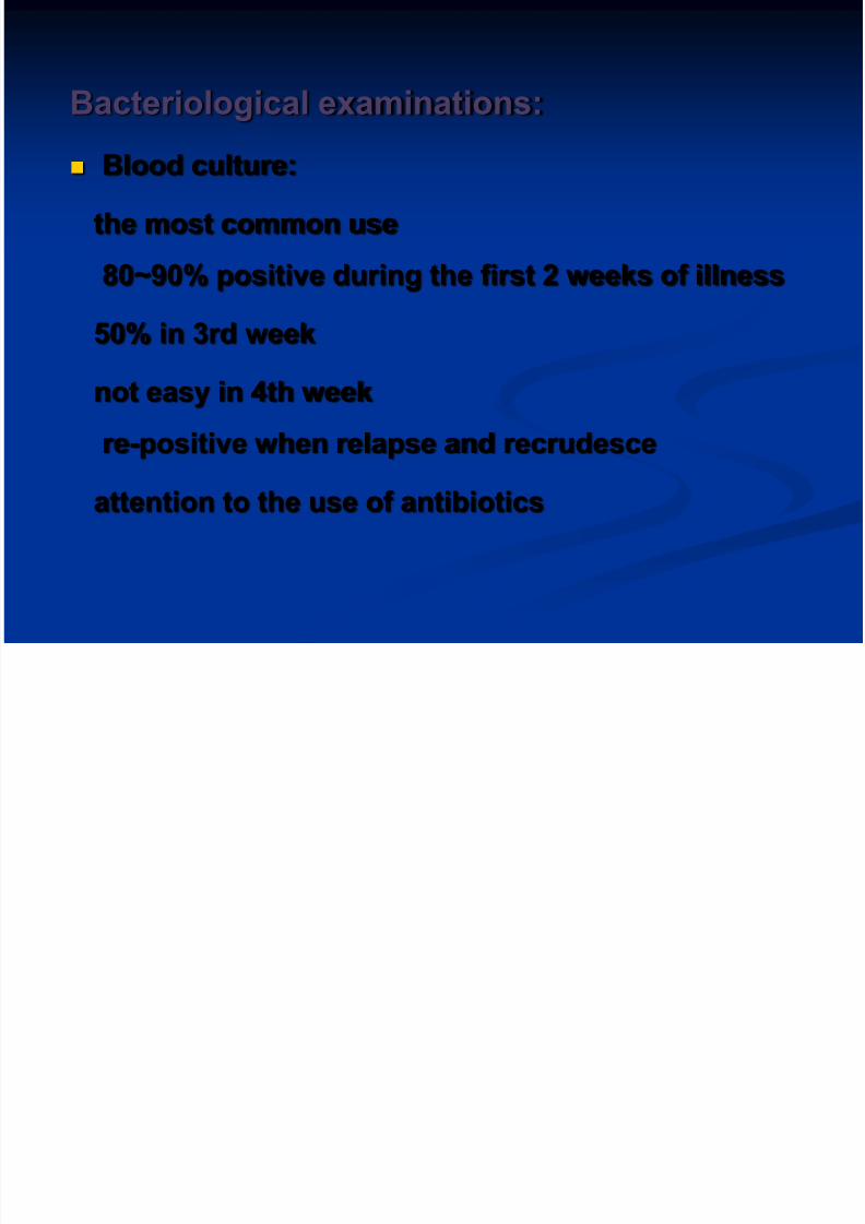
Bacteriological examinations:
Blood culture:
the most common use
80~90% positive during the first 2 weeks of illness
50% in 3rd week
not easy in 4th week
re-positive when relapse and recrudesceattention to the use of antibiotics

8/13/2019 thyphoid fever.ppt
http://slidepdf.com/reader/full/thyphoid-feverppt 38/66
The bone marrow culture
the most sensitive testspecially in patients pretreated with antibiotics.
Urine and stool cultures
increase the diagnostic yieldpositive less frequently
stool culture better in 3~4 weeks
The duodenal string test to culture bileuseful for the diagnosis of carriers.
Rose spots: Not use routinely

8/13/2019 thyphoid fever.ppt
http://slidepdf.com/reader/full/thyphoid-feverppt 39/66
Serological tests(Vidal test):
five types of antigens:
somatic antigen(O),flagella(H) antigen, and paratyphoid feverflagella(A,B,C) antigen.
Antibody reaction appear during first week
70% positive in 3~4 weeks and can prolong toseveral months
in some cases, antibodies appear slowly, or
remain at a low level,
some(10~30%) not appear at all.
8/13/2019 thyphoid fever.ppt
http://slidepdf.com/reader/full/thyphoid-feverppt 40/66
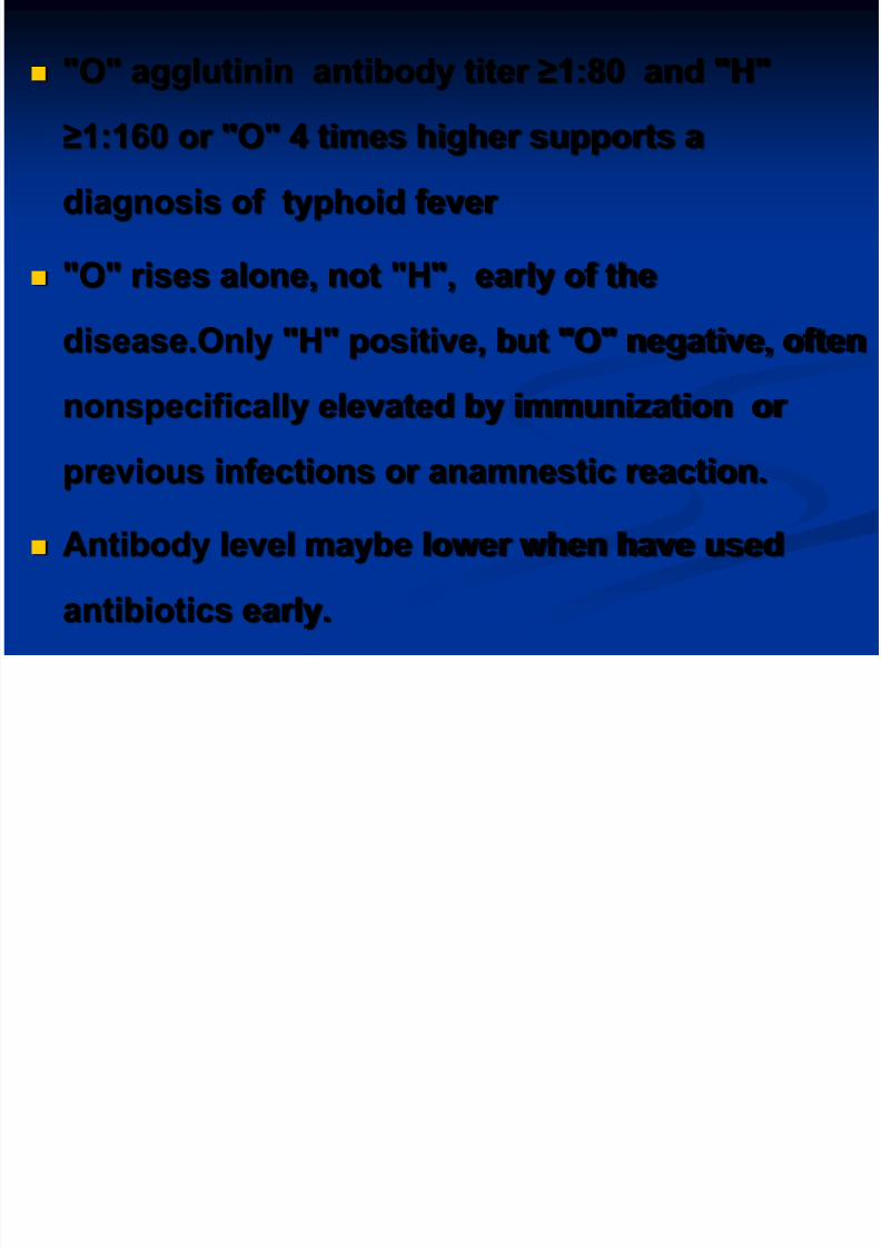
"O" agglutinin antibody titer ≥1:80 and "H"
≥1:160 or "O" 4 times higher supports a
diagnosis of typhoid fever
"O" rises alone, not "H", early of the
disease.Only "H" positive, but "O" negative, often
nonspecifically elevated by immunization or
previous infections or anamnestic reaction.
Antibody level maybe lower when have used
antibiotics early.
8/13/2019 thyphoid fever.ppt
http://slidepdf.com/reader/full/thyphoid-feverppt 41/66
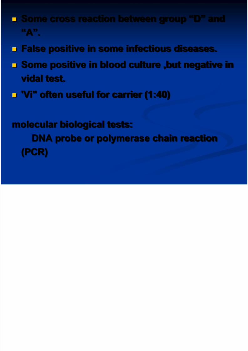
Some cross reaction between group “D” and
“A”.
False positive in some infectious diseases.
Some positive in blood culture ,but negative in
vidal test.
'Vi" often useful for carrier (1:40)
molecular biological tests:
DNA probe or polymerase chain reaction
(PCR)

8/13/2019 thyphoid fever.ppt
http://slidepdf.com/reader/full/thyphoid-feverppt 42/66
Complications
Intestinal hemorrhage Commonly appear during the second-third week of
illness
difference between mild and greater bleeding
often caused by unsuitable food, diarrhea et al
serious bleeding in about 2~8%
a sudden drop in temperature rise in pulse and
signs of shock followed by dark or fresh blood in the
stool.

8/13/2019 thyphoid fever.ppt
http://slidepdf.com/reader/full/thyphoid-feverppt 43/66
Intestinal perforation:
The more serious .Incidence,1-4%
Commonly appear during 2-3 weeks. Take place at the lower end of ileum.
Before perforation,abdominal pain or
diarrhea,intestinal bleeding .
When perforation, abdominal pain, sweating, drop in
temperature, and increase in pulse rate, then, rebound
tenderness when press abdomen,
abdomen muscle entasia, reduce or disappear in thesonant extent of liver, leukocytosis .
Temperature rise .peritonitis appear.
celiac free air under x-ray.
8/13/2019 thyphoid fever.ppt
http://slidepdf.com/reader/full/thyphoid-feverppt 44/66
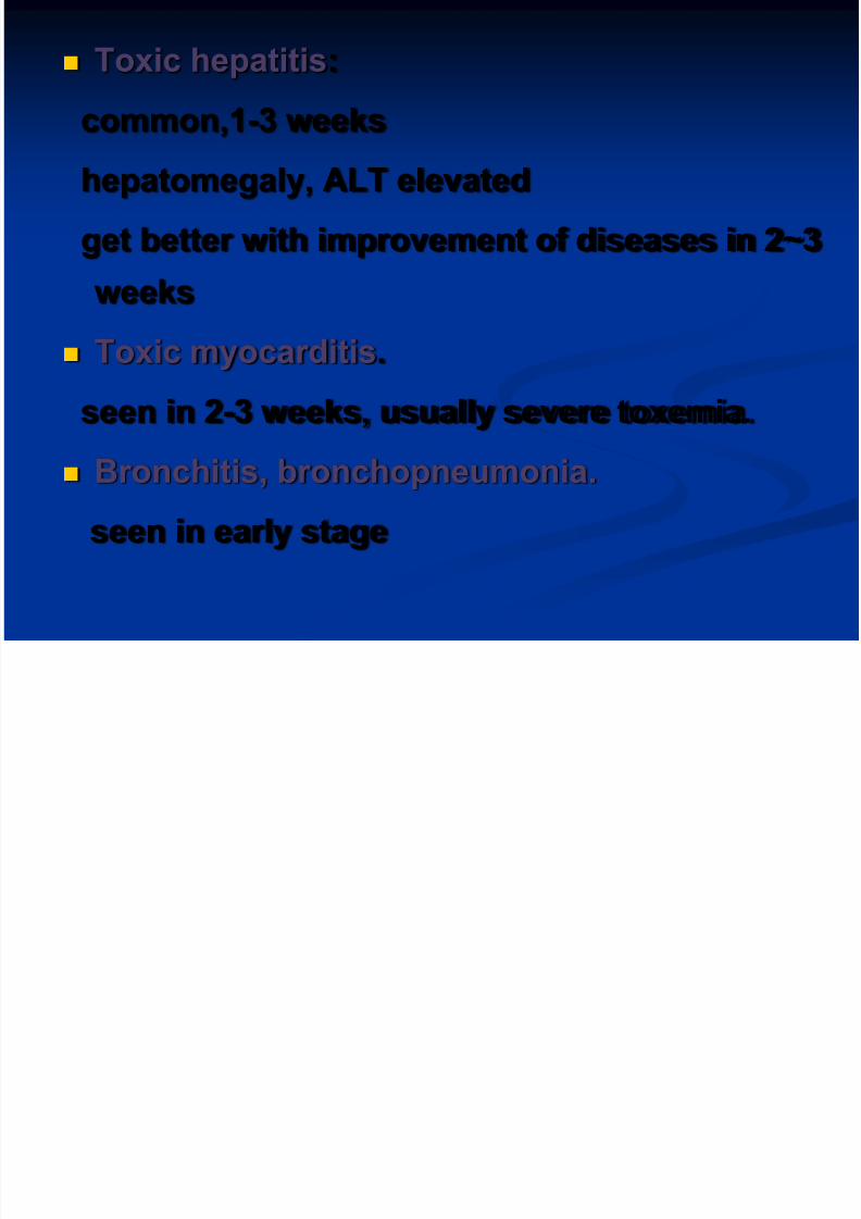
Toxic hepatitis:
common,1-3 weeks
hepatomegaly, ALT elevated
get better with improvement of diseases in 2~3
weeks Toxic myocarditis.
seen in 2-3 weeks, usually severe toxemia.
Bronchitis, bronchopneumonia.
seen in early stage
8/13/2019 thyphoid fever.ppt
http://slidepdf.com/reader/full/thyphoid-feverppt 45/66
Other complications:
toxic encephalopathy.
Hemolytic uremic syndrome.
acute cholecystitis
meningitis
nephritis et al.
8/13/2019 thyphoid fever.ppt
http://slidepdf.com/reader/full/thyphoid-feverppt 46/66
DIAGNOSTIK TYPHOID Typhoid Cardinal Sign
Febris continua Bradikardi relatif
Organomegali
Typhoid tounge
Roseole
Konstipasi/diare
Pemeriksaan lab hanya penunjang
Leukopenia Trombositopenia ringan
SGOT/SGPT meningkat
Widal Test + (dapat + pada 6 bulan-1 tahun post typhoid)

8/13/2019 thyphoid fever.ppt
http://slidepdf.com/reader/full/thyphoid-feverppt 47/66
Differential diagnosis
Viral infections:such as upper respiratory tract infection.
abrupt onset with fever, headache, leucopenia,
sore throat, cough, coryza.
no rose spots, no enlargement of liver & spleen.
The course of illness no more than 2 wks.differential diagnosis depends on typical
manifestations and blood culture.
8/13/2019 thyphoid fever.ppt
http://slidepdf.com/reader/full/thyphoid-feverppt 48/66
Malariahistory of exposure to malaria.
Paroxysms(often periodic) of sequential
chill,high fever and sweating.
Headache, anorexia, splenomegaly, anemia,leukopenia
Characteristic parasites in
erythrocytes,identified in thick or thin blood
smears.

8/13/2019 thyphoid fever.ppt
http://slidepdf.com/reader/full/thyphoid-feverppt 49/66
Leptospirosis
Endemic area,contacted with urine of mice.Abrupt fever,chills,severe headache,and myalgias,
especially of the calf muscles.
Leptospires can be isolated from
blood,cerebrospinal fluid.
Special agglutination titers develop after 7 days
and may persist at high levels for many years.

8/13/2019 thyphoid fever.ppt
http://slidepdf.com/reader/full/thyphoid-feverppt 50/66
Epidemic Louse-Borne typhus
prodromal of malaise and headache followed by
abrupt chills and fever.
headaches,prostration,persisting high fever.
Maculopapular rash appears on the forth toseventh days on the trunk and in the axillas,
spreading to the rest of the body but sparing the
face,palms,and soles. Laboratory confirmation by proteins OX19
agglutination and specific serologic tests.

8/13/2019 thyphoid fever.ppt
http://slidepdf.com/reader/full/thyphoid-feverppt 51/66
Tuberculosis
continuous high or low fever,fatigue,weight
loss,night sweats.
Mild cough
pulmonary infiltration on chest radiograph positive tuberculin skin test reaction(most
cases)
acid-fast bacilli on smear of sputum
sputum culture positive for mycobacterium
tuberculosis
8/13/2019 thyphoid fever.ppt
http://slidepdf.com/reader/full/thyphoid-feverppt 52/66
Septicemia of Gram-negative bacilli
abrupt onset,high fever,symptom oftoxemia.
Chill,sweats.
Shock.
Positive of gram-negative bacilli fromblood culture.

8/13/2019 thyphoid fever.ppt
http://slidepdf.com/reader/full/thyphoid-feverppt 53/66
Prognosis:
Case fatality 0.5~1%.
but high in old ages infant and serious
complications
Have immunity for ever after diseases
About 3% of patients become fecalcarriers .
TREATMENT
8/13/2019 thyphoid fever.ppt
http://slidepdf.com/reader/full/thyphoid-feverppt 54/66
TREATMENT
General treatment
isolation and rest
good nursing care and supportive
treatment
close observation T,P,R,BP,abdominal
condition and stool .
suitable diet include easy digested food orhalf-liquid food.drink more water
intravenous injection to maintain water and
acid-base and electrolyte balance

8/13/2019 thyphoid fever.ppt
http://slidepdf.com/reader/full/thyphoid-feverppt 55/66
Symptomatic treatment:
for high fever: physical measures firstly
antipyretic drugs such as aspirin should be
administrated with caution
delirium,coma or shock,2-4mg
dexamethasone in addition to antibioticsreduces mortality.

8/13/2019 thyphoid fever.ppt
http://slidepdf.com/reader/full/thyphoid-feverppt 56/66
Etiologic and special treatment
1.Quinolones:
first choice
it’s highly against S.typhi
penetrate well into macrophages,and achieve high
concentrations in the bowel and bile lumens
Norfloxacin (0.1 0.2 tid qid/10 14 days).
Ofloxacin (0.2 tid 10 14days).
ciprofloxacin (0.25 tid)
caution: not in children and pregnant
2 Chloramphenicol:

8/13/2019 thyphoid fever.ppt
http://slidepdf.com/reader/full/thyphoid-feverppt 57/66
2.Chloramphenicol:
For cases without multiresistant S.typhi.
Children in dose of 50~60mg/kg/per day.
adult 1.5~2g/day. tid.
Unable to take oral medication, the same dosage
given introvenously
after defervescence reduced to a half. complete a
10~14 day course.
But ,drug resistance, a high relapse rate,bone
marrow toxicity.
3 C h l i

8/13/2019 thyphoid fever.ppt
http://slidepdf.com/reader/full/thyphoid-feverppt 58/66
3.Cephalosporines:
Only third generation effective
Cefoperazone and Ceftazidime.
2~4g/day .10~14 days.
4.Treatment of complication.
Intestinal bleeding:
bed rest, stop diet,close observation T,P,R,BP.
intravenous saline and blood transfusion,andattention to acid-base balances.
sometimes,operative.

8/13/2019 thyphoid fever.ppt
http://slidepdf.com/reader/full/thyphoid-feverppt 59/66
Perforation:
early diagnosis.
stop diet.
decrease down the stomach pressure.
intravenous injection to maintain electrolyte
and acid-base balances.
use of antibiotics.sometimes operative.
Toxic myocarditis:

8/13/2019 thyphoid fever.ppt
http://slidepdf.com/reader/full/thyphoid-feverppt 60/66
Toxic myocarditis:
bed rest, cardiac muscle protection drugs,
dexamethasone, digoxin.
5.Chronic carrier:
Ofloxacin 0.2 bid or ciprofloxacin 0.5 bid, 4~6
weeks.
Ampicillin 3~6g/day tid plus probenecid 1~
1.5g/day. 4~6 weeks.
TMP+SMZ
2 tabs. Bid. 1~3 months.
Cholecystitis may require cholecystectomy.
P h l i

8/13/2019 thyphoid fever.ppt
http://slidepdf.com/reader/full/thyphoid-feverppt 61/66
Prophylaxis
1.control source of infection
Isolation and treatment of patients
stool culture one time per 5 days.
if negative continued two times ,without isolation.Control of carriers.
observation of 25 days(15 days in paratyphoid)
when close contact
8/13/2019 thyphoid fever.ppt
http://slidepdf.com/reader/full/thyphoid-feverppt 62/66
2. Cut of course of transmission
key way
avoid drinking untreated water and
food.
3.Vaccination
side-effect more, less use

8/13/2019 thyphoid fever.ppt
http://slidepdf.com/reader/full/thyphoid-feverppt 63/66
Paratyphoid fever A,B,C
Caused by Salmonella paratyphoid
A,B,C.respectively.
in no way different from typhoid fever in
epidemiology, pathogenesis,
pathology,clinical manifestations,
diagnosis, treatment and
Prophylaxis
Paratyphoid A B:
8/13/2019 thyphoid fever.ppt
http://slidepdf.com/reader/full/thyphoid-feverppt 64/66
Paratyphoid A,B:
incubation period 2~15days, in genaral,8~10 days.
milder in severity
fewer in complications.
Better in prognosis,
relapse more common in Paratyphoid A.
Treatment same as in typhoid fever.
Paratyphoid C:
8/13/2019 thyphoid fever.ppt
http://slidepdf.com/reader/full/thyphoid-feverppt 65/66
Paratyphoid C:
Always sudden onset.
Rapid rise of temperature.
Presented in different forms-- Septicemia,
Gastroenteritis and Enteric fever
Complications--arthritis, abscess formation,
cholecystitis, pulmonary complications are
commonly seen.
Intestinal hemorrhage and perforation not as
common as in typhoid fever.

8/13/2019 thyphoid fever.ppt
http://slidepdf.com/reader/full/thyphoid-feverppt 66/66