thromboelastographic evaluation of coagulation in a dog...
TRANSCRIPT
Thromboelastographic Evaluation of Coagulation in a Dog with Anticoagulant Rodenticide Intoxication
(Antikoagülant Rodentisit Zehirlenmesi Olan Bir Köpekte Koagülasyonun Tromboelastografik Olarak Değerlendirilmesi)
Oya ERALP İNAN 1 Meriç KOCATÜRK 2 Zafer MECİTOĞLU 2 Zeki YILMAZ 2
1
2
Medical and Surgical Experimental Animal Practice and Research Center, Eskişehir Osmangazi University, TR-26480 Eskisehir - TURKEYDepartment of Internal Medicine, Faculty of Veterinary Medicine, Uludag University, TR-165059 Bursa - TURKEY
Dear Editor,
As known, clinicians have a responsibility to make a true diagnosis and to provide effective treatment plan as soon as possible in the clinical setting. For this purpose, alternative diagnostic methods have been used to minimize time consuming effect during the diagnostic work-up in human and veterinary medicine. Thus, this case presentation provides a novel diagnostic approach by use of Thromboelastography (TEG) to evaluate coagulation cascade in a dog with anticoagulant rodenticid intoxication.
Anticoagulant rodenticids are leading to multiple clinical and laboratory disorders including primarily internal/ external bleeding, anemia and thrombocytopenia [1]. Traditional methods to evaluate coagulation system include active coagulation time, prothrombin time (PT) and activated partial thromboplastin time (aPTT) [2]. These procedures need not only to comprehensive and high technology laboratory equipment, but also to have some disadvantages due to long measurement time and economic reason in veterinary medicine. To solve this problem, TEG provides valuable information of haemostasis by measuring graphically and numerous the clotting time, clot firmness and fibrinolysis of the patient. TEG is used in veterinary medicine [3], but there is no report yet of the TEG evaluation of anticoagulant rodenticid intoxication in dogs.
A pointer (18 kg, 9 months old and female) was presented to the Animal Hospital (Uludag University, Faculty of Veterinary Medicine, Bursa - Turkey) with the complaint of severe depression and inappetence. Anticoagulant rodenticid (brodifacoum) intoxication was considered as a possible cause according to the owner’s history. In the physical examination, a muffled heart sound, anaemic mucous membranes and an echimosis at the medial right
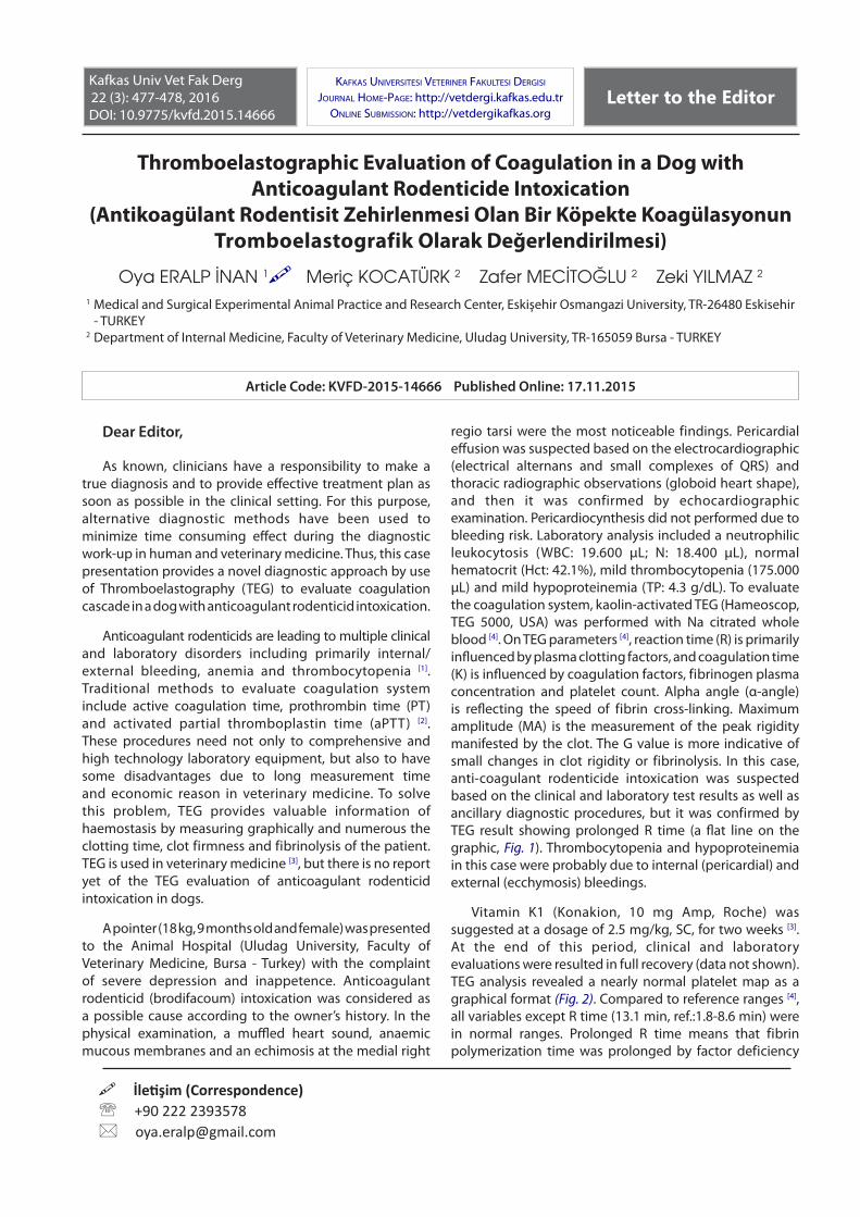
regio tarsi were the most noticeable findings. Pericardial effusion was suspected based on the electrocardiographic (electrical alternans and small complexes of QRS) and thoracic radiographic observations (globoid heart shape), and then it was confirmed by echocardiographic examination. Pericardiocynthesis did not performed due to bleeding risk. Laboratory analysis included a neutrophilic leukocytosis (WBC: 19.600 μL; N: 18.400 μL), normal hematocrit (Hct: 42.1%), mild thrombocytopenia (175.000 μL) and mild hypoproteinemia (TP: 4.3 g/dL). To evaluate the coagulation system, kaolin-activated TEG (Hameoscop, TEG 5000, USA) was performed with Na citrated whole blood [4]. On TEG parameters [4], reaction time (R) is primarily influenced by plasma clotting factors, and coagulation time (K) is influenced by coagulation factors, fibrinogen plasma concentration and platelet count. Alpha angle (α-angle) is reflecting the speed of fibrin cross-linking. Maximum amplitude (MA) is the measurement of the peak rigidity manifested by the clot. The G value is more indicative of small changes in clot rigidity or fibrinolysis. In this case, anti-coagulant rodenticide intoxication was suspected based on the clinical and laboratory test results as well as ancillary diagnostic procedures, but it was confirmed by TEG result showing prolonged R time (a flat line on the graphic, Fig. 1). Thrombocytopenia and hypoproteinemia in this case were probably due to internal (pericardial) and external (ecchymosis) bleedings.
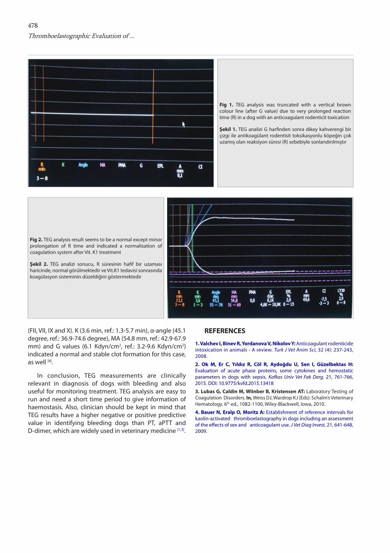
Vitamin K1 (Konakion, 10 mg Amp, Roche) was suggested at a dosage of 2.5 mg/kg, SC, for two weeks [3]. At the end of this period, clinical and laboratory evaluations were resulted in full recovery (data not shown). TEG analysis revealed a nearly normal platelet map as a graphical format (Fig. 2). Compared to reference ranges [4], all variables except R time (13.1 min, ref.:1.8-8.6 min) were in normal ranges. Prolonged R time means that fibrin polymerization time was prolonged by factor deficiency
İletişim (Correspondence) +90 222 2393578 [email protected]
Article Code: KVFD-2015-14666 Published Online: 17.11.2015
Letter to the EditorKafkas Univ Vet Fak Derg22 (3): 477-478, 2016DOI: 10.9775/kvfd.2015.14666
KafKas Universitesi veteriner faKUltesi Dergisi
JoUrnal Home-Page: http://vetdergi.kafkas.edu.tronline sUbmission: http://vetdergikafkas.org
478Thromboelastographic Evaluation of ...
(FII, VII, IX and X). K (3.6 min, ref.: 1.3-5.7 min), α-angle (45.1 degree, ref.: 36.9-74.6 degree), MA (54.8 mm, ref.: 42.9-67.9 mm) and G values (6.1 Kdyn/cm2, ref.: 3.2-9.6 Kdyn/cm2) indicated a normal and stable clot formation for this case, as well [4].
In conclusion, TEG measurements are clinically relevant in diagnosis of dogs with bleeding and also useful for monitoring treatment. TEG analysis are easy to run and need a short time period to give information of haemostasis. Also, clinician should be kept in mind that TEG results have a higher negative or positive predictive value in identifying bleeding dogs than PT, aPTT and D-dimer, which are widely used in veterinary medicine [1,3].
REFERENCES
1. Valchev I, Binev R, Yordanova V, Nikolov Y: Anticoagulant rodenticide intoxication in animals - A review. Turk J Vet Anim Sci, 32 (4): 237-243, 2008.
2. Ok M, Er C, Yıldız R, Cöl R, Aydoğdu U, Sen I, Güzelbektas H: Evaluation of acute phase proteins, some cytokines and hemostatic parameters in dogs with sepsis. Kafkas Univ Vet Fak Derg, 21, 761-766, 2015. DOI: 10.9775/kvfd.2015.13418
3. Lubas G, Caldin M, Wiinber B, Kristensen AT: Laboratory Testing of Coagulation Disorders. In, Weiss DJ, Wardrop KJ (Eds): Schalm’s Veterinary Hematology. 6th ed., 1082-1100, Wiley-Blackwell, Iowa, 2010.
4. Bauer N, Eralp O, Moritz A: Establishment of reference intervals for kaolin-activated thromboelastography in dogs including an assessment of the effects of sex and anticoagulant use. J Vet Diag Invest, 21, 641-648, 2009.
Fig 1. TEG analysis was truncated with a vertical brown colour line (after G value) due to very prolonged reaction time (R) in a dog with an anticoagulant rodenticit toxication
Şekil 1. TEG analizi G harfinden sonra dikey kahverengi bir çizgi ile antikoagülant rodentisit toksikasyonlu köpeğin çok uzamış olan reaksiyon süresi (R) sebebiyle sonlandırılmıştır
Fig 2. TEG analysis result seems to be a normal except minor prolongation of R time and indicated a normalisation of coagulation system after Vit. K1 treatment
Şekil 2. TEG analizi sonucu, R süresinin hafif bir uzaması haricinde, normal görülmektedir ve Vit.K1 tedavisi sonrasında koagülasyon sisteminin düzeldiğini göstermektedir