throat swab culture lab 1 - humsc
TRANSCRIPT

Throat culture
Throat swab culture
LAB 1

Throat swabA throat swab culture is a laboratory test done to isolate and identify
organisms that may cause infection in the throat mainly group A beta-
hemolytic streptococci.
Collection of SpecimenWho will collect the specimen
Physician Or Medical technologist, Microbiologist, experiencednurse.
Type of specimenTwo Swabs from posterior pharynx, tonsils, or other inflamed area.
StorageMaintain specimen swab at room temperature.
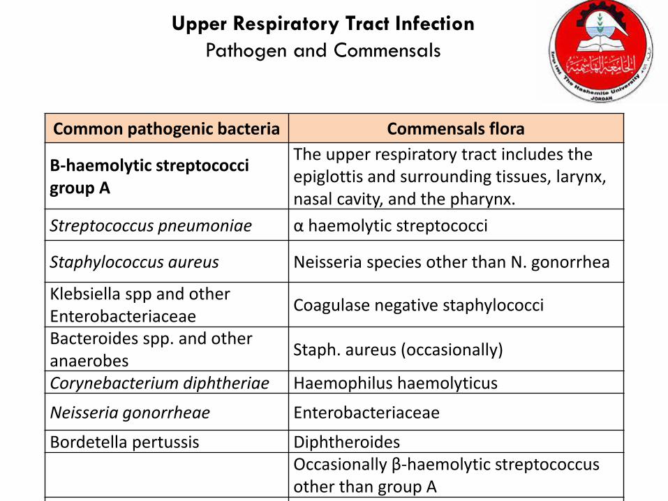
Common pathogenic bacteria Commensals flora
Β-haemolytic streptococci group A
The upper respiratory tract includes the epiglottis and surrounding tissues, larynx, nasal cavity, and the pharynx.
Streptococcus pneumoniae α haemolytic streptococci
Staphylococcus aureus Neisseria species other than N. gonorrhea
Klebsiella spp and other Enterobacteriaceae
Coagulase negative staphylococci
Bacteroides spp. and other anaerobes
Staph. aureus (occasionally)
Corynebacterium diphtheriae Haemophilus haemolyticus
Neisseria gonorrheae Enterobacteriaceae
Bordetella pertussis DiphtheroidesOccasionally β-haemolytic streptococcus other than group A
Upper Respiratory Tract Infection
Pathogen and Commensals

Throat Swabs collection procedure …I. Turn the patients face against the light, ask the patient to open his
mouth wide and phonate an “ah” gently depress the patients
tongue with a tongue blade so that the throat is well exposed and
illuminated.
II. Guide a swab over the tongue into the posterior pharynx.
III. Rub the swab firmly over the back of the throat, both tonsils and
any areas of inflammation, exudation or ulceration. Care should be
taken to avoid touching the tongue, cheeks or lips with the swab.
IV. Place the swab in the transport medium and push it down to the
bottom.

Specimen
Collection Time and Temp
GuidelinesDevice and
or minimum vol.
Transport Storage
Throat
1- Depress tongue with tongue depressor.2- Sample posterior pharynx, tonsils, and inflamed areas with sterile swab.3- Collect two swabs 0ne for gram stain and the other for culturing.
Swab transport medium
≤ 2h,RT ≤ 24h,RT
Criteria of specimen rejectionInappropriate specimen transport device.
mislabeled specimen.
Unlabeled specimen.
Dried samples.
Specimen received after prolonged delay (usually more than 2 hours).
Specimen received in expired transport media.
Throat Swabs collection procedure
S. pyogens is highly
resist to desiccation and
remains viable on a dry
swab for as long as 48
to 72 hours.

Specimen Processing
Culture plates should be incubated for at least 48 hours before reporting
as negative for group A streptococci . In addition, the incubation of plates
in 5% to 10% CO2. (also aerobic and anaerobic conditions are used but CO2
condition is perfect)

Gram StainDirect smear:
A gram stain from the swab noting the predominant organism.
Throat Swab CulturingCulture
Because Streptococcus pyogenes is the primary case of pharyngitis
most laboratories routinely screen throat cultures for this organism.
Classically throat swabs plated on 5% sheep Blood agar plates
and Columia C.N.A, streak the swab across first quadrant of blood
agar plate and using a sterile loop streak to produce isolated
colonies, make few stabs in the agar plates also, Group A
Streptococcus (S. pyogenes) are usually ß- hemolytic the activity of
hemolysin enzyme will increased by the stabbing.
NoteInoculate another Chocolate and MacConkey agar plates also are
recommended if organisms other than S. pyogenes is suspected.

Columbia C.N.A. agar with Blood
Peptone Colistin sulphate
Tryptic digest of beef heart Nalidixic Acid
Corn starch Agar
Sodium chloride Sheep Blood
Ingredients :
Nalidixic Acid and Colistin sulphate are the antimicrobics suppressing
the growth of Enterobacteriaceae and Pseudomonas spp., and allowing
yeast, Staphylococci, Streptococci, and Enterococci to grow.
Certain Gram-negative organisms, such as Gardnerella vaginalis and
certain Bacteriodes spp., can grow very well on Columbia CNA Agar
with blood.
Colistin disrupts the cell membrane of Gram-negative organisms,
particularly effective against Pseudomonas spp.
Nalidixic Acid blocks DNA replication in susceptible bacteria and acts
against many Gram-negative bacteria.

Make few stabs in the agar plates
Note: Make few stabs to increased hemolysin activity of group A Strepto.
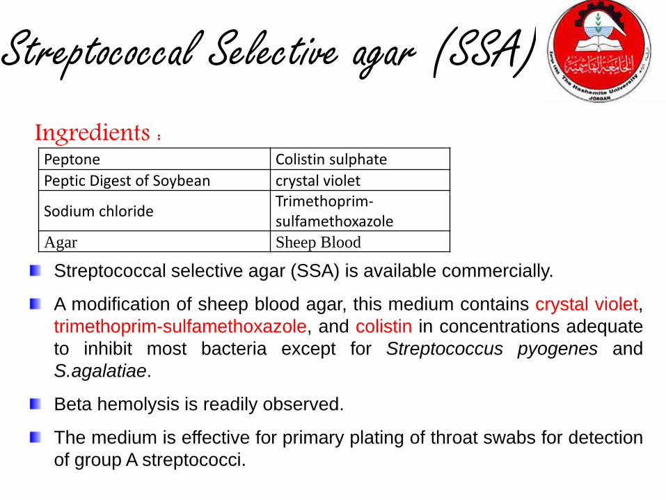
Streptococcal Selective agar (SSA)
Streptococcal selective agar (SSA) is available commercially.
A modification of sheep blood agar, this medium contains crystal violet,
trimethoprim-sulfamethoxazole, and colistin in concentrations adequate
to inhibit most bacteria except for Streptococcus pyogenes and
S.agalatiae.
Beta hemolysis is readily observed.
The medium is effective for primary plating of throat swabs for detection
of group A streptococci.
Ingredients :Peptone Colistin sulphate
Peptic Digest of Soybean crystal violet
Sodium chlorideTrimethoprim-sulfamethoxazole
Agar Sheep Blood

Specimen Processing continue…
Direct antigen detectionIdentification of group A Streptococcal antigen in throat specimens
are available now by using different methods including latex
agglutination, enzyme immunoassay and gene probe technology,
that allow detection of Streptococcal group A antigen within at little
as 10 minutes.
Antistreptolysin O titre (ASOT).

Rapid strep test1. Lateral flow testThe sample is applied to a strip of nitrocellulose film and, if GAS antigens are
present, these will migrate along the film to form a visible line of antigen bound to
labeled antibodies.
2. ImmunoassayThe newest and more expensive test. It involves mixing the sample with labeled
antibodies and then with a special substrate on a film which changes colour to
signal the presence or absence of GAS antigen
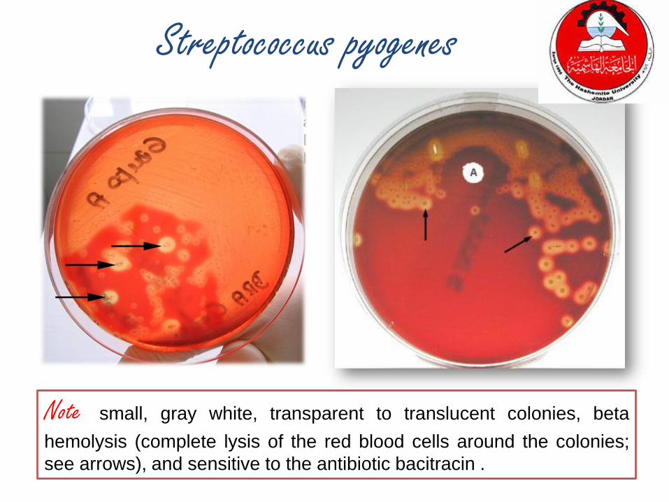
Note small, gray white, transparent to translucent colonies, beta
hemolysis (complete lysis of the red blood cells around the colonies;
see arrows), and sensitive to the antibiotic bacitracin .
Streptococcus pyogenes

Outline of differentiation between Gram-
Positive cocci
e.g. S. epidermidis

Differentiation between -hemolytic
streptococci
• The following tests can be used to differentiate
between -hemolytic streptococci
– Lanciefield Classification
– Bacitracin susceptibility Test
• Specific for S. pyogenes (Group A)
– CAMP test
• Specific for S. agalactiae (Group B)
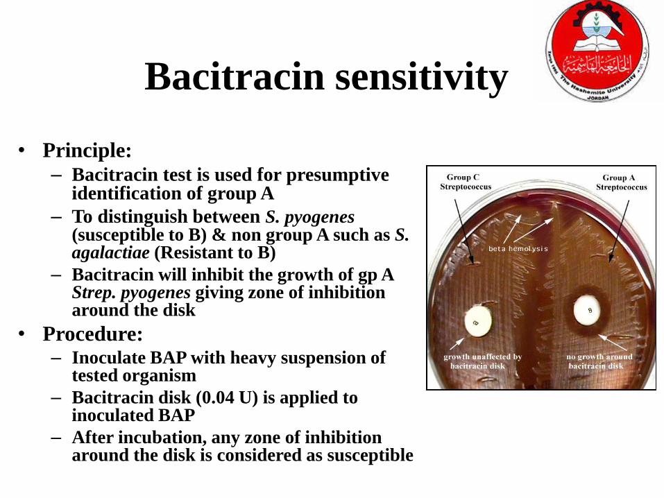
Bacitracin sensitivity
• Principle:– Bacitracin test is used for presumptive
identification of group A
– To distinguish between S. pyogenes(susceptible to B) & non group A such as S. agalactiae (Resistant to B)
– Bacitracin will inhibit the growth of gp A Strep. pyogenes giving zone of inhibition around the disk
• Procedure:– Inoculate BAP with heavy suspension of
tested organism
– Bacitracin disk (0.04 U) is applied to inoculated BAP
– After incubation, any zone of inhibition around the disk is considered as susceptible
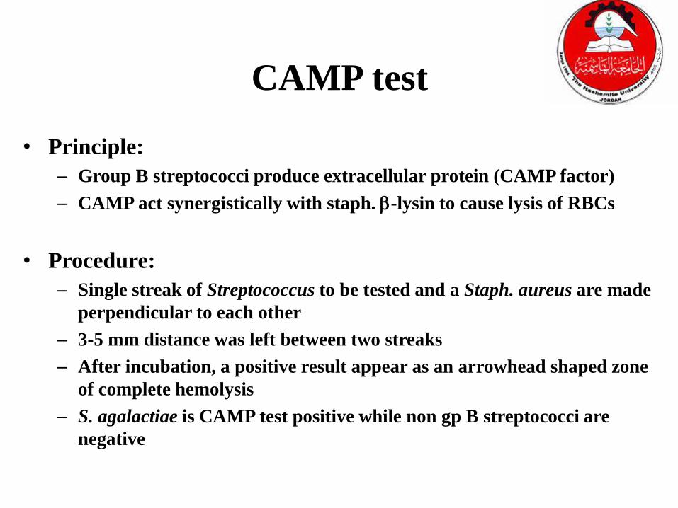
CAMP test
• Principle:
– Group B streptococci produce extracellular protein (CAMP factor)
– CAMP act synergistically with staph. -lysin to cause lysis of RBCs
• Procedure:
– Single streak of Streptococcus to be tested and a Staph. aureus are made
perpendicular to each other
– 3-5 mm distance was left between two streaks
– After incubation, a positive result appear as an arrowhead shaped zone
of complete hemolysis
– S. agalactiae is CAMP test positive while non gp B streptococci are
negative

CAMP Factor TestS. aureus
(Spingomyelinase C)Group B Streptococcus
(CAMP Factor)
Group A Streptococcus
Enhanced Zone of
Hemolysis
CAMP is an acronym for the authors of this test (Christie, Atkinson, Munch, Peterson). The CAMP test
takes advantage of the capacity of GBS-group B strep to produce this CAMP factor; most other hemolytic streptococci do not produce CAMP factor. Enhances the ability of S. aureus to produce Beta hemolysis
Differentiation between -hemolytic
streptococci
• The following definitive tests used to differentiate between S. pneumoniae & viridans streptococci
– Optochin Test
– Bile Solubility Test

Optochin Susceptibility Test
• Principle:
– Optochin (OP) test is presumptive test that is used to identify S. pneumoniae
– S. pneumoniae is inhibited by Optochin reagent (<5 µg/ml) giving a inhibition zone ≥14 mm in diameter.
• Procedure:
– BAP inoculated with organism to be tested
– OP disk is placed on the center of inoculated BAP
– After incubation at 37oC for 18 hrs, accurately measure the diameter of the inhibition zone by the ruler
– ≥14 mm zone of inhibition around the disk is considered as positive and ≤13 mm is considered negative
• S. pneumoniae is positive (S) while S. viridans is negative (R)

Optochin Susceptibility Test
Optochin susceptible
S. pneumoniae
Optochin resistant
S. viridans

Bile Solubility test
• Principle:
– S. pneumoniae produce a self-lysing enzyme (autolysin) to inhibit the growth
– The presence of bile salt accelerate this process
• Procedure:– Add ten parts (10 ml) of the broth culture of the organism to be
tested to one part (1 ml) of 2% Na deoxycholate (bile) into the test tube
– Negative control is made by adding saline instead of bile to the culture
– Incubate at 37oC for 15 min
– Record the result after 15 min

Bile Solubility test
• Results:
– Positive test appears as clearing
in the presence of bile while
negative test appears as turbid
– S. pneumoniae soluble in bile
whereas S. viridans insoluble

Differentiation between -hemolytic streptococci
CAMP testBacitracin
sensitivity
Hemolysis
NegativeSusceptibleS. pyogenes
PositiveResistantS. agalactiae
Inulin
Fermentation
Bile
solubility
Optochin
sensitivity
Hemolysis
Not fermentSolubleSensitive (≥
14 mm)
S. pneumoniae
FermentInsolubleResistant
(≤13 mm)
Viridans strep
Differentiation between -hemolytic streptococci

Outline of differentiation between
Gram-Negatives
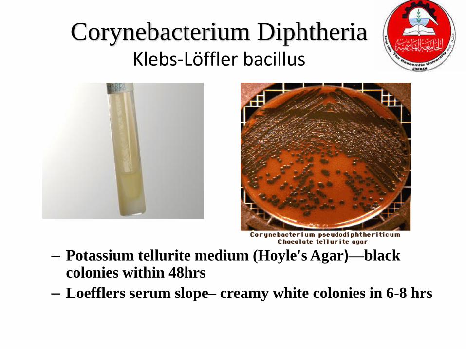
Corynebacterium DiphtheriaKlebs-Löffler bacillus
– Potassium tellurite medium (Hoyle's Agar)—black colonies within 48hrs
– Loefflers serum slope– creamy white colonies in 6-8 hrs

Bordetella pertussis
Bordet-Gengou agar
blood, potato extract, and glycerol, with an antibiotic such as
cephalexin or penicillin and sometimes nicotinamide
Regan-lowe charcoal agar
charcoal, blood, and antibiotic (cephalexin)
Medium of choice

Haemophilus
influenza b
Chocolate agar
M. catarrhalis
Chocolate or blood agar

Legionella
buffered charcoal yeast extract
(BCYE) agar
Cysteine, Iron
incubation for up to 10 days
N. gonorrhoeae
Thayer martin agar
Chocolate sheep blood plus
antibiotics (vancomycin, colistin,
nystatin, and TMP-SMX)
Post specimen processingInterfering factors:
Patient on antibiotic therapy.
Improper sample collection.
Result reporting:Report Gram stain finding as an initial report.
Report the isolated and its sensitivity pattern as a final report.
Turn around time:Gram stain result should be available half hour after specimen
receipt.
Isolation of a possible pathogen can be expected after 2-4 days.
Negative culture will be reported out 1-2 days after the receipt of
the specimen.

LAB 2
Sputum sample collection

33
1. It is a secretion that is produced in the lungs and the bronchi (tubes that carry the air to the lung), also known as phlegm
2. This mucus-like secretion may become infected, blood stained, or contain abnormal cells that may lead to a diagnosis
3. Tracheobronchial sections are an inconstant mixture of plasma, water, electrolytes and mucin
4. As these mixture pass through the lower and upper respiratory tract, they become contaminated with cellular exfoliations, nasal and salivary gland secretions and normal bacterial flora of the oral cavity
Sputum Definition

Sputum specimens
Ordered to identify organisms growing in sputum
➢C&S
➢AFB➢3 consecutive, early am
➢Cytology ➢Abnormal lung cancer by cell type
➢3 early am

Sputum collection
Sputum (Expectorate):
1. Collect early morning specimen under the direct supervision of a
nurse or a physician.
2. Have patient rinse or gargle with water to remove superficial flora.
3. Instruct patient to cough deeply to produce a lower respiratory
specimen.
4. Exam specimen to make sure it contains thick mucus. Do not submit
saliva.
Sputum (Induced):
1. Have patient rinse mouth with water after brushing gums and tongue.
2. With the aid of a nebulizer, have patients inhale about 25 mL of 3 to
10% sterile saline.
3. Collect the induced sputum in a sterile container.

36
Sputum Collection
1. Drinking a lot of water and other fluids the night before the test may help to get the sample
2. To be asked to cough deeply and spit any sputum in a sterile cup
3. The sputum is then taken to the laboratory
4. There, it is placed in a special substance (medium) under conditions that allow the organisms to grow

37
Physical Properties of Sputum
1. Appearance
• It may be described as liquid (serous), mucoid, purulent, bloody or combination of theses
2. Color• Normal sputum is either white or colorless.
• Yellow to green sputum can be an indication of pus, infection such as pneumonia.
• Blood in sputum is called hemoptysis which could be due to e.g ; lung cancer, tuberculosis, lung abscess , hemorrhage .
• Rust color is due to decomposed Hemoglobin and it is typical for S. pneumonia.
• Very thick (viscose) sputum is a characteristic of cystic fibrosis .
• Parasites in sputum can occur as in Ascaris .
38
3. Odor• Usually no odor is present in normal and
pathological sputum, but if bacterial decomposition has been taken place within the body or after expectoration, a variety of odor will be present
Physical Properties of Sputum

39
Sputum Chemical Composition
Component % of Total Weight
Water 95
Solid 5
% of Total Solids
Carbohydrates Variable
Proteins Variable
Lipids Variable
DNA Variable
Enzymes, -antitrypsin, LDH,
lysozyme, lactoferrin
Variable

40
Sputum Analysis: Pneumonia
• Moraxella catarrhalis, a large number of Gram negative (red) cocci (di-) are seen and many appear to be attaching to or residing within the PMNs
• Some physicians confuse these organisms with the Gram negative coccobacillary

41
Sputum Analysis: Pneumonia
• Hemophilus influenzae pneumonia demonstrating the typical Gram negative coccobacillary forms
• Because of the red background produced by the Gram stain method, these organisms can be difficult to see (oil immersion, 1000x)

42
Sputum Analysis: Pneumonia
• Klebsiella pneumoniaedemonstrating Gram negative bacillary organisms. (oil immersion, 1000x)
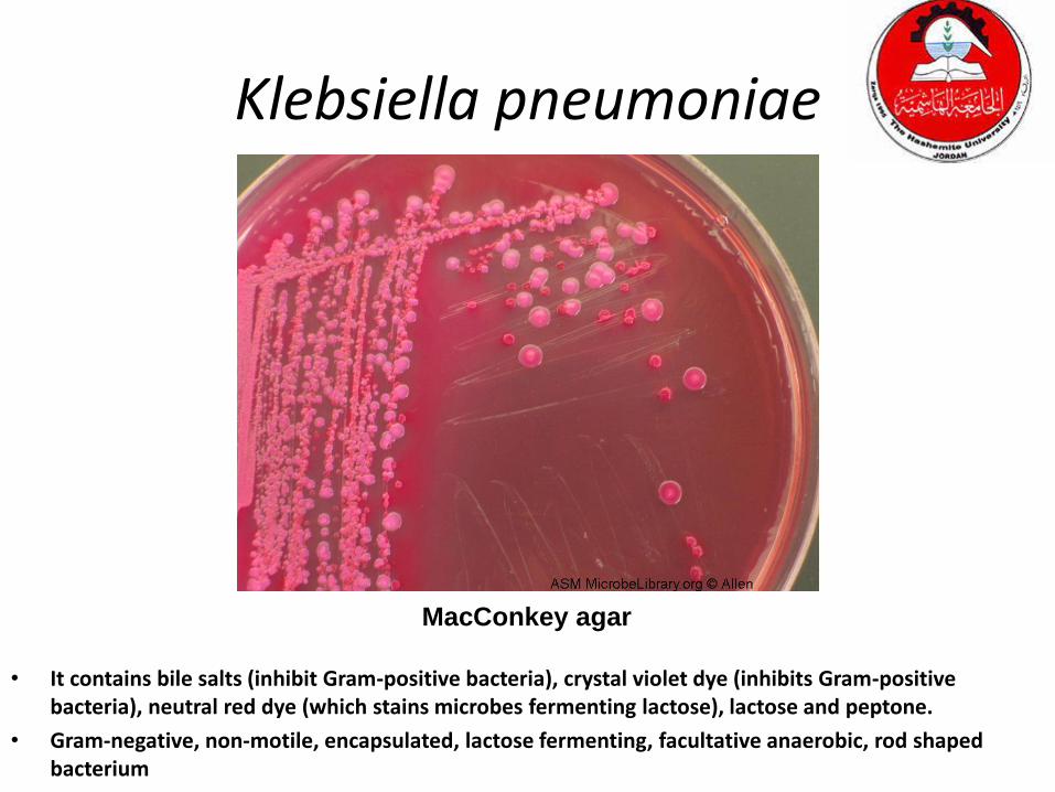
Klebsiella pneumoniae
• It contains bile salts (inhibit Gram-positive bacteria), crystal violet dye (inhibits Gram-positive bacteria), neutral red dye (which stains microbes fermenting lactose), lactose and peptone.
• Gram-negative, non-motile, encapsulated, lactose fermenting, facultative anaerobic, rod shaped bacterium
MacConkey agar

44
Sputum Analysis: Pneumonia
• Gram stain of the sputum from a patient with Staphylococcus aureus pneumonia demonstrating clusters of Gram positive cocci some of which are associated with the PMNs (oil immersion, 1000x)
45
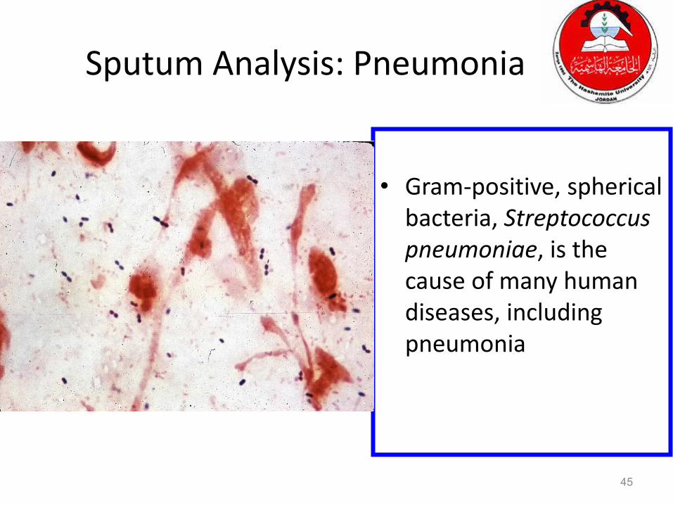
Sputum Analysis: Pneumonia
• Gram-positive, spherical bacteria, Streptococcus pneumoniae, is the cause of many human diseases, including pneumonia

• At 25C forming macroconidia • At 37 yeast form
Sputum Analysis: PneumoniaHistoplasma capsulatum
Grows on Blood agar, Chocolate agar and Sabouraud’s agar (dextrose and
peptone; PH5.6). Takes few weeks to grow
Mold
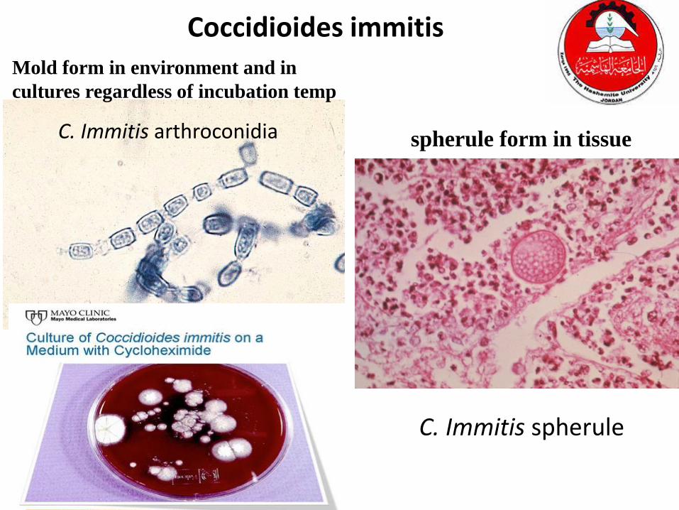
C. Immitis arthroconidia
C. Immitis spherule
Coccidioides immitis
spherule form in tissue
Mold form in environment and in
cultures regardless of incubation temp

• Conidial head
Sputum Analysis: PneumoniaAspergillus fumigatus
Aspergillus require 1-3 weeks for growth.
The colony begins as a dense white
mycelium which later assumes a variety of
colors, according to species

Ziehl Nielsen's Stain
1. Smear the sputum
2. Fix by Heating
3. Pour carbol fuchsin and heat it from below, Keep for 5
min.
4. Wash with water, Decolorize with 20%H2SO4
5. Wash with Loffler's methylene blue for 1 min.
6. Wash & dry
7. Mount under oil immersion


Mycobacterium tuberculosis Ziehl-Neelsen stain

Löwenstein–Jensen medium
• The usual composition as
applicable to Mycobacterium
tuberculosis is:
• Malachite green
• Glycerol
• Asparagine
• Potato starch
• Coagulated eggs
• Mineral salt solution
• Penicillin and nalidixic acid
Alternative culture media
• Egg-based – Petragnini medium and Dorset medium
• Middlebrook 7H10 Agar
• Middlebrook 7H9 broth