three dimensional high resolution mri …eprints.usm.my/37839/1/pages_from_husbani_mohd_amin... ·...
TRANSCRIPT

i
THREE DIMENSIONAL HIGH RESOLUTION
MRI MYELOGRAPHY OF CERVICAL SPINE
IN PATIENTS WITH
CERVICAL SPONDYLOTIC RADICULOPATHY
USING MODERATELY T2-WEIGHTED 3D TSE-FS SEQUENCE
By:
DR. HUSBANI BT MOHD AMIN REBUAN
Dissertion Submitted In
Partial Fulfillment Of The
Requirements For The Degree Of
Master of Medicine
(Radiology)
UNIVERSITI SAINS MALAYSIA
2011
ii
ACKNOWLEDGEMENT
The author would like to express deepest gratitude to the following individuals for their advice,
guidance, comments, cooperation and support during the preparation of this dissertation, and
thus, with their patience, help and encouragement making this dissertation possible.
• Ass.Prof. Dr.Mohd Ezane b. Aziz - Supervisor and lecturer in Radiology Department,
Hospital University Sains, Malaysia, Kubang Kerian, Kelantan, Malaysia.
• Dr.Elinah bt. Ali - Supervisor and Head of Radiology Department, Hospital Sultanah Nur
Zahirah, Kuala Terengganu, Malaysia.
• Mr.Ahmad Tajuddin - Head of Orthopedic Department, Hospital Sultanah Nur Zahirah,
Kuala Terengganu, Malaysia.
• All lecturers in Radiology Department, HUSM.
• All radiologists and radiographers in Hospital Sultanah Nur Zahirah,Kuala Terengganu,
Terengganu that involved in this study.

iii
TABLE OF CONTENTS
Acknowledgement i
Table of contents ii
List of Figures vii
List of Tables viii
Abbreviations ix
Abstract
Abstract (Malay) x
Abstract(English) xiii
Chapter I : Introduction
1.0 Introduction 1
Chapter II : Literature review
2.1 Cervical spondylotic radiculopathy 5
2.1.1 Epidemiology of cervical spondylotic radiculopathy 6
2.1.2 Clinical examination in cervical radiculopathy 7

iv
2.2 Anatomy of cervical spine
2.2.1 Body of cervical vertebra and vertebral arch 8
2.2.2 Intervertebral disc 8
2.2.3 Facet joint 9
2.2.4 Exit foramina 10
2.3 Imaging in cervical spondylotic radiculopathy
2.3.1 Conventional myelogram 10
2.3.2 CT myelogram 11
2.3.3 Magnetic Resonance Imaging 11
2.3.4 Association between conventional MRI with physical examination
12
2.3.5 Association between conventional MRI findings with intra-operative
findings 13
v
2.3.6 Conventional MRI vs CT myelogram 13
2.3.7 MRI myelogram 14
2.3.8 Association between MRI myelograms with physical findings 15
2.3.9 Association between MRI myelogram findings with intra- operative
findings 14
2.3.10 Added value in MRI myelogram 16
2.3.11 Various MRI myelogram (MRM) techniques 18
2.3.12 Current imaging technique for MRI Myelogram (MRM) 19
2.4 Rationale of study 21
Chapter 3 : Objectives
3.1 General objective 22
3.2 Specific objectives 22
3.3 Null hypothesis 22
Chapter 4: Methodology
4.1 Study design 23

vi
4.2 Patient’s criteria
4.2.1 Inclusion criteria 23
4.2.2 Exclusion criteria 24
4.3 Materials and Method
4.3.1 Information 24
4.3.2 Imaging procedure 26
4.3.3 Interpretation of MRI 29
4.4 Population and sample
4.4.1 Reference population 29
4.4.2 Source population 30
4.4.3 Sampling method 30
4.4.4 Research tools 30
4.4.5 Informed consent 30
4.5 Statistical analysis 31
Chapter 5: Result
5.1 General results 34
5.2 Association of clinical variables with MRI findings 40
5.3 Agreement of nerve root compression between conventional MRI and MRI
myelogram 43
5.4 Interobserver variability 45
vii
Chapter 6: Discussion
6.1 Dermographic data 47
6.2 Clinical symptoms and signs 49
6.3 Conventional MRI findings 50
6.4 Clinical findings vs conventional MRI findings 51
6.5 Clinical findings vs MRI myelogram findings 54
6. 6 Conventional MRI vs MRI myelogram 54
6.7 Interobserver variability 62
6.8 MRI myelogram findings using special sequence 64
Chapter 7: Conclusion 66
Chapter 8:
8.1 Limitation 67
8.2 Recommendations 68
Chapter 9 : References and appendices 69
viii
List of Figures
Figure 5.1.1 Histogram showing the age distribution of patients 32
Figure 5.1.2 Bar chart showing the various clinical symptoms 33
Figure 5.1.3(a) Bar chart showing reduced sensation on physical examination 34
Figure 5.1.3(b) Bar chart showing reduced power on physical examination 35
Figure 5.1.4(a) Pie chart showing nerve root compression on conv.MRI 36
Figure 5.1.4(b) Bar chart showing nerve root compression according to dermatome 37
Figure 5.1.5(a) Pie chart of nerve root compression in MRI myelogram in observer A 38
Figure 5.1.5(b) Pie chart shows nerve root compression in MRI myelogram in observer B 39
Figure 5.1.5(c) Bar chart shows nerve root compression in MRI myelogram according to level
of cervical spine in observer B
Figure 5.1.6(a-h) Bar chart showing physical examination vs conventional MRI and MRI
myelogram findings 41
Figure 6.6.1(a) T2 SPACE 3D MRI myelogram image shows marginal osteophyte 57
Figure 6.6.1(b) T2 SPACE 3D MRI myelogram image shows marginal osteophyte 58
Figure 6.6.1(c) T2 SPACE 3D MRI myelogram image shows posterolateral disc bulge 59

ix
Figure 6.8.1 (a) Normal T2 SPACE 3D MRI myelogram 63
Figure 6.8.1 (b) Normal T2 HASTE 3D MRI myelogram 63
Figure 6.8.1 (c) MRI myelogram T2 SPACE and T2 HASTE 3D MRI myelogram showing
disc bulge 64
Figure 6.8.1 (d) MRI myelogram T2 SPACE and T2 HASTE 3D MRI myelogram showing
bilateral pseudomeningocele 65
x
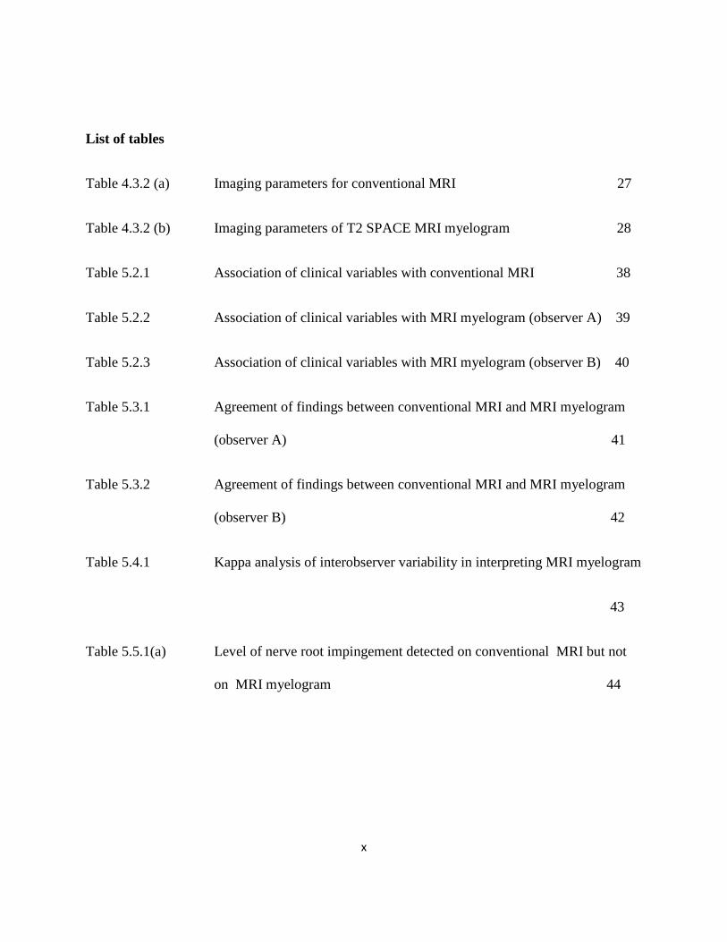
List of tables
Table 4.3.2 (a) Imaging parameters for conventional MRI 27
Table 4.3.2 (b) Imaging parameters of T2 SPACE MRI myelogram 28
Table 5.2.1 Association of clinical variables with conventional MRI 38
Table 5.2.2 Association of clinical variables with MRI myelogram (observer A) 39
Table 5.2.3 Association of clinical variables with MRI myelogram (observer B) 40
Table 5.3.1 Agreement of findings between conventional MRI and MRI myelogram
(observer A) 41
Table 5.3.2 Agreement of findings between conventional MRI and MRI myelogram
(observer B) 42
Table 5.4.1 Kappa analysis of interobserver variability in interpreting MRI myelogram
43
Table 5.5.1(a) Level of nerve root impingement detected on conventional MRI but not
on MRI myelogram 44

xi
ABBREVIATIONS AND TERMS
MRI - Magnetic Resonance Imaging
TSE - Turbo Spin Echo
FS - Fat saturation
TR - Repetition time
TE - Echo time
AT - Acquisition time
T1WI - T1 Weighted Images
T2WI - T2 Weighted Images
PD - Proton density

xii
KAJIAN MENGENAI PENGGUNAAN 3 DIMENSI MR MYELOGRAM YANG
MENGGUNAKAN SEKUENS ‘T2-WEIGHTED 3D TSE-FS” DALAM MENJALANKAN
UJIKAJI TERHADAP PESAKIT YANG MENGALAMI MASALAH URAT SARAF
TERTEKAN DI BAHAGIAN TULANG BELAKANG BAHAGIAN SERVIKAL.
Dr.Husbani bt.Mohd Amin Rebuan
M.Med Radiology
Department of Radiology
School of Medical Sciences, Universiti Sains Malaysia
Health Campus, 16150 Kelantan, Malaysia
Pengenalan: Golongan tua yang berusia lebih daripada 50 tahun adalah berisiko untuk
mendapat perubahan di bahagian tulang belakang di mana ia juga turut mula mengalami proses
penuaan. Ini akan memberi kesan kepada urat saraf yang keluar daripada saraf tunjang di
bahagian tengkuk di mana ia akan mengalami himpitan akibat daripada proses penuaan yang
berlaku pada bahagian ini. Akibat daripada himpitan ini akan menimbulkan kesakitan pada
bahagian tengkuk dan sebagainya. Keadaan ini memerlukan satu alat yang dapat mengenalpasti
dan mendiagnosa masalah yang berlaku pada urat tengkuk ini dan seterusnya masalah ini dapat
diatasi dengan segera. Dengan adanya kemajuan dari segi bidang perubatan, berbagai kaedah
telah dilakukan untuk penyakit ini didiagnosa dengan tepat tanpa memberi kemudaratan kepada
pesakit. Sekiranya sebelum ini “konvensional myelogram” atau “CT myelogram” digunakan
untuk mendiagnosa penyakit ini tetapi dengan kemajuan teknologi terkini ia diganti dengan MRI.
xiii
Penggunaan MRI adalah baik memandangkan pesakit tidak perlu terdedah kepada sinar x-ray
dan gambar yang dihasilkan secara amnya menyerupai imej “konvensional myelogram” .
Walaubagaimanapun, penggunaan konvensional MRI setakat ini masih ada kekurangan dan MRI
myelogram diharap boleh menggantikan konvensional MRI pada masa akan datang. Ini
secaratidak langsung dapat membantu pakar radiologi secara specific dan membantu pesakit
secara amnya.
Objektif: Untuk menentukan adakah terdapat persamaandi antara persembahan klinikal
dengan penekanan urat saraf di dalam konvensional MRI atau MRI myelogram, menentukan
adakah terdapat persamaan keputusan diantara “konvensional MRI” dan “MRI myelogram” dan
melihat adakah terdapat perbezaan keputusan di antara dua pakar radiologi dalam menilai
penekanan urat saraf di dalam “ MRI myelogram”.
Tatacara dan bahan-bahan: Ini adalah satu kajian keratan rentas secara rawak di mana
pesakit-pesakit yang didiagnosa sebagai “cervical spondylotic radiculopathy” di antara bulan
Januari 2009 hingga bulan Januari 2010 diperlukan menjalani pemeriksaan “MRI konvensional”
dan “MRI myelogram”. Maklumat pesakit seperti umur, bangsa dan jantina dicatatkan. Imej imej
yang diperolehi daripada kedua-dua “MRI myelogram” dan “konvensional MRI” kemudiannya
akan dilihat oleh dua orang pakar radiologi (pemerhati) yang tidak mengetahui berkenaan
persembahan klinikal dan pemeriksaan fizikal pesakit. Pemerhati perlu membuat pemerhatian
berdasarkan kriteria-kriteria yang telah ditetapkan untuk mengatakan terdapat himpitan kepada
xiv
urat saraf yang keluar daripada tengkuk. Keputusan ini kemudiannya akan dibandingkan dan
dianalisa.
Keputusan: Majoriti pesakit yang menghadapi masalah ini adalah daripada golongan
yang berproduktiviti tinggi dengan purata umur 46.9 tahun. Didapati persembahan klinikal
pesakit mempunyai kaitan yang tinggi dengan penekanan urat saraf yang keluar daripada saraf
tunjang di dalam kedua-dua pemeriksaan “konvensional MRI” dan “MRI myelogram”. Terdapat
persamaan keputusan terdapat penekanan urat saraf di antara kedua dua teknik juga adalah
sangat tinggi.Di antara kedua-dua pemerhati yang membaca imej MRI myelogram didapati kadar
persamaan keputusan yang tinggi dari segi penekanan urat saraf.
Kesimpulan: MRI myelogram mengubah interpretasi penekanan urat saraf di dalam 22
urat saraf daripada 47 himpitan urat saraf yang dikesan oleh konvensional MRI. Nilai ini adalah
sangat bermakna dimana MRI myelogram sesuai digunakan sebagai pemeriksaan bersama
dengan konvensional MRI dalam mengesan urat saraf yang tertekan.MRI dapat memberi
maklumat tambahan (8 urat saraf yang tertekan) di mana tidak dapat di kesan oleh konvensional
MRI. Walaupun nilai ini adalah minimum untuk menjadikan MRI myelogram sebagai teknik
yang tidak bergantung kepada konvensional MRI, tetapi ia sangat berguna kepada pesakit.
Prof. Madya Dr. Mohd Ezane Aziz: Supervisor
xv
THREE DIMENSIONAL HIGH RESOLUTION MR MYELOGRAPHY OF CERVICAL
SPINE IN PATIENTS WITH CERVICAL SPONDYLOTIC RADICULOPATHY USING
MODERATELY T2-WEIGHTED 3D TSE-FS SEQUENCE.
Dr Husbani bt.Mohd Amin Rebuan
M.Med Radiology
Department of Radiology
School of Medical Sciences, Universiti Sains Malaysia
Health Campus, 16150 Kelantan, Malaysia
Introduction: Neck pain is the most frequent cause of consultation in primary care
worldwide. The most common cause of neck pain in adult more than 50 years of age is cervical
spondylosis. These degenerative changes causing impingement of the nerve root that exit from
the foramina producing the patient’s clinical symptoms. MRI myelogram is a non invasive
radiation free procedure. Its special sequence is a new technique that can complement
conventional MRI in making diagnosis by detecting nerve root impingement. The advantage of
this new technique over the conventional MRI is still under investigation. The agreement of the
findings between these procedures can give an additional information in the process of making
MR myelography as an effective screening tool in the future.
xvi
Objectives: The objective of this study is to prospectively associate the clinical variables
with nerve root impingement in both conventional MRI and MRI myelogram, to determine the
agreement of findings (demonstration of foraminal nerve root impingement in cervical
spondylotic radiculopathy) between these two procedures and to determine the interobserver
variability between the two observers in depicting the nerve root impingement.
Materials and method: A randomised cross sectional prospective study to depict the nerve
root impingement in patients with clinical diagnosis of cervical spondylotic radiculopathy using
both conventional MRI and MRI myelogram of the cervical spine. Images from both two
imaging findings of each patient were reviewed by two experienced radiologists. They
interpretation of the images were done independently without knowing the symptoms and
clinical findings of the involved patients. The agreement of findings between the observers were
compared.
Results: Cervical spondylotic radiculopathy affects mainly of high productivity age
group. There was significant correlation between clinical symptoms and signs with nerve root
compression in both imaging techniques. There were moderate agreement of findings between
MRI myelogram with conventional MRI and there were moderate agreement of findings between
two observers in depicting nerve root impingement.
xvii
Conclusion: MRI myelogram altered the interpretation of nerve root impingement in 22
cases out of 47 nerve roots (approximately 50% of the cases). This value is very significant that
MRI myelogram can be used as a complementary test to conventional MRI in detecting nerve
root impingement in patient with cervical spondylotic radiculopathy. MRI myelogram gave
additional information (8 nerve roots) that appeared to impinge on MRI myelogram but did not
appear on conventional MRI. Even though this value is minimal to make MRI myelogram as an
independent imaging technique, it gives a big value to the patients.
Prof. Madya Dr. Mohd Ezane Aziz: Supervisor

1
CHAPTER I: INTRODUCTION:
Symptomatic cervical spondylotic radiculopathy is a prevalent condition worldwide.
World Federation of Neurology Research Group on Malaysian Medical Education reported in
1996, out of 75% of the patient that encountered the neurology clinic per week, 13% of the cases
were cervical spondylosis with radiculopathy. In United States, in 2009 reported that cervical
spondylosis with radiculopathy was the most common primary diagnosis among elderly admitted
to the hospital for surgical treatment of a degenerative cervical spine between 1992 and 2005.
Cervical spondylosis has created an enormous burden on medical and imaging resources.
Thus an effective imaging strategy for the demonstration of clinically significant cervical
degenerative disease is a fundamental requirement for the management of this condition.
There are several imaging diagnostic tests for this condition. MRI imaging is now widely
accepted as the most accurate imaging modality for screening of these patients as it is a non
invasive and radiation free procedure. Previously, conventional MRI followed by CT
myelography had been used to diagnose nerve root impingement in cervical spondylotic
radiculopathy. There is moderate degree of concordance between CT myelography and MRI due
to differentiation of disc and bony pathology (Shafaie FF et.al, 1999). MRI correctly predicted
88% of all surgically proven lesions compared to 81% for CT myelography (Brown et al, 1988).
Some study however reported superiority of CT myelography for pre-operative evaluation over
conventional MRI (Karnaze MG, et.al 1987). Many centers consider MRI and CT myelography
to be complementary technique, using MRI as an initial screening technique (D Birchall et.al,
2003).

2
Nowadays, MRI generally has replaced CT myelography as the primary diagnostic tool,
because of high soft tissue discrimination, multiplanar capability and does not utilize ionizing
radiation. CT myelography is generally reserved for the evaluation of patients who cannot
undergo MR examination and the delineation of osseous foraminal stenosis (Bartlett RJ, et.al,
1996 and Karnaze MG et.al, 1987). There is continuing effort to improve the diagnostic accuracy
of MRI in this patient group in order to reduce the necessity for subsequent CT myelography.
Even though MRI is widely used, there is still limitation in the diagnostic accuracy of
MRI in cervical spondylotic radiculopathy in the assessment of foraminal nerve root
impingement (Modic et.al, 1986, Bartlett,et.al 1996). MRI has a false negative rate for the
detection of foraminal nerve root compression because of suboptimal demonstration of foraminal
disc and osteophyte (British Journal of Radiology 2003). Nerve root impingement is
underestimated in 28-29% of the cases with conventional MR imaging (Taher et.al; 1996).
MRI myelogram is a new technique to overcome the problem. This technique is
generating myelogram-like images which is more sensitive in visualization of the nerve roots
like conventional myelogram. Many techniques had been invented to produce a high quality
image MRI myelogram in the cervical spine. It is difficult to produce good image in the cervical
area because the image is often degraded by artifacts arising from cerebrospinal fluid pulsatile
flow and background signal contributed by fat or paravertebral veins (Masako Nagayama et.al
2002).
New technique using a particular set of MR pulse sequences which utilizes moderate T2
weighted sequences to produce high signal from the fluid including cerebrospinal fluid (CSF)
3
inside the thecal sac has produced a highly contrasted images that are similar in appearance to
conventional myelograms (Krudy AG, 1992 and el-Gammal T,et.al 1995).
Three-dimensional MR myelography is a current technique with special sequence for
generating myelogram-like images. It uses the highly T2-weighted fast spin-echo imaging with
fat suppression which enhances the signal intensity of cerebrospinal fluid (Masako Nagayama
et.al,2002). It has been tested in a limited number of patients mainly for the evaluation of
degenerative disease of the lumbar spine generally (Roberto Gasparotti et.al, 1997).
By using this new technique, it is able to complement the conventional MRI in depicting
nerve root impingement and has been applied to the imaging of lumbar degenerative disease.
Several authors have reported it to be valuable supplement for demonstration of lumbar thecal
sac and dural sleeves (Thornton MJ,et.al, 1999, Pui MH,et.al,2000, Kuroki et.al, 1998 and
Hergan et.al,1996). And in the cervical area, MR myelography increased the diagnostic yield of
MRI examination for the detection of nerve root impingement in cervical spondylotic
radiculopathy (D Birchall, 2003). By using this ability, MR myelography can be used as a fast
screening tool and decrease the false negative rate of MR examinations.
Our aim in this study is to assess the diagnostic accuracy of this new technique using a
special sequence in depicting the nerve root impingement in cervical spondylotic radiculopathy.
The advantage of this technique include its ability to produce a myelogram-like images without
substracting the background structures. We will see is there any agreement of findings between
conventional MRI and MRI myelography which were then correlated with the physical

4
examination. The results obtained from this study can provide information which is very useful
in order to determine this new technique as a standard in MRI protocol for cervical spine.
CHAPTER II: LITERATURE REVIEW
2.1 CERVICAL SPONDYLOTIC RADICULOPATHY
Cervical spondylosis is a common degenerative condition of the cervical spine. Age related
wear and tear is the basic cause of cervical spondylosis. The term radiculopathy refers to pain,
weakness or dysaesthesia in the distribution of a spinal nerve due to compression of the affected
nerve root. The clinical diagnosis of cervical spondylotic radiculopathy is made when the patient
had signs and symptoms related to cervical nerve root impingement. These include neck pain,
shoulder pain and arm pain distal to the elbow associated with worsening of the pain by neck
movements. On physical examination, there is reduced sensation in one or more adjacent
dermatomes, diminished deep tendon reflexes in the affected arm and reduced power in one or
more adjacent myotomes which is approved by medical ethics committees of the hospital.
There are many causes of nerve root impingement which can give rise to radiculopathy. It
can be due to disk herniation, bony spur (osteophytes), or from thickening of surrounding
ligaments. Disc herniation is due to age-related changes that occur in the intervertebral discs. As
disks age, they lose water and fragmented lose and finally collapse. The annulus fibrosus bulge
outward and laterally and cause the compression of the corresponding exiting nerve roots. The
body sees the collapsed disk as a possible weak area and responds by forming more bone called
spurs around the disk to strengthen it.
5
Up to 90% of nerve root impingement in cervical spondylotic radiculopathy is due to
osteophyte encroaching the ventrolateral portion of foramina secondary to facet or neurocentral
joint hypertrophy. These cause narrowing of the foraminas and pinch the nerve roots (Walter
R.Frontera et.al, 2008). The nerve root may be impinged from its origin along the way its course,
within the exit foramina and extraforaminally.
2.1.1 EPIDEMIOLOGY OF CERVICAL SPONDYLOTIC RADICULOPATHY
In seventh and eighth decade of life, most of the individuals will display diffuse
degenerative changes of the cervical spine (Walter R. Frontera et.al, 2008). Cervical spondylosis
is one of the commonest causes of morbidity in patients older than 55 years. It is a degenerative
condition of the cervical spine. In 90% of men older than 50 years and 90% of women older than
60 years have evidence of cervical spondylosis (Ayman Ali Galhom, 2005).
An epidemiological survey of cervical radiculopathy done in Rochester, Minnesota from
1976 to 1990 in 561 patients (332 males and 229 females) in patient’s age ranging from 13 to 91
years; they found that the mean age ± SD for the cervical radiculopathy was 47.6±13.1 years for
males and 48.2±13.8 years for females.
A monoradiculopathy involving cervical nerve root seven (C7) was the most frequent,
followed by C6 (Sandeep S Rana, 2010). A confirmed disc protrusion was responsible for
cervical radiculopathy in 21.9% of patients; 68.4% were related to spondylosis, disc or both
(Kurupath Radhakrishnan et.al, 1994). Other study found that the C6 nerve root is the most
6
commonly affected one because of the predominant degeneration at the C5-C6 interspace and the
next most common sites are at C7 and C5 (Hassan Ahmad, et.al 2009).
Intermittent neck and shoulder pain, or cervicalgia, is the most common symptoms in
cervical spondylotic radiculopathy (McCormack, 1996) and the most common cause of neck
pain in patients older than 55 years old (Ayman Ali Galhom, 2005). In general population, the
lifetime prevalence of neck pain may be as high as 66% (Walter R. Frontera et.al, 2008).
Apart from old age changes, occupations that place increased loads on the head
predispose individuals to the development of cervical spondylosis. Strenous activities such as
rugby, soccer may predispose individuals to the early development of cervical spondylosis (Raj D
Rao, 2007).
2.1.2 CLINICAL EXAMINATION IN CERVICAL RADICULOPATHY
Depending on the nerve root involved, physical examination may reveal sensory and
reflex changes in dermatomal distribution and weakness of the muscles in myotomal distribution
and. C5 nerve root impingement will compromise shoulder abduction, C6 elbow flexors, C7
elbow extensors and C8 finger flexors. Radicular pain may frequently occur without weakness,
reflex or apparent sensory changes. Impingement of multiple cervical nerve roots will produce
various clinical syndromes.
2.2 ANATOMY OF CERVICAL SPINE
The cervical spine consists of seven cervical vertebras. The first seven vertebrae which
make up the cervical spine are abbreviated as C1, C2, C3, C4, C5, C6 and C7. The C1 vertebral

7
body connects to the bottom of skull (occiput) and ends at C7 where it joins the top of thoracic
spines (chest area).
2.2.1 Body of cervical vertebra and vertebral arch
The cervical vertebrae are smaller in size when compared to other spinal vertebrae. The
purpose of the cervical spine is to contain and protect the spinal cord, support the skull, and
enable diverse head movement. Each cervical vertebra, from C2 to C7, is formed by a round
block of bone, called the vertebral body.
The vertebral arch is made of lamina and pedicles. Lamina forms the back of the bony
arch, located in between transverse process and spinous process. Pedicles are located in between
the transverse process and vertebral body. The transverse processes project laterally and
posteriorly from the junction of pedicles and lamina. The spinous process is the bony portion of
the vertebral body that project backwards.
2.2.2 Intervertebral disc
The intervertebral disc is a pad of fibrocartilage that joins two adjacent vertebral bodies.
The disc is a hydrostatic load-bearing structure. Besides providing stability and allowing force
transmission, it also allows spinal movement. It has two main components: a central confined
semifluid mass, the nucleus pulposus, and a peripheral laminar fibrous structure, the annulus
fibrosis (Modic et.al, 2004). The nucleus pulposus is a remnant of embryonic notochord. It is
composed of a proteoglycan matrix and type II collagen. It is made up of approximately 88% of
8
water in young and 70% in the elderly. The function of nucleus pulposus is to redistribute
compressive forces (Morgan & Saifuddin, 1999).
The second component of intervertebral disc is the annulus fibrosus. The annulus fibrosus
has an outer and inner layer. The outer layer is tough and composed of bundles of tightly packed
type-I collagen fibres laid down in concentric fashion forming thin lamella that are thickest
anteriorly. They are attached to the adjacent hyaline cartilages, and more firmly to the right
apophysis periosteum as Sharpey fibres. The inner layer of annulus fibrosus is composed of
fibrocartilage and contains high proportion of type-II collagen. The annulus fibrosus unites the
vertebral bodies and functions as the limiting capsule of the central nucleus pulposus. Its main
purpose is to withstand tension (Modic et.al, 1984) and to resist radial tension induced by axial
loading force. It is subject to degeneration, extrusion, protrusion and herniation resulting in the
development of intervertebral disk disease known in humans as slipped disk.
The intervertebral disc is located directly in front of the exit foramina. A bulged or
herniated disc can narrow the opening and put pressure on the nerve.
2.2.3 Facet joint
The cervical facet joints are synovial joints formed by the articulation of the superior and
inferior articular processes. Its articular surface is covered by articular cartilage which is smooth
and rubbery material to prevent friction. Superior facet is anterolaterally located and faces
posteromedially; inferior facet is posteromedially and faces anterolaterally. Their capsules are
thick and fibrous and cover the dorsal aspect of joint. Its ventral capsule is made of an extension
of the ligamentum flavum. A facet joint sits in back of the foramen. Bone spurs that form on the
9
facet joint can project into exit foramina and causing nerve root compression.
2.2.4 Exit foramina
On the left and right side of each vertebra, there are small tunnels called foramina. The
two nerves that leave the spine at each vertebra go through these foramina.
2.3 HISTORY IN IMAGING OF CERVICAL SPONDYLOTIC RADICULOPATHY
2.3.1 Conventional myelogram
Traditionally, conventional myelogram was the imaging of choice for investigation of
cervical radiculopathy. It involves the introduction of a spinal needle into the spinal canal to
inject the contrast material into the subarachnoid space to outline the nerve roots. The images
were then taken using fluoroscopy. It was found that the sensitivity of myelography is almost as
equal as CT myelography (Kormano, 1989).
Surgical correlation with conventional myelography findings range from 80% to 90%
(Modic et. al, 1988). However, due to its invasive procedure and higher risk due to
administration of contrast material into the subarachnoid space via lumbar puncture, alternative
way to minimize the complication had been invented.
2.3.2 CT myelogram
As the next step in the development of imaging modalities, CT scan has been used for
better visualization of the nerve root in spinal disorders. CT scan gives excellent cross sectional
spinal images. It can reliably detect bony lesion as well as soft tissue component such as disc

10
bulge, herniation and calcification. It is the best imaging technique for facet joint pathology
(Kormano, 1989).
However, it also carries similar risk to conventional myelogram as it also need for
intrathecal injection of contrast media prior to CT scan examination for better visualization of the
nerve roots. Other disadvantage of CT scan is poor soft tissue contrast compared to MRI and
potential beam hardening artifact.
Nowadays, this procedure is generally reserved in those patients with contraindication for
MRI, for the equivocal findings and delineation of osseous foraminal stenosis (Bartlett RJ,et.al,
1996 and Karnaze MG et al, 1987).
2.3.3 Magnetic Resonance Imaging
MRI is the next step in imaging of the cervical spondylotic radiculopathy. It is a safe, non
invasive technique for evaluating of the cervical spine pathology. It does not involve radiation
and can be performed as an outpatient basis and it provides high quality images of the spine and
adjacent neural structures. Currently, it is becoming a popular technique in the study of spine.
With the advance of coil technology, it has enabled the use of thin slice and 3-dimensional
imaging (Yong Pei Yee, 2002).
MRI has generally replaced CT myelography as the primary diagnostic tool in this
condition because of its advantage [D.Birchall, 2003].
In the study on MRI sequences of spine, T2-weighted sequence, especially Fast Spin
Echo technique is very important in differentiating the nucleus pulposus from the annulus
fibrosus and annulus fibrosus from the subarachnoid space. Thus, in evaluation of spine, three
sequences obtained: a) sagittal T1 weighted images (T1WI); b) sagittal T2 weighted images

11
(T2WI) and c) axial images. T1WI image is best for evaluation of anatomy while the T2WI
images can depict the disc bulge. Axial images are helpful in identify disc herniation and its
relation to the nerve roots.
On T1WI, the nerve root sheaths within neural foramen can be demonstrated as they have
lower signal intensity in contrast to the higher signal intensity of the surrounding fat. On T2WI,
cerebrospinal fluid has high signal intensity. The spinal cord has lower signal intensity than
cerebrospinal fluid.
However, the diagnostic accuracy of MRI in cervical spondylotic radiculopathy is still
limited (has false negative rate), especially in the assessment of foraminal nerve root
impingement because of suboptimal demonstration of foraminal disc and osteophyte (Modic
et.al, 1986, Bartlett, et.al 1996, British Journal of Radiology 2003) and the susceptibility artifact .
2.3.4 Association between Conventional MRI with physical examination
In a study done by Aithala P Janardhana et.al 2010, found that the level of disc prolapse
correlated well with clinical level of nerve root impingement (Kappa 0.8). However they also
found that not all disc bulge produce symptoms. Root compression observed in MRI did not
produce neurological symptoms or deficits in all patients but when deficits were present, they
correlated well with presence of root compression in MRI. Multiple level disc herniations with
foramen compromise were strongly associated with presence of neurological signs. They also
found that MRI may not be essential for clinical diagnosis, and MRI is definitely essential when
surgery is planned.

12
2.3.5 Association between Conventional MRI findings with Intra-operative findings
Brown et.al (1988) found that MRI correctly predicted 88% of the lesion on opposed to
81% for CT myelography, 57% for plain myelography and 50% for CT. MRI replaced invasive
evaluation by myelography and CT myelography in 32% of pre-operative patients. They
conclude that MRI offers an accurate non invasive test for pre-operative evaluation of cervical
radiculopathy.
Several papers have correlated MR appearances with surgical findings in patients with
compressive cervical spondylotic radiculopathy and have reported diagnostic accuracy close to
90% [Brown et.al, 1988, Van de Kelft E et.al, 1995].
2.3.6 Conventional MRI vs CT myelogram
Bartlett et al, 1998 reported a diagnostic accuracy of 89% of three-dimensional T2
weighted MRI for the detection of foraminal nerve root compression. However, Yousem, et al in
1991, reported a diagnostic accuracy rate of CT myelography ranging from 73% to 82%.
Other groups have reported considerably lower diagnostic sensitivity for MR, with
several studies conformed that CT myelography has greater sensitivity for the demonstration of
foraminal entrapment. Modic et al, 1986 demonstrated that MRI corresponded to surgical
findings in a patient group with cervical spondylotic radiculopathy in only 74% of cases,
13
whereas CT myelography correctly predicted the surgical findings in 85%. In the report, a
combination of MRI and CT myelography increased the diagnostic accuracy to over 90%.
2.3.7 MRI myelogram
MRI myelogram is a great invention. It shares common points with conventional
myelography and CT myelography: the similar way in which the thecal sac, dural sleeves and
nerve roots are shown (Krudy AG, 1995). Major advantages of MR myelography include its
noninvasive nature, lack of ionizing radiation and of intrathecal contrast material (Van de Kelft E
et.al, 1995). Moreover, it allowed full and panoramic visualization of the subarachnoid spaces
and appears to be easy, rapid and noninvasive support to conventional MRI (Scarabino T et.al,
1996).
Since 1995, MR myelography using a particular set of MRI pulse sequences which
utilizes a heavily T2 weighted sequences to produce high signal from the fluid including
cerebrospinal fluid (CSF) inside the thecal sac (Boutin RD et.al, 2000, Tsuruda JS et.al 1989).
This results in highly contrasted images that are similar in appearance to conventional
myelograms. This technique has been applied to the imaging of lumbar degenerative disease and
several authors have reported it to be a valuable supplement for the demonstration of the lumbar
thecal sac and dural sleeves (Thornton MJ et.al, 1999, Pui MH et al, 2000, Kuroki et.al, 1998,
Hergan et.al, 1996).
Advantage of MR myelography include its ability to depict multi-level nerve root
impingement and also spinal canal stenosis (Van de Kelft E et.al, 1995, Pui MH, 2000). This fact
is related to the inability of the contrast agent in conventional myelography to reach those
14
regions situated distal to stenosis (Tsuruda JS, 1989). MR-myelography has also been used in the
evaluation of traumatic injuries of brachial plexus (Tsurada JS, 1991). Moreover, MR
myelography depicts spinal canal dural sleeves, intradural roots and spinal cord within large
areas and from different orientations without moving the patient (Modic MT,et.al 1993, Pui
MH,et.al, 2000).
The accuracy of these techniques vary, myelography being considered very specific but
with lower sensitivity. The confidence in the diagnosis is increased when some of these tests are
used jointly (Magma, 2004).
2.3.8 Association between MRI myelogram with physical findings
There is no study until now showing clinical correlation between MRI myelogram
(MRM) finding with physical examination in cervical spine. In lumbar region, M.J. O’Connel,
et.al 2003 had studied correlation between low back pain and MRI myelogram findings. They
concluded that MRM when employed in routine practice were of limited value, assisting in
establishing a diagnosis in minority of cases (6%). However, in a lumbar imaging study done to
see the correlation of clinical symptoms and MRM by Thornton MJ.et.al, in 1999 and D.Birchall
et.al, in 2003, they found that MRM is a useful adjunct to conventional MRI final diagnosis.
2.3.9 Association between MRI myelogram findings with Intra-operative findings
The value of MRI myelogram in the diagnosis of disc herniation and spinal stenosis had
been evaluated by two groups of peoples in 2003. MH Pui, YA Husen (2003) had evaluated 72
patients who underwent MRI myelogram (MRM) prior to surgery to determine its value. They

15
concluded that MRI myelogram did not significantly improve the diagnostic accuracy of
conventional MRI.
2.3.10 Added value in MRI myelogram
In a study done by Magma et.al, in 2004, they found that MR-myelography will show
lesions not detected in conventional MR in 3% of the cases. The data will not justify the routine
use of MR-myelography as an independent diagnostic technique. However, they suggested that
MR-myelography should be included as a sequence within the MR spinal protocol because of the
high added value to the final radiological diagnosis. Out of 228 patients, 22 patients had positive
information which were considered relevant and not clearly depicted in the conventional MR
examination. They noted that MR-myelography increases the confidence of radiologists in the
interpretation of conventional MR findings, confirms the site of spinal stenosis, depicts nerve
root compression, and immediately focuses on the most severely affected level.
In the cervical spine and in patient with cervical spondylotic radiculopathy, it has been
demonstrated that the addition of MR-myelography increased the diagnostic yield of the MR
examination for the detection of foraminal stenotic disease (Birchall D et.al, 2003) by increased
the number of positive findings (El.Gammal T.Brooks, et.al, 1994). The contribution of MR
myelography was similar on the different spinal levels (Magma 2004).
The main role of MR-myelography is to complement the diagnostic usefulness of
conventional MR of the spine, decrease the false negative rate of MR examinations and also help
the surgeon in preoperative planning (Shafaie FF et.al, 1999). In particular, documentation of

16
nerve root impingement is underestimated in 28-29% of the cases with conventional MR
imaging (Taher et.al; 1996).
MR myelography altered the interpretation of the conventional MR images in 22 of 400
exit foramina (5.5%) when viewed in combination. The addition of MR myelography to
conventional MRI of cervical spondylotic radiculopathy increased the number of compressive
foraminal stenoses positively identified and it has the potential to reduce the need for subsequent
CT myelographic examination in a proportion of this patient group (D Birchall,2003).
MR myelography when viewed in isolation had an insufficient diagnostic accuracy to
justify its use as an independent imaging technique for the evaluation of cervical foraminal
disease (D Birchall, 2003).
2.3.11 Various MRI myelogram (MRM) techniques
Prior to the current MR myelographic techniques, previously they used a non
tomographic projection approach using the RARE (Rapid Acquisition with Relaxation
Enhancement) pulse sequence, proposed by Hennig et al. in 1986, and a three dimensional
volume technique with Maximum Intensity Projection (MIP) using a Fast Imaging with Steady-
state Precession (FISP) gradient sequence, proposed by Ross et al. in 1991. However this
technique can be applied in the lumbar region only and it was reported that in thoracic and
cervical regions, images obtained with this technique generally have poor quality because the
increased CSF flow in this area results in signal loss (Krudy, 1992).
Another method of generating a myelogram-like image was presented later by Adrian
G.Krudy in 1994. The method was based on suppressing background signal by using heavily T2-

17
weighted fast spin-echo pulse sequences and obliterating fat signal by presaturation. This
sequence enhanced the signal intensity of CSF with subtraction of the surrounding background
signal. However, fully suppression of the background signals had disadvantage of unable to fully
identify between disc and marginal osteophyte in cervical spondylotic radiculopathy.
In the cervical area, the MRI images film quality will be degraded by cerebrospinal fluid
flow however Taher A.M.El Gammal et al; 1996 believed that this factor was not a major factor
to obtain good images. It was found that a modification of fast spin echo sequence in coronal
images yielded good quality of cervical MR myelograms in 72% of their patients. The
background signals that had been suppressed with this technique due to enhancement of the
magnetization transfer effect of the fast spin echo. They showed that using this technique had
improved the diagnostic capabilities of MR evaluation of the cervical spine (Taher A.M.El
Gammal et al; 1996).
Multiple slice imaging technique which was based on sequential plane imaging technique
produce better myelographic image which only add a short time and can be readily add to routine
MR examination of the spine without patient need to change position (Masako Nagayama et.al,
2002).
2.3.12 Current imaging technique for MRI Myelogram (MRM)
In the era of new technology, new sequences are growing exponentially. The latest
techniques of MRI myelogram are the three dimensional high resolution MRI myelography using
moderately T2-weighted Half Fourier Acquisition Single Shot Turbo Spin Echo (HASTE) and
Sampling Perfection with Application optimized Contrasts using different flip angle Evolution
(SPACE). Both of these two current techniques are able to produce MRI myelogram images
equivalent to conventional myelographic images.

18
In three dimensional T2-HASTE technique, the images are obtained when the raw data is
stored in the MR imaging system (k-space) (Twieg DB, 1983) will be transformed using Partial
Fourier technique. This technique used the modification of using one half of the information in
k-space to generate the data on the other half. Thus the acquisition of data will take fast time.
Half Fourier Acquisition Single Shot Turbo Spin Echo (HASTE) is a trade name for Siemens
machine which is equivalent to Single Shot Fast Spin Echo (SSFE) in GE Healthcare and
UltraFast Spin Echo (UFSE) in Philips Medical System (Richard Bitar et.al, 2006).
In three dimensional T2-SPACE, using this technique, unlike in T2-HASTE, the
background signals will not fully suppressed the background images thus the differentiation
between disc and osteophyte will be clearer in cervical spondylotic radiculopathy. This is the
advantage of T2-SPACE compared to T2-HASTE technique. In T2-SPACE, the data will be
obtained using variable excitation pulse which greatly reduced the acquisition time as compared
to conventional spin echo sequences.
The adjustment of time to repeat (TR) and time to echo (TE) determines the type of
weighted image in these two techniques. The use of single-shot turbo spin-echo pulse sequences,
are faster to complete which is in seconds rather than several minutes. The use of fat suppression
technique is effective in reducing the signals from the subcutaneous and adipose tissue. This
technique had been applied initially in 1989 first to localize the cortical and subcortical lesions in
the brain. The findings reported were correlated well with surgery (Kazuhiro et.al, 1996). And
the application of this technique is currently being applied as an imaging technique in cervical
spine.
19
Due to its qualitative information about the subarachnoid spaces and rapid acquisition time,
these techniques are useful technique to be included as a standard protocol in spinal magnetic
resonance imaging. Basically, the advantages of moderately T2-weighted three dimensional
turbo spin echo-fat saturation (T2-SPACE) sequences are:
1. Can be applied to the whole spine mainly cervical spine.
2. Can produce multiple images ie: T1WI, T2WI, PDWI and more
3. Had high signal-to-noise ratio due to the 3D acquisition thus the images are better.
4. Had better spatial resolution due to isotropic acquisition.
5. Images can be obtained in short time due to short acquisition times due to high turbo factors
and thin slice thickness.
6. Able to reformat data into many planes due to high resolution isotropic acquisition.

20
2.4 Rationale of the study
The aim in this study is to determine the agreement of findings between conventional MRI
and MRI myelogram (MRM) in depicting the nerve root impingement in patients with cervical
spondylotic radiculopathy. The agreement of findings between conventional MRI and MRI
myelography that were obtained from this study can provide informations which are very useful
in order to determine the usefulness of this new technique as a standard MRI protocol for
cervical spine and if possible to replace the conventional MRI.

21
CHAPTER III: OBJECTIVES AND HYPOTHESIS
3.1 General objective
To compare the findings of conventional MRI and MR myeloraphy in detecting of nerve root
impingement in patients with cervical spondylotic radiculopathy.
3.2 Specific objectives
3.2.1 To determine the association between conventional MRI and MR myelogram (MRM)
findings with clinical findings in cervical spondylotic radiculopathy.
3.2.2 To determine the agreement of findings between conventional MRI and MR myelogram
in depicting nerve root compression in patients with cervical spondylotic radiculopathy.
3.2.3 To study the interobserver variability between the two radiologists in interpreting the MR
myelography.
3.3 Null hypothesis
1. There is no association of findings between conventional MRI and MR myelography of the
cervical spine with clinical symptoms in patient with cervical spondylotic radiculopathy.
2. There is no agreement of findings between conventional MRI of the cervical spine and MR
myelography in cervical spondylotic radiculopathy.

22
CHAPTER IV: METHODOLOGY
4.1 Study design:
This was a cross sectional evaluation study done over 18 months period from January
2009 to May 2010. It was performed at Hospital Sultanah Nur Zahirah (HSNZ), Kuala
Terengganu, Malaysia. Patients were selected from clinical diagnosis of cervical spondylotic
radiculopathy who had been referred to undergo conventional MRI imaging of the cervical spine
to look at the level of nerve root impingement. Thirty patients were included in this study based
on the inclusion criteria. This study had received approval from the Institutional Human and
Ethical Committee in May 2010.
4.2 Patient’s criteria
4.2.1 Inclusion criteria
i. Patients who had been diagnosed as cervical spondylotic radiculopathy by the
orthopedic team. Criteria for diagnosis of cervical spondylotic radiculopathy
include:
a) Neck pain, shoulder pain or arm pain distal to the elbow associated with worsening of
the pain by neck movements.
b) Reduction of sensation on physical examination in one or more adjacent dermatomes.
c) Reduction of deep tendon reflexes in the affected arm.

23
d) Reduction of power or muscle weakness in one or more adjacent myotomes.
(approved by Hospital Sultanah Nur Zahirah medical ethics committees).
ii. Underwent conventional MRI and MR myelography of the cervical spine which
was done in Hospital Sultanah Nur Zahirah (HSNZ) Kuala Terengganu,
Terengganu, Malaysia.
4.2.2 Exclusion criteria
i. Patients who had contraindication to MRI.
ii. Presented with myelopathic symptoms secondary to spinal cord compression (ie:
tumour infiltration, demyelinating disease of the spinal cord, etc).
iii. History of previous cervical surgery which causes anatomical distortion at the
cervical region.
iv. Patients who has congenital or traumatic injury of cervical spine.
v. Missing MRI film series either all or part of its series.

24
4.3 Materials and Methods
4.3.1 Information
Patient’s symptoms and physical examination were obtained from the application form
and the following data were recorded using data collection sheet (Appendix 2) and the clinical
findings were compared with the imaging findings.
• Age
• Gender
• Symptoms of cervical spondylotic radiculopathy:
a) The symptoms include neck pain, shoulder pain or arm pain distal to the elbow
associated with worsening of the pain by neck movements of any duration.
b) On physical examination, there is reduced in sensation in one or more adjacent
dermatomes.
c) Reduced deep tendon reflexes in the affected arm.
d) Reduced power /muscle weakness in one or more adjacent myotomes.
(approved by Hospital Sultanah Nur Zahirah medical ethics committees)
• Signs of cervical spondylotic radiculopathy: