three-dimensional evaluation of upper airway following rapid maxillary expansion: a cbct study
TRANSCRIPT

Original Article
Three-dimensional evaluation of upper airway following rapid
maxillary expansion
A CBCT study
Hakan Ela; Juan Martin Palomob
ABSTRACTObjectives: To evaluate, by using cone beam computed tomography, the skeletal, dental,oropharyngeal (OP) airway volume, and nasal passage (NP) volume changes that occur after rapidmaxillary expansion (RME).Materials and Methods: Two groups were selected, each with 35 patients (15 males, 20 females),an RME group (mean age, 14.02 6 1.46 years) and a control group (mean age, 14.10 6 1.44 years).The RME group consisted of patients with maxillary constriction who were treated with Hyrax palatalexpanders, and the control group comprised age- and sex-matched patients who underwentcomprehensive orthodontic treatment without the use of a rapid maxillary expander.Results: All of the transverse skeletal (medial orbital width, lateral nasal width, maxillary width, andmandibular width) and interdental (intermolar, interpremolar, and intercanine) parameters weresignificantly enlarged in the RME group. A statistically significant increase in airway variables wasseen in both groups between pretreatment (T0) and final records (T1). The mean increase of NPairway volume for the RME group (1719.9 6 1510.7 mm3) was twofold compared with the controlgroup (813.6 6 1006.7 mm3), and no intergroup significant difference was found for the OPvolume.Conclusions: Rapid maxillary expansion creates a significant increase in nasal passage airwayvolume but no significant change in the oropharyngeal airway volume. (Angle Orthod.2014;84:265–273.)
KEY WORDS: CBCT; Airway; Expansion; RME; Three-dimensional; Cone beam CT
INTRODUCTION
Rapid maxillary expansion (RME) is a well-documentedorthodontic treatment modality for correcting trans-verse maxillary deficiency. It was first introduced byAngell in the 19th century, and it was suggested as atreatment for respiratory disturbances and cited for its
effects over the maxilla.1 Over the years, the methoddescribed by Angell was attempted with varyingsuccess, and it was finally reintroduced by Haas,2,3
who suggested that the application of RME createdconsiderable changes in the nasomaxillary complex.Wertz4 also stated that patients presenting with nasalstenosis located in the anteroinferior portion of thenasal chambers would benefit from maxillary sutureopening. Another detrimental effect of maxillary con-striction is its role in the pathophysiology of obstructivesleep apnea, where the retro position of the tonguemay cause a narrow oropharyngeal airway.5,6 Further-more, RME is known to affect the position of themandible, which may also change the size and volumeof the oropharyngeal (OP) airway.7
The effects of RME over nasal size and volume havebeen researched with various procedures such aslateral and anteroposterior radiographs,8–12 acousticrhinometric methods13–16 and multislice computedtomography.17–20 Recent advances in cone beamcomputed tomography (CBCT) and related software
a Associate Professor, Hacettepe University, Faculty ofDentistry, Department of Orthodontics, Ankara, Turkey.
b Associate Professor and Program Director of Orthodontics,Director of the Craniofacial Imaging Center, Case WesternReserve University, School of Dental Medicine, Department ofOrthodontics, Cleveland, Ohio.
Corresponding author: Dr J. Martin Palomo, Case WesternReserve University, School of Dental Medicine, 2124 Cornell Rd,Cleveland, OH 44106(e-mail: [email protected])
Accepted: June 2013. Submitted: January 2013.Published Online: July 18, 2013G 2014 by The EH Angle Education and Research Foundation,Inc.
Partially funded by The Scientific and Technological ResearchCouncil of Turkey (TUBITAK).
DOI: 10.2319/012313-71.1 265 Angle Orthodontist, Vol 84, No 2, 2014

have made it possible to visualize and measure theupper airway as a solid structure. Lower costs, lowerradiation dose, shorter scanning time, and overallaccuracy have made CBCT technology a preferredmethod to assess the airway.21–24 Studies have alsoused this technology to evaluate the effects of RME
over nasal airway.25–27 However, most of the RMEstudies have not taken into account nasal andoropharyngeal airway volumes.28,29 The objectives ofthis study were to evaluate the skeletal, dental, OPairway volume, and nasal passage (NP) volumechanges that occur after RME treatment.
Table 1. Comparison of Ages and Treatment Times of RME and Control-Group Subjects by Gender
Treatment Gender n
Age (Years) (T0)
P
Treatment Time
(Months)
PMean 6 SD Mean 6 SD
RME Male 15 14.00 6 1.50
.814
27.00 6 3.40
.612
Female 20 14.03 6 1.46 24.70 6 3.89
Total 35 14.02 6 1.46 25.69 6 3.83
Control Male 15 13.96 6 1.78 26.80 6 4.12
Female 20 14.32 6 0.78 25.70 6 5.46
Total 35 14.10 6 1.44 26.23 6 5.06
Figure 1. Cephalometric tracing illustrating measurements performed. 1, SNA in degrees; 2, SNB in degrees; 3, ANB in degrees;, 4, maxillary
depth (FH-NaPerp) in degrees; 5, maxillary skeletal (A-NaPerp) in millimeters; 6, skeletal profile convexity (NaApOG) in degrees; 7, midface
length (Co-A) in millimeters; 8, effective mandibular length (Co-Gn) in millimeters; 9, lower facial height (ANS-Me) in millimeters, 10, GoGnSN in
degrees; 11, palatal plane-facial height in degrees; 12, palatal plane-mandibular plane in degrees.
266 EL, PALOMO
Angle Orthodontist, Vol 84, No 2, 2014
MATERIALS AND METHODS
The experimental protocol used in this study wasapproved by the Institutional Review Board of CaseWestern Reserve University in Cleveland, Ohio. Allparticipants were obtained from the existing patientdatabase of the Department of Orthodontics and weretreated in the same facility. A total of 264 patientrecords consisting of initial and final records, includingCBCT scans, photographs, and medical history forms,were available.
All CBCT images were acquired using a customHitachi CB Mercuray scanner (Hitachi Medical Sys-tems America Inc, Twinsburg, Ohio). The custommodification allowed the low settings of 2 mA, 120 kVp,using a 12-inch field of view (F Mode).21,23 Each
patient’s image data consisted of 512 slices, with aslice thickness of 0.377 mm, a resolution of102431024 pixels, and 12 bits per pixel (4096 grayscale). The images were taken in natural headposition, with teeth in maximum intercuspation, andat the end of the exhalation period when the patientwas not swallowing. Each scan was 9.6 seconds long,with a single rotation around the patient’s head.
Inclusion criteria for the experimental group werecomplete records, nonextraction treatment plan, anduse of a Hyrax maxillary expander as part of thetreatment provided. Exclusion criteria were history ofcraniofacial deformities, pharyngeal pathology and/ornasal obstruction, snoring, obstructive sleep apnea,adenoidectomy, and tonsillectomy. Any CBCT scans inwhich the airways were not clear, were not fullycontained in the volume, or contained artifacts werealso excluded. From the selected records, a group of35 patients (15 females and 20 males) who underwentRME and a control group of 35 patients (15 femalesand 20 males) who underwent comprehensive ortho-dontic treatment were selected. All participants hadbilateral Class I malocclusion. The RME groupconsisted of patients with maxillary constriction treatedwith Hyrax maxillary expanders, and the control groupwas a sample matched for age, sex, and treatmentduration who underwent regular orthodontic treatmentwithout expanders (Table 1). Expansion protocolconsisted of twice per day screw activation until aslight amount of overcorrection was achieved. Screwswere then stabilized, and the expander was passivelyleft in place for 4–6 months. All subjects were treatedwith fixed edgewise appliances and had good occlu-sion at the end of treatment.
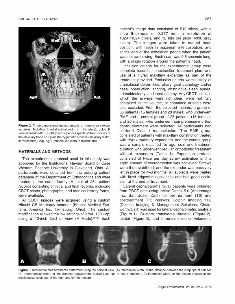
Lateral cephalograms for all patients were obtainedfrom CBCT data using InVivo Dental 5.0 (AnatomageInc, San Jose, Calif) for pretreatment (T0) andposttreatment (T1) intervals. Dolphin Imaging 11.0(Dolphin Imaging & Management Solutions, Chats-worth, Calif) was used for lateral cephalometric analysis(Figure 1). Custom transverse skeletal (Figure 2),dental (Figure 3), and three-dimensional volumetric
Figure 2. Three-dimensional measurements of transverse skeletal
variables. MoL-Mor (medial orbital width in millimeters), LnL-LnR
(lateral nasal width), JL-JR (most superior aspects of the concavity of
the maxillary bone as it joins the zygomatic process (maxillary width)
in millimeters), AgL-AgR (mandibular width in millimeters).
Figure 3. Interdental measurements performed using the coronal view. (A) Intercanine width, or the distance between the cusp tips of canines
(B) Interpremolar width, or the distance between the buccal cusp tips of first premolars. (C) Intermolar width, or the distance between the
mesiobuccal cusp tips of the right and left first molars.
RME AND THE 3D AIRWAY 267
Angle Orthodontist, Vol 84, No 2, 2014

(Figure 4) measurements were performed using InVivoDental 5.0. Mesiobuccal cusp tips of first molars, buccalcusp tips of first premolars, and cusp tip of canines wereused for interdental measurements (Figure 3). Thesuperior limit of the OP airway is the palatal plane(ANS-PNS), extending to the posterior wall of thepharynx, and the inferior limit is a line parallel to thepalatal plane, touching the most anteroinferior point ofthe second cervical vertebrae. The superior limit of theNP airway is the last slice before the nasal septum fuseswith the posterior wall of the pharynx, viewed on theaxial slice first and then projected to the sagittal view.The inferior limit is the palatal plane (Figure 4).22 Twoconstriction measurements also calculated were theposterior airway space (PAS), defined as the mostconstricted space behind the base of the tongue andlimited by soft tissues, and the minAx, defined as therepresentation of PAS on the axial slice (Figure 4). Allmeasurements were completed by an experiencedorthodontist.
To estimate operator reliability, 15 randomly selectedrecords were reevaluated after a week of preliminary datacollection. The error for linear and angular measurementswas measured using the Dahlberg formula30 as follows:ffiffiffiffiffiffiffiffiffiffiffiffiP
d2
2n
r
Independent sample t-tests were used to validate theconsistency of both groups at T0 in terms of age and tocompare the outcome variables at T1. Paired sample t-tests were used to compare changes from T0 to T1. TheSPSS Statistics 17.0 (SPSS Inc, Chicago, Ill) softwarewas used for all statistical analyses.
RESULTS
Reliability results showed an error of 0.47 mm,2.85 mm2, and 0.50u for linear, areal, and angularvariables, respectively, and an intraclass correlation of0.93 for the OP and 0.89 for the NP volumes. The
Figure 4. Superior and inferior limits of OP and NP volume, PAS, and minAx (area of the most constricted region at the base of the tongue). The
circle on the left illustrates the last axial slice before the nasal septum fuses with the posterior wall of the pharynx. In the center image, the top line
represents the mentioned axial slice on the sagittal view (superior border of NP). In the lower right image, the area in evidence represents the
PAS on the axial view; pp (line passing from palatal plane), 2cv (line passing from the most anteroinferior aspect of the second cervical vertebrae
and parallel to pp).
268 EL, PALOMO
Angle Orthodontist, Vol 84, No 2, 2014

Kolmogorov–Smirnov test was applied and showedthat all data were normally distributed.
Table 1 shows the sample demographics and confirmsthat both groups were matched on age, sex, andtreatment duration. Table 2 shows all variables separatefor males and females, showing the males having largervalues, probably because of size differences.
All of the transverse skeletal (medial orbital width,lateral nasal width, maxillary width and mandibular width)and interdental (intermolar, interpremolar, and interca-nine) parameters were enlarged significantly in the RMEgroup, whereas significant enlargement was also ob-served for all interdental variables and for lateral nasalwidth, maxillary width, and mandibular width for thecontrol group. A statistically significant increase for airwayvariables was also evident in both groups between T0and T1, which can probably be attributed to growth.
Table 3 shows no skeletal transverse differencesbetween the groups at either T0 or T1, but maxillaryvalues in the control group were larger at T0 andsmaller at T1. The dental transverse measurementsshow that the RME group was significantly smaller atT0 and transversely normalized after expansion; therewere no significant differences at T1.
Table 4 shows that the increases in lateral nasalwidth and maxillary width were significantly higher in theRME group. This was also true for the interdentalvariables. Although both groups displayed a significantincrease for OP and NP airway, only NP airway showeda significant difference between the groups. The meanincrease of NP airway volume in the RME group (1719.96 1510.7 mm3) was twofold compared with the controlgroup (813.6 6 1006.7 mm3) (Table 4).
No significant difference was observed between thegroups for PAS, minAx, and OP and NP airway volumeat T0 and for PAS, minAx, and OP airway volume atT1. At T1 the mean NP airway volume was significantlylarger in the RME group (Table 5).
DISCUSSION
Skeletal and dental effects of RME have beenextensively researched in the literature. The currentstudy analyzed three-dimensional skeletal and dentalchanges after RME treatment in teenage subjects. Thepresent study used images taken before and aftercomprehensive orthodontic treatment in order toanalyze the effects of a Hyrax maxillary expander.Given that all experimental subjects had orthodontictreatment in addition to RME, treatment may also haveplayed a significant role in the results seen. To factorout changes introduced by comprehensive orthodontictreatment, a matched control sample was used.
Weissheimer et al.31 compared the immediateeffects of RME in the transverse plane using CBCT
and reported an increase in all maxillary transversedimensions, which coincides with the findings of thepresent study. A systematic review by Lagravere etal.32 concluded that RME did not produce clinicallysignificant anteroposterior or vertical changes in thelong term compared with results for control subjects.Although in the present study there were significantchanges in the skeletal parameters for both RME andcontrol subjects, the intergroup comparison did notreveal any significant differences, which is in agree-ment with the reported systematic review.
The immediate and long-term effects of RME over theupper airway have been shown in previous stud-ies.9,18,27,28,32,33 The literature shows that patients pre-senting with maxillary constriction tend to have a highernasal airway resistance.34 The present study does notshow a difference in nasal air passage volume at T0, butthis may be due to the fact that patients with clinicallynormal respiratory functions for selected for both groups.The maxilla forms most of the lateral walls of the nasalcavity; therefore, an increase in volume in the nasalcavity would be an expected RME effect. The series ofevents that cause this phenomenon is mainly thetriangular11,35 or parallel36 opening of the median palatalsuture, which increases the width of the nasal floor andresults in an increased volume of the nasal cavity. Thepresent study shows a twofold increase in the NPvolume after RME. This finding suggests that RME maybe able to improve the breathing pattern by reducingnasal resistance, but further studies are needed toconfirm such anatomical and functional correlations.
Farronato et al.7 reported that the mandibularposition changes in various directions when RME isapplied to patients with different malocclusions. Thesedifferences in mandibular position may affect the OPairway size, shape, and volume.37 The present studydid not find significant changes in mandibular positionbetween the RME and control groups.
Zhao et al.28 assessed the changes of the OP airwayon 24 patients with maxillary constriction treated withRME and compared them to 24 age- and sex-matchedpatients and found no significant increase. Theyconcluded that RME would not enlarge OP airwayvolume. Malkoc et al.29 evaluated the effects ofmandibular symphyseal distraction osteogenesis fol-lowed by RME on pharyngeal size and concluded thatRME did not significantly affect the pharyngeal dimen-sions. The present study confirms such findings and alsofound no effect on pharyngeal airway when using RME.
CONCLUSION
N Rapid maxillary expansion creates a significant in-crease in nasal passage volume, but no significantchange is observed in the oropharyngeal airway region.
RME AND THE 3D AIRWAY 269
Angle Orthodontist, Vol 84, No 2, 2014
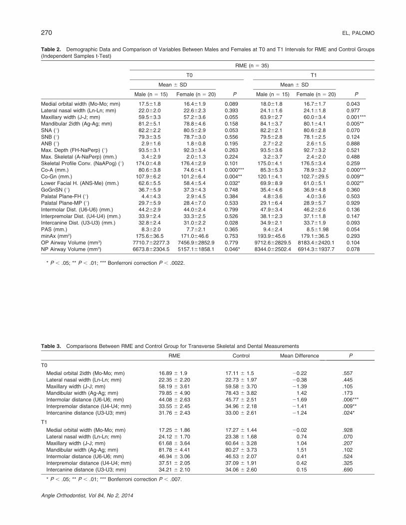
Table 2. Demographic Data and Comparison of Variables Between Males and Females at T0 and T1 Intervals for RME and Control Groups
(Independent Samples t-Test)
RME (n 5 35)
T0 T1
Mean 6 SD
P
Mean 6 SD
PMale (n 5 15) Female (n 5 20) Male (n 5 15) Female (n 5 20)
Medial orbital width (Mo-Mo; mm) 17.561.8 16.461.9 0.089 18.061.8 16.761.7 0.043
Lateral nasal width (Ln-Ln; mm) 22.062.0 22.662.3 0.393 24.161.6 24.161.8 0.977
Maxillary width (J-J; mm) 59.563.3 57.263.6 0.055 63.962.7 60.063.4 0.001***
Mandibular 2idth (Ag-Ag; mm) 81.265.1 78.864.6 0.158 84.163.7 80.164.1 0.005**
SNA (u) 82.262.2 80.562.9 0.053 82.262.1 80.662.8 0.070
SNB (u) 79.363.5 78.763.0 0.556 79.562.8 78.162.5 0.124
ANB (u) 2.961.6 1.860.8 0.195 2.762.2 2.661.5 0.888
Max. Depth (FH-NaPerp) (u) 93.563.1 92.363.4 0.263 93.563.6 92.763.2 0.521
Max. Skeletal (A-NaPerp) (mm.) 3.462.9 2.061.3 0.224 3.263.7 2.462.0 0.488
Skeletal Profile Conv. (NaAPog) (u) 174.064.8 176.462.9 0.101 175.064.1 176.563.4 0.259
Co-A (mm.) 80.663.8 74.664.1 0.000*** 85.365.3 78.963.2 0.000***
Co-Gn (mm.) 107.966.2 101.266.4 0.004** 120.164.1 102.7629.5 0.009**
Lower Facial H. (ANS-Me) (mm.) 62.665.5 58.465.4 0.032* 69.968.9 61.065.1 0.002**
GoGnSN (u) 36.765.9 37.364.3 0.748 35.464.6 36.964.8 0.360
Palatal Plane-FH (u) 4.464.3 2.964.5 0.384 4.863.6 4.063.6 0.503
Palatal Plane-MP (u) 29.765.9 28.467.0 0.533 29.166.4 28.965.7 0.929
Intermolar Dist. (U6-U6) (mm.) 44.262.9 44.062.4 0.799 47.963.4 46.262.6 0.136
Interpremolar Dist. (U4-U4) (mm.) 33.962.4 33.362.5 0.526 38.162.3 37.161.8 0.147
Intercanine Dist. (U3-U3) (mm.) 32.862.4 31.062.2 0.028 34.962.1 33.761.9 0.093
PAS (mm.) 8.362.0 7.762.1 0.365 9.462.4 8.561.98 0.054
minAx (mm2) 175.6636.5 171.0646.6 0.753 193.9645.6 179.1636.5 0.293
OP Airway Volume (mm3) 7710.762277.3 7456.962852.9 0.779 9712.662829.5 8183.462420.1 0.104
NP Airway Volume (mm3) 6673.862304.5 5157.161858.1 0.046* 8344.062502.4 6914.361937.7 0.078
* P , .05; ** P , .01; *** Bonferroni correction P , .0022.
Table 3. Comparisons Between RME and Control Group for Transverse Skeletal and Dental Measurements
RME Control Mean Difference P
T0
Medial orbital 2idth (Mo-Mo; mm) 16.89 6 1.9 17.11 6 1.5 20.22 .557
Lateral nasal width (Ln-Ln; mm) 22.35 6 2.20 22.73 6 1.97 20.38 .445
Maxillary width (J-J; mm) 58.19 6 3.61 59.58 6 3.70 21.39 .105
Mandibular width (Ag-Ag; mm) 79.85 6 4.90 78.43 6 3.82 1.42 .173
Intermolar distance (U6-U6; mm) 44.08 6 2.63 45.77 6 2.51 21.69 .006***
Interpremolar distance (U4-U4; mm) 33.55 6 2.45 34.96 6 2.18 21.41 .009**
Intercanine distance (U3-U3; mm) 31.76 6 2.43 33.00 6 2.61 21.24 .024*
T1
Medial orbital width (Mo-Mo; mm) 17.25 6 1.86 17.27 6 1.44 20.02 .928
Lateral nasal width (Ln-Ln; mm) 24.12 6 1.70 23.38 6 1.68 0.74 .070
Maxillary width (J-J; mm) 61.68 6 3.64 60.64 6 3.28 1.04 .207
Mandibular width (Ag-Ag; mm) 81.78 6 4.41 80.27 6 3.73 1.51 .102
Intermolar distance (U6-U6; mm) 46.94 6 3.06 46.53 6 2.07 0.41 .524
Interpremolar distance (U4-U4; mm) 37.51 6 2.05 37.09 6 1.91 0.42 .325
Intercanine distance (U3-U3; mm) 34.21 6 2.10 34.06 6 2.60 0.15 .690
* P , .05; ** P , .01; *** Bonferroni correction P , .007.
270 EL, PALOMO
Angle Orthodontist, Vol 84, No 2, 2014

Control (n 5 35)
T0 T1
Mean 6 SD
P
Mean 6 SD
PMale (n 5 15) Female (n 5 20) Male (n 5 15) Female (n 5 20)
17.261.4 17.061.5 0.684 17.561.6 17.161.3 0.500
22.861.7 22.762.2 0.854 23.361.1 23.562.0 0.678
60.863.5 58.763.5 0.073 62.363.3 59.462.6 0.011*
79.163.3 77.964.0 0.334 81.563.0 79.263.8 0.051
82.362.5 82.863.9 0.628 82.663.2 81.167.0 0.380
79.663.0 79.664.1 0.998 79.764.1 79.363.6 0.767
2.762.1 3.261.2 0.395 2.961.8 3.261.3 0.631
92.963.5 93.163.9 0.824 92.463.8 93.663.5 0.317
2.762.6 2.962.8 0.841 1.461.5 2.562.4 0.339
173.564.0 174.163.3 0.613 173.764.8 174.263.3 0.728
81.866.2 81.065.6 0.603 84.264.5 83.167.5 0.576
105.666.2 105.866.9 0.918 111.565.9 110.668.0 0.689
59.664.9 58.765.3 0.588 64.664.1 62.466.0 0.189
32.566.3 33.565.8 0.651 33.266.9 33.965.3 0.752
1.462.6 0.962.3 0.738 1.161.9 1.762.4 0.637
23.864.9 23.965.1 0.935 25.164.3 25.265.6 0.945
47.162.3 44.862.2 0.005** 47.862.2 45.661.3 0.001***
36.262.1 34.262.0 0.008** 38.161.8 36.261.5 0.002**
34.362.8 32.362.2 0.021* 35.562.4 32.962.0 0.002**
7.662.3 8.262.8 0.478 9.062.3 9.263.6 0.838
163.6645.7 171.8646.9 0.608 182.9643.3 178.6648.9 0.788
7598.262190.4 8400.462971.7 0.351 9486.662689.7 9389.563163.5 0.932
5573.762216.3 5648.461623.8 0.910 6736.262451.4 6196.162085.2 0.485
Table 2. Extended.
Table 4. Mean Differences Between T0 and T1 Intervals
RME Control RME vs Control
D Mean 6 SD (T1-T0) P D Mean 6 SD (T1-T0) P P
Medial orbital width (Mo-Mo; mm) 0.4 6 0.3 .022* 0.2 6 0.7 .172 .293
Lateral nasal width (Ln-Ln; mm) 1.8 6 1.2 .000*** 0.7 6 1.7 .024* .003**
Maxillary width (J-J; mm) 3.5 6 2.0 .000*** 1.1 6 2.4 .011* .000***
Mandibular width (Ag-Ag; mm) 1.9 6 1.6 .000*** 1.8 6 2.1 .000*** .878
SNA (u) 0 6 1.6 .966 20.8 6 5.6 .362 .327
SNB (u) 20.3 6 1.4 .235 20.1 6 2.6 .790 .632
ANB (u) 0.3 6 1.4 .200 0.1 6 1.3 .688 .233
Maxillary depth (FH-NaPerp) (u) 0 6 2.6 .959 0.1 6 2.7 .811 .721
Maxillary skeletal (A-NaPerp) (u) 0.1 6 2.5 .846 20.8 6 2.5 .056 .090
Skeletal profile convexity (NaAPog) (u) 0.5 6 2.4 .238 0.1 6 2.8 .770 .853
Co-A (mm) 4.9 6 4.4 .238 5.3 6 5.1 .000*** .053
Co-Gn (mm) 4.4 6 3.6 .000*** 2.3 6 2.1 .002*** .406
Lower facial height (ANS-Me; mm) 4.6 6 4.2 .000*** 4.3 6 2.5 .000*** .805
GoGnSN (u) 20.8 6 3.3 .158 0.5 6 3.6 .397 .140
Palatal plane-facial height (u) 0.8 6 3.1 .128 0.3 6 3.9 .664 .420
Palatal plane-mandibular plane (u) 0.1 6 4.2 .937 1.3 6 3.2 .016* .221
Intermolar distance (U6-U6; mm) 2.9 6 1.9 .000*** 0.8 6 1.3 .001** .000***
Interpremolar distance (U4-U4; mm) 4.0 6 1.9 .000*** 2.0 6 1.7 .000*** .000***
Intercanine distance (U3-U3; mm) 2.4 6 1.6 .000*** 0.8 6 2.3 .031* .006**
PAS (mm) 0.87 6 1.42 .001*** 1.15 6 2.12 .003** .518
minAx (mm2) 12.46 6 10.45 .021* 12.16 6 16.06 .044* .970
OP airway volume (mm3) 1273.1 6 1675.9 .000*** 1447.7 6 2464.4 .001*** .730
NP airway volume (mm3) 1719.9 6 1510.7 .000*** 833.1 6 1032.2 .000*** .006**
* P , .05; ** P , .01; *** Bonferroni correction P , .0022.
RME AND THE 3D AIRWAY 271
Angle Orthodontist, Vol 84, No 2, 2014

REFERENCES
1. Angell EC. Treatment of irregularities of the permanent oradult teeth. Dental Cosmos. 1860;1:540–544.
2. Haas AJ. Rapid expansion of the maxillary dental arch andnasal cavity by opening the mid palatal suture. AngleOrthod. 1961;31:73–89.
3. Haas AJ. The treatment of maxillary deficiency by openingthe midpalatal suture. Angle Orthod. 1965;35:200–217.
4. Wertz RA. Changes in nasal airflow incident to rapidmaxillary expansion. Angle Orthod. 1968;38:1–11.
5. Johal A, Conaghan C. Maxillary morphology in obstructivesleep apnea: a cephalometric and model study. AngleOrthod. 2004;74:648–656.
6. Schmidt-Nowara W, Lowe A, Wiegand L, Cartwright R,Perez-Guerra F, Menn S. Oral appliances for the treatmentof snoring and obstructive sleep apnea: a review. Sleep.1995;18:501–510.
7. Farronato G, Giannini L, Galbiati G, Maspero C. Sagittal andvertical effects of rapid maxillary expansion in Class I, II, andIII occlusions. Angle Orthod. 2011;81:298–303.
8. Chung CH, Font B. Skeletal and dental changes in the sagittal,vertical, and transverse dimensions after rapid palatal expan-sion. Am J Orthod Dentofacial Orthop. 2004;126:569–575.
9. Kurt G, Altug-Atac AT, Atac MS, Karasu HA. Changes innasopharyngeal airway following orthopedic and surgicallyassisted rapid maxillary expansion. J Craniofac Surg. 2010;21:312–317.
10. Buccheri A, Dilella G, Stella R. Rapid palatal expansion andpharyngeal space. Cephalometric evaluation. Prog Orthod.2004;5:160–171.
11. Basciftci FA, Mutlu N, Karaman AI, Malkoc S, KucukkolbasiH. Does the timing and method of rapid maxillary expansionhave an effect on the changes in nasal dimensions? AngleOrthod. 2002;72:118–123.
12. Altug-Atac AT, Atac MS, Kurt G, Karasud HA. Changes innasal structures following orthopaedic and surgically assist-ed rapid maxillary expansion. Int J Oral Maxillofac Surg.2010;39:129–135.
13. Halicioglu K, Kilic N, Yavuz I, Aktan B. Effects of rapidmaxillary expansion with a memory palatal split screw on themorphology of the maxillary dental arch and nasal airwayresistance. Eur J Orthod. 2010;32:716–720.
14. Bicakci AA, Agar U, Sokucu O, Babacan H, Doruk C. Nasalairway changes due to rapid maxillary expansion timing.Angle Orthod. 2005;75:1–6.
15. Babacan H, Sokucu O, Doruk C, Ay S. Rapid maxillaryexpansion and surgically assisted rapid maxillary expansioneffects on nasal volume. Angle Orthod. 2006;76:66–71.
16. Gordon JM, Rosenblatt M, Witmans M, et al. Rapid palatalexpansion effects on nasal airway dimensions as measuredby acoustic rhinometry. A systematic review. Angle Orthod.2009;79:1000–1007.
17. Deeb W, Hansen L, Hotan T, Hietschold V, Harzer W,Tausche E. Changes in nasal volume after surgically assistedbone-borne rapid maxillary expansion. Am J Orthod Dento-facial Orthop. 2010;137:782–789.
18. Haralambidis A, Ari-Demirkaya A, Acar A, Kucukkeles N,Ates M, Ozkaya S. Morphologic changes of the nasal cavityinduced by rapid maxillary expansion: a study on 3-dimensional computed tomography models. Am J OrthodDentofacial Orthop. 2009;136:815–821.
19. Habersack K, Karoglan A, Sommer B, Benner KU. High-resolution multislice computerized tomography with multi-planar and 3-dimensional reformation imaging in rapidpalatal expansion. Am J Orthod Dentofacial Orthop. 2007;131:776–781.
20. Tausche E, Deeb W, Hansen L, Hietschold V, Harzer W,Schneider M. CT analysis of nasal volume changes aftersurgically-assisted rapid maxillary expansion. J OrofacOrthop. 2009;70:306–317.
21. Palomo JM, Rao PS, Hans MG. Influence of CBCTexposure conditions on radiation dose. Oral Surg OralMed Oral Pathol Oral Radiol Endod. 2008;105:773–782.
22. El H, Palomo JM. Measuring the airway in 3 dimensions: areliability and accuracy study. Am J Orthod DentofacialOrthop. 2010;137(4 suppl):S50e51–59.
23. Kwong JC, Palomo JM, Landers MA, Figueroa A, Hans MG.Image quality produced by different cone-beam computedtomography settings. Am J Orthod Dentofacial Orthop.2008;133:317–327.
24. Osorio F, Perilla M, Doyle DJ, Palomo JM. Cone beamcomputed tomography: an innovative tool for airwayassessment. Anesth Analg. 2008;106:1803–1807.
25. Gohl E, Nguyen M, Enciso R. Three-dimensional computedtomography comparison of the maxillary palatal vaultbetween patients with rapid palatal expansion and ortho-dontically treated controls. Am J Orthod Dentofacial Orthop.2010;138:477–485.
26. Christie KF, Boucher N, Chung CH. Effects of bonded rapidpalatal expansion on the transverse dimensions of the maxilla:a cone-beam computed tomography study. Am J OrthodDentofacial Orthop. 2010;137(4 suppl):S79–S85.
27. Gorgulu S, Gokce SM, Olmez H, Sagdic D, Ors F. Nasalcavity volume changes after rapid maxillary expansion inadolescents evaluated with 3-dimensional simulation andmodeling programs. Am J Orthod Dentofacial Orthop. 2011;140:633–640.
Table 5. Mean OP and NP Volumes at T0 and T1 Intervals
RME Control Mean Difference P
T0
PAS (mm) 8.00 6 2.05 7.98 6 2.60 0.02 .999
minAx (mm2) 172.98 6 42.04 168.33 6 45.88 4.65 .660
OP volume (mm3) 7565.7 6 2588.5 8053.5 6 2658.5 2487.8 .400
NP volume (mm3) 5807.1 6 2167.0 5616.1 6 1874.3 191.0 .648
T1
PAS (mm) 8.95 6 2.28 9.14 6 3.10 0.19 .768
minAx (mm2) 185.44 6 40.69 180.48 6 45.96 4.96 .635
OP volume (mm3) 8838.8 6 2675.7 9431.5 6 3556.3 2592.7 .425
NP volume (mm3) 7527.0 6 2278.6 6429.6 6 2234.5 1097.4 .043*
* P , .05; ** P , .01; *** P , .001.
272 EL, PALOMO
Angle Orthodontist, Vol 84, No 2, 2014

28. Zhao Y, Nguyen M, Gohl E, Mah JK, Sameshima G,Enciso R. Oropharyngeal airway changes after rapidpalatal expansion evaluated with cone-beam computedtomography. Am J Orthod Dentofacial Orthop. 2010;137:S71–S78.
29. Malkoc S, Usumez S, Iseri H. Long-term effects of symphy-seal distraction and rapid maxillary expansion on pharyngealairway dimensions, tongue, and hyoid position. Am J OrthodDentofacial Orthop. 2007;132:769–775.
30. Dahlberg G. Statistical Method for Medical and BiologicalStudents. New York, NY: Interscience Publications; 1940.
31. Weissheimer A, de Menezes LM, Mezomo M, Dias DM, deLima EM, Rizzatto SM. Immediate effects of rapid maxillaryexpansion with Haas-type and hyrax-type expanders: arandomized clinical trial. Am J Orthod Dentofacial Orthop.2011;140:366–376.
32. Lagravere MO, Heo G, Major PW, Flores-Mir C. Meta-analysis of immediate changes with rapid maxillary expan-sion treatment. J Am Dent Assoc. 2006;137:44–53.
33. Baratieri C, Alves M Jr, de Souza MM, de Souza Araujo MT,Maia LC. Does rapid maxillary expansion have long-term
effects on airway dimensions and breathing? Am J OrthodDentofacial Orthop. 2011;140:146–156.
34. Cistulli PA, Sullivan CE. Influence of maxillary morphologyon nasal airway resistance in Marfan’s syndrome. ActaOtolaryngol. 2000;120:410–413.
35. Timms DJ. A study of basal movement with rapid maxillaryexpansion. Am J Orthod. 1980;77:500–507.
36. Ballanti F, Lione R, Baccetti T, Franchi L, Cozza P.Treatment and posttreatment skeletal effects of rapidmaxillary expansion investigated with low-dose computedtomography in growing subjects. Am J Orthod DentofacialOrthop. 2010;138:311–317.
37. Kim YJ, Hong JS, Hwang YI, Park YH. Three-dimensionalanalysis of pharyngeal airway in preadolescent children withdifferent anteroposterior skeletal patterns. Am J Orthod Dento-facial Orthop. 2010;137:306. e301–e311; discussion 306–307.
38. El H, Palomo JM. Airway volume for different dentofacialskeletal patterns. Am J Orthod Dentofacial Orthop. 2011;139:e511–e21.
39. El H, Palomo JM. An airway study of different maxillary andmandibular sagittal positions. Eur J Orthod. 2013;35:262–270.
RME AND THE 3D AIRWAY 273
Angle Orthodontist, Vol 84, No 2, 2014