three-dimensional domain swapping as a mechanism to lock the active conformation in a super-active...
TRANSCRIPT

RESEARCH ARTICLE
Three-dimensional domain swapping as amechanism to lock the active conformation ina super-active octamer of SARS-CoV mainprotease
Shengnan Zhang1,2*, Nan Zhong1,2*, Fei Xue3*, Xue Kang1,2, Xiaobai Ren1,2, Jiaxuan Chen1,4,Changwen Jin1,2,4, Zhiyong Lou3✉, Bin Xia1,2,4✉
1 Beijing Nuclear Magnetic Resonance Center, Peking University, Beijing 100871, China2 College of Chemistry and Molecular Engineering, Peking University, Beijing 100871, China3 Structural Biology Laboratory, Tsinghua University, Beijing 100084, China4 College of Life Sciences, Peking University, Beijing 100871, China✉ Correspondence: [email protected] (B. Xia), [email protected]. edu. cn (Z. Lou)Received February 26, 2010 Accepted March 18, 2010
ABSTRACT
Proteolytic processing of viral polyproteins is indispen-sible for the lifecycle of coronaviruses. The mainprotease (Mpro) of SARS-CoV is an attractive target foranti-SARS drug development as it is essential for thepolyprotein processing. Mpro is initially produced as partof viral polyproteins and it is matured by autocleavage.Here, we report that, with the addition of an N-terminalextension peptide, Mpro can form a domain-swappeddimer. After complete removal of the extension peptidefrom the dimer, the mature Mpro self-assembles into anovel super-active octamer (AO-Mpro). The crystal struc-ture of AO-Mpro adopts a novel fold with four domain-swapped dimers packing into four active units withnearly identical conformation to that of the previouslyreported Mpro active dimer, and 3D domain swappingserves as a mechanism to lock the active conformationdue to entanglement of polypeptide chains. Comparedwith the previously well characterized form of Mpro, inequilibrium between inactive monomer and active dimer,the stable AO-Mpro exhibits much higher proteolyticactivity at low concentration. As all eight active sitesare bound with inhibitors, the polyvalent nature of theinteraction between AO-Mpro and its polyprotein sub-strates with multiple cleavage sites, would make AO-Mpro
functionally much more superior than the Mpro active
dimer for polyprotein processing. Thus, during the initialperiod of SARS-CoV infection, this novel active form AO-Mpro should play a major role in cleaving polyproteins asthe protein level is extremely low. The discovery of AO-Mpro provides new insights about the functional mechan-ism of Mpro and its maturation process.
KEYWORDS SARS-CoV, main protease, crystal struc-ture, 3D domain swapping, polyprotein processing
INTRODUCTION
SARS coronavirus (SARS-CoV) was identified as theetiological agent of the pandemic transmissible diseasesevere acute respiratory syndrome (SARS). During theoutbreak in 2003, SARS-CoV infected more than 8400people worldwide with a high fatality rate of about 10%(Chan et al., 2003; Kuiken et al., 2003; Leng and Bentwich,2003). SARS-CoV is a positive-sense, single-stranded RNAvirus, with the 5¢ two-third genome of the virus encoding twooverlapping polyproteins pp1a (486 kDa) and pp1ab(790 kDa). During infection, the two polyproteins are proteo-lytically processed into 16 nonstructural proteins (nsp1–16)required for viral replication and transcription (Snijder et al.,2003). The proteolytic process is mediated by two virus-encoded proteinases, a papain-like proteinase (nsp3) and a3C-like proteinase (nsp5) (Snijder et al., 2003). The 3C-like
*These authors contributed equally to the work.
© Higher Education Press and Springer-Verlag Berlin Heidelberg 2010 371
Protein Cell 2010, 1(4): 371–383DOI 10.1007/s13238-010-0044-8
Protein & Cell
proteinase, also known as the main protease (Mpro), ismatured from polyproteins through autocleavage and furthercleaves all 11 downstream nonstructural proteins. This makesMpro essential for the viral life cycle, and thus an attractivetarget for anti-SARS drug development (Anand et al., 2003;Yang et al., 2003, 2005).
It has been reported that SARS-CoV Mpro exists in solutionas an equilibrium between monomeric and dimeric forms(Hsu et al., 2005; Graziano et al., 2006a, b), and only thedimeric form is enzymatically active (Fan et al., 2004; Chenet al., 2005, 2006; Shi and Song, 2006; Chen et al., 2008; Linet al., 2008; Shi et al., 2008; Hu et al., 2009). Crystalstructures of SARS-CoV Mpro revealed a symmetric homo-dimer which has a similar fold to other reported coronavirusMpro structures (Anand et al., 2002, 2003; Yang et al., 2003;Xue et al., 2007). Each protomer in the dimeric structurecontains an N-terminal domain (residues 1–184) with achymotrypsin-like fold and a unique C-terminal domain(residues 201–303) containing five α-helices. The substratebinding pocket is located in a cleft of the Mpro N-terminaldomain with the catalytic dyad consisting of residues His41and Cys145 (Yang et al., 2003). The N-terminal residues 1–7(N-finger) of each protomer are squeezed in between the twoprotomers and make contacts with both the N-terminal and C-terminal domain of the other protomer (Yang et al., 2003).Therefore, many studies focused on elucidating the roles ofN-finger and the C-terminal domain in the dimerization of Mpro
(Shi et al., 2004; Chen et al., 2005; Wei et al., 2006; Zhong etal., 2008; Hu et al., 2009; Zhong et al., 2009).
We have demonstrated that the N-finger is not only criticalfor the dimerization of SARS-CoV Mpro, but also essential forit to form the right quaternary structure which is theenzymatically active form (Zhong et al., 2008). The Mpro C-terminal domain alone (Mpro-C) is expressed in E. coli as astable monomer and a stable dimer in solution, with noapparent exchange (Zhong et al., 2009). The stable dimeradopts a novel fold which is characterized by 3D domainswapping with two helices of the two molecules interchangetheir positions. The N-finger deletion mutant of Mpro (Mpro-Δ7)can also form a stable dimer due to 3D domain swapping of itsC-terminal domain. However, it is not clear whether 3Ddomain-swapping plays a role in wild-type Mpro.
Three-dimensional domain swapping is a special way ofprotein association to generate stable multimers, by whichidentical domains from different molecules exchange theirpositions (Liu and Eisenberg, 2002; Gronenborn, 2009). Inthis way, the core tertiary structures of the domain-swappedmultimers are similar to those of monomers. Up to date,domain-swapped structures of more than 60 proteins areavailable in the Protein Data Bank. The biologic roles of 3Ddomain swapping have been widely discussed and somefunctions have been elucidated. 3D domain swapping isrecognized as a general mechanism for the formation ofamyloid fibers which cause conformational diseases
(Yamasaki et al., 2008). HIV-1 capsid dimerization via 3Ddomain swapping is proposed to be a critical event inimmature viral particle assembly (Ivanov et al., 2007). Bovinepancreatic ribonuclease A can oligomerize via 3D domainswapping, and acquires novel biologic functions such asantitumor activity (Libonati et al., 2008).
Here, we report that fusion of an extension peptide to the Nterminus of Mpro can also result in the formation of a 3Ddomain-swapped dimer. After proteolytic removal of theextension peptide, we discovered the existence of apreviously unknown active form of mature Mpro, which is astable homo-octamer self-assembled by four domainswapped dimers. Each protomer of the active octamer hasexactly the same sequence as the previously well character-ized matured form of Mpro. Crystal structure reveals that, dueto 3D domain swapping, the active conformation is locked inthe octamer by forming four active units, each adopting thesame conformation as the Mpro active dimer. At low proteinconcentration, the proteolytic activity of the Mpro activeoctamer is much higher than that of Mpro active dimer whichis in equilibrium with an inactive monomer.
RESULTS
Mpro can form a 3D domain-swapped dimer with N-terminal extension peptides
It has been pointed out that extra residues to the N terminus ofmature Mpro result in lower dimer ratio and enzymatic activity(Xue et al., 2007). We constructed an N-terminal extensionmutant of Mpro (Mpro-NE) by fusing a 26-residue peptide to theN terminus of Mpro, with an enterokinase cleavage site right infront of the N terminus.
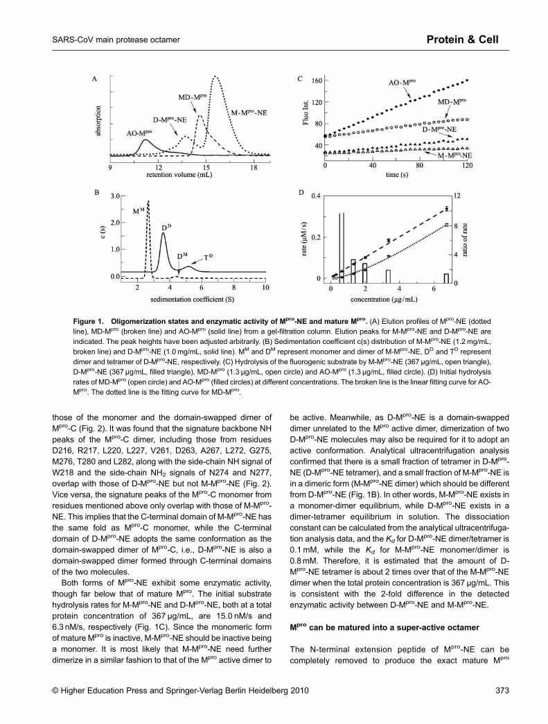
Mpro-NE produced in E. coli exists as two forms that can beseparated by gel-filtration, with retention volumes of 15.5 mLand 13.7 mL (Fig. 1A). The apparent molecular weightcalculated based on retention volume is 34.5 kDa for the15.5-mL fraction, and 71.1 kDa for the 13.7-mL fraction. Thetwo forms should correspond to a monomeric form (15.5-mLfraction, M-Mpro-NE) and a dimeric form (13.7-mL fraction, D-Mpro-NE) of Mpro-NE, as the theoretical molecular weights ofthe Mpro-NE monomer is 36.7 kDa. Both forms migrated on anSDS-PAGE gel at the same position as the Mpro-NEmonomer, with or without DTT, suggesting that the formationof D-Mpro-NE is not through a disulfide bond, even thoughMpro-NE has 12 free cysteine residues. Similar to the N-fingerdeletion mutant Mpro-∆7, no exchange between monomericand dimeric forms was observed for days.
Most of the NH peaks in 2D 1H-15N HSQC spectra of M-Mpro-NE and D-Mpro-NE overlap well, suggesting that mostparts of the structures are the same between the two forms.Using the same strategy as for characterizing the domain-swapped dimer of Mpro-∆7 (Zhong et al., 2008), we compared2D 1H-15N HSQC spectra of M-Mpro-NE and D-Mpro-NE with
372 © Higher Education Press and Springer-Verlag Berlin Heidelberg 2010
Shengnan Zhang et al.Protein & Cell
those of the monomer and the domain-swapped dimer ofMpro-C (Fig. 2). It was found that the signature backbone NHpeaks of the Mpro-C dimer, including those from residuesD216, R217, L220, L227, V261, D263, A267, L272, G275,M276, T280 and L282, along with the side-chain NH signal ofW218 and the side-chain NH2 signals of N274 and N277,overlap with those of D-Mpro-NE but not M-Mpro-NE (Fig. 2).Vice versa, the signature peaks of the Mpro-C monomer fromresidues mentioned above only overlap with those of M-Mpro-NE. This implies that the C-terminal domain of M-Mpro-NE hasthe same fold as Mpro-C monomer, while the C-terminaldomain of D-Mpro-NE adopts the same conformation as thedomain-swapped dimer of Mpro-C, i.e., D-Mpro-NE is also adomain-swapped dimer formed through C-terminal domainsof the two molecules.
Both forms of Mpro-NE exhibit some enzymatic activity,though far below that of mature Mpro. The initial substratehydrolysis rates for M-Mpro-NE and D-Mpro-NE, both at a totalprotein concentration of 367 μg/mL, are 15.0 nM/s and6.3 nM/s, respectively (Fig. 1C). Since the monomeric formof mature Mpro is inactive, M-Mpro-NE should be inactive beinga monomer. It is most likely that M-Mpro-NE need furtherdimerize in a similar fashion to that of the Mpro active dimer to
be active. Meanwhile, as D-Mpro-NE is a domain-swappeddimer unrelated to the Mpro active dimer, dimerization of twoD-Mpro-NE molecules may also be required for it to adopt anactive conformation. Analytical ultracentrifugation analysisconfirmed that there is a small fraction of tetramer in D-Mpro-NE (D-Mpro-NE tetramer), and a small fraction of M-Mpro-NE isin a dimeric form (M-Mpro-NE dimer) which should be differentfrom D-Mpro-NE (Fig. 1B). In other words, M-Mpro-NE exists ina monomer-dimer equilibrium, while D-Mpro-NE exists in adimer-tetramer equilibrium in solution. The dissociationconstant can be calculated from the analytical ultracentrifuga-tion analysis data, and the Kd for D-M
pro-NE dimer/tetramer is0.1 mM, while the Kd for M-Mpro-NE monomer/dimer is0.8mM. Therefore, it is estimated that the amount of D-Mpro-NE tetramer is about 2 times over that of the M-Mpro-NEdimer when the total protein concentration is 367 μg/mL. Thisis consistent with the 2-fold difference in the detectedenzymatic activity between D-Mpro-NE and M-Mpro-NE.
Mpro can be matured into a super-active octamer
The N-terminal extension peptide of Mpro-NE can becompletely removed to produce the exact mature Mpro
Figure 1. Oligomerization states and enzymatic activity of Mpro-NE and mature Mpro. (A) Elution profiles of Mpro-NE (dottedline), MD-Mpro (broken line) and AO-Mpro (solid line) from a gel-filtration column. Elution peaks for M-Mpro-NE and D-Mpro-NE areindicated. The peak heights have been adjusted arbitrarily. (B) Sedimentation coefficient c(s) distribution of M-Mpro-NE (1.2 mg/mL,
broken line) and D-Mpro-NE (1.0 mg/mL, solid line). MM and DM represent monomer and dimer of M-Mpro-NE, DD and TD representdimer and tetramer of D-Mpro-NE, respectively. (C) Hydrolysis of the fluorogenic substrate by M-Mpro-NE (367 μg/mL, open triangle),D-Mpro-NE (367 μg/mL, filled triangle), MD-Mpro (1.3 μg/mL, open circle) and AO-Mpro (1.3 μg/mL, filled circle). (D) Initial hydrolysis
rates of MD-Mpro (open circle) and AO-Mpro (filled circles) at different concentrations. The broken line is the linear fitting curve for AO-Mpro. The dotted line is the fitting curve for MD-Mpro.
© Higher Education Press and Springer-Verlag Berlin Heidelberg 2010 373
SARS-CoV main protease octamer Protein & Cell

through enterokinase digestion. As expected, the retentionvolume of the mature Mpro from M-Mpro-NE showed anobvious concentration dependence on a gel filtration column,and its enzymatic activity is dependent on both the proteinconcentration and the dissociation constant (Fig. 1D). Similarmethods have been used in previous studies for generatingmature Mpro (Kuo et al., 2004; Xue et al., 2007; Verschuerenet al., 2008).
Surprisingly, a higher order oligomer is observed afterremoval of the N-terminal extension peptide from D-Mpro-NE,and its retention volume (11.2 mL) is not concentration-dependent, indicating the absence of exchange. The appar-ent molecular weight calculated based on this retentionvolume is 271 kDa, which is close to eight times thetheoretical molecular weight of Mpro (8 × 33.4 kDa), suggest-ing that this higher-order oligomer is a homo-octamer.
This newly identified Mpro octamer is also enzymaticallyactive. At a total protein concentration of 16.9 µg/mL, theinitial hydrolysis rate of Mpro octamer (0.92 μM/s) is compar-able to that of the previously well-studied form of mature Mpro
(0.94 μM/s) that is in equilibrium between an inactivemonomer and an active dimer (MD-Mpro). Therefore, we
have discovered a previously unknown form of mature Mpro,which is a novel active octamer (AO-Mpro). However, incontrast to MD-Mpro whose enzymatic activity is dependenton both the enzyme concentration and the dissociationconstant, the initial substrate hydrolysis rate of AO-Mpro isonly linearly dependent on protein concentration. When thetotal protein concentration is reduced, the activity of MD-Mpro
decreases much more significantly than that of AO-Mpro, andthe initial hydrolysis rate of AO-Mpro is about 11 timeshigher than that of MD-Mpro at a total protein concentrationof 0.68μg/mL. Furthermore, below a concentration of0.34 μg/mL, the enzymatic activity of MD-Mpro is notdetectable, while AO-Mpro still shows obvious hydrolysisactivity (Fig. 1D). Therefore, we discovered a novel super-active octameric form of mature Mpro.
Octameric Mpro is assembled due to 3D domain swapping
The crystal structure of AO-Mpro in complex with thepreviously reported Michael acceptor inhibitor N3 (Yanget al., 2005) was determined using the molecular replacement(MR) method and refined at 3.2-Å resolution with a final Rwork
Figure 2. Overlay of 2D 1H-15N HSQC spectra of monomeric and dimeric Mpro-NE and Mpro-C. Black peaks belong to D-Mpro-NE, green peaks belong to M-Mpro-NE, red peaks are from Mpro-C dimer, and blue peaks are from Mpro-C monomer. Signature NH
peaks of Mpro-C dimer are indicated by orange squares. Six areas of the spectra are enlarged and displayed for clarity, in whichsignature NH peaks of Mpro-C dimer are labeled with one-letter amino acid code and residue number.
374 © Higher Education Press and Springer-Verlag Berlin Heidelberg 2010
Shengnan Zhang et al.Protein & Cell
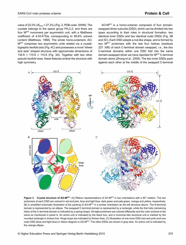
value of 22.0% (Rfree = 27.2%) (Fig. 3, PDB code: 3IWM). Thecrystals belongs to the space group P41212, and there arefour Mpro monomers per asymmetric unit, with a Matthewscoefficient of 4.04 Å3/Da, corresponding to 69.6% solventcontent (Matthews, 1968). The whole homo-octameric AO-Mpro comprises two asymmetric units related via a crystal-lographic twofold axis (Fig. 4C) and possesses a novel “wheeland axle” shaped structure with approximate dimensions of135 Å × 115 Å × 110 Å (Fig. 3A). Together with two otherpseudo twofold axes, these features endow the structure withhigh symmetry.
AO-Mpro is a homo-octamer composed of four domain-swapped dimer subunits (DSD), which can be divided into twotypes according to their roles in structural formation: twoidentical inner DSDs and two identical outer DSDs (Fig. 3Band 3C). Each DSD adopts a rod-like shape, and is formed bytwo Mpro protomers with the last four helices (residues227–306) of each C-terminal domain swapped, i.e., the twoC-terminal domains within one DSD fold into the samedomain-swapped dimer we have reported for Mpro C-terminaldomain alone (Zhong et al., 2009). The two inner DSDs packagainst each other at the middle of the swapped C-terminal
Figure 3. Crystal structure of AO-Mpro. (A) Ribbon representations of AO-Mpro in two orientations with a 90° rotation. The twoprotomers of each DSD are colored in red and pink, blue and light blue, dark green and pale green, orange and yellow, respectively.(B) A simplified schematic illustration of the packing of AO-Mpro in a similar orientation as the left structure above. The N-terminal
domain is represented by an ellipse. The swapped C-terminal domain is represented by a rectangle, while the first helix (remaininghelix) of the C-terminal domain is indicated by a spring shape. All eight protomers are colored differently and the color scheme is thesame as mentioned in panel A. An active unit is indicated by the black box, and a monomer-like structural unit is marked by therounded rectangle in broken line. Hinge loops are indicated by thicker lines. (C) Illustration of one inner DSD (red and pink) and one
outer DSD (blue and light blue) in ribbon diagram in AO-Mpro. The other DSDs are shown in gray dots. An active unit is indicated bythe orange ellipse.
© Higher Education Press and Springer-Verlag Berlin Heidelberg 2010 375
SARS-CoV main protease octamer Protein & Cell
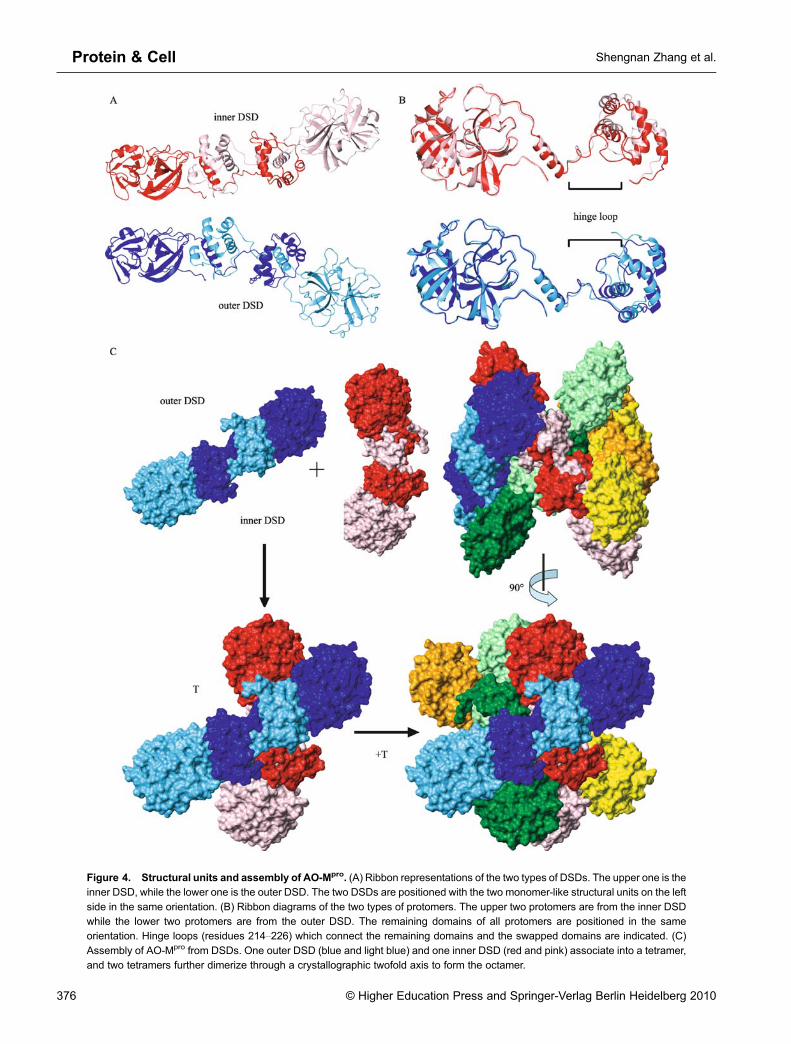
Figure 4. Structural units and assembly of AO-Mpro. (A) Ribbon representations of the two types of DSDs. The upper one is theinner DSD, while the lower one is the outer DSD. The two DSDs are positioned with the two monomer-like structural units on the left
side in the same orientation. (B) Ribbon diagrams of the two types of protomers. The upper two protomers are from the inner DSDwhile the lower two protomers are from the outer DSD. The remaining domains of all protomers are positioned in the sameorientation. Hinge loops (residues 214–226) which connect the remaining domains and the swapped domains are indicated. (C)
Assembly of AO-Mpro from DSDs. One outer DSD (blue and light blue) and one inner DSD (red and pink) associate into a tetramer,and two tetramers further dimerize through a crystallographic twofold axis to form the octamer.
376 © Higher Education Press and Springer-Verlag Berlin Heidelberg 2010
Shengnan Zhang et al.Protein & Cell
domains which define the axle region, while two N-terminaldomains from both inner DSDs form one wheel together withone outer DSD on either side (Fig. 3A and 3B).
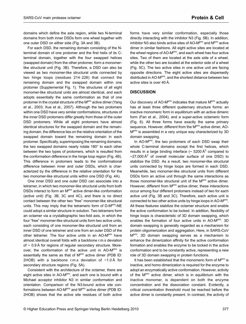
For each DSD, the remaining domain consisting of the N-terminal domain of one protomer and the first helix of its C-terminal domain, together with the four swapped helices(swapped domain) from the other protomer, form a monomer-like structural unit (Fig. 3B). Therefore, a DSD can also beviewed as two monomer-like structural units connected bytwo hinge loops (residues 214–226) that connect theremaining domain and the swapped domain within oneprotomer (Supplemental Fig. 1). The structures of all eightmonomer-like structural units are almost identical, and eachadopts essentially the same conformation as that of oneprotomer in the crystal structure of the Mpro active dimer (Yanget al., 2003; Xue et al., 2007). Although the two protomerswithin one DSD have very similar conformations, structures ofthe inner DSD protomers differ greatly from those of the outerDSD protomers. While all eight protomers have almostidentical structures for the swapped domain and the remain-ing domain, the difference lies on the relative orientation of theswapped domain toward the remaining domain in eachprotomer. Specifically, superimposing the remaining domains,the two swapped domains nearly rotate 180° to each otherbetween the two types of protomers, which is resulted fromthe conformation difference in the hinge loop region (Fig. 4B).This difference in protomers leads to the conformationaldifference between inner and outer DSDs, which is char-acterized by the difference in the relative orientation for thetwo monomer-like structural units within one DSD (Fig. 4A).
One inner DSD and one outer DSD can associate into atetramer, in which two monomer-like structural units from bothDSDs interact to form an Mpro active dimer-like conformation(active unit) (Fig. 3B, 3C and 4C), and there is no directcontact between the other two “free” monomer-like structuralunits. This may imply that the tetrameric form of D-Mpro-NEcould adopt a similar structure in solution. Two tetramers forman octamer via a crystallographic two-fold axis, in which thefour “free”monomer-like structural units form two active units,each consisting of one monomer-like structural unit from aninner DSD of one tetramer and one from an outer DSD of theother tetramer. The four active units in an AO-Mpro havealmost identical overall folds with a backbone r.m.s deviationof ~ 0.9 Å for regions of regular secondary structure. More-over, the conformation of the active unit in AO-Mpro isessentially the same as that of Mpro active dimer (PDB ID:2HOB) with a backbone r.m.s deviation of ~1.0 Å forsecondary structure regions (Fig. 5A).
Consistent with the architecture of the octamer, there areeight active sites in AO-Mpro, and each one is bound with aMichael acceptor inhibitor N3 in similar conformation andorientation. Comparison of the N3-bound active site con-formations between AO-Mpro and Mpro active dimer (PDB ID:2HOB) shows that the active site residues of both active
forms have very similar conformation, especially thosedirectly interacting with the inhibitor N3 (Fig. 5B). In addition,inhibitor N3 also binds active sites of AO-Mpro and Mpro activedimer in similar fashions. All eight active sites are located atthe wheel regions of AO-Mpro, and each wheel has four activesites. Two of them are located at the axle side of a wheel,while the other two are located at the exterior side of a wheel(Fig. 5C). The two active sites in one active unit are facingopposite directions. The eight active sites are dispersedlydistributed in AO-Mpro, and the shortest distance between twoactive sites is over 40 Å.
DISCUSSION
Our discovery of AO-Mpro indicates that mature Mpro actuallyhas at least three different quaternary structure forms: aninactive monomeric form in equilibrium with an active dimericform (Fan et al., 2004), and a super-active octameric form(Fig. 6). All three forms have exactly the same primarysequence. However, different from the Mpro active dimer, AO-Mpro is assembled in a very unique way characterized by 3Ddomain swapping.
In AO-Mpro, the two protomers of each DSD swap theirwhole C-terminal domains except the first helices, whichresults in a large binding interface (~ 5200 Å2 compared to~27,000 Å2 of overall molecular surface of one DSD) tostabilize the DSD. As a result, two monomer-like structuralunits connected by hinge loops are formed in each DSD.Meanwhile, two monomer-like structural units from differentDSDs form an active unit through the same interactions asthose monomer-like structural unit of the Mpro active dimer.However, different from Mpro active dimer, these interactionsoccur among four different protomers instead of two for eachactive unit (Fig. 3B and 3C). Therefore, each active unit isconnected to two other active units by hinge loops in AO-Mpro.All these features stabilize the octamer structure and enablethe active conformation to be locked. In addition, flexibility ofhinge loops is characteristic of 3D domain swapping, whichenables the formation of four active units in AO-Mpro. 3Ddomain swapping is generally regarded as a mechanism forprotein oligomerization and aggregation. Here, in SARS-CoVMpro, 3D domain swapping serves as a mechanism toenhance the dimerization affinity for the active conformationformation and enables the enzyme to be locked in the activeconformation and to be constantly active, representing a newrole of 3D domain swapping in protein functions.
It has been established that the monomeric form of Mpro isinactive, and homo dimerization is required for the enzyme toadopt an enzymatically active conformation. However, activityof the Mpro active dimer, which is in equilibrium with theinactive monomer, is dependent on both the enzymeconcentration and the dissociation constant. Evidently, acritical concentration threshold must be reached before theactive dimer is constantly present. In contrast, the activity of
© Higher Education Press and Springer-Verlag Berlin Heidelberg 2010 377
SARS-CoV main protease octamer Protein & Cell

AO-Mpro only depends on concentration, and it would remainactive once formed. As a result, AO-Mpro exhibits significantlyhigher activity than MD-Mpro at low concentrations.
Moreover, as there are four active units and thus eightactive sites in AO-Mpro and the substrates of Mpro havemultiple Mpro cleavage sites (7 for pp1a and 11 for pp1ab), theinteraction between AO-Mpro and pp1a/pp1ab could very wellbe a unique case of polyvalent interaction between enzymeand substrate. Polyvalent interactions are characterized bythe simultaneous binding of multiple ligands on one biologicentity (a molecule, a surface, etc) to multiple receptors onanother (Mammen et al., 1998), and have been implicated toplay pivotal roles in some important biologic processes, suchas cancer metastasis (Cattaruzza and Perris, 2005), virus andbacterial infections (Mammen et al., 1998), wound healing
(Minor and Peterson, 2002), chromatin modifications(Ruthenburg et al., 2007), and transcription regulations(Mammen et al., 1998). In a polyvalent interaction, the bindingaffinity between a multivalent ligand and a multiplevalentreceptor increases with the increasing number of ligands andreceptors. As the eight active sites are dispersedly distributedand are all accessible, it is quite possible for AO-Mpro tosimultaneously recognize and digest multiple cleavage sitesof one polyprotein substrate. Thus, compared to Mpro activedimer which has only two active sites, the binding affinitybetween AO-Mpro and its polyprotein substrates could bemuch higher, with the potential to increase the proteolyticactivity of AO-Mpro toward pp1a/pp1ab. Furthermore, it wasreported that Mpro exhibits quite different affinities andactivities toward the 11 different Mpro cleavage sequences
Figure 5. Active conformation and active sites. (A) Superposition of one active unit of AO-Mpro (the same color scheme inFig. 4C) with Mpro active dimer (2HOB, gray). (B) Comparison of one inhibitor N3 bound active pocket in AO-Mpro (cyan) with that inMpro active dimer (yellow, PDB ID: 2HOB). The corresponding inhibitors are in red and blue, respectively. (C) Ribbon diagrams of AO-
Mpro with eight inhibitors shown in stick display. The four inhibitors located at inside wheel are colored blue, while the other four atoutside wheel are colored red.
378 © Higher Education Press and Springer-Verlag Berlin Heidelberg 2010
Shengnan Zhang et al.Protein & Cell

in the polyprotein substrates (Fan et al., 2004). It is expectedthat the polyvalent interaction nature and the simultaneouscleavage ability of AO-Mpro should significantly increase theaffinity and activity of Mpro toward low efficiency cleavagesites by decreasing the dissociation rate (koff) of the enzyme/substrate complex. In addition, with 8 active sites, it is alsopossible for AO-Mpro to hydrolyze more than one polyproteinsubstrate at the same time. Therefore, although we have onlyshown that AO-Mpro has higher activity for a single sitesubstrate than Mpro active dimer at low protein concentra-tions, it is theoretically conceivable that the actual activity ofAO-Mpro toward polyprotein substrates could be even muchhigher than that of the well-known Mpro active dimer with onlytwo active sites.
In all crystal structures of SARS-CoV Mpro, the C terminusis exposed while the N-finger is buried in the dimer interfacewith several important interactions (Yang et al., 2003; Leeet al., 2005; Tan et al., 2005; Xu et al., 2005; Xue et al., 2007).Xue et al. (2007) have reported that extra residues to the Nterminus of Mpro, but not the C terminus, can significantlyreduce the activity of SARS-CoV Mpro. This is consistent withthe low enzymatic activity of Mpro-NE we observed, whichmay imply that mature Mpro is quite different from Mpro inpolyproteins prior to self-cleavage. It has been proposed thatMpro has to be activated by auto-cleavage from this inactivepolyprotein precursor before it can cleave the polyprotein atother cleavage sites (Anand et al., 2005). In a sense, theprocess for generating mature Mpro from Mpro-NE is some-what analogous to that of the Mpro maturation process, and
AO-Mpro matured from D-Mpro-NE should be as biologicallyrelevant as the commonly studied forms of the mature Mpro.
Although we don’t have evidence that AO-Mpro exists inSARS-CoV infected cells, it can be predicted that AO-Mpro
should be functionally superior to MD-Mpro for SARS-CoV if itdoes exist, especially during the initial period of infectionwhen the production level of mature Mpro is extremely low.Therefore, it is tempting to speculate that AO-Mpro may playan important role in polyprotein maturation at an early stage ofSARS-CoV infection when concentrations of pp1a, pp1b, andmature Mpro are extremely low.
SARS-CoV Mpro has been extensively investigated viastructural and biochemical studies (Anand et al., 2005;Bartlam et al., 2005; Po-Huang, 2006; Yang et al., 2006).Almost all these studies are carried out with mature Mpro, andlittle is clearly known about Mpro in the viral polyproteins andits maturation process. Recently, Chen et al. reported that twomonomeric mutants of Mpro can still perform N-terminalautocleavage, while the dimerization of mature proteaseand trans-cleavage activity following auto-processing arecompletely lost (Chen et al., 2010). They proposed that theauto-processing of Mpro from polyproteins is through an“intermediate dimer” structure which does not strictly dependon the active conformation existing in mature Mpro, and thepossible “intermediate dimer” may be formed through C-terminal domain dimerization. It is of interest to find outwhether this “intermediate dimer” is related to the Mpro-NEdomain-swapped dimer we observed. The finding by Chenet al. and our discovery of the super-active octameric form of
Figure 6. A schematic diagram illustrating the generation of the three different quaternary structure forms of mature Mpro.
© Higher Education Press and Springer-Verlag Berlin Heidelberg 2010 379
SARS-CoV main protease octamer Protein & Cell

SARS-CoV main protease indicate that the functionalmechanism of this important enzyme may be far morecomplicated than we thought, and that more studies arenecessary to reveal its genuine biologic function mechanism.
MATERIALS AND METHODS
Cloning, protein production, and purification
To construct the expression plasmid of Mpro-NE, the coding sequencefor the mature Mpro was amplified by PCR with a forward primer (5¢-GGCCGCCATATGGACGACGACGACAAAAGTGGTTTTAG-
GAAAATG-3¢) and a reverse primer (5¢-CGCACGATCTCGAGT-TATTGGAAGGTAACACCAG-3¢). The PCR product was cloned intothe pET28a vector between NdeI and XhoI sites. The resulting
plasmid encodes Mpro-NE which has an extra 26-residue fusionpeptide (MGSSHHHHHHSSGLVPRGSHMDDDDK) including anenterokinase cleavage site right in front of the N terminus of mature
Mpro. The plasmid was transformed into the E. coli BL21 (DE3) strainfor protein production. Cells were grown in LB medium containing100 μg/mL of kanamycin at 35°C until A600 reached 0.8, and wereinduced with 0.5 mM IPTG. After 8 h, the cells were harvested by
centrifugation at 5000 g for 10 min. The pelleted cells weresuspended in 50mM potassium phosphate buffer with 300mMNaCl at pH 8.0 (buffer A), and then stored at −80°C. After cell lysisby sonication and removal of cell debris by centrifugation at 24,000 gfor 20min, the supernatant was applied onto a Ni-NTA columnequilibrated with buffer A, and then washed by 50mM imidazole in
buffer A. Mpro-NE was eluted with 250mM imidazole in buffer A andfurther purified with gel-filtration (Superdex 200 10/300GL) on anÄKTA fast protein liquid chromatography system (FPLC) (GE, USA)
with 50mM Tris buffer (pH 8.0, 1 mM DTT). Thus, M-Mpro-NE and D-Mpro-NE were completely separated. At a concentration of 1 mg/mL,purified M-Mpro-NE or D-Mpro-NE was digested by enterokinase in areaction buffer of 50mM Tris-HCl (pH 8.0, 1 mM CaCl2, 0.1% Tween-
20, 1 mM DTT) at room temperature for 12 h, and then the matureMpro was purified using Superdex 200 column with 50mM Tris buffer(pH 7.4, 1 mM EDTA, 1mM DTT).
Analytical ultracentrifugation
The sedimentation velocity experiments were carried out with a
Beckman Coulter ProteomeLabTM XL-I at the Institute of Biophysics,Chinese Academy of Sciences (Beijing, China). All AUC runs werecarried out at a speed of 50,000 rpm at 16°C. The sample volume was400 μL with a protein concentration of ~ 1mg/mL in 50mM Tris buffer
(pH 7.4, 1 mM EDTA, 0.5 mM DTT). A wavelength of 280 nm wasused to record the UV absorption of the cells which were scannedevery 5min for 5 h. The data were analyzed with the SedFit program
(version 11.71 from 07/2008).The dissociation constant of M-Mpro-NE was calculated from the
peak area integrals of the monomer (AM) and dimer (AD) peaks in
AUC analysis data. As the total protein concentration (c) is known, themonomer concentration can be calculated using equation M = c/(1 +AD/AM), and the dimer concentration can be calculated as D = c/2(1 +
AM/AD). Therefore, the dissociation constant can be calculated as Kd
= M2/D. The dissociation constant between D-Mpro-NE dimer andtetramer was calculated in the same way.
Enzymatic activity assay
The enzymatic activity was determined by the peptide cleavage
assay using a peptide substrate with a sequence of MCA-AVLQSGFR-Lys (Dnp)-Lys-NH2 (more than 95% purity; GL Biochem(Shanghai) Ltd.). The reaction was monitored with a Hitachi (Tokyo,Japan) F-4500 fluorescence spectrophotometer at 25°C using
wavelengths of 320 nm and 405 nm for excitation and emission,respectively. The reaction was initiated by adding varied concentra-tions of enzyme to a 600 μL system containing 60 µM substrates with
the reaction buffer of 50mM Tris-HCl (pH 7.4) with 1mM EDTA and1mM DTT. When the enzyme concentration is less than 0.34 µg/mL,the reaction was recorded for 10min. Initial hydrolysis rates were
calculated by linearly fitting the linear portion of the data usingMicrosoft Office Excel 2003. For mature Mpro, enzyme activities weremeasured for concentrations range from 0.17 to 3.4 µg/mL, and theapparent dimer-monomer dissociation constant Kd values were
obtained by fitting the plot of reaction rate versus enzyme concentra-tion to the following equation, assuming the dimer is active and themonomer is inactive:
v ¼ A½Kd þ 4c− sqrtðKd2 þ 8Kd
�cÞ�=8 ð1ÞIn the equation, v is the observed hydrolysis rate, A is the
hydrolysis rate of the active dimer, and c is the total protein
concentration.
NMR sample preparation and NMR spectroscopy
E. coli cells were allowed to grow in M9 minimal medium prepared
with D2O, containing 15N-labeled ammonium chloride (Marley et al.,2001). The uniformly 15N, 2H-labeled D-Mpro-NE and M-Mpro-NEproteins were purified by the abovementioned methods. NMR
samples of D-Mpro-NE and M-Mpro-NE were at a concentration ofabout 1mM, with a buffer containing 50mM potassium phosphate(pH 7.0), 1 mM EDTA, 0.03% NaN3, in 90% H2O/10% D2O, plusComplete, an EDTA-free protease inhibitor cocktail (Roche, Ger-
many).All 2D NMR experiments were performed at 298 K on a Bruker
Avance 800MHz spectrometer. NMR spectra were processed with
NMRPipe (Delaglio et al., 1995), and analyzed using NMRView(Johnson and Blevins, 1994). The chemical shift in the 1H dimensionwas referenced directly to 2,2-dimethyl-2-silapentanesulfonic acid
(DSS), whereas the chemical shifts in the 15N dimensions wereindirectly referenced to DSS (Wishart et al., 1995).
Crystallization
The freshly prepared protein was incubated with inhibitor N3 with amolar ratio of 1:1 at 4°C and then concentrated to 30mg/mL in 20mMTris buffer (pH 7.0). Crystallization was performed by the sitting-drop
vapor-diffusion method at 16°C in 48-well plates. The initial crystal-lization conditions were screened using Hampton Research CrystalScreen Kits (Hampton Research Corporation). One microliter of theprotein solution was mixed with 1 μL reservoir solution and
equilibrated against 100 μL reservoir solution. Small crystals couldbe found in several conditions within three days. A series ofcrystallization grids was used to optimize the crystallization condition
at 18°C using the hanging-drop vapor-diffusion method by mixing1 µL AO-Mpro protein in the storage buffer with an equal volume of the
380 © Higher Education Press and Springer-Verlag Berlin Heidelberg 2010
Shengnan Zhang et al.Protein & Cell

reservoir solution containing 0.2 M sodium chloride, 0.1M Bis-Tris(pH 5.5) and 25% (w/v) PEG3350. Fine shaped and good qualitycrystals (size ~30×50×200 μm) suitable for data collection appeared
in 14 d and were soaked in a cryo-protectant solution consisting of 4M sodium formate. Crystals were flash-frozen in liquid nitrogen andthen transferred into a dry nitrogen stream at 100K for X-ray diffraction
data collection.
Data collection and structure determination
A 3.2 Å resolution diffraction data set was collected at 100 K from asingle AO-Mpro crystal using an ADSC Q270 detector on the BL17Abeamline at Photon Factory, KEK (Japan). A total of 360 frames ofdata were collected with 0.5° oscillation width. Processing of
diffraction images and scaling of the integrated intensities wereperformed using the HKL2000 software package (Otwinowski andMinor, 1997). The crystal belongs to space group P41212, with unit
cell parameters a = b = 161.9 Å, c = 166.4 Å, a = b = g = 90°. Weassumed that there are four Mpro molecules in the asymmetric unit,corresponding to a Matthews coefficient of 1.92 and solvent content
of 37% (Matthews, 1968). Initial phases were obtained by molecularreplacement with PHASER (McCoy et al., 2007) with the crystal
structure of SARS-CoV main protease (PDB code: 2H2Z) as thesearch model. The final manual structure rebuilding and refinementwere performed in COOT (Emsley and Cowtan, 2004), Refmac
(Murshudov et al., 1997) and Phenix (Adams et al., 2002) with theguidance of the 2Fo-Fc and Fo-Fc density maps. During the laterstages of positional refinement, restraints were relaxed and a bulk
solvent correction was applied under the guidance of Rfree. Modelgeometry was verified using the program PROCHECK (Laskowskiet al., 1993). Final refinement statistics are summarized in Table 1.
Figures were created by using MOLMOL (Koradi et al., 1996).
ACKNOWLEDGMENTS
This work was supported by Grant No. 2003CB514104 from the
National Basic Research Program (973 Program) and Grant No.30125009 from National Natural Science Foundation of China to BinXia, Grant No. 2006AA02A323 from the National Programs for High
Technology Research and Development Program (863 Program) toChangwen Jin, and Grant No. 2009ZX09311-001 to Zhiyong Lou fromthe National Major Projects of China. We gratefully thank Dr. ZengyiChang for his assistance on gel filtration analysis, and thank Ms.
Xiaoxia Yu for assisting the analytical ultracentrifugation experiment.
Table 1 Data collection and refinement statistics
data collection statistics
cell parametersa = b = 161.9 Å, c = 166.4 Å
a = b = g = 90º
space group P41212
wavelength used (Å) 1.000
resolution (Å) 50.0(3.3)c − 3.2
No. of all reflections 839,197
No. of unique reflections 37,177
completeness (%) 89.9 (64.6)
average I/σ (I) 8.4 (2.2)
Rmergea (%) 15.5 (65.2)
refinement statistics
No. of reflections used (σ(F) > 0) 33,573
Rworkb (%) 22.0
Rfreeb (%) 27.2
r.m.s.d. bond distance (Å) 0.009
r.m.s.d. bond angle (º) 1.526
average B-value (Å2) 97.1
ramachandran plot (excluding Pro & Gly)
Res. in most favored regions 727 (69.8%)
Res. in additionally allowed regions 249 (23.9%)
Res. in generously allowed regions 40 (3.8%)
a Rmerge ¼X
h
Xl Ieih < Ih >
������=X
h
Xl < Ih >, where < Ih > is the mean of the observations Iih of reflection h.
b Rwork = Σ( ||Fp(obs)~||Fp(calc)||)/ Σ|Fp(obs)|; Rfree = R factor for a selected subset (5%) of the reflections that was not included in prior
refinement calculations.c Numbers in parentheses are corresponding values for the highest resolution shell.
© Higher Education Press and Springer-Verlag Berlin Heidelberg 2010 381
SARS-CoV main protease octamer Protein & Cell

ABBREVIATIONS
DSD, domain-swapped dimer subunits; DSS, 2,2-dimethyl-2-silapen-tanesulfonic acid; DTT, 1,4-dithiothreitol; EDTA, ethylene diaminetetraacetic acid; FPLC, fast protein liquid chromatography system;HSQC, heteronuclear single quantum coherenc; MR, molecular
replacement; Mpro, main protease; NMR, nuclear magnetic reso-nance; RMSD, root mean square deviation; SARS, severe acuterespiratory syndrome; SARS-CoV, SARS coronavirus; WT, wild-type
REFERENCES
Adams, P.D., Grosse-Kunstleve, R.W., Hung, L.-W., Ioerger, T.R.,McCoy, A.J., Moriarty, N.W., Read, R.J., Sacchettini, J.C., Sauter,
N.K., and Terwilliger, T.C. (2002). PHENIX: building new softwarefor automated crystallographic structure determination ActaCrystallogr D 58, 1948–1954.
Anand, K., Palm, G.J., Mesters, J.R., Siddell, S.G., Ziebuhr, J., andHilgenfeld, R. (2002). Structure of coronavirus main proteinasereveals combination of a chymotrypsin fold with an extra α-helical
domain. EMBO J 21, 3213–3224.
Anand, K., Yang, H., Bartlam, M., Rao, Z. & Hilgenfeld, R. (2005).Coronavirus main proteinase: target for antiviral drug therapy. In:
Coronaviruses with special emphasis on first insights concerningSARS, A. Schmidt, M.H. Wolff, and O.F. Weber, ed. (Switzerland,Basel; Birkhauser Verlag). pp. 173–199.
Anand, K., Ziebuhr, J., Wadhwani, P., Mesters, J.R., and Hilgenfeld,R. (2003). Coronavirus main proteinase (3CLpro) structure: basisfor design of anti-SARS drugs. Science 300, 1763–1767.
Bartlam, M., Yang, H., and Rao, Z. (2005). Structural insights intoSARS coronavirus proteins. Curr Opin Struct Biol 15, 664–672.
Cattaruzza, S., and Perris, R. (2005). Proteoglycan control of cellmovement during wound healing and cancer spreading. Matrix Biol24, 400–417.
Chan, H.L., Tsui, S.K., and Sung, J.J. (2003). Coronavirus in severeacute respiratory syndrome (SARS). Trends Mol Med 9, 323–325.
Chen, S., Chen, L., Tan, J., Chen, J., Du, L., Sun, T., Shen, J., Chen,K., Jiang, H., and Shen, X. (2005). Severe acute respiratorysyndrome coronavirus 3C-like proteinase N terminus is indispen-
sable for proteolytic activity but not for enzyme dimerization.Biochemical and thermodynamic investigation in conjunction withmolecular dynamics simulations. J Biol Chem 280, 164–173.
Chen, H., Wei, P., Huang, C., Tan, L., Liu, Y., and Lai, L. (2006). Onlyone protomer is active in the dimer of SARS 3C-like proteinase. JBiol Chem 281, 13894–13898.
Chen, S., Hu, T., Zhang, J., Chen, J., Chen, K., Ding, J., Jiang, H., andShen, X. (2008). Mutation of Gly11 on the dimer interface results inthe complete crystallographic dimer dissociation of SARS-CoV
3CLpro: Crystal structure with molecular dynamics simulations. JBiol Chem 283, 554–564.
Chen, S., Jonas, F., Chen, C., and Higenfiled, R. (2010). Liberation of
SARS-CoV main protease from the viral polyprotein: N-terminalautocleavage does not depend on the mature dimerization mode.Protein Cell 1, 59–74.
Delaglio, F., Grzesiek, S., Vuister, G.W., Zhu, G., Pfeifer, J., and Bax,A. (1995). NMRPipe: a multidimensional spectral processingsystem based on UNIX pipes. J Biomol NMR 6, 277–293.
Emsley, P., and Cowtan, K. (2004). Coot: model-building tools for
molecular graphics. Acta Crystallogr D Biol Crystallogr 60,2126–2132.
Fan, K., Wei, P., Feng, Q., Chen, S., Huang, C., Ma, L., Lai, B., Pei, J.,Liu, Y., Chen, J., et al. (2004). Biosynthesis, purification, andsubstrate specificity of severe acute respiratory syndrome cor-onavirus 3C-like proteinase. J Biol Chem 279, 1637–1642.
Graziano, V., McGrath, W.J., DeGruccio, A.M., Dunn, J.J., andMangel, W.F. (2006a). Enzymatic activity of the SARS coronavirus
main proteinase dimer. FEBS Lett 580, 2577–2583.
Graziano, V., McGrath, W.J., Yang, L., and Mangel, W.F. (2006b).SARS CoV main proteinase: The monomer-dimer equilibrium
dissociation constant. Biochemistry 45, 14632–14641.
Gronenborn, A.M. (2009). Protein acrobatics in pairs—dimerizationvia domain swapping. Curr Opin Struct Biol 19, 39–49.
Hsu, W.C., Chang, H.C., Chou, C.Y., Tsai, P.J., Lin, P.I., and Chang,G.G. (2005). Critical assessment of important regions in the
subunit association and catalytic action of the severe acuterespiratory syndrome coronavirus main protease. J Biol Chem280, 22741–22748.
Hu, T., Zhang, Y., Li, L., Wang, K., Chen, S., Chen, J., Ding, J., Jiang,H., and Shen, X. (2009). Two adjacent mutations on the dimerinterface of SARS coronavirus 3C-like protease cause different
conformational changes in crystal structure. Virology 388,324–334.
Ivanov, D., Tsodikov, O.V., Kasanov, J., Ellenberger, T., Wagner, G.,
and Collins, T. (2007). Domain-swapped dimerization of the HIV-1capsid C-terminal domain. Proc Natl Acad Sci U S A 104,4353–4358.
Johnson, B.A., and Blevins, R.A. (1994). NMR View: A computerprogram for the visualization and analysis of NMR data. J BiomolNMR 4, 603–614.
Koradi, R., Billeter, M., and Wuthrich, K. (1996). MOLMOL: a programfor display and analysis of macromolecular structures. Journal ofmolecular graphics 14, 51–55.
Kuiken, T., Fouchier, R.A., Schutten, M., Rimmelzwaan, G.F., vanAmerongen, G., van Riel, D., Laman, J.D., de Jong, T., van
Doornum, G., Lim, W., et al. (2003). Newly discovered coronavirusas the primary cause of severe acute respiratory syndrome. Lancet362, 263–270.
Kuo, C.-J., Chi, Y.-H., Hsu, J.T.-A., and Liang, P.-H. (2004).Characterization of SARS main protease and inhibitor assayusing a fluorogenic substrate. Biochem Biophys Res Commun
318, 862–867.
Laskowski, R., MacArthur, M., Moss, D., and Thornton, J. (1993).PROCHECK: a program to check the stereochemical quality of
protein structures. J Appl Cryst 26, 283–291.
Lee, T.W., Cherney, M.M., Huitema, C., Liu, J., James, K.E., Powers,J.C., Eltis, L.D., and James, M.N. (2005). Crystal structures of the
main peptidase from the SARS coronavirus inhibited by asubstrate-like aza-peptide epoxide. J Mol Biol 353, 1137–1151.
Leng, Q., and Bentwich, Z. (2003). A novel coronavirus and SARS. NEngl J Med 349, 709.
Libonati, M., Gotte, G., and Vottariello, F. (2008). A novel biological
actions acquired by ribonuclease through oligomerization. CurrPharm Biotechnol 9, 200–209.
Lin, P.Y., Chou, C.Y., Chang, H.C., Hsu, W.C., and Chang, G.G.
(2008). Correlation between dissociation and catalysis of SARS-CoV main protease. Arch Biochem Biophys 472, 34–42.
382 © Higher Education Press and Springer-Verlag Berlin Heidelberg 2010
Shengnan Zhang et al.Protein & Cell
Liu, Y., and Eisenberg, D. (2002). 3D domain swapping: as domains
continue to swap. Protein Sci 11, 1285–1299.
Mammen, M., Choi, S.-K., and Whitesides, G.M. (1998). Polyvalentinteractions in biological systems: implications for design and use
of multivalent ligands and inhibitors. Angew Chem Int Ed 37,2754–2794.
Marley, J., Lu, M., and Bracken, C. (2001). A method for efficient
isotopic labeling of recombinant proteins. J Biomol NMR 20,71–75.
Matthews, B.W. (1968). Solvent content of protein crystals. J Mol Biol33, 491–497.
McCoy, A., Grosse-Kunstleve, R., Adams, P., Winn, M., Storoni, L.,
and Read, R. (2007). Phaser crystallographic software. J ApplCryst 40, 658–674.
Minor, K.H., and Peterson, C.B. (2002). Plasminogen activator
inhibitor type 1 promotes the self-association of vitronectin intocomplexes exhibiting altered incorporation into the extracellularmatrix. J Biol Chem 277, 10337–10345.
Murshudov, G.N., Vagin, A.A., and Dodson, E.J. (1997). Refinementof macromolecular structures by the maximum-likelihood method.Acta Crystallogr D 53, 240–255.
Otwinowski, Z., and Minor, W. (1997). Processing of X-ray diffractiondata collected in oscillation mode. In Macromolecular Crystal-
lography, part A, C.W. Carter Jr., and R.M. Sweet, eds. (AcademicPress), pp. 307–326.
Po-Huang, L. (2006). Characterization and inhibition of SARS-
coronavirus main protease. Curr Top Med Chem 6, 361–176.
Ruthenburg, A.J., Li, H., Patel, D.J., and Allis, C.D. (2007). Multivalentengagement of chromatin modifications by linked bindingmodules.
Nat Rev Mol Cell Biol 8, 983–994.
Shi, J., and Song, J. (2006). The catalysis of the SARS 3C-likeprotease is under extensive regulation by its extra domain. FEBS J
273, 1035–1045.
Shi, J., Wei, Z., and Song, J. (2004). Dissection study on the severe
acute respiratory syndrome 3C-like protease reveals the criticalrole of the extra domain in dimerization of the enzyme: defining theextra domain as a new target for design of highly specific protease
inhibitors. J Biol Chem 279, 24765–24773.
Shi, J., Sivaraman, J., and Song, J. (2008). Mechanism for controllingthe dimer-monomer switch and coupling dimerization to catalysis
of the severe acute respiratory syndrome coronavirus 3C-likeprotease. J Virol 82, 4620–4629.
Snijder, E.J., Bredenbeek, P.J., Dobbe, J.C., Thiel, V., Ziebuhr, J.,
Poon, L.L., Guan, Y., Rozanov, M., Spaan, W.J., and Gorbalenya,A.E. (2003). Unique and conserved features of genome andproteome of SARS-coronavirus, an early split-off from the
coronavirus group 2 lineage. J Mol Biol 331, 991–1004.
Tan, J., Verschueren, K.H., Anand, K., Shen, J., Yang, M., Xu, Y., Rao,Z., Bigalke, J., Heisen, B., Mesters, J.R., et al. (2005). pH-dependent conformational flexibility of the SARS-CoV main
proteinase (M(pro)) dimer: molecular dynamics simulations andmultiple X-ray structure analyses. J Mol Biol 354, 25–40.
Verschueren, K.H.G., Pumpor, K., Anemüller, S., Chen, S., Mesters,
J.R., Hilgenfeld, R., (2008). A structural view of the inactivation ofthe SARS coronavirus main proteinase by benzotriazole esters.Chem Biol 15, 597–606.
Wei, P., Fan, K., Chen, H., Ma, L., Huang, C., Tan, L., Xi, D., Li, C., Liu,Y., Cao, A., et al. (2006). The N-terminal octapeptide acts as adimerization inhibitor of SARS coronavirus 3C-like proteinase.
Biochem Biophys Res Commun 339, 865–872.
Wishart, D.S., Bigam, C.G., Yao, J., Abildgaard, F., Dyson, H.J.,
Oldfield, E., Markley, J.L., and Sykes, B.D. (1995). 1H, 13C and15N chemical shift referencing in biomolecular NMR. J BiomolNMR 6, 135–140.
Xu, T., Ooi, A., Lee, H.C., Wilmouth, R., Liu, D.X., and Lescar, J.(2005). Structure of the SARS coronavirus main proteinase as anactive C2 crystallographic dimer. Acta Crystallogr Sect F Struct
Biol Cryst Commun 61, 964–966.
Xue, X., Yang, H., Shen, W., Zhao, Q., Li, J., Yang, K., Chen, C., Jin,Y., Bartlam, M., and Rao, Z. (2007). Production of authentic SARS-
CoV M(pro) with enhanced activity: application as a novel tag-cleavage endopeptidase for protein overproduction. J Mol Biol 366,965–975.
Yamasaki, M., Li, W., Johnson, D.J., and Huntington, J.A. (2008).Crystal structure of a stable dimer reveals the molecular basis ofserpin polymerization. Nature 455, 1255–1258.
Yang, H., Yang, M., Ding, Y., Liu, Y., Lou, Z., Zhou, Z., Sun, L., Mo, L.,Ye, S., Pang, H., et al. (2003). The crystal structures of severeacute respiratory syndrome virus main protease and its complex
with an inhibitor. Proc Natl Acad Sci U S A 100, 13190–13195.
Yang, H., Xie, W., Xue, X., Yang, K., Ma, J., Liang,W., Zhao, Q., Zhou,
Z., Pei, D., Ziebuhr, J., et al. (2005). Design of wide-spectruminhibitors targeting coronavirus main proteases. PLoS Biol 3, e324.
Yang, H., Bartlam, M., and Rao, Z. (2006). Drug design targeting the
main protease, the Achilles' heel of coronaviruses. Curr PharmDes 12, 4573–4590.
Zhong, N., Zhang, S., Zou, P., Chen, J., Kang, X., Li, Z., Liang, C., Jin,
C., and Xia, B. (2008). Without its N-finger, SARS-CoV mainprotease can form a novel dimer through its C-terminal domain. JVirol 82, 4227–4234.
Zhong, N., Zhang, S., Xue, F., Kang, X., Zou, P., Chen, J., Liang, C.,Rao, Z., Jin, C., Lou, Z., et al. (2009). C-terminal domain of SARS-CoV main protease can form a 3D domain-swapped dimer. Protein
Sci 18, 839–844.
© Higher Education Press and Springer-Verlag Berlin Heidelberg 2010 383
SARS-CoV main protease octamer Protein & Cell