thoracic surgery i. outline terms anatomy & physiology pathology diagnosis anesthesia...
TRANSCRIPT
Thoracic Surgery I
Outline Terms Anatomy & Physiology Pathology Diagnosis Anesthesia Medications Supplies, Instruments, Equipment Patient preparation Prepping and Draping Procedures: Bronchoscopy, Mediastinoscopy, & Thoracostomy Post-operative Considerations/Complications

Purpose of Thoracic Surgery
Diagnose by endoscopic or open biopsy Treat disease by resection or repair of tissue Correct structural deformity Traumatic injury repair

Terms Bronchial washings-secretions obtained from
bronchi by injection and aspiration of small amounts of NS for cell identification
Empyema-pus in the pleural cavity Flail chest-rib fracture where are not attached
creating paradoxic movement during inspiration and expiration
Hemothorax-blood in the pleural cavity from trauma, pneumonia, TB, or malignancy that has caused vessel rupture
Hypoxia-insufficient oxygen intake upon inspiration Intercostal space-space between two ribs
Terms Lobes-well defined portions (Lungs: 2 left and 3 right) Pectus carinatum (pigeon-breast) -abnormal protrudence of the
sternum (congenital) Pectus excavatum-abnormal funnel-shaped depression of the
lower sternum (congenital) Pleural effusion-abnormal fluid accumulation in the pleural space Pneumothorax-accumulation of air or gas in the pleural cavity
resulting in collapse of the affected lung (may be closed or open) Thoracentesis-aspiration of fluid from the pleura via the chest
wall by inserting a needle

Anatomy & Physiology
of the
Respiratory System

Organs of Respiratory System
Nose Pharynx Larynx Trachea Lungs

Nose
Lined with goblet cells that produce mucous Air hits conchae/turbinates and tumbles
which allows warming, filtration, and moistening of the air
External nose from face out Internal nose face back to sinuses Upper posterior portion of nose lined with
receptors for olfactory sense

Pharynx
Starts at back of sinuses to top of larynx Hallway/Opening/Passageway: where nasal
cavity, oral cavity, eustacian tubes, esophagus, and trachea open to
Three sections: Nasopharynx, Oropharynx, Laryngopharynx
Larynx Voicebox 9 pieces of hyalin cartilage On top flap called epiglottis Epiglottis function to protect larynx when swallowing Epiglottis closed when swallow and open when
breath Vocal cords: False are on top (superior) and True
are on bottom (inferior) Tissue folds made of elastic fibers that produce a
sound and change pitch, loudness produced in the resonating chamber called the sinuses
Trachea
Anterior to esophagus 1” diameter and 4 ½” long Outside of 16-20 C-shaped rings of hyalin cartilage
which function to hold trachea open Between the C’s opening and the esophagus is an
open area with the trachealis muscle This muscle tissue relaxes when swallowing Passageway for oxygen into the lungs & carbon
dioxide out of the lungs Begins at larynx ends at bifurcation of the bronchi
Primary Bronchi Trachea branches into right and left primary bronchi Right larger, wider and shorter than left Right more vertical than left Result tend to aspirate things into right bronchi verses left Right and left (each called a bronchus) Also transport oxygen and carbon dioxide Lined with goblet cells that secrete mucus to trap particles in the
air we breath Contain cilia that sweep these trapped particles up and out to be
expelled or swallowed Primary bronchi divide into secondary bronchi each of which
goes to its own lobe
Secondary Bronchi/Lobar Bronchi
3 on right 2 on left Go to respective lobes of lungs: 3 lobes on right and 2 lobes on left Secondary bronchi divide into tertiary bronchi
that supply the lung in segments

Tertiary Bronchi
Terminal points of tertiary bronchi End at alveolar ducts, each of which is
surrounded by alveoli Alveoli encased in arteries and veins Alveoli are where the actual gas exchange
takes place (oxygen coming in and carbon dioxide going out)
Total alveoli surface area is actually as large as a tennis court (300 million)
Respiratory System Functions
1. Ventilation Movement of gas in and out of lungs Filter, moisten, and warm gases Inspiration and Expiration
Respiratory System Functions External Respiration Movement of gases from lungs to blood and back to lungs Exchange of gases takes place between alveoli and capillary Both membranes are thin which allow for great amount of
diffusion Diffusion is movement from greater concentration to lower
concentration Alveoli O2 level/pressure 105mm/Hg verses capillary O2 pressure
40 mm/Hg Alveoli CO2 pressure 40mm/Hg and capillary CO 2 pressure
45mm/Hg So external respiration driven by law of diffusion

Respiratory System Functions
Internal Respiration Movement of gases from blood to all other
tissues and back 3% O2 dissolved into plasma 97% O2 picked up by Fe portion of
hemoglobin (Hgb) molecule becomes oxyhemoglobin
O2 released as gets to area or tissue in need and Hgb releases

Respiratory System Functions
Internal Respiration Movement of gases from blood to all other
tissues and back 5-7% CO2 dissolved in plasma
23-25% CO2 attaches to protein portion of Hgb becomes carbaminohemoglobin
70% used in buffer system to maintain acid base balance of body

Respiration
4 Functions of : Provide oxygen to the body tissues and
organs Remove carbon dioxide waste Maintain homeostasis (acid-base balance)
through the oxygen and carbon dioxide exchange
Maintain heat exchange

Control of Respiratory System
Nervous System’s Respiratory Centers Medulla Oblongata = subconscious Pons = subconscious Cerebrum voluntary/conscious =can
temporarily over-ride medulla and pons

Regulation of Breathing
Receptors in PNS detecting CO2 levels not O2
levels Chemical receptors in carotid artery and
aorta are monitoring your pH blood levels Lower pH (acidic)= ↑ breathing rate Higher pH levels (alkaline) = ↓ breathing rate

The Thoracic Cavity
Sternum: xiphoid, body, and manubrium Ribs: 12 ribs attached to thoracic vertebrae
posteriorly 7 true ribs attached to the sternum by coastal
cartilage Next 3 false ribs indirectly are attached to the
sternum by costal cartilage Last 2 false or floating ribs, do not attach to
the sternum at all only the thoracic vertebrae
Anatomy & Physiology of the Thoracic Cavity
Mediastinum Middle of thoracic cavity Contains esophagus, trachea, heart, and
great vessels Pericardial cavity is where heart actually
located Pleural Cavities To left and right of mediastinum Contain the lungs

Lungs
Contained in the pleural cavity Each surrounded by the parietal pleura, a serous
membrane lining the chest wall and diaphragm Potential space between parietal and visceral pleura
is the pleural space or intrapleural space Against the lungs themselves and against the
parietal pleura is the visceral pleura, a thin membrane that covers each lung
Beneath the visceral pleura is lung tissue

Lungs
Right Lung Has three lobes (RUL, RML, RLL) Shorter than left due to liver beneath it Left Lung Has two lobes (LUL and LLL) Longer than right because the heart pushes
left*Apex of the lungs is above the clavicles
and the base is resting on the diaphragm


Respiration/Breathing Inspiration Passive result of skeletal muscle action Diaphragm causes the thoracic cavity to increase in
size as it descends (contracts) Diaphragm is the 1˚ muscle responsible for
inspiration External intercostals play a part in inspiration by
elevating the ribs Volume of chest cavity increases and pressure
decreases, so air moves in Atmospheric pressure in chest cavity low, outside is
high pressure negative pressure is established

Respiration
Expiration Passive As volume decreases pressure increases and air is
forced out Lungs are never completely empty Positive pressure causes air to come out Muscles involved in expiration: Internal intercostal muscles depress the ribs External oblique depress lower ribs Abdominus rectus muscles depress ribs and viscera

Boyle’s Law
Respiration is an inverse relationship between pressure and volume
Inspiration=V↑ P↓ Expiration = V↓ P↑

Pathology Mediastinum Children: neurogenic (resulting from nervous tissue)
tumors Adults: thymomas ( thymus gland tumor),
lymphomas (originating from lymphatic system, can be malignant or benign), and cysts (may be solid or fluid filled)
40% asymptomatic 60% symptomatic (cough, dyspnea, chest pain) Of 60% that are symptomatic, 60% of those will
have a malignant lesion or tumor
Pathology Lungs Carcinoma=a new growth or malignant tumor Lung cancer #1 cause of death r/t cancer Tumors Divided into 4 Groups: Small Cell Carcinoma or Oat Cell (malignant) Large Cell Carcinoma (malignant) Adenocarcinoma (malignant) of bronchi = primarily smokers of bronchioles = 50%smokers &
50%nonsmokers Squamous Cell Carcinoma (benign) formed from epithelial or
squamous cells which line mucous membranes) 90% malignant lung cancers r/t smoking

Pathology
All tumor types with the exception of small cell (oat cell), have a good prognosis with medical and or surgical intervention
Surgical Interventions include: Wedge/Tumor Resection with margins Lobectomy Pneumonectomy Medical Interventions include: Chemotherapy Radiation

Initial Diagnosis
Cytology of sputum sample Will determine the type of cells that are
present in the respiratory system Will show presence of cancer cells but not
where they actually came from in the lungs Most preliminary of all tests Chest X-ray must follow to narrow down
location of tumor or mass
Initial Diagnosis
Chest X-ray may be found on routine exam
(asymptomatic) may be ordered after presents with
symptoms:
Cough
Bloody sputum (hemoptysis)
Dyspnea
Diagnosis Cell type determines the course of treatment Tumors are looked at in terms of “staging” Staging means,” how developed is the tumor”? Is it in the lymph nodes, has it metastasized to another area, or is
it localized Staging is accomplished by sending a tissue sample to pathology
and having it analyzed for type Tissue samples are obtained by biopsy Tissue samples can be of lymph nodes or lung tumor, done with
a biopsy needle or actual wedge resections of the lung Biopsy can be done by laryngoscopy, bronchoscopy or
mediastinoscopy

Specimens
Specimens must be handled appropriately Mishandling could damage a sample causing
it to not be analyzable There are two types of tissue samples in the
OR related to node or tissue:
Fresh frozen
Permanent

Specimens Fresh Frozen Identifies type of tumor Determines margins, did you obtain the entire tumor Will entail a waiting period in the OR until pathology
has determined this Depending on results may have the tumor in its
entirety and close or not have it all and will require going in for more tissue
Frozen sent when tumor has not been previously identified by laryngoscopy, bronchoscopy, mediastinoscopy, or needle biopsy

Permanent Must ID the type of tumor before it can be
stained to determine staging There are different stains required for
different types of tumors Would send a wedge or lobe for permanent if
the tumor type had already been identified by a previous biopsy (from mediastinoscopy, bronchoscopy, or needle biopsy)

Specimens
Sometimes may hear send this for Fresh and the doctor will want cytology run
Cytology identifies an infectious process: Fungal Bacterial AFB (acid fast bacillus) checks for TB
Other Diagnostic Tests for Review
CT scan or MRI Shows location of tumor so that if a
thoracotomy is done, the surgeon knows where to operate to excise the lesion

Anesthesia
Local with IV sedation for straight laryngoscopy or bronchoscopy
Mediastinoscopy, tracheotomy, thoracoscopy General Epidural catheter may be placed for post-op
pain management (thoracoscopy) May use local injection at wound site at
closure to manage post-op pain

Medications
Sterile NS Sterile water (presence of malignant tumors) Antibiotic for irrigant Surgicel, Gelfoam and Thrombin (available) Avitene (available) Available for possible open thoracotomy:
Bone wax or focal-seal
Preoperative Patient Preparation
Chest X-ray, MRI, and or CT Scans should be in the OR before the patient arrives. They may accompany the patient. They should be displayed in the x-ray box for the surgeon.
Type & Cross should be done in the event that the patient experiences extreme blood loss and needs blood replacement during surgery
These procedures are risky in that large vessels are present in the thorax and mediastinum and could be accidentally injured
Positioning Mediastinoscopy Bronchoscopy Laryngoscopy Trachestomy Antero-lateral thoracotomy incision (following + add rolled blanket or
sandbag under operative side from scapula to buttocks)
Supine Arms tucked or on armboards Shoulder roll Pillow under knees Headrest (donut + towels) Heel protectors Safety strap
Laryngoscopes
L-shaped – intubation Flexible – assist with intubation, diagnostic,
biopsy Rigid U-shaped – biopsy, foreign body
removal, vocal cord procedures

Microlaryngoscopy
Laryngoscopy Microscope (400mm focal length=40cm focal length) Microlaryngeal instruments (22cm) Laser attached to microscope CO2 single beam, more precise (used with helium-neon beam to
provide red beam for proper aiming) Vocal cord, tracheal, bronchial lesions Nd: YAG Laser tracheal or bronchial lesions
Supplies, Instrumentation, Equipment Bronchoscopy Flexible or rigid bronchoscope ET tube adaptor Biopsy forceps (flexible or rigid) Light source Light cable (fiberoptic) Gown, gloves Suction tip and tubing Sponges K-Y jelly or other water soluble lubricant Basin with saline or water May be a Bronch Cart available with first five listed supplies

Bronchoscopes
Flexible Rigid (preferred for foreign body removal) Longer than laryngoscopes Adaptor required for oxygenation Nd: YAG (prn)
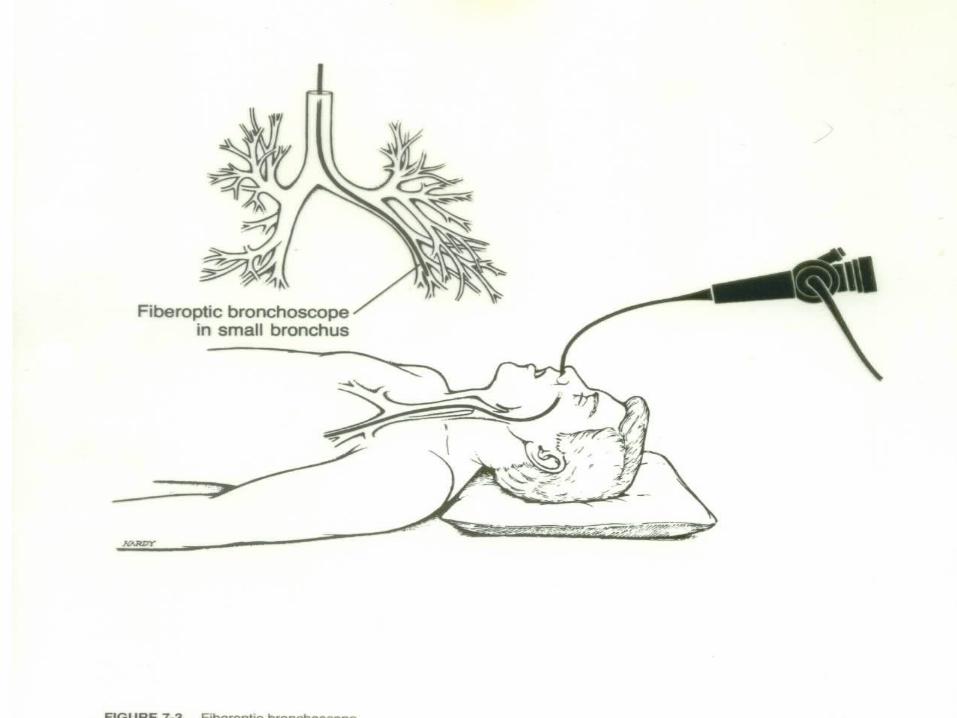

Procedure Bronchoscopy (clean procedure) Scope should be sterile (per institutional method) Prepare scope (lubricate if surgeon preference
attach light source) Give surgeon adaptor for ET tube, pass
bronchoscope Pass biopsy forceps prn Prepare to collect tissue samples on telfa (cut into
small squares) Identify with surgeon to communicate with circulator
for proper labeling and containing so it can be sent to the lab correctly

For cytology washings Attach sputum trap Surgeon will irrigate through the port on the scope
with a 10 to 30cc syringe filled with NS Pass to nurse clearly identifying the source and type
of cytology requested by the surgeon Remove scope Clean per institutional policy See pg 1060 Alexander’s

Positioning
Mediastinoscopy
Supine Pillow or donut under head Arms tucked or on armboards Pillow under knees Shoulder roll optional (surgeon preference) Heel protectors Safety strap

Prepping and Draping
Mediastinoscopy: Prep from incision site and out in a circle,
usually prep upper chest and anterior shoulders
Towels x 4, drying towel, Ioban, pediatric laparotomy sheet or thyroid sheet
Supplies, Instrumentation, Equipment
Mediastinoscopy Mediastinoscope Light cord (fiberoptic) Light source Suction tip and tubing Biopsy forceps Grasping forceps Clip applier ECU with special bovie tip
Minor instrument tray Raytex Telfa Biopsy needle Towels Pediatric lap sheet or a thyroid sheet
Procedure Mediastinoscopy (sterile procedure) Pass off bovie, suction tubing, light cord, & camera cord to
circulator Raytex up Knife or scalpel to surgeon (incision made 2 cm above
suprasternal notch) Cautery (may use bovie or knife or finger to create opening in
trachea) Mediastinoscope (requires assembly when setting up: attach
light cord, light carriers, camera) *Practice light cord safety/attach to mediastinoscope ASAP
Circulator should pay attention, but it is your responsibility too!

Procedure Mediastinoscopy Pass biopsy needle with syringe attached (10-30cc) This is so the surgeon can aspirate before he pulls out a tissue sample Checking for air or blood Getting blood back, especially bright red blood indicates arterial blood (Be prepared to open the sternum - need sternal saw available Pass biopsy forceps (be prepared to collect specimen on a small piece
of telfa) Have several pieces of telfa available-may send several specimens Make certain these go for frozen unless the surgeon tells you otherwise You are getting a preliminary node and tissue analysis that may lead to
medical or further surgical intervention
Procedure
Mediastinoscopy Scope removed upon completion of biopsies Wound closed with a 3-0 absorbable suture (Vicryl)
on a tapered needle (SH) Skin closed with 4-0 absorbable suture on a small
cutting needle (PS-2) Dressing applied (telfa, tegaderm) Disassemble mediastinoscope handling carefully
and clean per institutional policy See pg 1064 Alexanders

Indications For Tracheotomy or Tracheostomy
Vocal cord paralysis Neck surgery Trauma Prolonged intubation Secretion management Cannot intubate Stridor due to tracheal blockage Sleep apnea

Tracheotomy/Tracheostomy Tracheotomy temporary opening into the trachea to
facilitate breathing Tracheostomy permanent opening of the trachea
and creation of a tracheal stoma Must place tracheal tube with either Patient will be hooked up to a ventilator Long term tracheostomy may eventually be able to
ween off ventilator, but maintain stoma that will function as their nose did prior to surgery

Positioning Thoracoscopy or Thoracotomy For talc pleurodesis, decortication, wedge resection, lobectomy,
pneumonectomy
Full postero-lateral position Operative side up Vacuum-bag/beanbag under draw-sheet (surgeon preference) Axillary roll prevents brachial plexus damage Down arm on armboard Up arm on padded mayo or airplane sling device Pillow under head Pillows x 2 between knees (protects peroneal nerve) and feet Foam pad under down leg Safety strap and adhesive tape across pelvic girdle and
shoulders for stabilization
CHEST TUBE INSERTION“Thoracostomy”
Review of Normal Lung Function Negative intrathoracic pressure and elasticity
are required Requires intact pleural cavity and intact
visceral pleura

Chest Tube Insertion“Thorocostomy” Disruption of Normal Lung Function 1. PNEUMOTHORAX Interference with the negative intrathoracic pressure Air in the pleural space or visceral pleura causes partial lung
collapse The air takes up the space the lung needs to expand Requires chest tube placement to re-establish negative pressure
and intactness Cause: trauma (blunt or penetrating), tear or perforation in the
visceral lining Tear may result from an emphysematous bleb or lung abscess


CHEST TUBE INSERTION“Thoracostomy” Disruption of Normal Lung Function 2. Open Pneumothorax Large penetrating wound Cover wound with vaseline dressing Insert chest tube 3. Tension Pneumothorax Air coming out of bronchus into pleural space and can’t get back
out Requires decompression with a large needle and chest tube
insertion Urgent Intervention required due to potential shift that could
affect the opposite lung and heart (death)
Chest Tube Insertion“Thoracostomy”
Disruption of Normal Lung Function 4. Hemothorax Blood from small vessel rupture is leaking out
into the pleural space Causes: pneumonia
TB
malignancy
Chest Tube Insertion“Thoracostomy”
Prepping/Draping Pre-existing chest wound /work around site and
prep wound last (avoids further infection) Non-pre-existing wound prep site of chest tube first
and work around (remember prep axilla last if prepping that far out)
Towels x 4, laparotomy sheet, or universal sheet In emergency may only use towels In emergency may just pour anti-bacterial onto chest
and GO
Chest Tube Insertion“Thoracostomy” Procedure Knife Bovie Long kelly or tonsil Chest tube Heavy Silk (#1) on a cutting needle (are free-eyed
needles) Cut chest tube to protrude about 4” attach to
pleurevac with suction attached Band connection or tape Apply gauze dressing or drain sponge and tape
Chest Tube Insertion“Thoracostomy”
Chest Drainage System/Pleurevac Provides way for blood, fluid, or air to drain
from the mediastinal or pleural cavities re-establishing negative pressure
Drainage system work 3 ways:
positive expiratory pressure
suction
gravity or water seal


Chest Tube Insertion“Thoracostomy”
Keep drainage system or pleurevac below the patient’s body
Must be kept sterile Are usually taken out in 3-7 days depending
on the reason they were placed

Thoracoscopy
Visualization of the thoracic cavity by a thoracoscope
Used to obtain/evaluate biopsies, take wedge resections, and administer talc pleuredesis
*Talc Pleuredesis (tx. for spontaneous bleb rupture) Consent will have most often have “thoracoscopy
possible thoracotomy” in event larger incision is needed based on biopsy results and visibility
Will need to have instrumentation for a thoracotomy on field

Thoracoscopy Supplies Laparotomy sheet or universal sheets Gowns, gloves Minor or major basin set Blades #10 and #15 Bovie Chest tubes (surgeon preference) & Pleurevac FRED or anti-fog Scope warmer (place raytex in bottom to prevent scope damage/tell
circulator it is in there)
Trocar Insufflation tubing (available) Warm saline on field and in scope warmer Raytex Bovie Suction (sigmoid suction tip) Closing suture (vicryl 2-0 CT-2 and 4-0 PS-2) Endoscopic staplers (variety) Dressing post final counts

Thoracoscopy
Instrumentation 0° or 30° scope Camera Light cord Endoscopic instrument set (graspers, scissors,
bovie, clip appliers) CV Tray or Major Tray Chest Tray Pilling lung clamp tray available Long instrument set available
Thoracoscopy
Equipment ECU Suction Bair Hugger (lower body) Light source/Camera box Video monitor Thorax cannot be insufflated Double lumen ET tube allows for single lung
ventilation and collapse of affected lung Defibrillator (available)

Thoracoscopy
Prepping and Draping Begin at incision site and work outward in a
circular motion (axilla last) Towels x 4 or 5 Drying towels Ioban Laparotomy sheet or universal drapes

Thoracoscopy Procedure Pass off bovie, suction, light cord, and camera cord Knife Bovie Incisions x two or three Kelly or metz to open intercostal space Trocar (keep obturator available) Scope Trocar for manipulating device such as forceps, long pilling lung clamps Endoscopic staplers as requested as well as reloads Biopsy needle or culture swab as needed Talc available if pleuradesis Rough bovie pad cut and on a long sponge stick or pilling lung clamp

Thoracoscopy
Chest tubes (x 1 or 2) surgeon preference on size and type {come straight and right angled} Sizes #10F through 36F
Sew in chest tubes with #1 silk on a cutting needle Close with 2-0 Vicryl taper CT-2
and 4-0 cutting PS-2 Dress per surgeon preference Keep table sterile until all frozen results back and
patient ready to transport (r/o thoracotomy)

*Decortication (removal of exudate or scarring)
Scarring interfering with normal respiration Generally due to infectious process
(ex. pneumonia) Also called “membrane peel” If extensive will have to open = “thoracotomy”
Post-operative Considerations
Connect chest tube immediately to prevent clot formation in the tube or pneumothorax
Make sure chest tube attached securely so it does not come undone

Possible Complications
Atelectasis Pneumonia Respiratory insufficiency Pneumothorax Hemorrhage Pulmonary embolus Mediastinal shift Acute pulmonary edema Infection
Prognosis
Depends on patient’s post-operative status and the pathological process
Malignancies that have not progressed into the lymph nodes (negative nodes) have good prognosis with tumor removal
May undergo chemotherapy or radiation post-operatively
Summary Anatomy & Physiology Pathology Diagnosis Anesthesia Medications Patient preparation Positioning Supplies, Instruments, Equipment Prepping and Draping Procedures: Laryngoscopy, Bronchoscopy, Mediastinoscopy,
Tracheotomy, Thoracostomy & Thoracoscopy Post-operative Considerations/Complications