this is the first histology lecture for the respiratory...
TRANSCRIPT
This is the first histology lecture for the respiratory tract system .
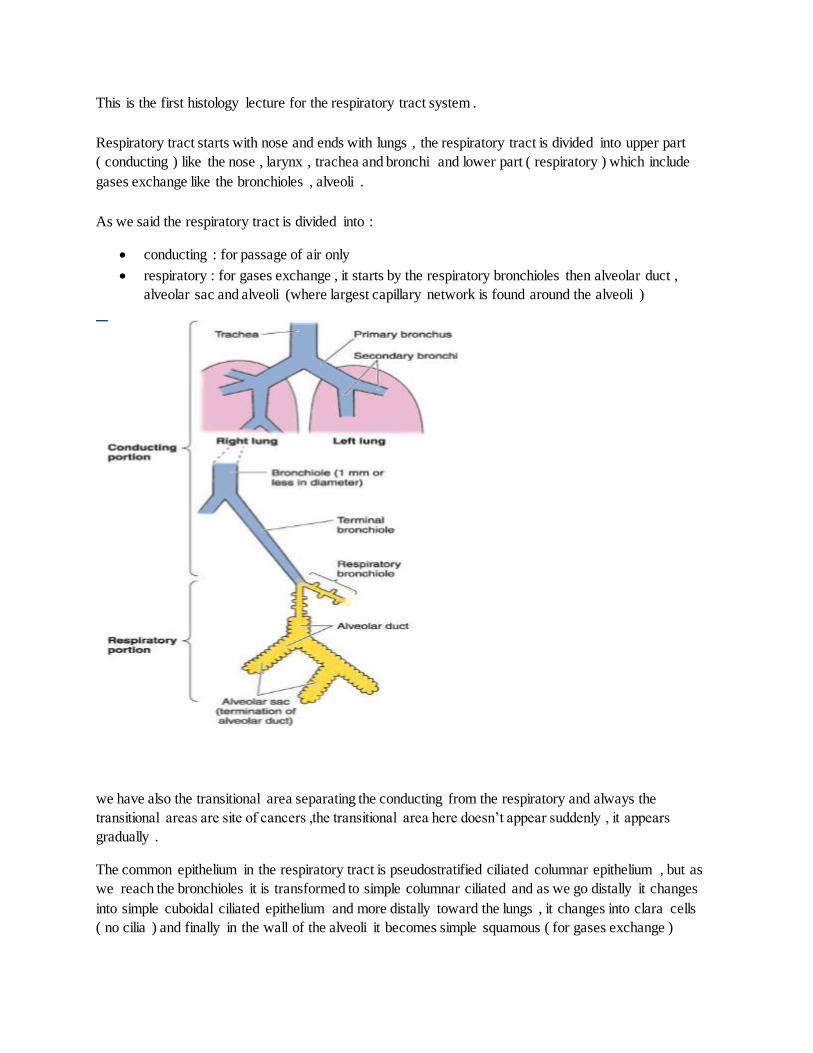
Respiratory tract starts with nose and ends with lungs , the respiratory tract is divided into upper part
( conducting ) like the nose , larynx , trachea and bronchi and lower part ( respiratory ) which include
gases exchange like the bronchioles , alveoli .
As we said the respiratory tract is divided into :
conducting : for passage of air only
respiratory : for gases exchange , it starts by the respiratory bronchioles then alveolar duct ,
alveolar sac and alveoli (where largest capillary network is found around the alveoli )
we have also the transitional area separating the conducting from the respiratory and always the
transitional areas are site of cancers ,the transitional area here doesn’t appear suddenly , it appears
gradually .
The common epithelium in the respiratory tract is pseudostratified ciliated columnar epithelium , but as
we reach the bronchioles it is transformed to simple columnar ciliated and as we go distally it changes
into simple cuboidal ciliated epithelium and more distally toward the lungs , it changes into clara cells
( no cilia ) and finally in the wall of the alveoli it becomes simple squamous ( for gases exchange )
Goblet cells start with large number but as we go distally , the number is reduced , and once we reach the
bronchioles , they become scattered and finally disappear . Also there are seromucous gland with the
same story , they decrease in number as we go distally .
Cartilage also changes , in the nose there is some cartilage and bone ( nasal bone ) , and in the larynx
there are hyaline cartilage and elastic cartilage , and in trachea there is C shaped hyaline cartilage to
prevent the collapse to keep the passage of air , but when we go distally until we reach the bronchioles
there are no cartilage , no goblet cells , no glands BUT the elastic fibers increase , and they are found with
high amount at the lungs because they are needed for inflation and deflation .
In the lungs we have lobes ( three lobes and two fissures in the right and two lobes with one fissure in the
left ) , primary and secondary bronchus enter the lobes , then the secondary becomes tertiary . Inside the
lobes , there are lobules and each lobule is surrounded by connective tissue , which has it’s own blood
supply , venous drainage and lymphatics so these days the surgeon is able to remove the lobule only not
the whole lobe .
the doctor is advising us not to SMOKE , 90% of lung cancers are due to smoking , because smoking
affects a protein called dynein , which is responsible for the movement of cilia ( from inside to outside ) ,
so by affecting the dynein will lead to prevent this movement , leading to some disease like bronchitis and
eventually to cancers .
again and again , the doctor is talking about the two parts of the respiratory tract , and he mentioned that
as we go distally the diameter is decreasing , starting from the trachea which is like tip of the finger and
reaching the large bronchioles which is 1 mm in diameter and eventually in terminal bronchioles .5 mm ,
in the respiratory part it is less than .5 mm and the wall here is simple squamous epithelium .
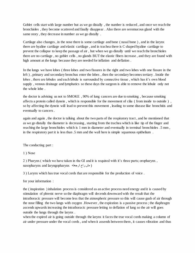
The conducting part :
1 ) Nose
2 ) Pharynx ( which we have taken in the GI and it is required with it’s three parts; oropharynx ,
nasopharynx and laryngopharynx مش راح ارجعله )
3 ) Larynx which has true vocal cords that are responsible for the production of voice .
for your information :
the ( inspiration ) inhalation process is considered as an active process need energy and it is caused by
stimulation of phrenic nerve so the diaphragm will decends downward with the result that the
intrathoracic pressure will become less that the atmospheric pressure so this will cause gush of air through
the nose filling the two lungs with oxygen .However , the expiration is a passive process ; the diaphragm
ascends upwards increasing the intrathoracic pressure letting to deflation of lung so the air will goes
outside the lungs through the larynx .
when the expired air is going outside through the larynx it faces the true vocal cords making a column of
air under pressure under the vocal cords , and when it assends between them , it causes vibration and thus
production of voice . Sudden opening after closure of these cords will cause the coughing ( a protection
process )
also when anyone tried to lift heavy object , the vocal cords will adduct to produce pressure column of air
so he won’t breath till putting it off .
4 ) Bronchi , and we have main bronchus on the right and other one on the left , each is going to it’s lung -
to the hilum - ( extrapulmonary) , the main bronchus is the primary , the secondary is lobar , three in the
right lung and two in the left lung ( inside the lung > intrapulmonary ) bronchi to the lobes , and then the
tertiary ( segmental or bronchopulmonary segment 10 on the right and 10 on the left but at birth , you will
find 8 on the left and 10 on the right ) which is also intrapulmonary .
5 ) Bronchioles , the diameter is smaller and they are intrapulmonary , no cartilage , no glands , no goblet
cells and there is changes in epithelium , they are of two kinds , conducting ends with terminal and
respiratory .
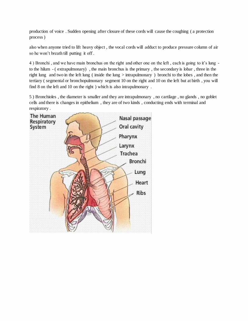
Section in the trachea :
any part of the respiratory tract has the following layers :
1 ) mucosa which has lining epithelium which is - pseudostratified ciliated columnar in general - , lamina
propia muscularis mucosa also
2 ) submucosa which has connecting tisse , glands ( seromucous ) , blood vessels , nerves and lymphatics
3 ) supportive layer which has cartilage like the hyaline cartilage , elastic fibers , blood vessels , glands ,
smooth muscles and lymphatics
( smooth muscles increase as we go distally , and they are spiral in bronchioles - the doctor mentioned the
cause of asthma that is contraction of the smooth muscles which will cause narrowing the lumen of
bronchioles with the result that the patient will have difficulty in breathing and wheezing during
breathing , treatment will be by taking smooth muscle relaxant like adrenaline )
4 ) adventitia which is connecting tissue and can be found as serosa in pleura which surrounds the lung
which is simple squamous epithelium ( mesothelium like the peritoneum ) .
there are more functions for the conducting part other than conduction of air , like cleaning , moistening
and warming .
we will notice that at the beginning , in the nose there is hair which will clean the air from the dust , and
also there is venous blood in the mucosa and submucosa which will cause warming and moistening to the
air as a protecting mechanism .
The doctor mentioned again the conducting and the respiratory part of the tract -,- and the alveolar duct
will end with the alveolar sac , and the whole wall is the alvioli which do the principle function of the
reparatory tract .
Gases Exchange :
The oxygen concentration which will reach the cappilary is low almost 40 , but after the gases exchange ,
the hemoglobin takes the oxygen ,so the oxygen concentration increase.
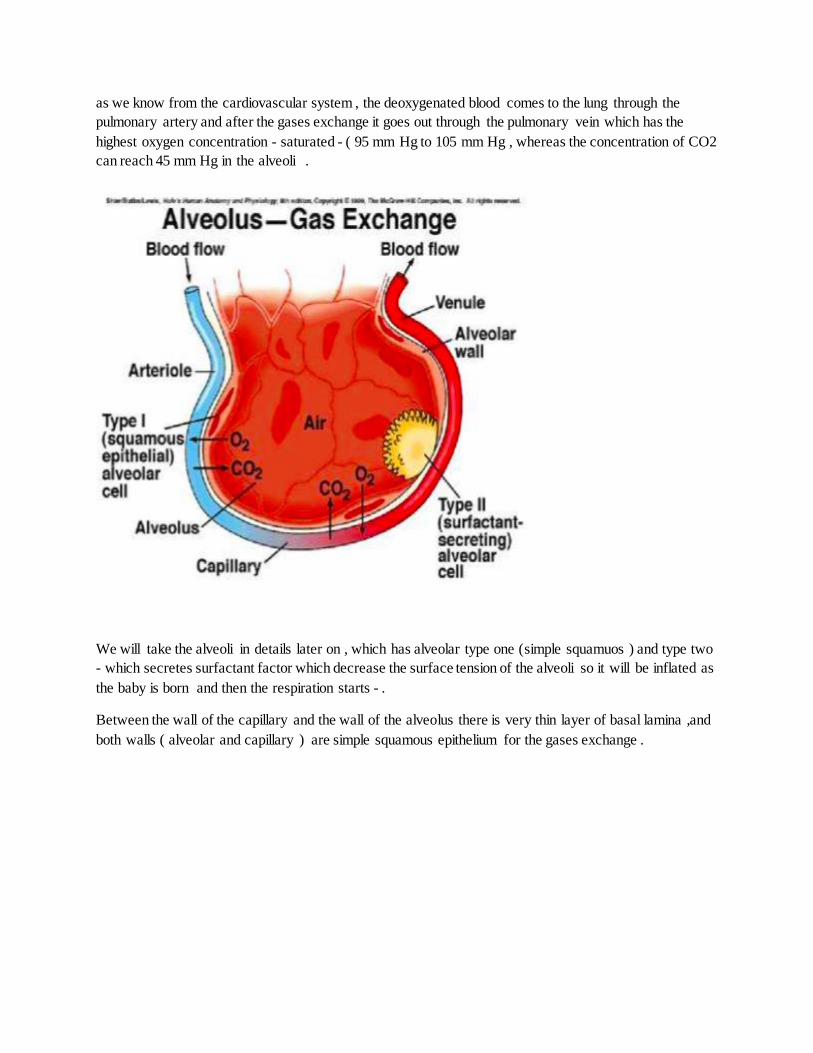
as we know from the cardiovascular system , the deoxygenated blood comes to the lung through the
pulmonary artery and after the gases exchange it goes out through the pulmonary vein which has the
highest oxygen concentration - saturated - ( 95 mm Hg to 105 mm Hg , whereas the concentration of CO2
can reach 45 mm Hg in the alveoli .
We will take the alveoli in details later on , which has alveolar type one (simple squamuos ) and type two
- which secretes surfactant factor which decrease the surface tension of the alveoli so it will be inflated as
the baby is born and then the respiration starts - .
Between the wall of the capillary and the wall of the alveolus there is very thin layer of basal lamina ,and
both walls ( alveolar and capillary ) are simple squamous epithelium for the gases exchange .
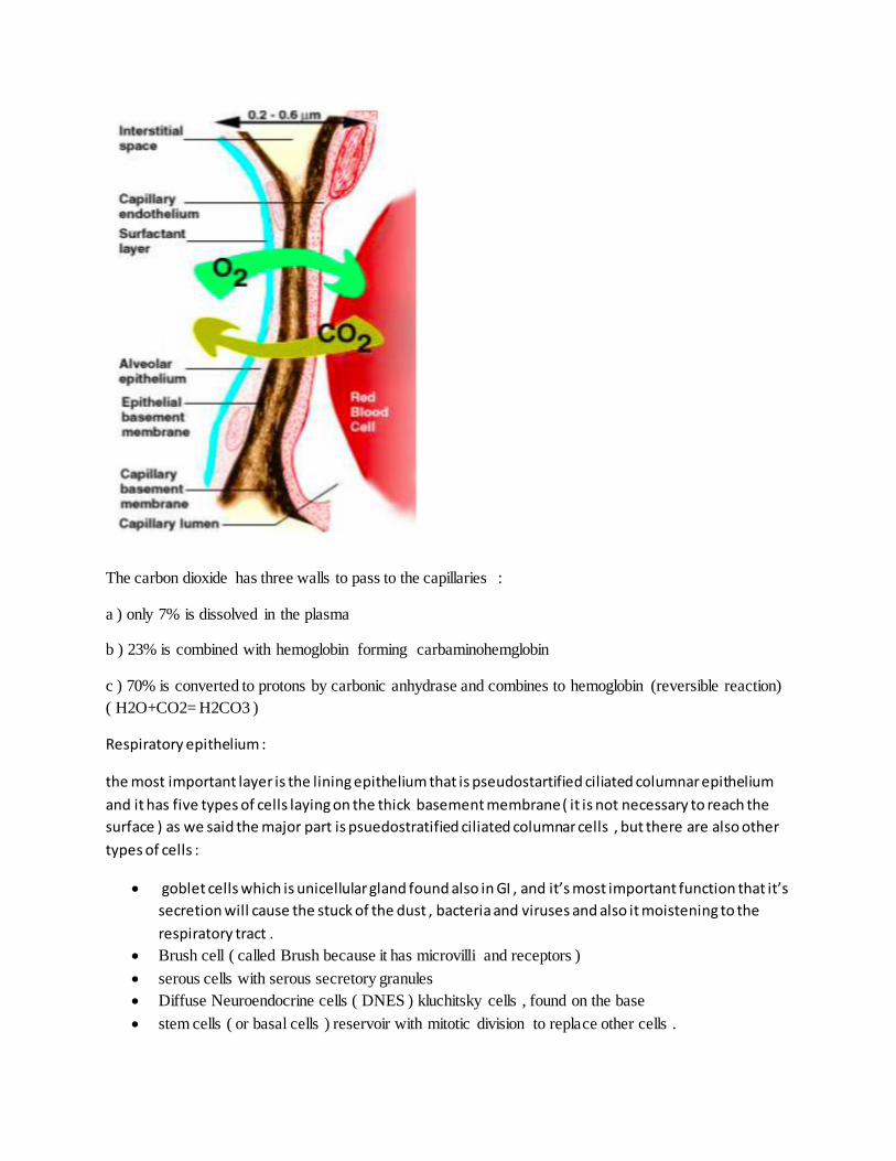
The carbon dioxide has three walls to pass to the capillaries :
a ) only 7% is dissolved in the plasma
b ) 23% is combined with hemoglobin forming carbaminohemglobin
c ) 70% is converted to protons by carbonic anhydrase and combines to hemoglobin (reversible reaction)
( H2O+CO2= H2CO3 )
Respiratory epithelium :
the most important layer is the lining epithelium that is pseudostartified ciliated columnar epithelium
and it has five types of cells laying on the thick basement membrane ( it is not necessary to reach the
surface ) as we said the major part is psuedostratified ciliated columnar cells , but there are also other
types of cells :
goblet cells which is unicellular gland found also in GI , and it’s most important function that it’s
secretion will cause the stuck of the dust , bacteria and viruses and also it moistening to the
respiratory tract .
Brush cell ( called Brush because it has microvilli and receptors )
serous cells with serous secretory granules
Diffuse Neuroendocrine cells ( DNES ) kluchitsky cells , found on the base
stem cells ( or basal cells ) reservoir with mitotic division to replace other cells .
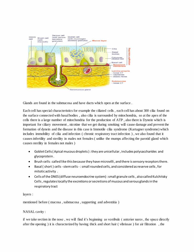
Glands are found in the submucosa and have ducts which open at the surface .
Each cell has special characteristics for example the ciliated cells , each cell has about 300 cilia found on
the surface connected with basal bodies , also cilia is surrounded by mitochondria, so at the apex of the
cells there is a large number of mitochondria for the production of ATP , also there is Dynein which is
important for ciliary movement , nicotine that we get during smoking will cause damage and prevent the
formation of dynein and the disease in this case is Immotile cilia syndrome (Kartagner syndrome) which
includes immobility of cilia and infection ( chronic respiratory tract infection ) , we also found that it
causes infertility and sterility in males not females ( unlike the mumps affecting the parotid gland which
causes sterility in females not males )
Goblet Cells ( Apical mucous droplets ) : they are unicellular , includes polysaccharides and
glycoprotein .
Brush cells : called like this because they have microvilli , and there is sensory receptors there.
Basal ( short ) cells - stem cells - : small rounded cells, and considered as reserve cells , for
mitotic activity .
Cells of the DNES (diffuse neuroendocrine system) : small granule cells , also called Kulchitsky
Cells , regulates locally the excretions or secretions of mucous and serous glands in the
respiratory tract
layers :
mentioned before ( mucosa , submucosa , supporting and adventitia )
NASAL cavity :
if we take section in the nose , we will find it’s beginning as vestibule ( anterior nares , the space directly
after the opening ) it is characterized by having thick and short hair ( vibrissae ) for air filtration , the

epithelium is stratified squamous non keratinized , and has sebaceous and sweat gland ( like the skin )
ONLY the vestibule .
P.s : the doctor said that it is non keratinized , but according to Wikipedia , it is keratinized .
Then the respiratory area , three conchae like shelf ( process ) , three meatuses ( groove under them ) and
one recess “ lateral wall “
The respiratory area is were the respiration occurs , the epithelium is pseudostratified ciliated columnar
with seromucos gland , and what characterize it is having venous blood plexus within the submucosa for
warming the air for protection.
The last area is olfactory which is responsible for smell sensation found under the cribriform plate of
ethmoid ( called like this because olfactory filaments enter through it ) plate of ethmoid , on the roof and
septum . And there is respiratory mucosa in addition to olfactory epithelium with bipolar cells which are
responsible to convert the odor stimulus from chemical to mechanical then to electrical impusle .
bipolar means it has two poles - two ends - , one has hair leads and the other is nerve fibers forming
olfactory filaments - appear from the base - then olfactory bulb then tract , around them there is
supportive or sustintacular cells supporting the bipolar , also there is gland called bowmans glad secrete
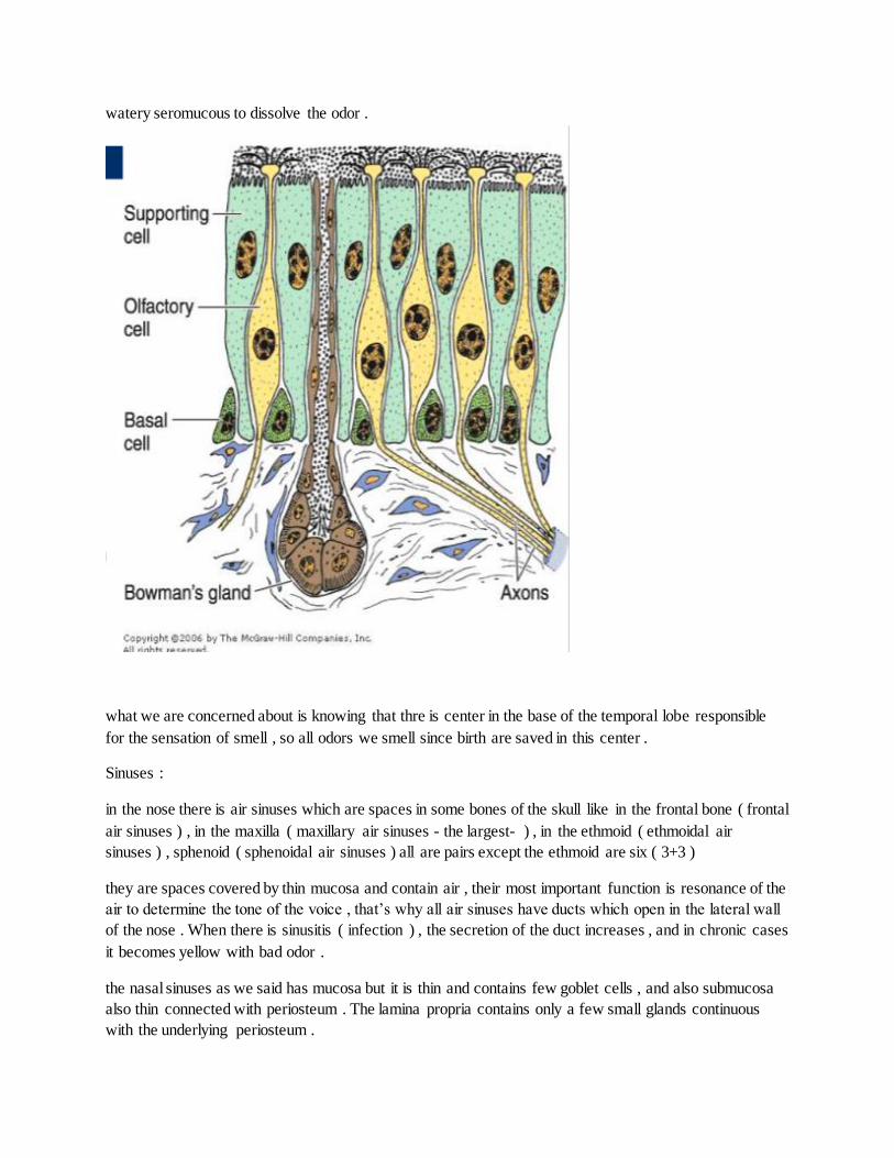
watery seromucous to dissolve the odor .
what we are concerned about is knowing that thre is center in the base of the temporal lobe responsible
for the sensation of smell , so all odors we smell since birth are saved in this center .
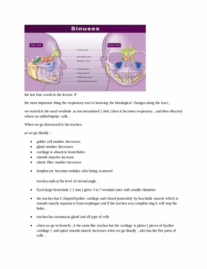
Sinuses :
in the nose there is air sinuses which are spaces in some bones of the skull like in the frontal bone ( frontal
air sinuses ) , in the maxilla ( maxillary air sinuses - the largest- ) , in the ethmoid ( ethmoidal air
sinuses ) , sphenoid ( sphenoidal air sinuses ) all are pairs except the ethmoid are six ( 3+3 )
they are spaces covered by thin mucosa and contain air , their most important function is resonance of the
air to determine the tone of the voice , that’s why all air sinuses have ducts which open in the lateral wall
of the nose . When there is sinusitis ( infection ) , the secretion of the duct increases , and in chronic cases
it becomes yellow with bad odor .
the nasal sinuses as we said has mucosa but it is thin and contains few goblet cells , and also submucosa
also thin connected with periosteum . The lamina propria contains only a few small glands continuous
with the underlying periosteum .
the last four words in the lecture :P
the most important thing the respiratory tract is knowing the histological changes along the tract ;
we started in the nasal vestibule as non keratinized ( skin ) then it becomes respiratory , and then olfactory
where we added bipolar cells .
When we go downward to the trachea :
as we go distally :
goblet cell number decreases
gland number decreases
cartilage is absent in bronchioles
smooth muscles increase
elastic fiber number increases
lymphocyte becomes nodules after being scattered
trachea ends at the level of sternal angle .
Each large bronchiole ( 1 mm ) gives 5 to 7 terminal ones with smaller diameter
the trachea has C shaped hyaline cartilage and closed posteriorly by brachialis muscle which is
smooth muscle separate it from esophagus and if the trachea was complete ring it will stop the
bolus .
trachea has seromucus gland and all type of cells
when we go to bronchi , it the same like trachea but the cartilage is plates ( pieces of hyaline
cartilage ) and spiral smooth muscle increases when we go distally , also has the five parts of
cells .

PLEASE REFER TO the slides especially the last parts , the doctor didn’t mention
everything .
P.S : excuse me for the bad editing but I have done this sheet online and the editing options are
not enough .
Done by : Mohammad Abu Dosh .
