this is an open access document downloaded from orca, … · of the basal ganglia were highly...
TRANSCRIPT

This is an Open Access document downloaded from ORCA, Cardiff University's institutional
repository: http://orca.cf.ac.uk/94878/
This is the author’s version of a work that was submitted to / accepted for publication.
Citation for final published version:
Bruce, Laura L., Erichsen, Jonathan Thor and Reiner, Anton 2016. Neurochemical
compartmentalization within the pigeon basal ganglia. Journal of Chemical Neuroanatomy 78 , pp.
65-86. 10.1016/j.jchemneu.2016.08.005 file
Publishers page: http://dx.doi.org/10.1016/j.jchemneu.2016.08.005
<http://dx.doi.org/10.1016/j.jchemneu.2016.08.005>
Please note:
Changes made as a result of publishing processes such as copy-editing, formatting and page
numbers may not be reflected in this version. For the definitive version of this publication, please
refer to the published source. You are advised to consult the publisher’s version if you wish to cite
this paper.
This version is being made available in accordance with publisher policies. See
http://orca.cf.ac.uk/policies.html for usage policies. Copyright and moral rights for publications
made available in ORCA are retained by the copyright holders.

1
Neurochemical Compartmentalization within the Pigeon Basal Ganglia
by
Laura L. Brucea, Jonathan T. Erichsenb and Anton Reinerc
aDepartment of Biomedical Sciences, Creighton University, Omaha NE, USA
bSchool of Optometry and Vision Sciences, Cardiff University, Cardiff, UK
cDepartment of Anatomy and Neurobiology, The University of Tennessee Health Science
Center, Memphis, TN, USA
Corresponding author:
Laura L. Bruce
Department of Biomedical Sciences
Creighton University School of Medicine,
Omaha, NE 68178
Email: [email protected]

2
Highlights
1. Sixteen distinct compartments were identified in the pigeon basal ganglia using multiple
neurochemical markers.
2. The striatum contains neurochemical regions comparable to the mammalian
somatomotor and associational striatum.
3. A neurochemically distinct area located in the medialmost striatum of pigeons appears
to be unique to birds.
4. The ventral striatum contains neurochemical regions similar to the mammalian
accumbens core, shell, and rostral areas.
5. Most of the main compartments of the basal ganglia were highly conserved during
tetrapod evolution, yet unique avian compartments representing diversification have also
evolved.
Key Words: (6 max)
striatum; accumbens; globus pallidus; ventral pallidum; bed nucleus of stria terminalis; avian

3
Abstract
The goals of this study were to use multiple informative markers to define and characterize the
neurochemically distinct compartments of the pigeon basal ganglia, especially striatum and
accumbens. To this end, we used antibodies against 12 different neuropeptides, calcium-
binding proteins or neurotransmitter-related enzymes that are enriched in the basal ganglia.
Our results clarify boundaries between previously described basal ganglia subdivisions in birds,
and reveal considerable novel heterogeneity within these previously described subdivisions.
Sixteen regions were identified that each displayed a unique neurochemical organization. Four
compartments were identified within the dorsal striatal region. The neurochemical
characteristics support previous comparisons to part of the central extended amygdala,
somatomotor striatum, and associational striatum of mammals, respectively. The medialmost
part of the medial striatum, however, has several unique features, including prominent pallidal-
like woolly fibers and thus may be a region unique to birds. Four neurochemically distinct
regions were identified within the pigeon ventral striatum: the accumbens, paratubercular
striatum, ventrocaudal striatum, and the ventral area of the lateral part of the medial striatum
that is located adjacent to these regions. The pigeon accumbens is neurochemically similar to
the mammalian rostral accumbens. The pigeon paratubercular and ventrocaudal striatal
regions are similar to the mammalian accumbens shell. The ventral portions of the medial and
lateral parts of the medial striatum, which are located adjacent to accumbens shell-like areas,
have neurochemical characteristics as well as previously reported limbic connections that are
comparable to the accumbens core. Comparisons to neurochemically identified compartments
in reptiles, mammals, and amphibians indicate that, although most of the basic compartments
of the basal ganglia were highly conserved during tetrapod evolution, uniquely avian
compartments may exist as well.

4
Abbreviations
Ac accumbens
BM nucleus basalis magnocellularis
BSTL bed nucleus of stria terminalis, lateral part
BJ juxtacapsular part of BSTL
Bv ventral part of the BSTL
GP globus pallidus
INP intrapeduncular nucleus
LFB lateral forebrain bundle
LOT lateral olfactory tract
LPS lamina pallio-subpallialis
LSt lateral striatum
MSt medial striatum
MStM medial part of the medial striatum
LStM lateral part of the medial striatum
StVC ventral striatum, ventrocaudal part
StP ventral striatum, paratubercular region
QF quintofrontal tract
TSM tractus septopallio-mesencephalicus
TuO tuberculum olfactorium
VP ventral pallidum

5
1. Introduction
The basal ganglia play a critical role in modulating motor functions. Similar types of
neurons and fibers have been identified in the basal ganglia of birds, reptiles, amphibians and
mammals, which co-express similar neuropeptides and appear to have similar functions
(Anderson and Reiner, 1990a; Reiner and Anderson, 1990). Homologues of the main
components of the basal ganglia have been identified in mammals, birds, and reptiles, including
striatum, nucleus accumbens, bed nucleus of stria terminalis, globus pallidus (or dorsal
pallidum), and ventral pallidum (Medina and Reiner, 1997; Smeets et al., 2000; Roberts et al.,
2002; Reiner et al., 2004b; Balint and Csillag, 2007; Kuenzel et al., 2011). The boundaries of
these major subdivisions have, however, not necessarily been clearly defined in all cases.
The main components of the avian striatum are the medial striatum, lateral striatum,
and accumbens, each of which has a distinct neurochemical expression pattern (Reiner et al.,
1994, 2004b). The medial striatum (previously named the lobus parolfactorius) has not been
associated with a specific homologous field with the mammalian striatum, although it has some
markers in common with the nucleus accumbens, and others in common with striatum proper
(Reiner et al., 2004b). Similarly, the lateral striatum (previously the paleostriatum
augmentatum) in birds is clearly striatal in nature but has not been definitively related to any
one specific part of mammalian striatum. Neurochemical and connectional studies have
suggested that the avian nucleus accumbens consists of regions corresponding to the
mammalian accumbens core, shell, and rostrum (Balint and Csillag, 2007), but the boundaries
of these regions have been elusive. Thus, in spite of numerous studies on avian basal ganglia,
boundaries between and within its major subdivisions remain poorly documented, particularly
in the ventral striatum, in part because only a limited number of neurochemical markers have
been used for identifying different compartments. Moreover, prior studies show regional
neurochemical heterogeneity throughout avian basal ganglia that needs to be better defined
for a clearer understanding of avian basal ganglia organization and to facilitate its comparison
to the basal ganglia in other vertebrate groups.
The goals of this study were to use multiple informative markers to define and
characterize the neurochemically distinct compartments of the pigeon basal ganglia, especially

6
those of the striatum and accumbens. To this end, we used antibodies against 10 different
neuropeptides, calcium-binding proteins or neurotransmitter-related enzymes known to be
enriched in the basal ganglia: (1) calbindin (CALB), (2) cholecystokinin (CCK), (3) choline acetyl
transferase (ChAT), (4) glutamic acid decarboxylase (GAD), (5) leucine-enkephalin-(ENK), (6)
neuropeptide Y (NPY), (7) parvalbumin (PARV), (8) substance P (SP), (9) tyrosine hydroxylase
(TH), and (10) vasoactive intestinal polypeptide (VIP). In addition, antibodies against calretinin
(CR) and the neuropeptide cocaine- and amphetamine-regulated transcript (CART) were used
to discriminate striatal accumbens territories. Our results clarify boundaries between
previously described basal ganglia subdivisions in birds, and reveal considerable novel
heterogeneity within these previously described subdivisions. Comparisons to neurochemically
identified compartments in reptiles, mammals, and amphibians indicate that although most of
the basic compartments of the basal ganglia were highly conserved during tetrapod evolution,
as previously noted, uniquely avian compartments may exist as well.
2. Materials and methods
The brains of sixteen adult homing pigeons (Columba livia), which were immunostained
for CCK, ChAT, CR, GAD, ENK, NPY, TH, SP, VIP, CALB or PARV, were used in these studies.
Sections from one of these were stained sequentially with the first 8 antibodies and provide the
images in figs. 2-8. All procedures employed in this study were approved by the Animal Care
Committees at State University of New York at Stony Brook and the University of Tennessee.
The pigeons were deeply anesthetized with sodium pentobarbital (70 mg/kg) and perfused
through the left ventricle with in 0.1M phosphate buffer (PB; pH 7.4) with 0.75% sodium
chloride followed by 4% paraformaldehyde in PB. Brains were removed from the skulls, placed
in fixative at 4oC for 3-4 hrs, and then transferred to a cryoprotective solution of 30% sucrose
and 0.1% sodium azide in PB for 3-4 days. Brains were sectioned at 30-40 m on a sliding,
freezing microtome in the transverse plane used in the Karten and Hodos (1967) brain atlas.
For each antibody, sections were washed in PB, immersed in 0.3% H2O2 in PB for 10 min, and
rinsed again in PB prior to antibody incubation.

7
Some of the tissue used in this study was used for analyses of other brain systems in
earlier reports (Erichsen et al., 1991; Krebs et al., 1991; Riters et al. 1999), and the
immunohistochemical procedures we employed are described there in greater detail. For the
immunohistochemical staining employed to generate the tissue illustrated in figures 2-8, the
following standard procedure was used (see Table 1 for optimal primary antibody
concentrations and antibody sources). All sections were incubated in a 1.5% solution of normal
serum in 0.1 M PB with 0.3% Triton X-100 (Sigma Chemical, St. Louis, MO) (PBX) appropriate for
the animal source of the primary antibody. Following a brief wash in PB as noted above, each
series of sections was then incubated in a primary antibody diluted in PBX containing 1.5%
normal serum for 65-89 hours at 4oC. Several washes preceded a 1 hour incubation in
biotinylated IgG (Vector Labs, Burlingame, CA) (diluted 1:200 in PBX) directed against the
species originating each of the primary antibodies used in the study. After another wash in PB,
the sections were preincubated in ABC solution (1:50 dilution; Vector Labs) for one hour before
being placed in a solution of DAB with H2O2 for an additional 15 min. Sections were then
washed, mounted onto slides, osmicated and coverslipped for subsequent examination. For
immunolabeling with CALB and PARV, the method described in Laverghetta et al. (2006) was
used. For SP, ENK and VIP, colchicine-treated material that had been prepared for use in prior
studies was also available (Anderson and Reiner, 1990a,b, 1991).
The distribution patterns of ChAT, VIP, ENK, TH, CCK, NPY, GAD, SP, CALB, PARV, CR, and
CART within the lateral wall of the pigeon subpallium were analyzed in a parallel series of
transverse sections through the forebrain (Figs. 2-10). Sections were imaged using a Nikon USB
5 megapixel CCD camera attached to a Nikon Optiphot microscope and connected to a High-
End Desktop M55 computer. The gray scale and image contrast were adjusted, and figures
labeled and formatted, using CorelDraw and Corel PhotoPaint 12. Six transverse planes
through the striatum rostral to the anterior commissure were selected to illustrate the
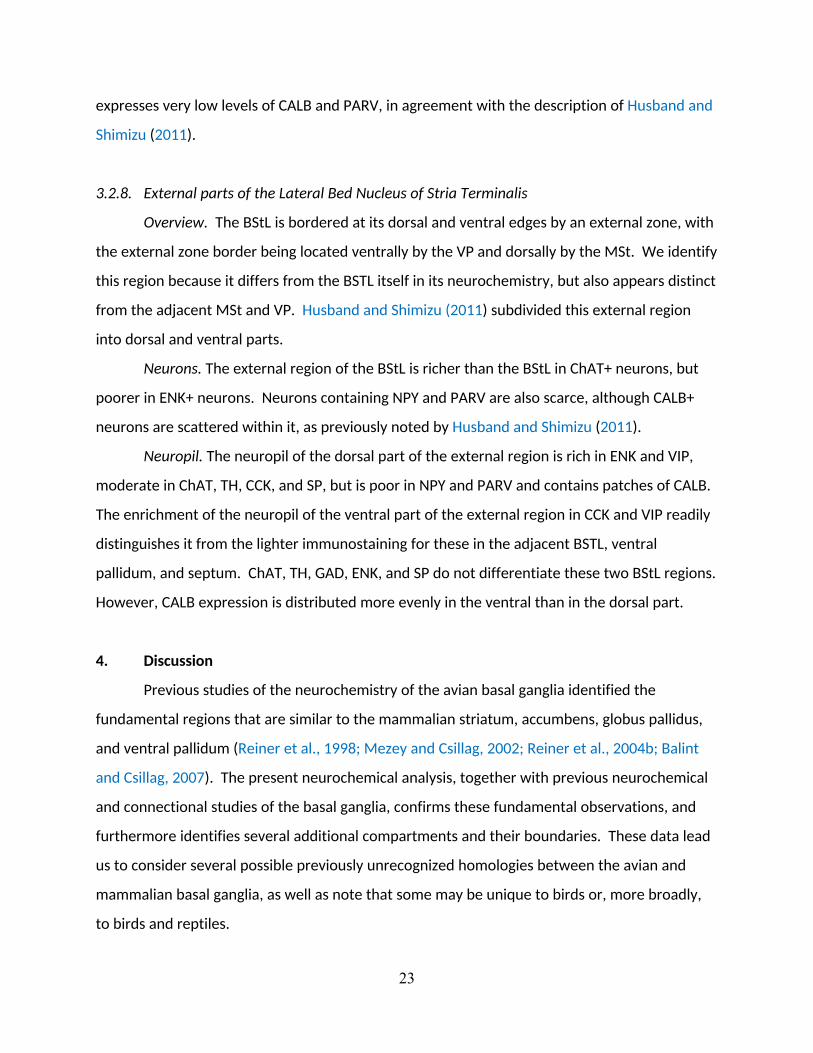
expression patterns within each neurochemical compartment using 8 stains (Fig. 1). The
regional intensity of immunolabeling was densitometrically measured to quantitatively
distinguish compartments (Table 2). The types of perikarya, and the dendrites and terminals
that contained the markers were qualitatively analyzed to define the limits of the known

8
subregions within the striatal and pallidal part of the subpallium, as well as to identify novel
subdivisions. Immunolabeling of neuropil intensity was quantitatively measured in images prior
to any contrast adjustments by recording intensity levels at three points within each
compartment and at each level using the Corel Photo-Paint eyedropper tool (Table 2).
Measurements were ranked at 4 levels: very intense or abundant immunolabeling, intense or
abundant immunolabeling, moderately abundant or intense immunolabeling, and background
or near background immunolabeling). Abundance of labeled perikarya was qualitatively rated
separately (Table 3). The revised avian nomenclature of Reiner et al. (2004b) is used, but
expanded upon where labeling patterns suggest there are previously unrecognized
compartments.
2.1 Antibody characterization and specificity
A list of all antibodies used in the present study is shown in Table 1, including the immunogens
and dilutions used and their sources. Each antibody produced characteristic patterns of immunostaining
that were expected based on previous reports in mammals or birds.
Western blot studies of the monoclonal anti-calbindin-D-28K by the manufacturer showed a
single band at 28 kD, and also showed that it does not react with other members of the EF-hand family.
It recognizes calbindin, but not PARV or CR (Conde et al., 1994). Our data are consistent with a study
that described the distribution pattern of CALB in the bed nucleus of stria terminalis and accumbens in
the pigeon using another CALB antibody (Husband and Shimizu, 2012). In mammals calbindin
immunostaining demarcates rostral associative and paralimbic subdivisions of the striatum (Morel,
2002), which it also does in the present study.
The polyclonal anti-calretinin antibody recognizes a 29 kDa protein in amphibians, lizards, and
chicks (Hack et al., 2000; Morona and González, 2008; Yan et al., 2010) and has been widely used in
studies of diverse vertebrate species. Within the rodent basal ganglia this antibody is a marker for
terminals in the olfactory tubercle, medial parts of nucleus accumbens, as well as numerous small
interneurons throughout the basal ganglia (Bubser et al., 2000). The present study focused on the
pattern of CR terminal labeling in the basal ganglia, which we found is consistent with results in rodents.
The immunogen for the polyclonal anti-cholecystokinin octapeptide antibody is sulfated CCK-8
(26-33) coupled to bovine thyroglobulin (BTg) with glutaraldehyde. Immunostaining with this antibody
is abolished by preadsorption with CCK-8. The antibody has been used studies of numerous vertebrate

9
and invertebrate species. Although glutaraldehyde was used to produce the immunogen, this antibody
works very well with paraformaldehyde-fixed tissues, as do other similarly produced antibodies against
immunogens (e.g., Veenman and Reiner, 1996). This antibody is a marker for terminals in the ventral
accumbens and the olfactory tubercle of rodents (Zaborszky et al., 1985; Zahm and Heimer, 1988),
which is consistent with the labeling pattern seen in pigeons in the present study.
The monoclonal cocaine- and amphetamine-regulated transcript peptide antibody was
generated against a rat CART (54-102) fragment (Thim et al., 1998). The specificity of this antibody has
been demonstrated by omission of primary antibodies and by immunoblot analysis showed a single
precipitin band that migrates at about 14 kD (Subhedar et al., 2011; Singru et al., 2007). Within the
basal ganglia, CART is a marker for neurons in the medial part of the accumbens shell in diverse
vertebrate species, (Smith et al., 1999; Lázár et al., 2004; Barsagade et al., 2011; Subhedar et al., 2011),
which is consistent with the labeling pattern observed in the present study.
The anti-glutamic acid decarboxylase polyclonal antibody preferentially recognizes GAD65 and
also binds to GAD67 (Oertel et al., 1981; Kaufman et al., 1991). It has been characterized by Western
blot analyses using zebra finch cerebellum and forebrain, revealing bands at 61 and 59kD (Spiro et al.,
1995). The rodent and pigeon basal ganglia contain numerous GAD-expressing neurons throughout, and
fibers and terminals in pallidal areas stain particularly heavily with anti-GAD (Veenman et al., 1995; Sun
et al. 2005), which is consistent with the labeling pattern in the present study.
The mouse anti-leu-enkephalin monoclonal antibody to leu-enkephalin has been previously
characterized for specificity by immunodot-blotting and by specific adsorption (Cuello et al., 1984;
Milner et al., 1989), and has been used extensively in a variety of vertebrate species. In the rodent and
pigeon basal ganglia anti-leu-encephalin antibodies have been used as a marker of neurons located
throughout the striatal part of the basal ganglia, as well as for terminals in pallidal territories, including
woolly fibers in the external segment of globus pallidus and in the ventral pallidum (Reiner et al., 1984a;
Anderson and Reiner, 1990a). The present study is confirms these labeling patterns. The pattern of
neuronal labeling is also consistent with prior studies using in situ hybridization histochemistry (Molnar
et al., 1994).
The rabbit polyclonal anti-neuropeptide Y antiserum was raised against a 36 amino acid
sequence that cross reacts with human, rat, and porcine NPY, but not other closely related peptides
(Kienzler et al., 2009). Within the mammalian basal ganglia, NPY is a marker for identifying the
accumbens shell and ventral pallidum (Bálint and Csillag, 2007; Brauer et al., 2000), which we found
useful in the present study.

10
The mouse parvalbumin monoclonal antibody reacts with parvalbumin (12 kDa) from a variety
of mammals as well as frog and fish but does not react with closely related peptides of the EF-hand
family such as calmodulin and intestinal calcium-binding protein. The specificity has been examined by
the manufacturer and researchers (Heizmann and Celio, 1987; Sigma-Aldrich). It has been used
previously in pigeons to describe the distribution of PARV neurons in the basal ganglia (Reiner and
Anderson, 1993; Lavergetta et al., 2006), and the present results are consistent with their findings. Our
data are qualitatively similar to the staining patterns shown in a previous study in in the pigeon using
another PARV antibody (Husband and Shimizu, 2012). In mammals, parvalbumin neuropil
immunostainings demarcates a caudolateral somatomotor subdivision of the striatum (Morel, 2002),
which it also does in the present study.
lmmunohistochemical localization of substance P utilized a monoclonal substance P antibody
(supplier: Sera-Lab, Crawley, England) raised in tissue culture from rat spleen hybridoma. The details of
the production of this antibody and the specificity have been described elsewhere (Cuello et al., 1979).
The specificity of the immunoreactivity for SP was assessed by the use of a blocked control in which the
primary antibody was preabsorbed with synthetic SP (Reiner et al., 1983). In prior studies in mammals
and birds, the striatum was found to contain numerous SP+ neurons and varying intensities of neuropil
stain depending on location, whereas the neuropil of the globus pallidus and ventral pallidum are
characterized by heavy SP staining but few SP+ neurons, consistent with the present results (Cuello et
al., 1979; Reiner et al., 1983).
The specificity of the immunostaining produced with the rabbit tyrosine hydroxylase polyclonal
antiserum is well established (Pickel et al., 1975; Hervonen et al., 1980; Armstrong et al., 1981).
Tyrosine hydroxylase was partially purified from bovine adrenal medulla by precipitation with
ammonium sulfate and column chromatography. The antibody specificity was based on
immunoelectrophoresis of the antibody run against either partially purified tyrosine hydroxylase from
bovine or rat adrenal medulla which yielded a single precipitin band. No precipitin bands formed when
the antibody was run against other catecholamine-synthesizing enzymes, including dopa-decarboxylase,
dopamine beta-hydroxylase, or phenylethanolamine N-methyltransferase (Pickel et al., 1975). TH is a
marker for dopaminergic and adrenergic neurons and fibers. For example, TH immunostaining detects
dopaminergic neurons of the substantia nigra pars compacta and their terminals in the striatum. In the
present study, TH immunostaining of dopaminergic terminals was used to delineate pigeon striatal
subterritories.

11
The rabbit polyclonal antiserum to vasoactive intestinal polypeptide was generously provided by
Dr. J. Walsh (UCLA) and has been widely used in numerous species. The VIP antiserum is specific for the
carboxyl terminal 18-28 region of VIP. Tests for specificity showed negligible reactivity with glucagon,
secretin, and gastric inhibitory polypeptide (Furness et al., 1981). VIP is widespread in the brain, and we
used it as a marker to delineate striatal and pallidal sub-territories.
3. Results
The avian subpallium is traditionally divided into dorsal and ventral subdivisions, each of
which contains striatal and pallidal territories (Reiner et al., 1994). The dorsal and ventral
subpallia are associated with somatic and limbic functions of the basal ganglia, respectively.
The divide between dorsal and ventral subpallial regions can be established most notably by the
rich dopamine beta-hydroxylase (DBH) neuropil in the ventral subpallial striatal areas, whereas
the dorsal subpallium is DBH poor (Reiner et al., 1994). In addition, we have found that a
subset of regions within the DBH-poor dorsal subpallium is rich in parvalbumin, whereas the
DBH-rich ventral subpallial regions, including the bed nucleus of the stria terminalis, are the
opposite. This rationale for the dorsal-ventral somatic/limbic distinction in birds does not
appear to apply for distinguishing mammalian dorsal and ventral subpallial regions. The
differential expression patterns of Islet1, cPax6, cLmo4, and cLmo3 are consistent with this
dorsal-ventral somatic/limbic distinction in birds (Abellán and Medina, 2009), and will be
considered further in the Discussion. Note that we focus on subpallial territories of the lateral
telencephalic wall anterior to the anterior commissure, and do not include amygdalar or septal
subpallial territories.
3.1. Dorsal Subpallium
Based on their neurochemistry, the medial and lateral striatum are each further divided
into medial and lateral zones. The nucleus intrapeduncularis is included here as a striatal part
of the dorsal subpallium, although this assignment is not unambiguous. Finally, we also
recognize distinct striatal cellular islets in the boundary territory between pallium and
subpallium. Only one large pallidal territory is recognized in the dorsal subpallium, namely the
globus pallidus.

12
3.1.1. Medial part of the medial striatum
Neurons. As is true of dorsal striatum in general, the medial MSt contains medium spiny
neurons expressing ENK, SP, and low levels of GAD immunolabeling (Fig. 11B). The sparse GAD
perikaryal immunolabeling is, however, deceptive, because in situ hybridization for GAD65 and
immunolabeling for GABA shows this region to be rich in GAD+/GABA+ perikarya (Veenman et
al., 1995; Sun et al. 2005). Scattered ChAT+ neurons are present, although less frequent than in
the more lateral parts of MSt. NPY+ and CART+ neurons are sparsely scattered throughout the
medial MSt (Fig. 11A). Some VIP+ perikarya are observed in colchicine-treated material. This
area has fewer CALB+ neurons than the adjacent lateral part of MSt. PARV+ neurons are also
sparse in the medial MSt.
Neuropil. The neuropil of the medialmost part of the MSt expresses very high levels of
SP, high levels of GAD and ENK, moderate levels of TH, CCK, NPY, CALB and PARV, and low
levels of ChAT and VIP (Figs. 2-8). The medialmost MSt contains subtle heterogeneously
distributed areas of slightly lighter and darker PARV expression, particularly near the transition
with the lateralmost MSt (Fig. 9A’-D’). The low TH and CALB and higher PARV expression
distinguishes the medialmost MSt, and show that it extends caudally to the level at which the
tractus septopallio-mesencephalicus (TSM) reaches the basal telencephalon. Within the
neuropil of medialmost MSt are “woolly fibers”, which are tight rows of terminals presumed to
be from the medium spiny neurons in the striatum that are rich in SP, ENK, NPY and GAD, and
that synapse along unlabeled dendrites (Fig. 11A,B). Based on prior findings for the globus
pallidus (Reiner and Caraway, 1987), these woolly fiber terminals are likely to end on long,
smooth dendrites of neurons that lightly express LANT6 and/or PARV. Whether these striatal
neurons are interneurons or projection neurons is unknown, although PARV+/LANT6+
expressing pallidal projection neurons, but not interneurons, receive woolly fiber inputs (Reiner
et al., 2004a,b).
3.1.2. Lateral part of the medial striatum

13
Neurons. Neurons containing ChAT are more plentiful in the lateral MSt than in more
medial or lateral striatal compartments. The lateral MSt, like the medial MSt, is rich in medium-
sized spiny projection neurons containing either SP or ENK, as evident in colchicine-treated
material. Scattered NPY+ neurons were also present throughout lateral MSt. Neurons
immunolabeled for GAD are scarce in the lateral MSt, although GABA immunolabeling and
GAD65 in situ hybridization studies have shown that GAD-synthesizing perikarya are in fact
abundant throughout both medial and lateral MSt (Veenman and Reiner, 1994; Sun et al.,
2005). A few neurons immunolabeled for VIP are observed in colchicine-treated cases.
Numerous PARV+ and CALB+ neurons are also present, in contrast with the medial MSt.
Neuropil. The striatal neuropil contains high levels of TH and GAD, moderate levels of
ChAT, ENK, CCK, NPY, SP, and CALB, and low levels of VIP and PARV. The neuropil of the
lateralmost MSt is distinguished from the more medial MSt by its higher expression of TH, ChAT
and CALB and lower expression of PARV. The lateralmost MSt extends medially to the ventricle
at its more caudal levels, that is from the level at which the TSM reaches the basal
telencephalon to the anterior commissure (Figs. 8, 9F,G,G’). The lateralmost medial striatum
contains some woolly fibers, which are strongly labeled for SP, NPY, and ENK, and which
emanate as finger-like extensions from globus pallidus into the lateral MSt. Woolly fibers are,
nonetheless, less abundant in the lateral than medial MSt. VIP and CCK are sparse in the
neuropil of both the medial and lateral MSt.
3.1.3. Medial part of lateral striatum
Neurons. Like other striatal territories, the medial LSt is rich in medium-sized spiny
projection neurons that contain either SP or ENK. The medial LSt is very poor in both ChAT and
NPY neurons, in contrast to the MSt. VIP+ neurons were rarely observed, even in colchicine-
treated tissue. The medial LSt contains numerous PARV+ neurons but few CALB+ neurons.
Large GAD+ neurons resembling the PARV+ neurons in size and frequency are present,
consistent with previous studies showing that GAD and PARV are co-expressed in the same
neurons of LSt (Reiner and Anderson, 1993).

14
Neuropil. The medial part of the LSt contains high levels of TH, GAD, and PARV,
moderate levels of ChAT, VIP, ENK, CCK, NPY, and SP, and low levels of CALB and (Figs. 2-8;
Table 2). The medial part of the LSt extends medially to the ventricle at levels near the anterior
commissure. Woolly fibers are rare, except along the border with the globus pallidus (GP),
where they extend from the GP into the striatum. Except when present in woolly fibers, NPY,
GAD, CCK, and VIP are expressed fairly evenly throughout the LSt neuropil. TH is present in a
graded pattern that is lowest in the medial most MSt, higher in the lateralmost MSt, slightly
lower in the medialmost LSt, and highest in the lateralmost LSt (Figs. 2-8). CALB is found in
higher levels in the lateral part of the medial striatum than in adjacent striatal territories,
whereas PARV and CART are less abundant in the lateral part of the medial striatum than in
adjacent striatal territories (Figs. 9, 10).
3.1.4. Lateral part of lateral striatum
Neurons. The lateral LSt is very similar to the medial LSt. It is rich in SP+ and ENK+
medium-sized spiny projection neurons, but is very poor in ChAT+, NPY+, VIP+, and CALB+
neurons. There are few CALB+ neurons. Numerous large PARV+ and GAD+ neurons likely
represent the same neuronal population (Reiner and Anderson, 1993).
Neuropil. The neuropil in the lateralmost part of the LSt contains very high levels of TH,
high levels of VIP, CCK, and GAD, moderate levels of ChAT, ENK, NPY, and SP, and low levels of
CALB and PARV. The lateralmost part of the LSt along the lateral margins of the globus pallidus
is a neurochemically unique area, distinguished by its much higher expressions of VIP, TH, and
CCK in its neuropil (Figs. 6-7), suggesting that this area may have a specialized function and thus
differ from the remainder of lateral LSt. Woolly fibers are rare, except along the border with
the globus pallidus (GP), where they extend from the GP into the striatum. Except when
present in woolly fibers, NPY, GAD, CCK, and VIP are expressed fairly evenly throughout the
lateral LSt neuropil. The lateral LSt can be readily distinguished from the medial LSt by the
paucity of PARV in its neuropil. Of the markers examined in this study, the localization of TH
and the calcium binding proteins provides the best means to distinguish the different striatal
areas (Figs. 2-8). In the case of the lateral striatum, CALB is found in higher levels in the

15
neuropil of the lateral striatum than in the medial striatum, whereas PARV is less abundant in
the neuropil of the lateral part of the lateral striatum than in the medial parts of the lateral
striatum (Fig. 9).
3.1.4. Intrapeduncular Nucleus (INP)
Neurons. The INP is located along the ventromedial border of globus pallidus and lateral
to the MSt (Figs. 7-8). The neuronal profile of INP is largely striatal. For example, many
medium-sized SP+ and ENK+ neurons are present in colchicine-treated material in INP,
resembling those seen in MSt and LSt. Prior in situ hybridization studies confirm that the INP
contains many SP+ neurons (Abellán and Medina, 2009) and scattered ENK+ neurons (Molnar et
al., 1994), as well as medium-sized GAD+ neurons (Sun et al., 2005). NPY+ and CALB+ neurons
are sparse in the INP, as they are in most other striatal areas. On the other hand, ChAT+
neurons are much more plentiful in the INP than in the medial or lateral striatum, whereas
PARV+ neurons are scarcer.
Neuropil. The neuropil of INP contains moderate levels of ChAT, TH, GAD, and PARV,
and low levels of VIP, ENK, CCK, NPY, SP, and CALB. The neuropil of the INP can be
distinguished from that of the globus pallidus by the absence of woolly fibers that contain GAD,
ENK and/or SP, and a more intensely PARV+ neuropil, whereas the GP neuropil is rich in woolly
fibers that contain GAD, ENK and/or SP. The neuropil of INP, in general, resembles that of the
striatum. The INP contains many ChAT+ fibers, likely reflecting its enrichment with cholinergic
neurons.
3.1.5. Cell islands within the lamina pallio-subpallialis
Neurons. The dorsal and lateral edge of the striatum is formed by a band of fibers, the
lamina pallio-subpallialis. Tightly clustered neurons form compact islands within it, which are
particularly large and prominent rostrally (Figs. 2-3), but smaller more caudally (Figs. 4-8).
Abellán and Medina have identified these islands in embryonic chick and termed them the
striatal capsule. Neurons within these islands appear to contain medium-sized striatal
projection neuron markers, notably SP+ and ENK+. According to Abellán and Medina (2009),

16
these cells also express the striatal gene cLmo4 and GAD67 (their fig. 13D-F). These neurons
are enriched in DARPP32+ as well (Reiner et al., 1998b).
Neuropil. The neuropil of the islands of the striatal capsule contain high levels of ChAT,
moderate levels of ENK, TH, CALB and PARV, and low levels of VIP, GAD, CCK, NPY, and SP. The
islands are most easily identified by their ChAT immunostaining because the adjacent striatum
stains less intensely, whereas immunostaining for other markers is either absent or similar to
that in the adjacent striatum.
3.1.6 Globus Pallidus
Neurons. The globus pallidus appears rostrally at approximately A11.75 as small finger-
like extensions into rostral striatum (Figs. 4-5) arising from the main body of the more caudal
pallidal zone (Figs. 6-8). The large GABAergic pallidal neurons are enriched in PARV, and so
PARV+ neurons are plentiful in globus pallidus (Laverghetta et al., 2006). ChAT-immunolabeled
neurons are relatively plentiful within the globus pallidus as well, although less numerous than
in the INP. Scattered spiny SP+ and ENK+ neurons are also seen in globus pallidus (Fig. 11C), as
noted previously (Reiner et al., 1983; Molnar et al., 1994), but NPY+, CCK+, and CALB+ neurons
are largely absent.
Neuropil. Woolly fibers dominate the neuropil of the globus pallidus and contain very
high levels of GAD, high levels of ENK and SP, and moderate levels of VIP (Fig. 11C). The woolly
fibers arise from medium spiny neurons in the LSt, which are likewise rich in GAD, SP, and ENK,
and the terminals of LSt neurons form rows of synapses along the sides of the dendrites of
globus pallidus neurons. Since the globus pallidus also contains some VIP+ woolly fibers, it
seems likely that some LSt medium spiny neurons do synthesize VIP, although VIP+ neurons
were not detected in it by immunolabeling. The globus pallidus contains few fibers labeled for
ChAT, TH, CCK, NPY, CALB or PARV, with the paucity of TH typical of a pallidal zone.
3.2. Ventral Subpallium
The ventral subpallium is divided into striatal, pallidal, and mixed populations as
determined by their neurochemical patterns (Abellán and Medina, 2009). Based on our

17
findings and prior studies, we identify the following four striatal regions within the ventral
subpallium: (1) nucleus accumbens, defined by Reiner et al. (2004b) as a ventral part of rostral
striatum, nearest the ventricle; (2) an area lateral to nucleus accumbens that we here refer to
as reticular ventral striatum; (3) a region deep to the olfactory tubercle but external to the
reticular ventral striatum, which we term the paratubercular striatum; (4) a region that extends
dorsal and caudal to the reticular striatum and ventral pallidum that we call the ventrocaudal
striatum (Stvc). Our paratubercular striatum and Stvc corresponds to the “accumbens shell” of
chicken embryos identified by Abellán and Medina (2009). A single pallidal territory of the
ventral subpallium, the ventral pallidum, is considered pallidal based on the presence of woolly
fibers, namely closely spaced parallel rows of synaptic terminals that immunostain strongly for
DARPP32, SP, ENK, CALB or neurotensin (Reiner and Carraway, 1987; Reiner et al., 1998b).
Finally, several subpallial regions have a mixture of striatal and pallidal characteristics: (1) the
lateral bed nucleus of stria terminals (BStL), which is adjacent to the tip of the lateral ventricle
and extends as a thin sheet rostrally along the ventricle at the medial edge of nucleus
accumbens; (2) the juxtacapsular (Bjx) subdivision, which forms the lateral border of the BStL
(N.B. earlier avian studies often called the BStL and Bjx the ‘nucleus accumbens’) (Reiner et al.,
2004b); and (3) the olfactory tubercle, which occupies the superficial surface of the lateral
subpallium and receives olfactory input (Abellán and Medina, 2009).
3.2.1. Nucleus accumbens
Neurons. Nucleus accumbens, as defined in Reiner et al. (2004b) is rich in SP+ and ENK+
neurons, as well as GABAergic neurons (Veenman and Reiner, 1994; Sun et al., 2005), as typical
of striatal domains. ChAT+, VIP+, NPY+, CALB+, and CART+ neurons also are scattered
throughout.
Neuropil. The neuropil of nucleus accumbens is heterogeneous, and largely
characterized by very high levels of VIP, ENK, NPY, and SP, high levels of CCK and GAD,
moderate levels of ChAT, TH, and CALB, and low levels of PARV (Figs. 2-5). The neurochemical
characteristics of its neuropil are particularly useful for defining nucleus accumbens. For
example, the rich levels of SP immunoreactivity distinguish it from adjacent dorsal striatal

18
regions, except the medialmost MSt, which also has high SP neuropil levels. In contrast, TH is
less abundant in the accumbens compared to the dorsal striatal regions, except for the
medialmost MSt. The accumbens neuropil is richer in VIP, ENK, CCK, and NPY than other parts
of the striatum. The VIP fibers, in particular, differentially delineate accumbens from striatal
territory above and lateral to it. The NPY and ENK fibers tend to be distributed in patches of
high and low abundance, not seen with other markers (Figs. 5 C,G). Caudally, the accumbens is
contiguous with the rostral BStL (Fig. 5), but can be easily distinguished by the much higher
levels of SP in accumbens compared to the BStL (Figs. 2-5). In addition, nucleus accumbens is
one of the few striatal areas with high levels of CR and CART in the neuropil (Fig. 10).
3.2.2. Paratubercular striatum
Overview. The paratubercular striatum (Stp) is located superficial (i.e. ventral or
external) to the accumbens and reticular striatum, but deep (i.e. ventral or internal) to the thin,
rostral extension of the ventral pallidum and to the olfactory tubercle, which itself is the thin
olfactory-recipient zone along the ventral surface of the subpallium. Its neuropil exhibits a
unique pattern because it immunostains intensely for most markers in contrast to other striatal
regions, as described below, which is the major reason for recognizing it as a distinct territory
(Figs. 2-6). However, it is difficult to distinguish it from the olfactory tubercle, as both have
similar staining characteristics and some neurons of Stp may receive olfactory bulb input, like
the olfactory tubercle, via dendrites that extend into the olfactory terminal zone of olfactory
tubercle. Nonetheless, olfactory terminals do not end in the Stp (Reiner and Karten, 1985; Atoji
and Wild, 2014), and for this reason, we do not include it as part of the olfactory tubercle. The
Stp expands dorsolaterally towards the MSt at the level of the reticular striatum (see below)
just anterior to the rostral pole of the ventral pallidum (Fig. 5). Around the lateral aspect of VP,
it blends with the Stvc (Fig. 6).
Neurons. The Stp contains SP+ and ENK+ neurons, but only scattered ChAT+, NPY+, and
PARV+ neurons. VIP+ neurons were rarely seen. CART neurons are also present within the Stp
(Fig. 10), which differentiates the Stp from most other striatal areas.

19
Neuropil. The Stp neuropil contains very high levels of VIP, ENK, TH, CCK, GAD, and SP,
high levels of ChAT and NPY, moderate levels of CALB, and low levels of PARV (Figs. 2-9).
Woolly fibers rich in VIP, ENK, GAD, and SP, and non-woolly fibers rich in TH and CCK are more
abundant rostromedially than caudolaterally. The medial Stp neuropil also displays high levels
of CART (Fig. 10H-I).
3.2.3. Reticular ventral striatum
Overview. The reticular part of the ventral striatum (StR) lies lateral to nucleus
accumbens of Reiner et al. (2004b) and anterior to the rostral pole of the ventral pallidum (Fig.
5). It is better distinguished by its neuropil traits than by its constituent neuronal populations,
as further detailed below. In particular, it is characterized by and named for the reticulated
appearance imparted by the numerous fiber bundles that traverse this region, as most evident
in the TH and CCK immunolabeled tissue.
Neurons. As a striatal region, it contains neurons rich in ENK or SP, which probably co-
contain GABA (Reiner and Anderson, 1993). ChAT+ neurons are scattered throughout, in a
somewhat greater abundance than in nucleus accumbens. Scattered NPY+ and CALB+ neurons
are also present.
Neuropil. The reticular part of the ventral striatum contains very high levels of VIP and
SP, high levels of ChAT, ENK, TH, CCK, NPY and GAD, but moderate levels of CALB and low levels
of PARV. The high SP and VIP combined with low GAD and NPY distributions especially define
this region. It exhibits a unique combination of features that distinguish it from the accumbens
and the overlying MSt. First, compared to the adjacent accumbens the StR neuropil is richer in
ChAT and TH, but poorer in ENK and NPY. Second, compared to the medialmost MSt, the
neuropil is much richer in VIP, TH, CCK, and GAD, but poorer in PARV. Third, it is poor in ENK
and NPY woolly fibers, and the more numerous ENK and NPY woolly fibers of the MSt form a
clear border with the StR. Finally, the abundance of neuropil zones with lower levels of ENK,
TH, CCK, NPY, GAD, and PARV, interspersed with zones displaying higher levels, distinguish it
from the olfactory tubercle and paratubercular striatum below (Figs. 2-6). The pattern of TH
and CCK immunolabeled fibers also characterizes this reticular region, with small zones of richly

20
labeled neuropil interdigitating with small, poorly labeled zones, in contrast to the uniform
neuropil labeling for TH and CCK throughout the dorsal striatum.
3.2.4. Ventrocaudal part of the striatum
Overview. The ventrocaudal part of the striatum (Stvc) appears rostrally just above the
reticular striatum and extends caudally above the ventral pallidum and medial to the globus
pallidus, forming a cup-shaped zone at the caudoventral pole of the lateral MSt (Figs. 7-8). It is
usually included in the lateral part of the MSt because of its location just below it. Abellán and
Medina (2009), however, considered this region as the caudal continuation of the
paratubercular striatum, and the similar immunohistochemical labeling patterns we observed
are consistent with their grouping. Although the ventrocaudal region and the MSt have some
neuronal and neuropil characteristics in common, many other features readily distinguish the
two regions, suggesting it is distinct from the MSt.
Neurons. The ventrocaudal part of MSt, like the other parts of MSt, is rich in medium-
sized spiny projection neurons containing either SP or ENK, as evident in colchicine-treated
material. Scattered NPY+ neurons are present, but there are few PARV+ and CALB+ neurons,
similar to the medial MSt but in contrast to the lateral MSt. Neurons immunolabeled for GAD
are scarce, but GABA immunolabeling and GAD65 in situ hybridization studies have shown that
GAD-synthesizing perikarya are abundant in this area (Veenman and Reiner, 1994; Sun et al.,
2005). Rare neurons immunolabeled for VIP are observed in colchicine-treated cases. The Stvc
has scattered ChAT+ neurons, which are more abundant than in the adjacent lateral part of
MSt, but far less abundant than in the adjacent globus pallidus or INP.
Neuropil. The neuropil of the ventrocaudal part of the striatum contains very high levels
of TH and CCK, high levels of ChAT, VIP, ENK, NPY and GAD, moderate levels of CALB, and low
levels of PARV. It is highly enriched in SP and ChAT. A number of these features distinguish the
ventrocaudal striatum from the MSt. Notably, woolly fibers enriched with SP, ENK and/or VIP
are more numerous in the Stvc. GAD woolly fibers, however, are less intensely labeled in the
Stvc than in the MSt. The non-woolly part of the neuropil contains more ChAT, VIP, TH, and
NPY compared to the MSt. Unlike the medial part of the MSt, but like the lateral part of the

21
MSt, the neuropil of the ventrocaudal striatal region is rich in TH but poor in PARV. At its more
rostral levels (Figs. 6, 7), it has especially high levels of TH+ and CCK+ fibers and terminals. The
enrichment in CALB is similar to the lateral MSt. The similar distributions of TH and CCK in
terminals in the paratubercular, reticulated, and ventrocaudal striatal regions are consistent
with the possibility of their co-localization in the terminals of dopaminergic midbrain neurons in
these regions. CCK has been colocalized to dopaminergic terminals in mammals (Seroogy et al.,
1988).
3.2.5. Ventral Pallidum
Neurons. The ventral pallidum (VP) appears rostrally as a very thin band between the
striatum and olfactory tubercle, and is best distinguished by its very low TH expression
compared to these adjacent regions (Figs. 5D-7D). At approximately A10.75 in the Karten and
Hodos atlas (1967), the VP expands dorsally, and occupies an oval zone ventrolateral to the bed
nucleus of the stria terminalis (Fig. 6). It is rich in large aspiny GABAergic and PARV+ neurons,
which are likely to be co-labeled projection neurons. Very few neurons with medium spiny
projection neuron markers such as SP, ENK, and CALB are present, although ChAT+ neurons are
scattered throughout the VP.
Neuropil. The neuropil of the VP contains very high levels of ENK, NPY, GAD, and SP,
high levels of VIP, moderate levels of ChAT, CCK, and CALB, and low levels of PARV. The SP,
ENK, GAD and NPY immunolabeling take the form of woolly fibers, which occur throughout the
ventral pallidum, whereas VIP+ woolly fibers are only seen in the medial VP. ChAT, TH, and CCK
immunolabeling are conspicuously poorer in VP than in adjacent regions, and thereby serve to
delineate it. The complementarity between the few TH+ or CCK+ fibers, and the many SP+,
ENK+, and NPY+ woolly fibers is especially striking.
3.2.6. Olfactory Tubercle
Overview. The olfactory tubercle is defined here as the thin subpallial region at the base
of the telencephalon that receives olfactory bulb input (Figs. 2-6).

22
Neurons. The rostral and ventral olfactory tubercle contain neurons that express striatal
markers, including ENK and SP. GAD+ neurons are also present in the rostral and ventromedial
olfactory tubercle as expected for a striatal region. ChAT+, NPY+, VIP+, CALB+ and PARV+
neurons are scarce in this territory.
Neuropil. Like other striatal regions, the neuropil of the rostral ventral olfactory tubercle
is rich in ENK, TH, CCK, NPY, GAD, SP, and CALB, moderate in VIP, and poor in PARV. More
caudally and laterally, the olfactory tubercle has some pallidal characteristics, since it is poor in
TH.
3.2.7. Lateral part of the Bed Nucleus of Stria Terminalis
Overview. The lateral bed nucleus of the stria terminalis (BStL) is largest at levels caudal
to the nucleus accumbens (Figs. 6-8). It extends rostrally as a thin sheet along the ventricular
edge of the accumbens, and can be distinguished from the accumbens by its neurochemical
traits (Figs. 4-5, asterisk), as described below.
Neurons. The BStL is characterized by the presence of many ENK+ neurons, which are
also known to be GABAergic, whereas neurons containing ChAT, NPY, SP, CALB, and PARV are
scarce. By contrast, both SP+ and ENK+ neurons are abundant in nucleus accumbens.
Neuropil. Most of the BStL neuropil is rich in ENK but poor by comparison in VIP,
although it contains smaller patches that are poor in ENK and rich in VIP. Other markers are
distributed more homogeneously, and the BSTL is characterized by moderate levels of GAD and
NPY, and only very low levels of ChAT, TH, CCK, and SP. The neurochemistry of the neuropil of
BStL is distinctive, since it is conspicuously poor in SP, ChAT, CCK, and TH compared to the
surrounding striatum, although a TH+ neuropil zone is present at caudal levels (Figs. 7D, 8D).
The neuropil of the BStL and striatum express similar levels of GAD and NPY. Our results are
consistent with a previous suggestion (Abellán and Medina, 2009) that the BStL has both
pallidal and striatal traits, since we observed the pallidal traits of low TH and CCK, and the
striatal trait of many ENK+ neurons. The BStL pallidal traits stem from its development from
the pallidal sector of the lateral ventricle where it is located, and striatal neurons such as those
containing ENK migrate into the pallidal territory (Abellán and Medina, 2009). The BStL
23
expresses very low levels of CALB and PARV, in agreement with the description of Husband and
Shimizu (2011).
3.2.8. External parts of the Lateral Bed Nucleus of Stria Terminalis
Overview. The BStL is bordered at its dorsal and ventral edges by an external zone, with
the external zone border being located ventrally by the VP and dorsally by the MSt. We identify
this region because it differs from the BSTL itself in its neurochemistry, but also appears distinct
from the adjacent MSt and VP. Husband and Shimizu (2011) subdivided this external region
into dorsal and ventral parts.
Neurons. The external region of the BStL is richer than the BStL in ChAT+ neurons, but
poorer in ENK+ neurons. Neurons containing NPY and PARV are also scarce, although CALB+
neurons are scattered within it, as previously noted by Husband and Shimizu (2011).
Neuropil. The neuropil of the dorsal part of the external region is rich in ENK and VIP,
moderate in ChAT, TH, CCK, and SP, but is poor in NPY and PARV and contains patches of CALB.
The enrichment of the neuropil of the ventral part of the external region in CCK and VIP readily
distinguishes it from the lighter immunostaining for these in the adjacent BSTL, ventral
pallidum, and septum. ChAT, TH, GAD, ENK, and SP do not differentiate these two BStL regions.
However, CALB expression is distributed more evenly in the ventral than in the dorsal part.
4. Discussion
Previous studies of the neurochemistry of the avian basal ganglia identified the
fundamental regions that are similar to the mammalian striatum, accumbens, globus pallidus,
and ventral pallidum (Reiner et al., 1998; Mezey and Csillag, 2002; Reiner et al., 2004b; Balint
and Csillag, 2007). The present neurochemical analysis, together with previous neurochemical
and connectional studies of the basal ganglia, confirms these fundamental observations, and
furthermore identifies several additional compartments and their boundaries. These data lead
us to consider several possible previously unrecognized homologies between the avian and
mammalian basal ganglia, as well as note that some may be unique to birds or, more broadly,
to birds and reptiles.

24
4.1. Medial and Lateral Striatum
The dorsal striatum of birds comprises the dorsalmost part of most of the lateral
subpallium, and is composed of the medial and lateral striatum, formerly known as the lobus
parolfactorius and paleostriatum augmentatum, respectively (Karten and Hodos, 1967; Reiner
et al., 2002, 2004b). Our results illustrate striking differences in immunolabeling intensities
between the medial and lateral striatum, which confirms and extends the differences noted by
many other investigators (Reiner et al., 1983; Wynne and Gunturkun, 1995; Gallatioto et al.,
1998; Ballint and Csillag, 2007). The histochemical differences may reflect functional
differences, as for example reflected in connectional differences between the medial and
lateral striatum. The avian medial striatum is connected with areas often characterized as
limbic (Yamamoto and Reiner, 2005). For example, it receives its principal inputs from the
prehippocampal area, olfactory cortex, caudolateral nidopallium, and posterior amygdala, and
projects to the dopaminergic substantial nigra (Karten and Dubbeldam, 1973; Brauth et al.,
1978; Reiner et al., 1983; Bottjer et al., 1989; Veenman et al., 1995). The lateral striatum
receives inputs predominantly related to sensory and motor processing, including the
hyperpallium (visual and somatosensory cortical-like areas), and lateral nidopallium (Veenman
et al., 1995). The lateral striatum projects primarily to the globus pallidus, whereas the medial
striatum projects primarily to the substantia nigra pars compacta and ventral tegmental area
(Karten and Dubbeldam, 1973; Brauth et al., 1978; Lewis et al., 1981; Reiner et al., 1983, 1998a;
Bottjer et al., 1989; Bottjer, 1993; Castro and Ball, 1994; Grisham and Arnold, 1994; Reiner et
al., 1994; Medina and Reiner, 1995; Soha et al., 1996; Luo and Perkel, 1999; Sun and Reiner,
2000). As detailed below, however, our results support division of medial striatum into
neurochemically distinct medial and lateral zones, as suggested by Abellán and Medina (2009),
and that the lateral striatum of birds is also divided into neurochemically distinct medial and
lateral zones.
Most striatal regions are enriched with dopaminergic terminals, identifiable by intense
granular TH immunolabeling, and a high density of medium-sized GABAergic neurons with spiny
dendrites containing SP, ENK, GAD and dopamine-regulated neuronal phosphoprotein

25
(DARPP32) (Reiner et al., 1983; Anderson and Reiner 1990a,b; Reiner et al., 1994; Veenman and
Reiner, 1994). Medium-sized spiny projection neuronal perikarya containing either SP or ENK
are evident in colchicine-treated material (Anderson and Reiner, 1990a,b). Although all
medium-sized spiny neurons are thought to be GABAergic, their perikarya do not immunolabel
well for either GAD or GABA (Veenman et al., 1994), but can be detected by in situ hybridization
for GAD65 (Sun et al., 2005). Various striatal interneuron types are also characteristic of
striatum, including large neurons containing ChAT, large GABAergic neurons containing
parvalbumin, and medium-sized interneurons co-containing somatostatin, NPY and NADPHd
(nicotinamide adenine dinucleotide phosphate-diaphorase) (Reiner et al., 1998a). Note that
somatostatin, NPY and NADPHd are largely found in the same striatal interneurons in both
mammals and birds, but in birds, somatostatin and NPY also occur in many medium spiny
projection neurons (Anderson and Reiner, 1990b). Thus, NADPHd (which is known to represent
the enzymatic activity of nitric oxide synthase) selectively identifies the interneuron cells in
pigeons, while somatostatin and NPY immunolabeling do not. Other studies show that
NADPHd+ neurons are present in both medial and lateral striatum in birds (Brüning, 1993;
Brüning et al., 1994; Atoji et al., 2001). The lateral striatum with its paucity of cholinergic
interneurons differs from medial striatum due to its relative abundance in cholinergic
interneurons, as noted here and previously (Medina and Reiner, 1994), whereas both striatal
sectors contain NADPHd+ interneurons and PARV+ interneurons. The predominant outputs of
the medial and lateral striatum also differ, as the medial striatum largely projects to the
midbrain nigral region, whereas the lateral striatum projects mainly to globus pallidus (Karten
and Dubbeldam, 1973; Reiner et al., 2004b). Further details about medial and lateral striatum
are considered in the next section in which the avian dorsal striatum is compared to that in
mammals.
4.1.1. Comparisons of the avian striatum to striatum in mammals
The avian striatum, like the mammalian striatum, develops from a Dlx1/2-rich and
Nkx2.1-poor neuroepithelium (Fernandez et al., 1998; Puelles et al., 2000). The mammalian
striatum contains neurochemically and hodologically distinct striosomes (or patches) dispersed

26
within the striatal matrix, but such a segregation of patches within the striatal matrix is not
obvious in birds (Karten and Dubbeldam, 1973; Brauth et al., 1978; Reiner et al., 1983, 1994,
1998a; Bottjer et al., 1989; Bottjer, 1993; Castro and Ball, 1994; Grisham and Arnold, 1994;
Medina and Reiner, 1995; Soha et al., 1996; Durstewitz et al., 1999; Luo and Perkel, 1999). The
matrix compartment of mammalian striatum is further divided into functional territories
(termed T1-T3 by Morel et al., 2002) that are distinguished by their neuropil content of
different markers, in particular CALB and PARV (Morel et al., 2002, François et al., 1994; Holt et
al., 1997; Joel and Winer, 1997; Prensa et al, 2003; Riedel et al., 2002). The accumbens core
comprises the T4 region of Morel et al., (2002) and is discussed with the ventral striatum below.
The matrix of the caudal and lateral putamen (T1 region of Morel et al., 2002) of
mammals is characterized by low levels of CALB and very high levels of PARV. It receives its
dominant input from somatosensory and motor cortical areas, and for this reason is sometimes
called somatomotor striatum (Morel et al., 2002; Künzle, 1975; Parent and Hazrati, 1995;
Prensa 1999; Riedel et al., 2002). This compartment in mammals resembles the medial part of
the lateral striatum in pigeons, which likewise contains low CALB and high PARV levels (present
data). The medial part of the LSt receives a prominent input from the rostral Wulst of the
hyperpallium (a motor cortical area) and projects to globus pallidus, which together form the
motor output circuit of the avian basal ganglia (Veenman et al., 1995; Medina et al., 1997; Wild
and Williams, 2000; Shimizu et al., 1995). Additionally, the medial LSt and mammalian
somatomotor striatum have similar developmental origins (Abellán and Medina, 2009).
Although the medial LSt of pigeons is certainly at least analogous to the somatomotor striatum
of mammals, the immunohistochemical, connectional, and developmental similarities between
it and the mammalian somatomotor striatum suggest that they are most likely homologous
rather than independently derived similarities.
The matrix of the rostral dorsal caudoputamen (T2 of Morel et al., 2002) is characterized
by high levels of CALB and very little PARV; it receives its main inputs from associative cortical
territories such as the visual, sensory-motor, cingulate, and entorhinal cortices as well as the
basolateral amygdala, and projects to the globus pallidus (Mesulam, 1985; McGeorge and Faull,
1989; Parent and Hazrati, 1995; Haber and McFarland, 1999; Morel et al., 2002; Prensa et al.,

27
2003; Riedel et al., 2002; Künzle, 2005). The matrix of the rostral ventral caudoputamen (T3
region of Morel et al., 2002) is also characterized by high levels of CALB and very little PARV,
similar to the T2 region in mammals. In addition, the main inputs to T3 arise from paralimbic
cortical territories such as prefrontal, insular, perirhinal and entorhinal cortices, and amygdala
(Mesulam, 1985; Haber and McFarland, 1999; Morel et al., 2002; Prensa et al., 2003; Riedel et
al., 2002; Künzle, 2005). Our results show that immunochemistry of the lateral part of the
medial striatum of birds resembles both the rostral dorsal and rostral ventral matrix (T2 and T3)
of the caudoputamen, as it, also, has high CALB/ low PARV. The connections further support
these comparisons with associative-like inputs arising predominantly from the caudal Wulst of
the hyperpallium, a largely visual cortical area, from the caudolateral nidopallium, a limbic
associative-like pallial area and limbic-like projections from periolfactory and paralimbic pallial
areas and amygdalar areas (Veenman et al., 1995; Kröner and Güntürkün, 1999), and output
projections to the globus pallidus (Farries et al., 2005; Abellán and Medina, 2009; Kuenzel et al.,
2011). In addition to these hodological and immunohistochemical similarities, developmental
parallels also support the view that the lateral part of MSt is homologous to the mammalian
associative caudoputamen (Abellán and Medina, 2009; Kuenzel et al., 2011). Thus, the
immunohistochemical, connectional and developmental characteristics of the lateral part of the
MSt resemble both the associational and limbic striatal areas of mammals. Further work is
needed to determine if separate limbic and association striatal areas can be differentiated
within the lateral MSt, and to what degree the lateral MSt is homologous or independently
derived from the ancestral amniote striatal organization.
The projections to the medialmost part of the MSt resemble those to the mammalian T3
matrix, as medialmost MSt receives its input from such limbic-associated regions as the
prehippocampal area, the pyriform cortex, prepyriform cortex, and nucleus taeniae (olfactory
amygdala area), the core of the arcopallium, and the caudolateral nidopallium (Veenman et al.,
1995; Atoji and Wild 2014). Moreover, medialmost MSt projects to dopamine neurons in the
ventral tegmental area, as does the mammalian limbic striatum (Mezey and Csillag, 2002). The
medialmost MSt of pigeons, however, shows less CALB and more PARV than the mammalian
T3, and also shows fewer TH+ fibers and more SP+ woolly fibers than any of the mammalian

28
striatal matrix areas (Voorn et al., 1989; Morel et al., 2002; Holt et al., 1997). Thus, it is
uncertain if the two regions are homologous but with divergent neurochemistry, or if they are
merely similar in connectivity but evolutionarily separately derived (i.e., analogous). The
possibility has also been raised that medialmost MSt might correspond to a mammalian
striosome-like compartment that has not been interwoven into a matrix compartment (Bálint
and Csillag, 2007; Kuenzel et al., 2011). The low CALB and PARV, moderate TH, and heavy SP
levels of medial MSt are consistent with this possibility, as is its limbic-associated input. The
rostroventral striosomes near the ventricle appear late in neurogenesis in mammals, in
agreement with the late neurogenesis of the medial striatum in birds (Tsai et al., 1981a,b; Song
& Harlan, 1994). However, the medialmost MSt in birds is unique in its enrichment of woolly
fibers, which are absent in the mammalian striatal striosomes and matrix. Additionally, high
mu opiate receptor levels are a characteristic of striosomes, and the medial striatum possesses
low levels, although slightly higher than in more lateral striatal areas (Reiner et al., 1989; Wang
et al., 2007), which led Reiner et al. (1989) to suggest that neurons homologous to striosomes
may be homogeneously distributed in the striatum of birds. Thus, a clear mammalian
homologue of the medial MSt is uncertain and it appears to be a divergent region.
Finally, the caudal and lateralmost part of the lateral striatum of pigeons expresses low
levels of both PARV and CALB, and does not readily compare to any part of the mammalian
striatum. Several prior studies have suggested that at least part of the caudalmost part of LSt
may correspond to the central extended amygdala of mammals (Abellán and Medina, 2009;
Bruce, 2012).
4.1.2. Comparisons of the avian striatum to striatum in reptiles and amphibians
The reptilian striatum has a medial–lateral differentiation similar to most species that
have been studied. In Caiman, the subpallial region termed the ventrolateral area is
comparable to the striatum and contains heterogeneous distributions of markers that define at
least three regions comparable to those identified here in pigeons (Brauth, 1984; Brauth et al.,
1985; Brauth, 1988; Brauth et al., 1988). Those regions are: (1) a rostromedial region that is
CALB poor but contains high levels of ENK+, SP+ and ChAT+ in neuronal perikarya and neuropil,

29
and is thus comparable to the medial MSt of pigeons; (2) a dorsolateral region that has a CALB+,
ENK+, TH+, and serotonin+ neuropil, and is thus comparable to pigeon lateral medial striatum;
and (3) a ventrolateral region with a TH+ neuropil, comparable to pigeon lateral striatum. It is
uncertain whether the lateral striatum of Caiman can be further divided into sectors, as we
have here for pigeons. The striatum (paleostriatum augmentatum) of the turtle Pseudemys
contains a medial region with higher levels of ChAT+, ENK+, NPY+, SP+, and somatostatin+ cells
and neuropil and thus is comparable to avian medial striatum, and a lateral region with a similar
neuronal profile but higher levels of dopaminergic terminals in the neuropil and thus is
comparable to avian lateral striatum (Reiner et al., 1984; Reiner, 1987; Reiner and Oliver, 1987;
Smeets et al., 1987; Powers and Reiner, 1993). In lizards, the neuropil of the medial striatum
contains higher levels of ENK and somatostatin than the lateral striatum (Russchen et al., 1987;
Pérez-Clausell and Fredens, 1988), but other markers (dopamine and acetylcholinesterase) did
not reveal additional compartments (Smeets et al, 1986a,b). Studies of CALB and PARV
localization are needed to determine if the striatum of turtles and lizards possesses two medial
striatum and two lateral striatum sectors, as in pigeons.
The amphibian striatum can also be divided into neurochemical compartments. In
contrast to pigeons, the medial striatal region in amphibians contains higher levels of TH and
ChAT and lower levels of SP and ENK compared to the lateral striatal region, although
occasional patches that express high levels of ENK are present near the ventricle (Marin et al.,
1997, 1998; Mühlenbrock-Lenter et al., 2005). The neuropil at the border of medial and lateral
regions contains higher levels of CALB (Morona and González, 2008). Together, these data
suggest that there may be three neurochemical compartments in amphibian striatum. In
amphibians, however, the features of the medial and lateral striatal compartments do not
correspond to those of the similarly located compartments in birds. Moreover, striatal neurons
tend to be concentrated along the ventricle in amphibians, and few are located more laterally.
By contrast, in birds they are uniformly distributed in striatum. Thus, the compartments in
amphibians may be independently derived and unrelated to those in birds.
4.2. Ventral Striatum

30
The avian ventral striatum consists of the nucleus accumbens, the paratubercular
striatum, the reticular striatum, Stvc, and the olfactory tubercle. In addition, the lateral StM
extends ventrally so that it is adjacent to ventral striatal territories, and its ventralmost part will
be discussed as a possible ventral striatal territory. Each compartment is distinguished by
unique combinations of neurochemical characteristics, as noted above. The olfactory tubercle
is clearly similar to its mammalian namesake, but homologs of the other compartments are less
straightforward. In mammals, the accumbens core and accumbens shell regions are often
subdivided into medial and lateral regions based on their neurochemical properties.
4.2.1. Nucleus Accumbens: Core-like region
The human and marmoset nucleus accumbens can be divided into medial and lateral
compartments based on their CR+, CALB- and PARV-neuropil content (medial and lateral T4
region of Morel et al., 2002). The medial T4 region corresponds with the rodent rostral
accumbens (Meredith et al., 1996) and will be considered with the shell-like regions below. The
characteristics of the lateral subdivision (lateral T4 region of Morel et al., 2002) of the human
and rodent accumbens core closely resemble those seen in the ventralmost lateral MSt of
pigeons. The primate accumbens core contains a lateral compartment (lateral T4) that is
neurochemically similar to the adjacent (T2 and T3) striatal matrix, with high levels of CALB and
very low levels of PARV (Meredith et al., 1996; Morel et al., 2002). This region also contains
high levels of TH+ fibers, moderate levels of ENK and SP, and low levels of NPY (Zaborsky et al.,
1985; Voorn et al., 1989; Zahm and Brog, 1992; Heimer et al., 1997; Brauer et al., 2000; Riedel
et al., 2002, Prensa et al., 2003). Thus, the similar characteristics and location of the
ventralmost lateral MSt suggest it may be homologous to the lateral subdivision of the
accumbens core compartment of humans and rodents.
4.2.2. Nucleus accumbens: Shell-like regions
The accumbens shell region of rodents is distinguished from the core region by its low
levels of CALB (Voorn et al., 1989; Zahm and Brog, 1992; Jongen-Rêlo et al., 1994). By this
definition, the main shell region of rodents is often subdivided into medial and lateral divisions,

31
but also includes most of the rostral pole of accumbens. The rodent rostral pole is comparable
to the human medial striatal subdivision (medial T4 region of Morel et al., 2002), a medial core
region of humans, and is dealt with separately below.
The main part of the mammalian accumbens shell (medial and lateral divisions) contains
higher levels of TH+, SP+, VIP+, and CCK+ fibers than the accumbens core (Zaborsky et al., 1985;
Voorn et al., 1989; Zahm and Brog, 1992; Heimer et al., 1997; Brauer et al., 2000; Riedel et al.,
2002, Prensa et al., 2003). CART+ perikarya and a CR+ neuropil are located predominantly in
the medial accumbens shell (Bubser et al., 2000; Fagergren and Hurd 2007). In birds, the
homologue of the mammalian accumbens shell has been identified relatively recently (Roberts
et al., 2002; Reiner et al., 2004b; Ballint and Csillag, 2007; Abellán and Medina, 2009), although
its boundaries remained unclear. Based on similar neurochemical similarities, both the
paratubercular and Stvc regions resemble the mammalian shell of the accumbens, although the
Stvc immunostains slightly less for VIP, ENK, and GAD than the paratubercular region. The
Lmo4 gene has been identified as a marker for the accumbens shell in chicks and mammals
(Abellán and Medina, 2009), and this Lmo4+ region in chicks roughly coincides with the
paratubercular striatum plus Stvc of pigeons, consistent with a homology with the mammalian
medial and lateral shell regions. In pigeons, striatal CART+ cells are primarily located in the
medial part of the medial part of the paratubercular region, suggesting it may be comparable to
the mammalian medial accumbens shell. The location of the mammalian medial and lateral
accumbens shell also resembles the paratubercular and Stvc of pigeons, being sandwiched
between the accumbens core and the ventral pallidum. Interestingly, in pigeons, the Stvc forms
a cup around the caudal pole of the ventral part of the lateral MSt, much like the accumbens
shell that surrounds the caudal accumbens core in mammals (Zahm and Brog, 1992), giving this
homology a further topological similarity.
4.2.3 Nucleus accumbens: Rostromedial-like regions
In humans, the neuropil of the medial subdivision of the accumbens core (medial T4
region of Morel et al., 2002) immunostains at very high levels for CR, moderate levels of CALB,
and low levels of PARV (Morel, 2002). In rodents, this compartment appears to correspond to a

32
low CALB region in the rostral accumbens and the striosomal patches that extend from it into
the accumbens core, which label with very high levels of SP, low CALB and PARV (Voorn et al.,
1989; Zahm and Brog, 1992; Zahm and Heimer, 1993; Meredith et al., 1996; Riedel et al., 2002).
In rodents this region is often considered a shell-like region because of its low CALB+ neuropil
and its projections to the ventral pallidum, hypothalamus, and ventral tegmental area (Zahm
and Heimer, 1993). The region identified as the avian nucleus accumbens in Reiner et al.
(2004b) is similarly characterized by a neuropil with very high levels of SP and CR, moderate
levels of CALB, and low levels of PARV, compared to the overlying dorsal striatum. Thus, the
similar combination of neurochemical staining and the rostral medial location suggests that the
avian accumbens of Reiner et al. (2004b) may be comparable to the medial accumbens core of
humans and to the rostral accumbens clusters and striosome-like extensions of rodents.
4.2.4. Nucleus accumbens: Connectional and functional correlations
Connectional similarities further support these shell and core comparisons between
mammals and pigeons. For example, in mammals, the ventral pallidum receives projections
from the medial and lateral shell and core, and in pigeons, it receives projections from the
paratubercular region, the Stvc, and from the ventral part of the lateral MSt (Groenewegen et
al., 1999; Medina and Reiner, 1997; Bálint et al., 2011). Both the avian and mammalian
accumbens shell and core exhibit largely similar efferent projections, including their major
projections to the ventral pallidum, basal nucleus of Meynert, nucleus of the diagonal band,
lateral hypothalamus, lateral preoptic area, subthalamic nucleus, substantia nigra and
parabrachial region, although a projection from neurotensin-labeled neurons in the accumbens
(likely corresponding to our Stvc) and BSTL project to the parabrachial nucleus appears to be
unique to birds (Bálint et al., 2011, 2016). The accumbens core and shell of both birds and
mammals are distinguished by some of their afferent projections. The hippocampal subicular
and CA1 regions project predominantly to the rostral and medial parts of the shell
(Groenewegen et al., 1999; Groenewegen et al., 1987; Brog et al., 1993). Similarly, the pigeon
hippocampal formation projects to the accumbens and paratubercular regions (Atoji and Wild,
2004). Furthermore, the avian Stvc, ventral part of the lateral MSt (proposed accumbens shell

33
and core), and BSTL receive projections from the dorsal arcopallium, which may be comparable
in part to the mammalian projection from the pallial amygdala (Hanics et al., 2016).
The subdivisions of the mammalian accumbens shell and core have distinct connections
and distinct roles in processing appetitive behaviors, (e.g., Kelley, 1999; Zahm, 1999; Heimer et
al., 1997). The mammalian nucleus accumbens plays a critical role in learning and regulating
reward-associated motivational responses, with the shell and core regions contributing distinct
functions (Mogenson et al., 1980; Swerdlow and Koob, 1987; LeMoal and Simon 1991; Heimer
et al., 1991; Zahm and Brog, 1992; Kalivas et al., 1999; Haber and McFarland, 1999). The shell
receives motivationally relevant information from areas such as the ventral tegmental area, the
medial part of the ventral pallidum, and the prefrontal cortex. This information is integrated to
determine the intensity of the response and reaches the accumbens core. The accumbens core
then regulates the initiation of the motor response through projections to motor regions such
as the substantia nigra, subthalamic nucleus, and pedunculopontine nuclei. Comparable
studies in birds are important for resolving this possible homology. Further investigations are
needed to determine if the potential accumbens shell and core subterritories that we have
identified histochemically have functional interactions similar to those in mammals.
4.2.5. Striatal reticular area
The striatal reticular area is distinguished by small interdigitating zones that stain lightly
vs. darkly with ENK, TH, CCK, NPY, GAD, and PARV. The darker staining zones appear to
represent the neuropil of the paratubercular region as it passes over the rostral pole of the
ventral pallidum to fuse with the Stvc, which is located dorsal to the enlarged ventral pallidum.
The interdigitating lighter staining zones may represent the rostral pole of the ventral pallidum,
or an axonal tract that passes along the rostral pole of the ventral pallidum, or both. Because
we could not distinguish between these possibilities, we recognize this as a distinct region that
requires further investigation.
4.2.6. Comparisons of ventral striatum compartments to reptiles and amphibians

34
A mammalian-like accumbens region associated with the rostral striatum has been
identified in both reptiles and amphibians. In frogs it is characterized by very high levels of TH,
SP, and ENK (Marin et al., 1998), and thus appears to be comparable to the shell-like regions of
pigeons, StP and Stvc, and to the mammalian accumbens shell. In the lizard Psamodromus, two
accumbens regions have been identified (Guirado et al., 1999). The rostromedial accumbens
expresses high levels of TH, SP, GABA, whereas the caudolateral accumbens expresses levels
more similar to the adjacent striatum, suggesting a possible shell and core division,
respectively, similar to those in birds and mammals.
4.3. Olfactory tubercle
The similarities between the olfactory tubercle of birds, reptiles, and mammals are
striking. In birds, reptiles, and mammals, it receives a projection from, and projects to, the
olfactory bulb (Martinez Garcia et al., 1991; Lohman and Smeets, 1993; Lanuza and Halpern,
1998; Atoji and Wild, 2014). Embryologically, it is formed predominantly by pallidal derivatives
medially and striatal derivatives laterally (including the islands of Calleja), based on gene
expression in birds and mammals (Abellán and Medina, 2009). Most neurochemical markers
are richly abundant in the neuropil (ENK, TH, CCK, NPY, GAD and SP) and in cells (ENK, SP, and
GAD). This region has been identified in multiple studies, and its similarity to the like-named
mammalian region has been noted (Striedter et al., 1998; Puelles et al., 2000; Roberts et al.,
2002; García-López et al., 2008; Reiner et al., 2004b; Abellán and Medina 2009).
4.3. Globus Pallidus and Ventral Pallidum
The avian pallidum, like that of mammals, includes the globus (dorsal) pallidus and
ventral pallidum, which are distinguished from striatal areas by an abundance of woolly fibers
that at least contain substance P or enkephalin, and low levels of dopaminergic and cholinergic
fibers and acetylcholinesterase (Young et al, 1984; Heimer et al., 1987; Medina and Reiner,
1995; Heimer et al., 1999), which are largely pallidal characteristics. Our data confirm these
earlier findings. Furthermore, the pallidal nature of these groups is also indicated by their
origin from comparable Nkx2.1-expressing regions during development, the medial ganglionic

35
eminence in mammals or its equivalent in birds, as well as their expression of chicken Lhx6 and
chicken Lhx7/8 (Puelles et al., 2000; Abellán and Medina, 2009). Nonetheless, in reptiles and
birds the globus pallidus is not divided into internal and external pallidal segments as in
mammals, since SP+ and ENK+ woolly fibers have overlapping distributions in birds, unlike in
mammals, in which they are segregated to the internal and external pallidal segments,
respectively. Of interest, our data indicate the existence of a prominent population of VIP+
striatal neurons that project to GP, given its enrichment in VIP+ woolly fibers. This population
was not seen with colchicine treatment and will require sensitive in situ hybridization to detect.
Similarly, the presence of VIP+ and NPY+ woolly fibers in VP indicates the existence of VIP+ and
NPY+ striatal projection neurons projecting to VP. The present and prior studies indicate these
are likely to reside in MSt and nucleus accumbens (Anderson et al., 1990b). The boundaries of
the ventral pallidum have occasionally been misidentified in previous studies in birds, often
locating the ventral pallidum more rostral or medial than its true location as defined here by
woolly fiber localization, or mislabeling it as the accumbens shell (e.g., Kuenzel et al., 2011).
Recognizing the full extent of the VP boundary is a challenge even with histochemical markers
such as VIP, CCK and NPY, because they do not occupy the entire the ventral pallidum. In this
regard, SP, ENK, and GAD provide useful positive markers for identifying VP, as they heavily
stain the entire nucleus. TH and CCK define the VP and GP by their absence.
4.4. Intrapeduncular nucleus
The intrapeduncular nucleus was once considered a pallidal region because of its
location between the globus pallidus and the ventral pallidum (Karten and Dubbeldam, 1973;
Medina and Reiner, 1994), and suggested to be comparable to globus pallidus internus of
mammals. Indeed, it contains relatively few ChAT+ and TH+ terminals in its neuropil, giving it
this pallidal characteristic. However, the intrapeduncular nucleus is devoid of SP, ENK, and VIP,
and GAD woolly fibers, which is the most defining feature of the globus pallidus and ventral
pallidum (Reiner et al., 2004b). Moreover, it contains neurons expressing striatal markers
Lmo4, SP, and Cdh8, but has few cells expressing pallidal markers Nkx2.1, Lhx6, or Lhx7/8
(Abellán and Medina, 2009, Vicario et al., 2014). These findings suggest that INP is a dorsal

36
striatal structure that contains a small number of cells originating from pallidal areas during
development. A clear mammalian homologue is uncertain, and further studies are needed to
determine if this cell group represents a part of the central extended amygdalar complex, part
of the striatum, or part of the pallidum. As no clear homologue is evident in reptiles, INP may
be uniquely evolved in avians. Along these lines, it is important to note that INP is highly
enriched in ChAT+ neurons, as observed here and reported previously by Medina and Reiner
(1994). It may be that, in the absence of intrinsic cholinergic neurons, these are the source of
the cholinergic terminals in lateral striatum, and enable a role of the type of muscarinic
mechanisms in motor plasticity as seen in mammals (Pisani et al., 2007).
4.5. Bed nucleus of stria terminalis: Lateral and external parts
Although the location of the BSTL varied in earlier studies in pigeons, it is recognized and
clearly differentiated from the striatal and accumbens regions by its low levels of TH, ChAT,
NPY, SP, DARPP-32, parvalbumin and calbindin and its relative richness in ENK and calretinin
(Reiner et al., 1998b; Durstewitz et al., 1999; Reiner et al., 2004b; Balint and Csillag, 2007;
Husband and Shimizu, 2011; this study). Our data confirm the low levels of ChAT and SP and
show that the BSTL can also be identified by lower expression levels of VIP, TH and NPY, relative
to adjacent areas. The expression of VIP, ENK and CCK is more complex, with regions of higher
and lower intensity within the BSTL. Our BSTL appears to correspond to what was termed the
dorsomedial part of the BSTL (BSTLdm) in embryonic chicks, which contains lower levels of
cLmo4 and cSP, relative to adjacent areas (Abellán and Medina, 2009).
The external part of the BSTL forms the dorsal and ventral margins of the BSTL,
respectively. The dorsal part of this border region displays higher levels of ChAT (or AChE), CCK,
NPY, TH and VIP than the BSTL (present study). The ventral part displays higher levels of TH,
CCK, and NPY, but lower levels of ChAT (or AChE). The ventral external BSTL corresponds to an
area that expresses higher levels of calbindin, and the dorsal BSTL corresponds to a
heterogeneous area with clusters of parvalbumin, calretinin, and calbindin neuropil, called the
ventral and dorsal regions of nucleus accumbens by Husband and Shimizu (2011).

37
Based on these characteristics, the pigeon dorsal external BSTL is comparable to the
dorsal capsular and dorsal central parts of the rodent BSTL (Alheid et al., 1995). The pigeon
ventral external BSTL is comparable to rodent juxtacapsular and ventral BSTL groups (Riedel et
al., 2002; Alheid et al., 1995). The pigeon dorsal external BSTL appears to correspond to the
dorsolateral BSTL of embryonic chicks, in which cells express cLmo4, cPax6, cCdh8. The ventral
external BSTL corresponds to the ventral BSTL of embryonic chicks, with cells that express
cLmo4 (Abellán and Medina, 2009).
4.5.1. Bed nucleus of stria terminalis: Comparison to reptiles and amphibians
A homologue of the mammalian BSTL, a rostral part of the central extended amygdala,
has been recognized in amphibians, but not yet in reptiles, although labeling patterns suggest it
is present (Smeets et al., 1986a,b; Reiner et al., 1984b, 1987; Russchen et al., 1987; Marin et al.,
1998). In frogs, the bed nucleus of the stria terminalis expresses low levels of TH and high
levels of ENK, as in birds and mammals.
4.6. Summary
The analysis of a combination of markers in the pigeon basal ganglia provides a powerful
tool for identification of discrete, neurochemically distinct compartments and for comparisons
to compartments in other species. This study provides a detailed description of the distribution
of some of the major neurochemical markers of the basal ganglia. By combining multiple
markers known to discriminate different regions of the mammalian basal ganglia and
comparing their distribution over the full rostrocaudal extent of the subpallium, the present
study has yielded a more comprehensive view of the heterogeneous organization of the basal
ganglia in birds and a better resolution of the boundaries of the various groups. The
combination of neuronal markers used here distinguished sixteen compartments in the pigeon
basal ganglia. Four compartments were identified in the dorsal striatum, including a medial
compartment that appears to be unique to birds based in part on its high content of woolly
fibers, an associational striatal compartment, a lateral somatomotor striatal compartment, and
a lateralmost compartment that is part of the central extended amygdala. Regions within the

38
ventral striatum were identified that appear to be comparable to the rostral pole, shell, and
core of the mammalian accumbens (Fig. 12). The distinct compartments align in many cases
with particular sets of projections reported in various mammalian species. Comparative
analyses of our results demonstrate that most basal ganglia compartments are highly
conserved among tetrapods. However, there are also divergent areas that may have evolved
independently in birds, notably the medialmost MSt and the INP.
The compartments of the basal ganglia may have arisen from distinct genetic domains
that are recognized differently by ingrowing axons. It will be important to determine how the
neurochemical compartments relate to particular sets of projections, and to identify the genetic
mechanisms that control their origins and development.
5.0 Acknowledgements
Supported by NS-19620, NS-28721, and NS-57722 The Methodist Hospitals Endowed
Professorship in Neuroscience (AR). ). We are grateful for the technical assistance of Gary
Henderson, Sherry Cuthbertson, Ellen Karle, and Marion Joni.

39
6.0 References
1. Abellán, A., Medina, L., 2009. Subdivisions and derivatives of the chicken subpallium
based on expression of LIM and other regulatory genes and markers of neuron
subpopulations during development. J. Comp. Neurol. 515, 465-501.
2. Alheid, G.F., De Olmos, J.S., Beltramino, C.A., 1995. Amygdala and extended amygdala,
in: Paxinos, G., (Ed.), The Rat Nervous System. Academic Press, San Diego, pp. 495–
578.
3. Anderson, K.D., Reiner, A., 1990a. Extensive co-occurrence of substance P and
dynorphin in striatal projection neurons: an evolutionarily conserved feature of basal
ganglia organization. J. Comp. Neurol. 295, 339-369.
4. Anderson, K.D., Reiner, A., 1990b. Distribution and relative abundance of neurons in
the pigeon forebrain containing somatostatin, neuropeptide Y, or both. J. Comp.
Neurol. 299, 261-282.
5. Anderson, K.D., Reiner, A., 1991. Striatonigral projection neurons: a retrograde
labeling study of the percentages that contain substance P or enkephalin in pigeons. J.
Comp. Neurol. 303, 658-673.
6. Armstrong, D.M., Pickel, V.M., Joh, T.H., Reis, D.J., Miller, R.J., 1981.
Immunocytochemical localization of catecholamine synthesizing enzymes and
neuropeptides in area postrema and medial nucleus tractus solitarius of rat brain. J.
Comp. Neurol. 196, 505-517.
7. Atoji, Y., Yamamoto, Y., Suzuki, Y., 2001. Distribution of NADPH diaphorase-containing
neurons in the pigeon central nervous system. J. Chem. Neuroanat. 21, 1-22.
8. Atoji, Y., Wild, J.M., 2004. Fiber connections of the hippocampal formation and
septum and subdivisions of the hippocampal formation in the pigeon as revealed by
tract tracing and kainic acid lesions. J. Comp. Neurol. 475, 426-61.
9. Atoji, Y., Wild, J.M., 2014. Efferent and afferent connections of the olfactory bulb and
prepiriform cortex in the pigeon (Columba livia). J. Comp. Neurol. 522, 1728-1752.

40
10. Bálint, E., Csillag, A., 2007. Nucleus accumbens subregions: hodological and
immunohistochemical study in the domestic chick (Gallus domesticus). Cell Tissue
Res., 327, 221-230.
11. Bálint, E., Balázsa, T., Zachar, G., Mezey, S., Csillag, A., 2016. Neurotensin: revealing a
novel neuromodulator circuit in the nucleus accumbens-parabrachial nucleus
projection of the domestic chick. Brain Struct. Funct. 221, 605-616.
12. Bálint, E., Mezey, S., Csillag, A., 2011. Efferent connections of nucleus accumbens
subdivisions of the domestic chicken (Gallus domesticus): an anterograde pathway
tracing study. J. Comp. Neurol. 519, 2922-2953.
13. Barsagade, V.G., Mazumdar, M., Singru, P.S., Thim, L., Clausen, J.T., Subhedar, N.,
2010. Reproductive phase-related variations in cocaine- and amphetamine-regulated
transcript (CART) in the olfactory system, forebrain, and pituitary of the female catfish,
Clarias batrachus (Linn.). J. Comp. Neurol. 518, 2503–2524.
14. Bottjer, S.W., 1993. The distribution of tyrosine hydroxylase immunoreactivity in the
brains of male and female zebra finches. J, Neurobiol, 24, 51–69.
15. Bottjer, S.W., Halsema, K.A., Brown, S.A., Miesner, E.A., 1989. Axonal connections of a
forebrain nucleus involved with vocal learning in zebra finches. J. Comp. Neurol. 279,
312–326.
16. Brauer, K., Häusser, M., Härtig, W., Arendt, T., 2000. The core-shell dichotomy of
nucleus accumbens in the rhesus monkey as revealed by double-immunofluorescence
and morphology of cholinergic interneurons. Brain Res. 858, 151-162.
17. Brauth, S.E., 1984. Enkephalin-like immunoreactivity within the telencephalon of the
reptile Caiman crocodilus. Neuroscience 11, 345-358.
18. Brauth, S.E., 1988. The organization and projections of the paleostriatal complex of
Caiman crocodilus, in: Schwerdtfeger, W.K., Smeets, W.J.A.J. (Eds.), The Forebrain of
Reptiles. Int. Symp. Recent Advances in Understanding the Structure and Function of
the Forebrain in Reptiles. Karger (Basel), Frankfurt, pp. 60-76.
19. Brauth, S.E., Ferguson, J.L., Kitt, C.A., 1978. Prosencephalic pathways related to the
paleostriatum of the pigeon (Columba livia). Brain Res, 147, 205-221.

41
20. Brauth, S.E., Kitt, C.A., Gerfen, C.R., 1988. Calcium binding protein in the basal ganglia
system of a non-mammalian vertebrate: an immunohistochemical study in the reptile
Caiman crocodilus. Brain Res. 452, 367-372.
21. Brauth, S.E., Kitt, C.A., Price, D.L., Wainer, B.H., 1985. Cholinergic neurons in the
telencephalon of the reptile Caiman crocodilus. Neurosci. Lett. 58, 235-240.
22. Brog, J.S., Salyapongse, A., Deutch, A.Y., Zahm, D.S., 1993. The patterns of afferent
innervation of the core and shell in the "accumbens" part of the rat ventral striatum:
immunohistochemical detection of retrogradely transported fluoro-gold. J. Comp.
Neurol. 338, 255-278.
23. Bruce, L.L., 2012. Evolution of the amygdala, in: Yilmazer-Hanke, D., (Ed.), Insights into
the Amygdala. Nova Science, New York, pp. 1-24.
24. Brüning, G., 1993. Localization of NADPH-diaphorase in the brain of the chicken. J
Comp Neurol. 334, 192-208.
25. Brüning, G., Wiese, S., Mayer, B., 1994. Nitric oxide synthase in the brain of the turtle
Pseudemys scripta elegans. J. Comp. Neurol. 348, 183-206.
26. Bubser, M., Scruggs, J.L., Young, C.D., Deutch, A.Y., 2000. The distribution and origin of
the calretinin-containing innervation of the nucleus accumbens of the rat. Eur. J.
Neurosci. 12: 1591-1598.
27. Castro, J.M., Ball, G.F., 1994. Characterization and localization of D1 dopamine
receptors in the sexually dimorphic vocal control nucleus, area X, and the basal ganglia
of European starlings. J, Neurobiol. 25, 767–780.
28. Condé, F., Lund, J.S., Jacobowitz, D.M., Baimbridge, K.G., Lewis, D.A., 1994. Local
circuit neurons immunoreactive for calretinin, calbindin D-28k or parvalbumin in
monkey prefrontal cortex: distribution and morphology. J. Comp. Neurol., 341, 95-
116.
29. Cuello, A.C., Galfré, G., Milstein, C., 1979. Detection of substance P in the central
nervous system by a monoclonal antibody. Proc. Natl Acad. Sci. U.S.A. 76, 3532-3536.

42
30. Cuello, A.C., C. Milstein, R. Coutre, B. Wright, J.V. Priestley, and J. Jarvis (1984)
Characterization and immunocytochemical application of monoclonal antibodies
against enkephalins. J. Histochem. Cytochem., 32947- 957.
31. Durstewitz, D., Kroner, S., Gunturkun, O., 1999. The dopaminergic innervation of the
avian telencephalon. Prog. Neurobiol. 59, 161–195.
32. Erichsen, J.T., Bingman, V.P., Krebs, J.R., 1991. The distribution of neuropeptides in the
dorsomedial telencephalon of the pigeon (Columba livia): a basis for regional
subdivisions. J Comp Neurol. 314, 478-492.
33. Fagergren, P., Hurd, Y., 2000. Human cocaine- and amphetamine-regulated transcript
(CART) mRNA is highly expressed in limbic- and sensory-related brain regions. J. Comp.
Neurol. 425, 583-598.
34. Farries, M.A., Meitzen, J., Perkel, D.J., 2005. Electrophysiological properties of neurons
in the basal ganglia of the domestic chick: conservation and divergence in the
evolution of the avian basal ganglia. J Neurophysiol. 94, 454-467.
35. Fernandez, A.S., Pieau, C., Repérant, J., Boncinelli, E., Wassef, M., 1998. Expression of
the Emx-1 and Dlx-1 homeobox genes define three molecularly distinct domains in the
telencephalon of mouse, chick, turtle and frog embryos: implications for the evolution
of telencephalic subdivisions in amniotes. Development 125, 2099-2111.
36. François, C., Yelnik, J., Percheron, G., Tande, D., 1994. Calbindin D-28k as a marker for
the associative cortical territory of the striatum in macaque. Brain Res 633, 331–336.
37. Furness, J.B., Costa, M., Walsh, J.H., 1981. Evidence for and significance of the
projection of VIP neurons from the myenteric plexus to the taenia coli in the guinea
pig. Gastroenterology 80, 1557-1561.
38. Galatioto, S., Abbate, F., Laura, R., Naccari, F., Germanà, G., 1998. Morphological and
immunohistochemical considerations on the basal ganglia in pigeon (Columba livia).
Anat. Histol. Embryol. 27, 173-178.
39. García-López, M., Abellán, A., Legaz, I., Rubenstein, J.L., Puelles, L., Medina, L., 2008.
Histogenetic compartments of the mouse centromedial and extended amygdala based
on gene expression patterns during development. J Comp Neurol. 506, 46-74.

43
40. Grisham, W., Arnold, A.P., 1994. Distribution of GABA-like immunoreactivity in the
song system of the zebra finch. Brain Res. 651, 115–122.
41. Groenewegen, H.J., Wright, C.I., Beijer, A.V., Voorn, P., 1999. Convergence and
segregation of ventral striatal inputs and outputs. Ann. N. Y. Acad. Sci. 877, 49-63.
42. Groenewegen, H.J., Vermeulen-Van der Zee, E., te Kortschot, A., Witter, M.P., 1987.
Organization of the projections from the subiculum to the ventral striatum in the rat.
A study using anterograde transport of Phaseolus vulgaris leucoagglutinin.
Neuroscience. 23, 103-120.
43. Guirado, S., Dávila, J.C., Real, M.A., Medina, L., 1999. Nucleus accumbens in the lizard
Psammodromus algirus: Chemoarchitecture and cortical afferent connections. J.
Comp. Neurol. 405, 15-31.
44. Haber, S.N., McFarland, N.R., 1999. The concept of the ventral striatum in nonhuman
primates. Ann. N.Y. Acad. Sci. 877, 33–48.
45. Hack, N.J., Wride, M.C., Charters, K.M., Kater, S.B., Parks, T.N., 2000. Developmental
changes in the subcellular localization of calretinin. J. Neurosci., 20, RC67.
46. Hanics, J., Teleki, G., Alpár, A., Székely, A.D., Csillag, A., 2016. Multiple amygdaloid
divisions of arcopallium send convergent projections to the nucleus accumbens and
neighboring subpallial amygdala regions in the domestic chicken: a selective pathway
tracing and reconstruction study. Brain Struct. Funct. Apr 6. [Epub ahead of print].
47. Heimer, L., Zahm, D.S., Churchill, L., Kalivas, P.W., Wohltmann, C., 1991. Specificity in
the projection patterns of accumbal core and shell in the rat. Neuroscience. 41, 89-
125.
48. Heimer, L., Alheid, G.F., de Olmos, J.S., Groenewegen, H.J., Haber, S.N., Harlan, R.E.,
Zahm, D.S., 1997. The accumbens: beyond the core-shell dichotomy. J.
Neuropsychiatry Clin. Neurosci. 9, 354-381.
49. Heimer, L., de Olmos, J.S., Alheid, G.F., Pearson, J. Sakamoto, N., Shinoda, K.,
Marsteiner, J., Switzer, R.C. III, 1999. The human basal forebrain. Part II, in: Bloom,
F.E., Bjorklund, A., Hokfelt, T., (Eds.), Handbook of Chemical Neuroanatomy. Vol 15.
The Primate Nervous System, Part 3. Elsevier, Amsterdam, pp. 57-226.

44
50. Hervonen, A., Pickel, V.M., Joh, T.H., Reis, D.J., Linnoila, I., Kanerva, L., Miller, R.J.,
1980. Immunocytochemical demonstration of the catecholamine-synthesizing
enzymes and neuropeptides in the catecholamine-storing cells of human fetal
sympathetic nervous system. Adv. Biochem. Psychopharmacol. 25, 373-378.
51. Holt, D.J., Graybiel, A.M., Saper, C.B., 1997. Neurochemical architecture of the human
striatum. J. Comp. Neurol. 384, 1-25.
52. Husband, S.A., Shimizu, T., 2011. Calcium-binding protein distributions and fiber
connections of the nucleus accumbens in the pigeon (Columba livia). J. Comp. Neurol.
519, 1371-1394.
53. Joel, D., Weiner, I., 1997. The organization of the basal ganglia–thalamocortical
circuits: open interconnected rather than closed segregated. Neuroscience 63, 363–
379.
54. Jongen-Rêlo, A.L., Voorn, P., Groenewegen, H.J., 1994. Immunohistochemical
characterization of the shell and core territories of the nucleus accumbens in the rat.
Eur. J. Neurosci. 6, 1255-1264.
55. Kalivas, P.W., Churchill, L., Romanides, A., 1999. Involvement of the pallidal-
thalamocortical circuit in adaptive behavior. Ann. N. Y. Acad. Sci. 877, 64-70.
56. Karten, H.J., Dubbeldam, J.L., 1973. The organization and projections of the
paleostriatal complex in the pigeon (Columba livia). J. Comp. Neurol. 148, 61-89.
57. Karten, H.J., Hodos, W., 1967. A Stereotaxic Atlas of the Brain of the Pigeon, Columba
livia. The Johns Hopkins University Press, Baltimore.
58. Kaufman, D.L., Houser, C.R., Tobin, A., 1991. Two forms of the gamma-aminobutyric
acid synthetic enzyme glutamate decarboxylase have distinct intraneuronal
distributions and cofactor interactions. J. Neurochem. 56, 720-723.
59. Kelley, A.E., 1999. Functional specificity of ventral striatal compartments in appetitive
behaviors. Ann. N.Y. Acad. Sci. 877, 71-90.
60. Krebs, J.R., Erichsen, J.T., Bingman, V.P., 1991. The distribution of neurotransmitters
and neurotransmitter-related enzymes in the dorsomedial telencephalon of the
pigeon (Columba livia). J. Comp. Neurol. 314, 467-477.

45
61. Kröner, S., Güntürkün, O., 1999. Afferent and efferent connections of the caudolateral
neostriatum in the pigeon (Columba livia): a retro- and anterograde pathway tracing
study. J. Comp. Neurol. 407, 228-260.
62. Kuenzel, S.J., Medina, L., Csillag, A., Perkel, D.J., Reiner A., 2011. The avian subpallium:
new insights into structural and functional subdivisions occupying the lateral subpallial
wall and their embryological origins. Brain Res. 1424, 67-101.
63. Künzle, H., 1975. Bilateral projections from precentral motor cortex to the putamen
and other parts of the basal ganglia. An autoradiographic study in Macaca fascicularis.
Brain Res. 88, 195-209.
64. Künzle, H., 2005. The striatum in the hedgehog tenrec: Histochemical organization and
cortical afferents. Brain Res. 1034, 90-113.
65. Lanuza, E., Halpern, M., 1998. Efferents and centrifugal afferents of the main and
accessory olfactory bulbs in the snake Thamnophis sirtalis. Brain Behav. Evol. 51, 1-22.
66. Laverghetta, A.V., Toledo, C.A., Veenman, C.L., Yamamoto, K., Wang, H., Reiner, A.,
2006. Cellular localization of AMPA type glutamate receptor subunits in the basal
ganglia of pigeons (Columba livia). Brain Behav. Evol. 67, 10-38.
67. Lázár, G., Calle, M., Roubos, E.W., Kozicz, T., 2004. Immunohistochemical localization
of cocaine- and amphetamine-regulated transcript peptide in the central nervous
system of the frog Rana esculenta. J. Comp. Neurol., 477, 324–339.
68. Le Moal, M., Simon, H., 1991. Mesocorticolimbic dopaminergic network: functional
and regulatory roles. Physiol. Rev. 71, 155-234.
69. Lewis, J.W., Ryan, S.M., Arnold, A.P., Butcher, L.L., 1981. Evidence for a
catecholaminergic projection to area X in the zebra finch. J. Comp. Neurol. 196, 347-
354.
70. Lohman, A.H., Smeets, W.J., 1993. Overview of the main and accessory olfactory bulb
projections in reptiles. Brain Behav. Evol. 41, 147-55.
71. Luo, M., Perkel, D.J., 1999. Long-range GABAergic projection in a circuit essential for
vocal learning. J. Comp. Neurol. 403, 68–84.

46
72. Marin, O., Smeets, W.J., Gonzalez, A., 1997. Distribution of choline acetyltransferase
immunoreactivity in the brain of anuran (Rana perezi, Xenopus laevis) and urodele
(Pleurodeles waltl) amphibians. J. Comp. Neurol. 382, 499-534.
73. Marin, O., Smeets, W.J., Gonzalez, A., 1998. Basal ganglia organization in amphibians:
chemoarchitecture. J. Comp. Neurol. 392, 285-312.
74. Martinez-Garcia, F., Olucha, F.E., Teruel, V., Lorente, M.J., Schwerdtfeger, W.K., 1991.
Afferent and efferent connections of the olfactory bulbs in the lizard Podarcis
hispanica. J. Comp. Neurol. 305, 337-347.
75. McGeorge AJ, Faull RL. 1989. The organization of the projection from the cerebral
cortex to the striatum in the rat. Neuroscience 29, 503-537.
76. Medina, L., Reiner, A., 1994. Distribution of choline acetyltransferase
immunoreactivity in the pigeon brain. J. Comp. Neurol. 342, 497-537.
77. Medina, L., Reiner, A., 1995. Neurotransmitter organization and connectivity of the
basal ganglia in vertebrates: implications for the evolution of basal ganglia. Brain
Behav. Evol. 46, 235-258.
78. Medina, L., Reiner, A., 1997. The efferent projections of the dorsal and ventral pallidal
parts of the pigeon basal ganglia, studied with biotinylated dextran amine.
Neuroscience 81, 773-802.
79. Meredith, G.E., Pattiselanno, A., Groenewegen, H.J., Haber, S.N., 1996. Shell and core
in monkey and human nucleus accumbens identified with antibodies to calbindin-
D28k. J. Comp. Neurol. 365, 628-639.
80. Mesulam, M.M., 1985. Patterns in behavioral neuroanatomy: Association areas, the
limbic system, and hemispheric specialization, in: Plum, F., Baringer, J.R., Gilman, S.,
(Eds.), Principles of Behavioral Neurology. Davis, Philadelphia, pp. 1–192.
81. Mezey, S.E., Csillag, A., 2002. Selective striatal connections of midbrain dopaminergic
nuclei in the chick (Gallus domesticus). Cell Tissue Res. 308, 35-46.
82. Milner TA, Pickel VM, Reis DJ. 1989. Ultrastructural basis for interactions between
central opioids and catecholamines. I. Rostral ventrolateral medulla. J Neurosci. 9,
2114-2130.

47
83. Mogenson GJ, Jones DL, Yim CY., 1980. From motivation to action: functional interface
between the limbic system and the motor system. Prog Neurobiol., 14, 69-97.
84. Molnar, M., Casini, G., Davis, B.M., Bagnoli, P., Brecha, N.C., 1994. Distribution of
preproenkephalin mRNA in the chicken and pigeon telencephalon. J. Comp. Neurol.
348, 419-432.
85. Morel, A., Loup, F., Magnin, M., Jeanmonod, D., 2002. Neurochemical organization of
the human basal ganglia: Anatomofunctional territories defined by the distributions of
calcium-binding proteins and SMI-32. J. Comp. Neurol. 443, 86-103.
86. Morona, R., González, A., 2008. Calbindin-D28k and calretinin expression in the
forebrain of anuran and urodele amphibians: further support for newly identified
subdivisions. J. Comp. Neurol. 511, 187-220.
87. Mühlenbrock-Lenter, S., Endepols, H., Roth, G., Walkowiak, W., 2005.
Immunohistological characterization of striatal and amygdalar structures in the
telencephalon of the fire-bellied toad Bombina orientalis. Neuroscience 134, 705-719.
88. Oertel, W.H., Schmechel, D.E., Tappaz, M.L., Kopin, I.J., 1981. Production of a specific
antiserum to rat brain glutamic acid decarboxylase by injection of an antigen-antibody
complex. Neuroscience 61, 2689-2700.
89. Parent, A., Hazrati, L.N., 1995. Functional anatomy of the basal ganglia. I. The cortico-
basal ganglia-thalamo-cortical loop. Brain Res. Rev. 20, 91–127.
90. Pérez-Clausell, J., Fredens, K., 1988. Chemoarchitectonics in the telencephalon of the
lizard Podarcis hispanica, in: Schwerdtfeger, W.K., Smeets, W.J.A.J. (Eds.), The
Forebrain of Reptiles. Int. Symp. Recent Advances in Understanding the Structure and
Function of the Forebrain in Reptiles, Karger (Basel), Frankfurt, pp. 85-96.
91. Pickel, V.M., Joh, T.H., Reis, D.J., 1975. Ultrastructural localization of tyrosine
hydroxylase in noradrenergic neurons of brain. Proc. Natl. Acad. Sci. USA 72, 659-663.
92. Pisani, A., Bernardi, G., Ding, J., Surmeier, D.J., 2007. Re-emergence of striatal
cholinergic interneurons in movement disorders. Trends Neurosci. 30, 545-553.
93. Powers, A.S., Reiner, A., 1993. The distribution of cholinergic neurons in the central
nervous system of turtles. Brain, Behav. Evol. 41, 326-345.

48
94. Prensa, L., Giménez-Amaya, J.M., Parent, A., 1999. Chemical heterogeneity of the
striosomal compartment in the human striatum. J. Comp. Neurol. 413, 603-618.
95. Prensa, L., Richard, S., Parent, A., 2003. Chemical anatomy of the human ventral
striatum and adjacent basal forebrain structures. J. Comp. Neurol. 460, 345-367.
96. Puelles, L., Kuwana, E., Puelles, E., Bulfone, A., Shimamura, K., Keleher, J., Smiga, S.,
Rubenstein, J.L., 2000. Pallial and subpallial derivatives in the embryonic chick and
mouse telencephalon, traced by the expression of the genes Dlx-2, Emx-1, Nkx-2.1,
Pax-6, and Tbr-1. J. Comp. Neurol. 424, 409-438.
97. Reiner, A., 1987. The distribution of proenkephalin-derived peptides in the central
nervous system of turtles. J. Comp. Neurol. 259, 65-91.
98. Reiner, A., Anderson, K.D., 1990. The patterns of neurotransmitter and neuropeptide
co-occurrence among striatal projection neurons: conclusions based on recent
findings. Brain Res. Brain Res. Rev. 15, 251-265.
99. Reiner, A., Anderson, K.D., 1993. Co-occurrence of gamma-aminobutyric acid,
parvalbumin and the neurotensin-related neuropeptide LANT6 in pallidal, nigral and
striatal neurons in pigeons and monkeys. Brain Res. 624, 317-325.
100. Reiner, A., Brauth, S.E., Kitt, C.A., Quirion, R., 1989. Distribution of mu, delta, and
kappa opiate receptor types in the forebrain and midbrain of pigeons. J. Comp.
Neurol. 280, 359-382.
101. Reiner, A., Carraway, R.E., 1987. Immunohistochemical and biochemical studies on
Lys8-Asn9-neurotensin8-13 (LANT6)- related peptides in the basal ganglia of pigeons,
turtles, and hamsters. J. Comp. Neurol. 257, 453-476.
102. Reiner, A., Davis, B.M., Brecha, N.C., Karten, H.J., 1984a. The distribution of
enkephalinlike immunoreactivity in the telencephalon of the adult and developing
domestic chicken. J. Comp. Neurol. 228, 245-262.
103. Reiner, A., Karle, E.J., Anderson, K.D., Medina, L., 1994. Catecholaminergic perikarya
and fibers in the avian nervous system, in: Smeets, W.J.A.J., Reiner, A., (Eds.),
Phylogeny and Development of Catecholamine Systems in the CNS of Vertebrates.
Cambridge Univ, Cambridge, pp. 135-181.

49
104. Reiner, A., Karten, H.J., 1985. Comparison of olfactory bulb projections in pigeons and
turtles. Brain Behav. Evol. 27, 11-27.
105. Reiner, A., Karten, H.J., Solina, A.R., 1983. Substance P: localization within
paleostriatal-tegmental pathways in the pigeon. Neuroscience 9, 61-85.
106. Reiner, A., Krause, J.E., Keyser, K.T., Eldred, W.D., McKelvy, J.F., 1984b. The
distribution of substance P in turtle nervous system: a radioimmunoassay and
immunohistochemical study. J. Comp. Neurol. 226, 50-75.
107. Reiner, A., Laverghetta, A.V., Meade, C.A., Cuthbertson, S.L., Bottjer, S.W., 2004a. An
immunohistochemical and pathway tracing study on the striatopallidal organization of
area X in the male zebra finch. J. Comp. Neurol. 469, 239–261.
108. Reiner, A., Medina, L., Veenman, C.L. 1998a. Structural and functional evolution of the
basal ganglia in vertebrates. Brain Res. Rev. 28, 235-285.
109. Reiner, A., Oliver, J.R., 1987. Somatostatin and neuropeptide Y are almost exclusively
found in the same neurons in the telencephalon of turtles. Brain Res. 426, 149-156.
110. Reiner, A., Perera, M., Paullus, R., Medina, L., 1998b. Immunohistochemical
localization of DARPP32 in striatal projection neurons and striatal interneurons in
pigeons. J. Chem. Neuroanat. 16, 17-33.
111. Reiner, A., Perkel, D.J., Bruce, L.L., Butler, A.B., Csillag, A., Kuenzel, W., Medina, L.,
Paxinos, G., Shimizu, T., Striedter, G., Wild, M., Ball, G.F., Durand, S., Gunturkun, O.,
Lee, D.W., Mello, C.V., Powers, A., White, S.A., Hough, G., Kubikova, L., Smulders, T.V.,
Wada, K., Dugas-Ford, J., Husband, S., Yamamoto, K., Yu, J., Siang, C., Jarvis, E.D.; Avian
Brain Nomenclature Forum, 2004b. Revised nomenclature for avian telencephalon
and some related brainstem nuclei. J. Comp. Neurol. 473, 377-414.
112. Riedel, A., Härtig, W., Seeger, G., Gärtner, U., Brauer, K., Arendt, T., 2002. Principles of
rat subcortical forebrain organization: a study using histological techniques and
multiple fluorescence labeling. J. Chem. Neuroanat. 23, 75-104.
113. Riters, L.V., Erichsen, J.T., Krebs, J.R., Bingman, V.P., 1999. Neurochemical evidence for
at least two regional subdivisions within the homing pigeon (Columba livia)
caudolateral neostriatum. J. Comp. Neurol. 412, 469-487.

50
114. Roberts, T.F., Hall, W.S., Brauth, S.E., 2002. Organization of the avian basal forebrain:
chemical anatomy in the parrot (Melopsittacus undulatus). J. Comp. Neurol. 454, 383-
408.
115. Russchen, F.T., Smeets, W.J., Hoogland, P.V., 1987. Histochemical identification of
pallidal and striatal structures in the lizard Gekko gecko: evidence for
compartmentalization. J. Comp. Neurol. 256, 329-341.
116. Seroogy, K., Tsuruo, Y., Hökfelt, T., Walsh, J., Fahrenkrug, J., Emson, P.C., Goldstein,
M., 1988. Further analysis of presence of peptides in dopamine neurons.
Cholecystokinin, peptide histidine-isoleucine/vasoactive intestinal polypeptide and
substance P in rat supramammillary region and mesencephalon. Exp. Brain Res. 72,
523-534.
117. Shimizu T, Cox K, Karten HJ., 1995. Intratelencephalic projections of the visual wulst in
pigeons (Columba livia). J. Comp. Neurol. 359, 551-572.
118. Singru, P.S., Mazumdar, M., Sakharkar, A.M., Lechan, R.M., Thim, L., Clausen, T.J.,
Subhedar, N.K., 2007. Immunohistochemical localization of cocaine- and
amphetamine-regulated transcript peptide in the brain of the catfish, Clarias
batrachus (Linn.). J. Comp. Neurol. 502, 215–235.
119. Smeets, W.J., Hoogland, P.V., Lohman, A.H., 1986a. A forebrain atlas of the lizard
Gekko gecko. J. Comp. Neurol. 254, 1-19.
120. Smeets, W.J., Hoogland, P.V., Voorn, P., 1986b. The distribution of dopamine
immunoreactivity in the forebrain and midbrain of the lizard Gekko gecko: an
immunohistochemical study with antibodies against dopamine. J. Comp. Neurol. 253,
46-60.
121. Smeets, W.J., Jonker, A.J., Hoogland, P.V., 1987. Distribution of dopamine in the
forebrain and midbrain of the red-eared turtle, Pseudemys scripta elegans,
reinvestigated using antibodies against dopamine. Brain, Behav. Evol. 30, 121-142.
122. Smeets, W.J., Marin, O., Gonzalez, A., 2000. Evolution of the basal ganglia: new
perspectives through a comparative approach. J. Anat. 196 (Pt 4), 501-517.

51
123. Smith, Y., Kieval. J., Couceyro, P.R., Kuhar, M.J., 1999. CART peptide-immunoreactive
neurones in the nucleus accumbens in monkeys: ultrastructural analysis, colocalization
studies, and synaptic interactions with dopaminergic afferents. J. Comp. Neurol., 407,
491-511.
124. Soha, J., Shimizu, T., Doupe, A.J., 1996. Development of the catecholaminergic
innervation of the song system of the male zebra finch. J. Neurobiol. 29, 473–489.
125. Spiro, J.E., Dalva, M.B., Mooney, R., 1999. Long-range inhibition within the zebra finch
song nucleus RA can coordinate the firing of multiple projection neurons. J.
Neurophysiol. 81, 3007-3020.
126. Striedter, G.F., Marchant, T.A., Beydler, S., 1998. The "neostriatum" develops as part
of the lateral pallium in birds. J. Neurosci. 18, 5839-5849.
127. Subhedar, N., Barsagade, V.G., Singru, P.S., Thim, L., Clausen, J.T., 2011. Cocaine- and
amphetamine-regulated transcript peptide (CART) in the telencephalon of the catfish,
Clarias gariepinus: Distribution and response to Fasting, 2-Deoxy-D-glucose, glucose,
insulin, and leptin Treatments. J. Comp. Neurol., 519, 1281-1300.
128. Sun, Z., Reiner, A., 2000. Localization of dopamine D1A and D1B receptor mRNAs in
the forebrain and midbrain of the domestic chick. J. Chem. Neuroanat. 19, 211-224.
129. Sun, Z., Wang, H.B., Laverghetta, A., Yamamoto, K., Reiner, A., 2005. The distribution
and cellular localization of glutamic acid decarboxylase-65 (GAD65) mRNA in the
forebrain and midbrain of domestic chick. J. Chem. Neuroanat. 29, 265-281.
130. Swerdlow, N.R., Koob, G.F. 1987. Dopamine, schizophrenia, mania, and depression:
Toward a unified hypothesis of cortico-striatopallido-thalamic function. Behav. Brain
Sci. 10, 197-245.
131. Thim, L., Nielsen, P.F., Judge, M.E., Andersen, A.S., Diers, I., Egel-Mitani, M., Hastrup,
S., 1998. Purification and characterization of a new hypothalamic satiety peptide,
cocaine and amphetamine regulated transcript (CART), produced in yeast. FEBS Lett.,
428, 263–268.

52
132. Veenman, C.L., Albin, R.L., Richfield, E.K., Reiner, A., 1994. Distributions of GABAA,
GABAB, and benzodiazepine receptors in the forebrain and midbrain of pigeons. J.
Comp. Neurol. 344, 161-189.
133. Veenman, C.L., Reiner, A., 1994. The distribution of GABA-containing perikarya, fibers,
and terminals in the forebrain and midbrain of pigeons, with particular reference to
the basal ganglia and its projection targets. J. Comp. Neurol. 339, 209-250.
134. Veenman, C.L., Wild, J.M., Reiner, A., 1995. Organization of the avian "corticostriatal"
projection system: a retrograde and anterograde pathway tracing study in pigeons. J.
Comp. Neurol. 354, 87-126.
135. Vicario, A., Abellán, A., Desfilis, E., Medina, L., 2014. Genetic identification of the
central nucleus and other components of the central extended amygdala in chicken
during development. Front Neuroanat. 8, 90.
136. Voorn, P., Gerfen, C.R., Groenewegen, H.J., 1989. Compartmental organization of the
ventral striatum of the rat: immunohistochemical distribution of enkephalin,
substance P, dopamine, and calcium-binding protein. J. Comp. Neurol. 289, 189-201.
137. Wild, J.M., Williams, M.N., 2000. Rostral wulst in passerine birds. I. Origin, course, and
terminations of an avian pyramidal tract. J. Comp. Neurol. 416, 429-450.
138. Wynne, B., Güntürkün, O., 1995. Dopaminergic innervation of the telencephalon of
the pigeon (Columba livia): a study with antibodies against tyrosine hydroxylase and
dopamine. J. Comp. Neurol. 357, 446-464.
139. Yamamoto, K., Reiner, A., 2005. Distribution of the limbic system-associated
membrane protein (LAMP) in pigeon forebrain and midbrain. J. Comp. Neurol. 486,
221-242.
140. Yan, K., Tank, Y.-Z., Carr, C.E. 2010. Calcium-binding protein immunoreactivity
characterizes the auditory system of Gekko gecko. J. Comp. Neurol. 518, 3409-3426.
141. Young, W.S. 3rd, Alheid, G.F., Heimer, L., 1984. The ventral pallidal projection to the
mediodorsal thalamus: a study with fluorescent retrograde tracers and
immunohistofluorescence. J. Neurosci. 4, 1626-1638.

53
142. Zaborszky, L., Alheid, G.F., Beinfeld, M.C., Eiden, L.E., Heimer, L., Palkovits, M., 1985.
Cholecystokinin innervation of the ventral striatum: a morphological and
radioimmunological study. Neuroscience 14, 427-453.
143. Zahm, D.S., 1999. Functional-anatomical implications of the nucleus accumbens core
and shell subterritories. Ann N.Y. Acad. Sci. 877, 113-128.
144. Zahm, D.S., Brog, J.S., 1992. On the significance of subterritories in the "accumbens"
part of the rat ventral striatum. Neuroscience 50, 751-767.
145. Zahm, D.S., Heimer, L., 1993. Specificity in the efferent projections of the nucleus
accumbens in the rat: comparison of the rostral pole projection patterns with those of
the core and shell. J. Comp. Neurol. 327, 220-232.

54
Table 1. Antibodies Used in Immunohistochemistry 1
Antibody Immunogen Optimal [1o] Source
Mouse anti-CALB Bovine kidney
calbindin-D-28K
1:500 C9848; Sigma-Aldrich, St. Louis, MO
Mouse anti-CART Rat CART (54-102)
conjugated to
ovalbumin
1:2000 Dr. L. Thim, Novo Nordisk A/S, Bagsvaerd, Denmark
Rabbit anti-CCK Sulfated CCK-8 (26-
33)
1:1000 20078; Incstar, Stillwater, MN
Rabbit anti-CR recombinant human
calretinin
1:500 7699/3; Swant (Bellinzona, Switzerland);
Mouse anti-Leu-
ENK
Leu5-enkephalin
conjugated to
bovine serum
albumin
1:1000 MAS083; Sera-Lab; Crawley Down, UK; Cuello et al.,
1984
Sheep anti-GAD Partially purified rat
brain synaptosomes
1:3000-4000 1440-4; National Institutes of Health; Oertel et al.,
1981
Rabbit anti-NPY 36 amino acid
sequence
1:1,500 RAS7172N; Peninsula Labs
Mouse anti-PARV Purified frog muscle
parvalbumin
1:500-1:1000 P3088; Sigma-Aldrich, St. Louis, MO; Heizmann et al.,
1987
Rat anti-SP Carboxylic terminal
fragment of human
SP
1:1000 MAS035b; Sera-Lab, Crawley Down, Sussex, UK
Rabbit anti-TH TH purified from
bovine adrenal
medulla
1:1000-2000 TE101; Eugene Tech, Allendale, NJ; Armstrong et al.,
1981
Rabbit anti-VIP Carboxyl terminal
18-28 region of VIP
1:1000 7916; Dr. J.H. Walsh, Univ. California Los Angeles
1 For abbreviations, see text.
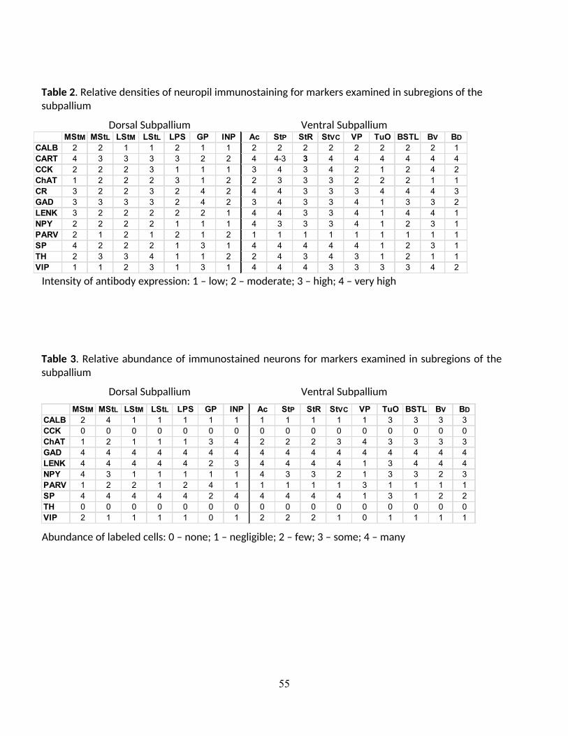
55
Table 2. Relative densities of neuropil immunostaining for markers examined in subregions of the
subpallium
Dorsal Subpallium Ventral SubpalliumMStM MStL LStM LStL LPS GP INP Ac StP StR StVC VP TuO BSTL BV BD
CALB 2 2 1 1 2 1 1 2 2 2 2 2 2 2 2 1
CART 4 3 3 3 3 2 2 4 4-3 3 4 4 4 4 4 4
CCK 2 2 2 3 1 1 1 3 4 3 4 2 1 2 4 2
ChAT 1 2 2 2 3 1 2 2 3 3 3 2 2 2 1 1
CR 3 2 2 3 2 4 2 4 4 3 3 3 4 4 4 3
GAD 3 3 3 3 2 4 2 3 4 3 3 4 1 3 3 2
LENK 3 2 2 2 2 2 1 4 4 3 3 4 1 4 4 1
NPY 2 2 2 2 1 1 1 4 3 3 3 4 1 2 3 1
PARV 2 1 2 1 2 1 2 1 1 1 1 1 1 1 1 1
SP 4 2 2 2 1 3 1 4 4 4 4 4 1 2 3 1
TH 2 3 3 4 1 1 2 2 4 3 4 3 1 2 1 1
VIP 1 1 2 3 1 3 1 4 4 4 3 3 3 3 4 2
Intensity of antibody expression: 1 – low; 2 – moderate; 3 – high; 4 – very high
Table 3. Relative abundance of immunostained neurons for markers examined in subregions of the
subpallium
Dorsal Subpallium Ventral Subpallium
MStM MStL LStM LStL LPS GP INP Ac StP StR StVC VP TuO BSTL BV BD
CALB 2 4 1 1 1 1 1 1 1 1 1 1 3 3 3 3
CCK 0 0 0 0 0 0 0 0 0 0 0 0 0 0 0 0
ChAT 1 2 1 1 1 3 4 2 2 2 3 4 3 3 3 3
GAD 4 4 4 4 4 4 4 4 4 4 4 4 4 4 4 4
LENK 4 4 4 4 4 2 3 4 4 4 4 1 3 4 4 4
NPY 4 3 1 1 1 1 1 4 3 3 2 1 3 3 2 3
PARV 1 2 2 1 2 4 1 1 1 1 1 3 1 1 1 1
SP 4 4 4 4 4 2 4 4 4 4 4 1 3 1 2 2
TH 0 0 0 0 0 0 0 0 0 0 0 0 0 0 0 0
VIP 2 1 1 1 1 0 1 2 2 2 1 0 1 1 1 1
Abundance of labeled cells: 0 – none; 1 – negligible; 2 – few; 3 – some; 4 – many

56
List of Abbreviations
Ac accumbens
BM nucleus basalis magnocellularis
BSTL bed nucleus of stria terminalis, lateral part
BD external capsule of BSTL, dorsal part
BV external capsule of BSTL, ventral part
GP globus pallidus
INP intrapeduncular nucleus
LFB lateral forebrain bundle
LOT lateral olfactory tract
LPS lamina pallio-subpallialis
LSt lateral striatum
MSt medial striatum
MStL lateral part of the medial striatum
MStM medial part of the medial striatum
LStM medial part of the lateral striatum
StVC ventral striatum, ventrocaudal part
StP ventral striatum, paratubercular region
QF quintofrontal tract
TSM tractus septopallio-mesencephalicus
TuO tuberculum olfactorium
VP ventral pallidum

57
Figure Legends
Fig. 1. Lateral view of the forebrain of Columbia livia. Numbers on top refer to the figure
number that illustrates transverse sections from that level.
Fig. 2. Series of transverse sections through the rostral basal ganglia comparing
immunostaining patterns of ChAT (A), VIP (B), ENK (C), TH(D), CCK (F), NPY G), GAD (H), and SP
(I) in different compartments of the striatum and ventral pallidum. These are illustrated in a
schematic diagram (E). The series is arranged from rostral (A) to caudal (I). Note that two
patches of neurons, the MI (open arrowhead) and islands of cells within the LPS (open arrows)
express different neurochemical patterns than the adjacent striatal matrix. Rostrocaudal level
is approximately A13.50.
Fig. 3. Series of transverse sections through the rostral basal ganglia at approximately A12.50,
arranged as explained in Fig. 2. Open arrows - islands of cells in the lamina pallio-subpallialis.
Fig. 4. Series of transverse sections through the rostral basal ganglia at approximately A11.75,
arranged as explained in Fig. 2. Asterisk - thin rostral extension of the BStL; closed arrows -
finger-like extensions of the GP into the medial striatum; open arrows - islands of cells within
the LPS.
Fig. 5. Series of transverse sections through the rostral basal ganglia at approximately A10.75,
arranged as explained in Fig. 2. Asterisk - thin rostral extension of the BStL; closed arrows -
finger-like extensions of the GP into the medial striatum; open arrows - islands of cells within
the LPS.
Fig. 6. Series of transverse sections through the rostral basal ganglia at approximately A10.00,
arranged as explained in Fig. 2. Closed arrows - finger-like extensions of the GP into the medial
striatum; open arrows - islands of cells in the LPS.
Fig. 7. Series of transverse sections through the rostral basal ganglia at approximately A9.50,
arranged as explained in Fig. 2. Closed arrows - finger-like extensions of the GP into the medial
striatum; open arrows - islands of cells in the LPS.

58
Fig. 8. Series of transverse sections through the rostral basal ganglia at approximately A9.00,
arranged as explained in Fig. 2. Open arrows - islands of cells in the LPS.
Fig. 9. Series of transverse sections through the rostral basal ganglia comparing
immunostaining patterns of CALB (A-G) and PARV (A’-G’). Images from the 7 levels shown in
Fig. 1 are arranged from rostral (A, A’) to caudal (G, G’).
Fig. 10. Transverse sections through the rostral striatum immunostaining patterns of CR (A-G)
and CART (H-N). Note that the neuropil of nucleus accumbens stains with high levels of CR and
CART, whereas the neuropil of the medial part of the paratubercular striatum stains with high
levels of CART+ but low levels of CR.
Fig. 11. Images of woolly fibers stained with NPY (A), GAD (B), and SP (C) in the medial MSt
(A,B) and GP (C). Note that NPY+ and GAD+ fibers (arrows), terminals, and cells (arrowheads)
are evident in the medial MST (A,B) and SP+ fibers (arrows) and cells (arrowheads) are evident
in the globus pallidus (C).
Fig. 12. Schematic drawings summarizing the neurochemical subdivisions observed in this
study, and proposed striatal and accumbens relationships. A, B, and C correspond to
rostrocaudal levels A12.50, A10.75, and A9.50, respectively.


Highlights
1. Sixteen distinct compartments were identified in the pigeon basal ganglia using multiple
neurochemical markers.
2. The striatum contains neurochemical regions comparable to the mammalian
somatomotor and associational striatum.
3. A neurochemically distinct area located in the medialmost striatum of pigeons appears
to be unique to birds.
4. The ventral striatum contains neurochemical regions similar to the mammalian
accumbens core, shell, and rostral areas.
5. Most of the main compartments of the basal ganglia were highly conserved during
tetrapod evolution, yet unique avian compartments representing diversification have
also evolved.