this document has been downloaded from
TRANSCRIPT

This document has been downloaded fromTampub – The Institutional Repository of University of Tampere
Publisher's version
Authors: Hyvärinen Anne K, Kumanto Mona K, Marjavaara Sanna K, JacobsHoward T
Name of article: Effects on mitochondrial transcription of manipulating mTERFprotein levels in cultured human HEK293 cells
Year ofpublication: 2010
Name of journal: BMC Molecular BiologyVolume: 11Number ofissue: 72
Pages: 1-12ISSN: 1471-2199Discipline: Medical and Health sciences / Medical biotechnologyLanguage: enSchool/OtherUnit: Institute of Biomedical Technology
URL: http://www.biomedcentral.com/1471-2199/11/72URN: http://urn.fi/urn:nbn:uta-3-506DOI: http://dx.doi.org/10.1186/1471-2199-11-72
All material supplied via TamPub is protected by copyright and other intellectual property rights, andduplication or sale of all part of any of the repository collections is not permitted, except that materialmay be duplicated by you for your research use or educational purposes in electronic or print form.You must obtain permission for any other use. Electronic or print copies may not be offered, whetherfor sale or otherwise to anyone who is not an authorized user.

RESEARCH ARTICLE Open Access
Effects on mitochondrial transcription ofmanipulating mTERF protein levels in culturedhuman HEK293 cellsAnne K Hyvärinen1, Mona K Kumanto1, Sanna K Marjavaara1,2, Howard T Jacobs1*
Abstract
Background: Based on its activities in vitro, the mammalian mitochondrial transcription termination factor mTERFhas been proposed to regulate mitochondrial transcription by favouring termination at its high-affinity bindingimmediately downstream of the rDNA segment of mitochondrial DNA, and initiation selectively at the PH1 site ofthe heavy-strand promoter. This defines an rDNA transcription unit distinct from the ‘global’ heavy-strandtranscription unit initiating at PH2. However, evidence that the relative activities of the two heavy-strandtranscription units are modulated by mTERF in vivo is thus far lacking.
Results: To test this hypothesis, we engineered human HEK293-derived cells for over-expression or knockdown ofmTERF, and measured the steady-state levels of transcripts belonging to different transcription units, namelytRNALeu(UUR) and ND1 mRNA for the PH2 transcription unit, and tRNAPhe plus 12S and 16S rRNA for the PH1transcription unit. The relative levels of 16S rRNA and ND1 mRNA were the same under all conditions tested,although mTERF knockdown resulted in increased levels of transcripts of 12S rRNA. The amount of tRNAPhe relativeto tRNALeu(UUR) was unaffected by mTERF over-expression, altered only slightly by mTERF knockdown, and wasunchanged during recovery from ethidium bromide-induced depletion of mitochondrial RNA. mTERFoverexpression or knockdown produced a substantial shift (3-5-fold) in the relative abundance of antisensetranscripts either side of its high-affinity binding site.
Conclusions: mTERF protein levels materially affect the amount of readthrough transcription on the antisensestrand of mtDNA, whilst the effects on sense-strand transcripts are complex, and suggest the influence ofcompensatory mechanisms.
BackgroundMammalian mitochondrial DNA is organized into threemulticistronic transcription units (reviewed in [1], Fig.1A), which give rise to the mature RNAs encoded bythe circular genome: two ribosomal RNAs, 22 tRNAsand 11 mRNAs (2 of them bicistronic). Each strand istranscribed in its entirety, employing closely spaced pro-moters located within the major non-coding region ofthe genome, namely LSP, the promoter of the light-strand, with a unique initiation site designated PL, andPH1 and PH2, the alternate transcription start sites ofthe heavy-strand promoter (HSP), which give rise to
partially overlapping transcripts. Based on metaboliclabeling studies, PH1 and PH2 have been inferred togive rise to distinct primary transcripts of the heavy-strand [2]. PH1 is located within the non-coding regionand generates a primary transcript comprising bothrRNAs and two tRNAs (-Phe and -Val), terminating atthe end of the rDNA region, mainly within the 5’ end ofthe tRNALeu(UUR) gene [3]. PH2 is located within thecoding sequence of tRNAPhe and generates a primarytranscript comprising all of the remaining heavy-strandencoded genes. PL generates a primary transcript com-prising the entire light strand.The mechanism by which the transcriptional machin-
ery selects between these different initiation sites, andalso effects selective termination at the end of therDNA, in the case of transcripts initiated at PH1, is
* Correspondence: [email protected] of Medical Technology and Tampere University Hospital, FI-33014University of Tampere, FinlandFull list of author information is available at the end of the article
Hyvärinen et al. BMC Molecular Biology 2010, 11:72http://www.biomedcentral.com/1471-2199/11/72
© 2010 Hyvärinen et al; licensee BioMed Central Ltd. This is an Open Access article distributed under the terms of the CreativeCommons Attribution License (http://creativecommons.org/licenses/by/2.0), which permits unrestricted use, distribution, andreproduction in any medium, provided the original work is properly cited.

incompletely understood. It can be manipulated in orga-nello by various drugs and by ATP [4-6]. The mitochon-drial RNA polymerase comprises a single catalyticsubunit, MTRPOL, plus an accessory factor, TFB2M,required for formation of the initiation complex in vitroat both HSP and LSP [7,8], together with mitochondrialtranscription factor A (TFAM), which is needed for pro-moter-dependent transcription in vitro [7,9]. TFAM hasa natural binding affinity for DNA and has been sug-gested also to play a more general role in organizing themitochondrial chromosome, analogous with bacterialHU or eukaryotic and archaeal histones. A third factor,mTERF, with sequence-specific binding affinity for a
sequence located within the tRNALeu(UUR) gene immedi-ately downstream of the rDNA [10,11], has been pro-posed to play a key role in both initiation andtermination of the PH1 transcription unit [12].mTERF has selective termination activity in vitro on
templates containing its high-affinity binding site in thetRNALeu(UUR) gene [10,13]. In crude extracts [14], aswell as in a reconstituted system based on recombinantproteins [15], this activity appears to be bidirectional,but operates in the latter case more efficiently in thereverse direction, i.e. to terminate transcription initiatingfrom the LSP side more efficiently than from HSP [15].Based on the fact that it has weak binding to other sites
Figure 1 Manipulation of mTERF expression has minimal effects on steady-state levels of mature mitochondrial RNAs. (A) Schematicdiagram of the promoter and rDNA region of human mtDNA. Because mTERF binding dictates the use of alternate transcriptional start sites andterminators, tRNAPhe and tRNALeu(UUR) fall into separate transcription units (PH1 and PH2 respectively). (B) Relative expression of mitochondrialtranscripts in cells overexpressing mTERF, based on phosphorimaging of Northern blots probed successively for mitochondrial tRNAPhe andtRNALeu(UUR) and for 5S rRNA. Data (means ± SD) are signal ratios of tRNAPhe to tRNALeu(UUR) (F/L) and tRNAPhe to 5S rRNA (F/5S) for the mTERF-overexpressing clones shown in Additional File 1, Fig. S1, normalized to the corresponding ratio in cells stably transfected with empty-vector.Bars shown alongside are based on single reference experiments, using HEK293T cells transiently transfected with the same construct, or mock-transfected. (C) Q-RT-PCR analysis (means ± SD) of mitochondrial transcript levels, plus cytosolic18S rRNA, as indicated, in Flp-In™ T-Rex™-293 cellsover-expressing mTERF-MycHis after doxycyclin induction for 3 d (or not induced). Data were normalized, in each case, to the correspondingratio for uninduced cells. (D) Relative expression of mitochondrial transcripts in cells knocked down for mTERF, as indicated. – denotes mock-transfection. Northern blot probed successively for 16S rRNA and ND1 mRNA, as shown. The panels represent non-adjacent pairs of lanes fromthe same exposure of the same gel. (E) Relative expression of mitochondrial transcripts in cells knocked down for mTERF, as indicated, calculatedfrom Northern blot data as in (B), normalized to the corresponding ratio in mock-transfected HEK293T cells. * indicates significant differencesfrom the corresponding mock-transfected cells, and # a significant difference between cell-lines (t-test, p values as in text). For original blots seeAdditional File 1, Fig. S1C. Note that additional Q-RT-PCR data on levels of 12S rRNA gene transcripts are shown in Fig. 3.
Hyvärinen et al. BMC Molecular Biology 2010, 11:72http://www.biomedcentral.com/1471-2199/11/72
Page 2 of 12

in mtDNA, including the promoter region [12,16,17], ithas been proposed that mTERF favours transcription ofthe PH1 transcription unit by simultaneous binding tothe promoter and to the terminator region, creating aloop structure that can be visualized in vitro [12]. Thelevel of active mTERF would thus act as a fine tuning ofthe relative production of rRNA and mRNA.There are, however, some problems associated with
this model. First, efficient transcription from PH1 invitro does not require mTERF (although does appear tobe stimulated by it [18]), whereas transcription fromPH2 in vitro is weak [18]. Second, measurements of therelative half-lives of mitochondrial rRNAs and mRNAsin cultured cells [19] indicate that post-transcriptionalregulation is substantial and may in fact be sufficient tomaintain the different transcript levels seen in vivo,without the need for any differential regulation of tran-scription from the PH1 and PH2 transcription units.Note that, although the synthesis rates of mitochondrialrRNAs and mRNAs appear to be very different in bothcultured cells [19] and rat liver [20], ‘synthesis rate’ hereincludes RNA processing as well as transcription. Inorganello, the combined rate of accumulation of pre-rRNA plus mature rRNA is, in fact, lower than that ofmRNA [6]. Third, no modulation of transcription fromthe two initiation sites correlating with mTERF activityhas ever been convincingly demonstrated in vivo.Fourth, in cells bearing the 3423A > G mutation, whichgreatly impairs mTERF binding in vitro, there is noalteration in the relative levels of 16S rRNA and ND1mRNA [21,22], and no alteration in site occupancy invivo, based on footprinting studies [21]. Fifth, decreasedlevels of mTERF expression in Mpv17 knockout miceare associated with globally increased mitochondrialtranscription [23], suggesting rather than mTERF mayfunction in vivo as a negative but general regulator oftranscription. Finally, whilst recombinant mTERF isactive in a reconstituted system in vitro [15], its activityin the presence of less pure mitochondrial extracts issubject to post-translational modifications and/or thepresence of other proteins [11-13,18,24], raising doubtsas to whether and how it influences transcription invivo.mTERF is a member of a family of organellar proteins
proposed to interact with DNA to produce a variety ofoutcomes [25]. In mammals, two homologues ofmTERF, MTERFD1 (mTERF3) and MTERD3(mTERF2), have been shown to influence mitochondrialRNA levels and have been proposed to act as regulatorsof transcription from LSP [26,27], with consequenteffects on oxidative phosphorylation mediated broughtabout by altered translation, as seen also in Drosophila[28]. However, neither mTERF homologue has beenconclusively demonstrated to have high-affinity
sequence-specific binding to DNA [26,27,29]. Homolo-gues of mTERF in invertebrates have been demonstratedto influence both RNA and DNA synthesis in vitro, buthere too, there is only weak evidence for a specific rolein vivo. The mTERF-homologue in sea urchins, mtDBP,binds to at least two sites in the mitochondrial genome[30] and exhibits bidirectional transcription terminationactivity in vitro in the presence of human mitochondrialRNA polymerase, although it acts unidirectionally incombination with phage polymerases [31]. It alsoimpedes the progress of DNA polymerase bidirection-ally, acting as a contrahelicase in vitro [32], suggesting apossible role in DNA replication. A role for mTERF inmammalian mtDNA replication is also suggested by theobservation that the level of mTERF expression in cul-tured human cells influences replication pausing in thevicinity of mTERF binding sites [16]. The DrosophilamTERF homologue mTTF binds to two putative tran-scriptional terminators [33], acting in vitro with similardirectional properties to mtDBP [34]. Manipulation ofDmTTF levels in cultured cells leads to effects on tran-script levels consistent with it acting in the mannerhypothesized for mTERF, i.e. as a regulator of termina-tion (bidirectionally) and also of promoter activity [35].The difficulty of interpreting in vitro experiments, and
the open questions regarding the role of mTERF in vivo,prompted us to address the issue of whether and howmTERF activity influences mitochondrial RNA levels incultured human cells. Clearly, if mTERF is a regulatorof mitochondrial transcription in vivo, via a model asproposed, up- or down-regulation of its expressionshould influence mitochondrial RNA levels in a predict-able fashion. We therefore undertook a study of mito-chondrial transcripts in cells over-expressing or knockeddown for mTERF. Surprisingly, we found that varyingthe level of mTERF over a wide range has only a smalleffect on the levels of sense-strand transcripts of themitochondrial genome in the rDNA region. Conversely,we detected a clear effect on the relative amounts ofantisense transcripts on the two sides of the high-affinitybinding site. These findings support a role for mTERFin influencing mitochondrial transcription in vivo, butnot in setting the levels of mature mitochondrialtranscripts.
ResultsOver-expression of mTERF does not alter steady-statelevels of mature mitochondrial RNAsTo evaluate whether the expression level of mTERFinfluences the steady-state levels of the mature mito-chondrial transcripts encoded on either side of its high-affinity binding site, we generated a series of transfectedHEK293T cell clones stably over-expressing the naturalmTERF protein. Expression of the mTERF transgene
Hyvärinen et al. BMC Molecular Biology 2010, 11:72http://www.biomedcentral.com/1471-2199/11/72
Page 3 of 12

was verified at the RNA level by Q-RT-PCR (AdditionalFile 1, Fig. S1A) and at the protein level by the substan-tial increase in DNA-binding capacity at the high-affinitymTERF binding site, as judged by EMSA (electrophore-tic mobility shift assay, Additional File 1, Fig. S1B).We analysed two parameters which we considered
diagnostic for the relative utilization of the two heavy-strand transcription units predicted by the classic modelof mammalian mtDNA transcription (Fig. 1A). The firstis the relative amounts of tRNAs -Phe and -Leu(UUR),which are exclusively produced by transcription fromPH1 and PH2, respectively, according to the classicalmodel. The second is the relative amounts of mature16S rRNA and ND1 mRNA. The latter is synthesizedvia transcription from PH2, whereas the former hasbeen proposed to be generated mainly or exclusivelyfrom transcription initiating at PH1, although it has notbeen formally excluded that transcription from PH2 alsocontributes some of the 16S rRNA. We found that therelative amounts of tRNAs -Phe and -Leu(UUR) in dif-ferent cell-clones over-expressing natural mTERF wasindistinguishable from that in control cells transfectedwith empty vector (Fig. 1B), and was also unchanged incells transiently transfected with the mTERF overexpres-sion construct (or mock transfected cells). The globalamount of mitochondrial transcription, as measured bythe ratio of tRNAPhe to cytosolic 5S rRNA was morevariable, but showed no systematic relation to mTERFoverexpression (Fig. 1B). We also found no detectablealteration in the relative amounts of mature NDI1mRNA and 16S rRNA between mTERF over-expressingclones and control cells, based on Northern blots (Fig.1D: compare lanes 1 of panels i and ii [control cells]with lanes 1 of panels iii and iv [over-expressing cells]).In an effort to quantify any such effect and avoid possi-ble influences of cell background, we also used Q-RT-PCR to analyse transcripts of the 16S and ND1 genes inRNA extracted from Flp-In™ T-Rex™-293 cells stablytransfected with the mTERF-MycHis construct, in whichexpression of mTERF can be induced by doxycycline(Fig. 1C). We found no differences in the relativeamounts of transcripts from these two genes, nor in theratio of either to cytosolic 18S rRNA transcripts.
Effects of mTERF knockdown on steady-state levels ofmature mitochondrial RNAsIn previous studies [16] we noted that transfection withan siRNA directed against mTERF suppressed most ofthe binding activity at the high-affinity mTERF bindingsite, as judged by EMSA [16]. We therefore comparedthe relative levels of mitochondrial transcripts in cellsknocked down for mTERF. Northern blots probed suc-cessively for 16S rRNA and ND1 mRNA showed no dif-ference in the relative levels of these mature transcripts
in HEK293T cells after prolonged treatment (7 d) withan mTERF-specific siRNA (Fig. 1D: compare lanes 1and 2 of panels i and ii), nor in mTERF-MycHis over-expressing cells knocked down for mTERF (Fig. 1D:compares lanes 1 and 2 of panels iii and iv). mTERFknockdown in HEK293T cells did, however, produce asmall but significant decrease in the relative amount oftRNAPhe compared with tRNALeu(UUR), (Fig. 1E, t-test, p< 0.05), accompanied by an increase in the overallamount of mitochondrial tRNAs, represented by theratio of mitochondrial tRNAPhe to cytosolic 5S rRNA (t-test, p < 0.01). siRNA treatment of mTERF-MycHisoverexpressing cells caused no significant alteration inmitochondrial tRNAs (Fig. 1E), compared with mock-transfected cells. Note also that cells overexpressingmTERF-MycHis showed no clear difference fromHEK293T cells in the relative levels of mitochondrialtRNAPhe and tRNALeu(UUR) (Fig. 1E), although mito-chondrial tRNA levels globally were lower than inuntransfected HEK293T cells (t-test, p < 0.01).
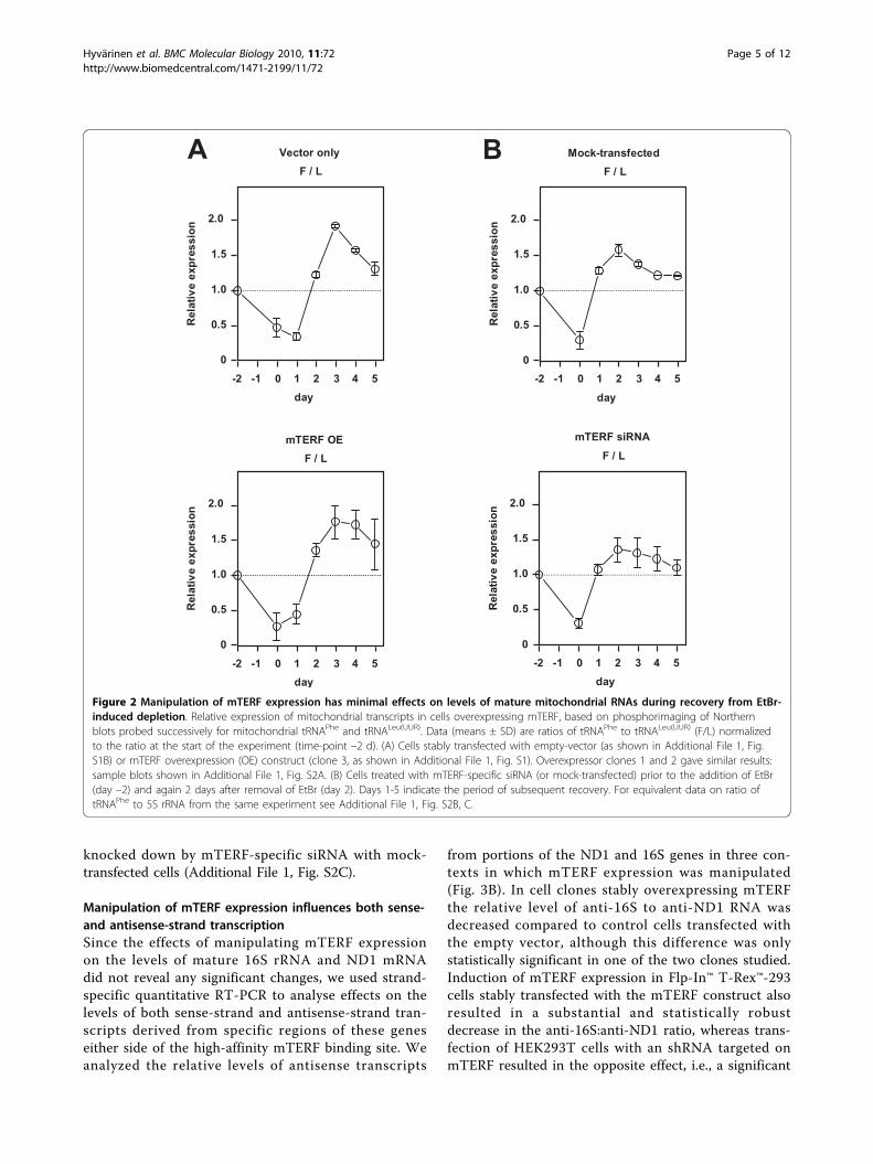
Manipulation of mTERF expression does not alter therelative levels of mitochondrial tRNAs during recoveryfrom mitochondrial RNA depletionReasoning that the steady-state levels of mature mito-chondrial transcripts may not accurately reflect theirtranscription rates in vivo, due to the influence of post-transcriptional processing, we set out to study whetherthe level of mTERF expression can influence the re-accumulation of tRNA transcripts belonging to the PH1and PH2 transcription units during recovery from ethi-dium bromide (EtBr)-induced depletion of mitochon-drial RNA. We compared the ratio of mitochondrialtRNAs -Phe and -Leu(UUR) in stably transfected cellsoverexpressing mTERF-with that in empty vector-trans-fected cells over 2 days of EtBr treatment followed by 5days of recovery (Fig. 2A, Additional File 1, Fig. S2A). Inboth cell lines the ratio fell substantially during deple-tion, reflecting the much shorter half-life of tRNAPhe,but then recovered to levels higher than those seen inuntreated cells, before decreasing again gradually,towards the starting value. This may indicate that thePH2 transcription unit is used preferentially duringrecovery from depletion. However, this did not appearto be influenced by the level of mTERF, since the samepattern was seen in control cells and in three separatelyanalysed overexpressor cell lines, as well as in cellsknocked down for mTERF by treatment with themTERF-specific siRNA, which behaved indistinguishablyfrom mock-transfected cells (Fig. 2B). The overallkinetics of recovery of mitochondrial transcripts com-pared with cytosolic 5S rRNA was also similar, compar-ing cells over-expressing mTERF with control cells(Additional File 1, Fig. S2B), and comparing cells
Hyvärinen et al. BMC Molecular Biology 2010, 11:72http://www.biomedcentral.com/1471-2199/11/72
Page 4 of 12
knocked down by mTERF-specific siRNA with mock-transfected cells (Additional File 1, Fig. S2C).
Manipulation of mTERF expression influences both sense-and antisense-strand transcriptionSince the effects of manipulating mTERF expressionon the levels of mature 16S rRNA and ND1 mRNAdid not reveal any significant changes, we used strand-specific quantitative RT-PCR to analyse effects on thelevels of both sense-strand and antisense-strand tran-scripts derived from specific regions of these geneseither side of the high-affinity mTERF binding site. Weanalyzed the relative levels of antisense transcripts
from portions of the ND1 and 16S genes in three con-texts in which mTERF expression was manipulated(Fig. 3B). In cell clones stably overexpressing mTERFthe relative level of anti-16S to anti-ND1 RNA wasdecreased compared to control cells transfected withthe empty vector, although this difference was onlystatistically significant in one of the two clones studied.Induction of mTERF expression in Flp-In™ T-Rex™-293cells stably transfected with the mTERF construct alsoresulted in a substantial and statistically robustdecrease in the anti-16S:anti-ND1 ratio, whereas trans-fection of HEK293T cells with an shRNA targeted onmTERF resulted in the opposite effect, i.e., a significant
Figure 2 Manipulation of mTERF expression has minimal effects on levels of mature mitochondrial RNAs during recovery from EtBr-induced depletion. Relative expression of mitochondrial transcripts in cells overexpressing mTERF, based on phosphorimaging of Northernblots probed successively for mitochondrial tRNAPhe and tRNALeu(UUR). Data (means ± SD) are ratios of tRNAPhe to tRNALeu(UUR) (F/L) normalizedto the ratio at the start of the experiment (time-point –2 d). (A) Cells stably transfected with empty-vector (as shown in Additional File 1, Fig.S1B) or mTERF overexpression (OE) construct (clone 3, as shown in Additional File 1, Fig. S1). Overexpressor clones 1 and 2 gave similar results:sample blots shown in Additional File 1, Fig. S2A. (B) Cells treated with mTERF-specific siRNA (or mock-transfected) prior to the addition of EtBr(day –2) and again 2 days after removal of EtBr (day 2). Days 1-5 indicate the period of subsequent recovery. For equivalent data on ratio oftRNAPhe to 5S rRNA from the same experiment see Additional File 1, Fig. S2B, C.
Hyvärinen et al. BMC Molecular Biology 2010, 11:72http://www.biomedcentral.com/1471-2199/11/72
Page 5 of 12
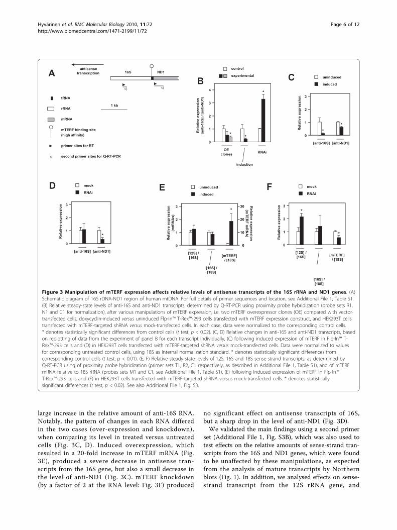
large increase in the relative amount of anti-16S RNA.Notably, the pattern of changes in each RNA differedin the two cases (over-expression and knockdown),when comparing its level in treated versus untreatedcells (Fig. 3C, D). Induced overexpression, whichresulted in a 20-fold increase in mTERF mRNA (Fig.3E), produced a severe decrease in antisense tran-scripts from the 16S gene, but also a small decrease inthe level of anti-ND1 (Fig. 3C). mTERF knockdown(by a factor of 2 at the RNA level: Fig. 3F) produced
no significant effect on antisense transcripts of 16S,but a sharp drop in the level of anti-ND1 (Fig. 3D).We validated the main findings using a second primer
set (Additional File 1, Fig. S3B), which was also used totest effects on the relative amounts of sense-strand tran-scripts from the 16S and ND1 genes, which were foundto be unaffected by these manipulations, as expectedfrom the analysis of mature transcripts by Northernblots (Fig. 1). In addition, we analysed effects on sense-strand transcript from the 12S rRNA gene, and
Figure 3 Manipulation of mTERF expression affects relative levels of antisense transcripts of the 16S rRNA and ND1 genes. (A)Schematic diagram of 16S rDNA-ND1 region of human mtDNA. For full details of primer sequences and location, see Additional File 1, Table S1.(B) Relative steady-state levels of anti-16S and anti-ND1 transcripts, determined by Q-RT-PCR using proximity probe hybridization (probe sets R1,N1 and C1 for normalization), after various manipulations of mTERF expression, i.e. two mTERF overexpressor clones (OE) compared with vector-transfected cells, doxycyclin-induced versus uninduced Flp-In™ T-Rex™-293 cells transfected with mTERF expression construct, and HEK293T cellstransfected with mTERF-targeted shRNA versus mock-transfected cells. In each case, data were normalized to the corresponding control cells.* denotes statistically significant differences from control cells (t test, p < 0.02). (C, D) Relative changes in anti-16S and anti-ND1 transcripts, basedon replotting of data from the experiment of panel B for each transcript individually, (C) following induced expression of mTERF in Flp-In™ T-Rex™-293 cells and (D) in HEK293T cells transfected with mTERF-targeted shRNA versus mock-transfected cells. Data were normalized to valuesfor corresponding untreated control cells, using 18S as internal normalization standard. * denotes statistically significant differences fromcorresponding control cells (t test, p < 0.01). (E, F) Relative steady-state levels of 12S, 16S and 18S sense-strand transcripts, as determined byQ-RT-PCR using of proximity probe hybridization (primer sets T1, R2, C1 respectively, as described in Additional File 1, Table S1), and of mTERFmRNA relative to 18S rRNA (probes sets M1 and C1, see Additional File 1, Table S1), (E) following induced expression of mTERF in Flp-In™T-Rex™-293 cells and (F) in HEK293T cells transfected with mTERF-targeted shRNA versus mock-transfected cells. * denotes statisticallysignificant differences (t test, p < 0.02). See also Additional File 1, Fig. S3.
Hyvärinen et al. BMC Molecular Biology 2010, 11:72http://www.biomedcentral.com/1471-2199/11/72
Page 6 of 12

determined the levels of sense-strand transcripts of bothmitochondrial rRNAs relative to cytosolic 18S rRNA(Fig. 3E, F). Under conditions of induced over-expres-sion of mTERF, sense-strand transcripts of 12S rRNAand of 16S rRNA were unchanged relative to each otherand to cytosolic 18S rRNA (Fig. 3E). However, we diddetect a significant increase in sense-strand 12S rRNAtranscripts in cells knocked down for mTERF (Fig. 3F).
DiscussionmTERF and heavy-strand promoter modulationIn this study we investigated the effects of manipulatingthe expression level of mTERF on the relative levels ofdifferent mitochondrial transcripts. Under all conditionstested we failed to detect any significant effects on therelative levels of mature 16S rRNA and ND1 mRNA(Fig. 1C, 1D, Additional File 1, S3C). Over-expression ofa tagged mTERF variant, which resulted in the greatestincrease in DNA-binding activity that we were able togenerate (Fig. 5C of [16]), produced no significantchange in the relative levels of the mitochondrial tRNAstested, with only a minor decrease in their overall abun-dance (Fig. 1E). Induced 20-fold over-expression of nat-ural mTERF in a controlled nuclear background also didnot alter the ratio of mature 16S rRNA to ND1 mRNA,nor were the levels of 16S or ND1 transcripts affectedrelative to transcripts of cytosolic 18S rRNA or 12SrRNA (Fig. 1C, 3E). Knockdown of mTERF resulted in avery modest decrease in the level of tRNAPhe relative totRNALeu(UUR). However, this was not sufficient to gener-ate any significant change in the kinetics of recovery ofmitochondrial tRNA levels following EtBr-induceddepletion.We did, however, obtain two piece of evidence that
mTERF knockdown is not inert as regards transcriptionof the mitochondrial heavy strand. Firstly, we observed,by Northern blots, a small increase in the amount ofmitochondrial tRNAs belonging to each of the heavy-strand transcription units, relative to cytosolic 5S rRNA(Fig. 1E) in normal cells after mTERF knockdown. Sec-ondly, the level of sense-strand 12S rRNA gene tran-scripts analysed by quantitative RT-PCR wassignificantly increased relative to sense-strand 16S rRNAor cytosolic 18S rRNA gene transcripts (Fig. 3F), in nor-mal cells knocked down for mTERF. However, the levelsof sense-strand 16S and ND1 transcripts relative to eachother or to 18S were not significantly affected (Fig. 3F).This suggests the existence of a compensatory mechan-ism, whereby decreased mTERF levels, which mightotherwise impair 16S rRNA biogenesis, generate a signalfor globally increased mitochondrial transcription (ordecreased turnover) to overcome any such defect. Itmay also be noted that the effects of knockdown may beunderestimated due to the rather limited decrease in
mTERF mRNA level that we were able to achieve inthese experiments. A 50% decrease is not untypical incultured mammalian cells in cases where knockdown ofa given gene may provoke a growth defect, even just atransient one, compared with untransfected cells in theculture. Thus, the effects we observed may likely repre-sent a combination of normal expression in almost halfthe cells, plus greatly reduced expression in the remain-ing cells.Nevertheless, our findings imply that the expression
level of mTERF does not determine, in a simple manner,the relative steady-state levels of transcripts belonging tothe two transcription units of the heavy-stand. AlthoughmTERF was previously shown to stimulate transcriptionin vitro from PH1 in a comparatively crude system[12,18], it may be noted that no such effect was seenwhen purified, recombinant proteins were used [15], oreven in crude extracts using DNA-affinity purifiedmTERF [18].Our results indicate that even if mTERF levels do
influence transcriptional readthrough, a compensatoryresponse nevertheless adjusts the relative output of dif-ferent transcripts belonging to the two heavy-strandtranscription units. This may involve the modulation oftranscriptional initiation, post-transcriptional processingor RNA turnover. Our findings are consistent with pre-vious reports of the action of thyroid hormone [36] orvariation in ATP supply [37], both of which can influ-ence the relative rates of transcriptional of initiation atPH1 and PH2 without any effect on that at the high-affinity mTERF binding site. It is also possible thatmTERF might have a different physiological function,and that its effects on transcription are accommodatedby modulating other components of the mitochondrialRNA synthesis machinery.
Is mTERF activity in HEK293 cells physiological?All of the current study was conducted in one cell-lineand its derivatives which, as a cancer cell-line, may notbehave in a physiologically normal manner. We consid-ered the hypothesis that mTERF levels may, in othercell-types, have a more profound effect on mitochon-drial transcription but that, in HEK293 cells, mTERFcould be present in such excess that neither over-expression nor any amount of knockdown achievable byRNAi technology influences its functional level. How-ever, from available gene expression data (biogps.gnf.org) the range of expression of mTERF in different cell-types in vivo, plus primary tumours and cell-linesincluding HEK293 and its derivatives, is only of theorder of 2-5 fold. Furthermore, in HEK293T cellsmTERF is expressed at very close to the median levelfor all cells investigated. Therefore, the range of expres-sion achieved in the present study (~40-fold at the RNA
Hyvärinen et al. BMC Molecular Biology 2010, 11:72http://www.biomedcentral.com/1471-2199/11/72
Page 7 of 12
level, Fig. 3E, F) far exceeds that known to be experi-enced in vivo.Another possibility, given the wealth of previous data
indicating possible post-translational regulation of thetranscriptional activity of mTERF, is that mTERF is con-stitutively inactivated in HEK293T cells, regardless of itsexpression level. Although we analysed DNA-bindingactivity as well as RNA levels, some mTERF prepara-tions that are competent for DNA binding are neverthe-less unable to influence transcription in vitro [11,24].This is unlikely, however, since the patterns of mito-chondrial transcripts in HEK cells, and their responsesto other manipulations, such as increases in the level ofTFAM [38], are similar to other cultured cells and invivo tissues.Thiamphenicol treatment, which alters the representa-
tion of PH1- and PH2-derived transcripts in a mannersimilar to thyroid hormone treatment, is able to modifythe EMSA signal at the high-affinity mTERF bindingsite, whilst leaving the actual levels of mTERF polypep-tide unaffected [39]. This may indicate that a post-trans-lational modification of mTERF could modulate both itsDNA-binding and its transcriptional properties in vivo,but is equally consistent with the notion that anotherfactor, capable of binding in this region, is involved.Final resolution of these issues will require the crea-
tion of an in vivo model in which mTERF levels can bemanipulated over at least as great a range in a tissue-selective manner. The possibility of redundancy betweenmTERF and other members of the mTERF family inregulating read-through transcription at the 16S/tRNA-Leu(UUR) gene boundary needs also to be considered.
Modulation of antisense-strand transcriptsWe found that alterations in mTERF expression pro-duced systematic changes in the extent of read-throughtranscription in the antisense direction, as inferred fromthe relative levels of anti-16S to anti-ND1 transcripts.Increased levels of mTERF, resulting from stable over-expression or from induction of Flp-In™ T-Rex™-293cells transfected with an mTERF expression construct,shifted the balance of antisense transcripts in the anti-ND1 direction, whereas mTERF knockdown had theopposite effect, shifting the balance in favour of anti-16S. These findings are consistent with the notion thatmTERF, bound to its high affinity binding site in thetRNALeu(UUR) gene, promotes termination of antisensetranscription initiated at PL, which has traversed mostof circular genome. Increased termination at this siteshould deplete the representation of anti-16S, whereasdecreased termination should increase the amount ofanti-16S, consistent with our observations. However, theeffects seen are more complex than implied by this sim-ple model. Specifically, the shift towards anti-ND1
under conditions of over-expression consists of a ratherdrastic decrease in the amount of stable anti-16S, com-bined with a much smaller decrease in the amount ofanti-ND1 (Fig. 3E). Since there are additional, weakerbinding sites for mTERF in the IQM tRNA cluster andND1 coding sequence [16], our finding supports theidea that a high level of mTERF leads to increased occu-pancy also of these weaker affinity binding sites,restraining readthrough into anti-ND1 as well as themore dramatic effect on readthrough into anti-16Sfurther downstream. On the other hand, mTERF knock-down resulted in a clear decrease in the level of anti-NDI1 but only a small change in anti-16S (Fig. 3F).These findings imply that maintenance of the physiolo-gical level of mTERF is important for the formation ofstable antisense transcripts of ND1, by preventing read-through into the rDNA. If this interpretation is correct,one in vivo role of mTERF is thus inferred to be theregulation of antisense transcriptional termination, foran unknown physiological reason.In vitro, mTERF exhibits bidirectional termination
activity [15]. If this applies also in vivo, it may be thatthe primacy of post-transcriptional processing, the stabi-lization of rRNA into ribosomal subunits, and compen-satory effects on transcriptional initiation or RNAstability, mask or complicate the effects on sense-strandtranscripts. Conversely, antisense transcripts, which aredestined only for turnover (or for some unknown phy-siological function) would appear to be regulated morestraightforwardly by mTERF.A somewhat different interpretation arises from the
recent, and thus far unexplained reports of hairpin-looptranscripts deriving from the 16S rRNA gene, whoselevels appear to reflect the proliferation status andtumorigenicity of cells [40,41]. It is not yet known howthese transcripts arise. Possibilities are that they are cre-ated post-transcriptionally by trans-splicing or RNAligation, or else that they arise by template strand-switching during transcription. Our antisense resultscould thus imply that mTERF influences the rate oftheir production in ways related to or even independentof its binding to mitochondrial DNA.
Physiological function(s) of mTERFGiven that the effects of mTERF manipulation on thelevels of mature mitochondrial transcripts in vivo appearto be negated or modified by compensatory mechan-isms, it may be that the principal physiological functionof this evolutionarily conserved protein is somethingother than transcriptional regulation as such. In our pre-vious study [16] we speculated that mTERF might playsome role in regulating collisions between oppositelymoving transcription and replication machineries, facili-tating their orderly passage, whilst minimizing the risk
Hyvärinen et al. BMC Molecular Biology 2010, 11:72http://www.biomedcentral.com/1471-2199/11/72
Page 8 of 12
of stalled replication giving rise to recombinogenic 3’ends. A requirement for such activity is well establishedin both prokaryotic and eukaryotic DNA replication[42,43], and other members of the mTERF family havebeen inferred to play a role in the completion of DNAreplication in human cells [44]. The presence of a tran-scriptional terminator at a replication pause site more-over provides a potential primer of lagging-strandsynthesis commencing immediately from the pausingsite, ensuring that no region remains single-strandedand hence susceptible to DNA damage during pausing.The RITOLS model of mtDNA replication [45] postu-lates that the entire lagging strand is laid down initiallyas RNA, which might be facilitated by such a mechan-ism. However, the lagging strand for mtDNA replicationis the same strand as the rRNA. Therefore, if boundmTERF were to deliver the 3’ end of a paused transcriptto an arriving replication complex, this would be as aresult of its activity in the sense direction. The role ofattenuation on the antisense strand is less clear,although this might provide a primer required for re-initiation of the replication machinery at a stalled repli-cation fork, especially since the former leading strand 3’end may be unavailable, e.g. due to fork regression. Arole for DnaG primase in replication restart at stalled,gapped forks has been identified in E. coli [46], servingas a precedent for primer-dependent restart. Codirec-tional collisions between the transcription and replica-tion machineries in E. coli also generate leading-strandgaps, with the nascent RNA being recruited as a newprimer by the replisome [47].Another possibility which should be seriously consid-
ered is that mTERF’s effects on nucleic acid metabolismare incidental to its real biological function inside mito-chondria, which may be something completely different.However, arguing against this is the fact that othermembers of the mTERF family also affect mitochondrialtranscript levels, including a recently reported case ofthe SOLDAT10 protein in Arabidopsis chloroplasts, amutation in which appears to activate retrograde signal-ing by decreasing plastid rRNA synthesis [48]. MOC1,an mTERF family homologue in Chlamydomonas, isrequired for maintaining mitochondrial RNA levels afterexposure to light, although its mechanism of action isunknown and the broader phenotype of the mutant sug-gests that the effect might be indirect [49].
ConclusionsIn summary, our findings support a role for mTERF ininfluencing mitochondrial transcription in vivo, eventhough it does not appear to set the levels of maturemitochondrial transcripts encoded by the PH1 and PH2heavy-strand transcription units in a simple manner. Itappears to modulate the levels of antisense transcripts,
by implication regulating the extent of readthrough bythe transcriptional machinery of its high-affinity bindingsite in the tRNALeu(UUR) gene, as well as other, weakermTERF binding sites in the vicinity. Further experi-ments will now be required to resolve the functional sig-nificance of this regulation, and its possible relevance toDNA replication and other processes.
MethodsCell-lines and cell cultureHEK293T cells and derivatives were cultured in Dulbec-co’s modified Eagle’s medium (DMEM, Sigma) as pre-viously [16]. HEK293T-derived cell-clones over-expressing natural mTERF were created by recloningthe mTERF coding sequence, including its natural stopcodon, into the expression vector pcDNA3.1/hygro(-)(Invitrogen) as a BamHI/HindIII fragment. Aliquots ofthe sequence-verified plasmid DNA (1 μg) were trans-fected into HEK293T cells using Lipofectamine™ (Invi-trogen) diluted in 1 ml of Opti-MEM® (Invitrogen)according to the manufacturer’s protocol. Twenty fourhours later cells were either harvested (for transienttransfection) or placed under hygromycin selection (Cal-biochem, 200 μg/ml). Hygromycin-resistant colonieswere grown up and tested for expression of the mTERFtransgene by reverse transcriptase (RT)-PCR and byelectrophoretic mobility shift assay (EMSA) as describedin Additional File 1. Flp-In™ T-Rex™ 293 cells trans-fected with expression constructs for natural mTERFand for epitope-tagged mTERF-MycHis, as well as theirinduction by doxycycline, were as described previously[16]. mTERF-specific RNA interference was induced bysiRNA for 48 h as described previously [16] or by trans-fection (using Lipofectamine™ 2000, Invitrogen, manu-facturer’s protocol) with a customized shRNA construct(10 μg) targeting the following sequence within mTERFmRNA (5’ to 3’): GCUGUAACUUGAGUACUUU, OpenBiosystems Expression Arrest™ pSM2 Retroviral shRNA-mir Library, Oligo ID V2HS_95064 (Thermo FisherScientific, Huntsville, AL, USA). shRNA-transfected cellswere harvested 48 h after transfection.
Depletion of mitochondrial RNACells were passaged one day before adding ethidiumbromide (EtBr) so that the 60 × 15 mm plates wereapproximately 50% confluent on the day of experiment.EtBr was added to the medium to 250 ng/ml and thecells were incubated for 48 h, after which the plateswere approximately 90% confluent. Cells were then pas-saged at different densities so that each re-seeded platewould reach approximately 70-80% confluence whenharvested for RNA extraction. RNA samples were col-lected before EtBr treatment (day -2), on the day whendrug was washed away (day 0) and 24, 48, 72, 96 and
Hyvärinen et al. BMC Molecular Biology 2010, 11:72http://www.biomedcentral.com/1471-2199/11/72
Page 9 of 12

120 h after removing EtBr (days 1-5). To ensure com-plete removal of EtBr the medium was changed 3 h and6 h after passaging the cells, and then again every day.Where depletion was carried out in combination withmTERF-directed RNA interference, siRNA transfectionwas carried out prior to the addition of EtBr (day -2)and was repeated 2 d after removal of the drug (day 2).
RNA extraction, electrophoresis and Northern blottingTotal RNA was extracted from cells using TRIzol®Reagent (Invitrogen) according to the manufacturer’sinstructions. Any traces of DNA were removed by treat-ment with RNase-free DNase I (GE Healthcare, manu-facturer’s recommended conditions), followed bystandard acid phenol/chloroform extraction and isopro-panol precipitation. For Northern blotting to tRNAprobes RNA samples were electrophoresed at 4°C over-night at 100 V in neutral 12% acrylamide/7 M urea gelsin TBE buffer, electroblotted onto Zeta-Probe GT mem-brane (Bio-Rad) at 4°C, u.v.-crosslinked and processedas described previously [50]. Oligonucleotide probes formitochondrial tRNAs and cytosolic 5S rRNA were radi-olabeled using T4 polynucleotide kinase (PNK, MBI Fer-mentas) according to the manufacturer’s protocol and[g-32P] ATP (Amersham Pharmacia Biotech, 3000 Ci/mmol) and purified using mini Quick Spin Columns(Roche). The probe oligonucleotide sequences were asfollows (all 5’ to 3’): 5S - GGGTGGTATGGCCGTA-GAC, tRNALeu(UUR) - GTTTTATGCGATTACCGGGCand tRNAPhe - CTAAACATTTTCAGTGTATTGC.Hybridization, washing, autoradiography and phosphori-maging (Phosphorimager SI, Molecular Dynamics) wereas described previously [51]. For re-probing, the mem-branes were stripped by boiling in 0.5% SDS solution for3 min and cooled to room temperature. For Northernblotting to 16S rRNA or ND1 probes, RNA sampleswere fractionated on formaldehyde agarose gels and pro-cessed for blotting and hybridization as described pre-viously [51], using probes labelled by random-priming[50]. The template used for synthesis of the ND1 probewas as described previously [50]; that for 16S rRNA wasthe shorter ApaI digestion product (230 bp) from thesame fragment.
Quantitative RT-PCRQuantitative RT-PCR was used to estimate the relativeamounts of 12S and16S rRNA, ND1 mRNA, cytosolic18S rRNA and mTERF mRNA. For cDNA synthesis, 5μg of RNA was reversed transcribed using 40 units ofM-MuLV reverse transcriptase (Fermentas), primed by0.2 μg random hexamers (Pharmacia) in a 20 μl reactionaccording to manufacturer’s instructions. Three dilu-tions of each cDNA sample (1:10, 1:20 and 1:50) wereanalysed, and each reaction was performed in three
technical replicates. PCR reactions were performed in aLightCycler™ apparatus using LightCycler FastStart DNAMaster SYBR Green I kit (Roche) according to the man-ufacturer’s instructions, with the following primer pairs(all 5’ to 3’) and annealing temperatures: for 18S rRNA,18Sfor3 - GACGATCAGATACCGTCGTA and 18Srev3-TGAGGTTTCCCGTGTTGAGT, 52°C; for 16S rRNA,16Sfor1 - GGTAGAGGCGACAAACCTACCG and16Srev1 - TTTAGGCCTACTATGGGTGT, 50°C; forND1 mRNA, ND1for1 - GGCCAACCTCCTACTCCand ND1rev1 - GATGGTAGATGTGGCGGGTT, 50°C.cDNA synthesized from 5 μg of RNA pooled from dif-ferent cell-lines was used to prepare the standard curve,based on a five-fold dilution series. The homogeneity ofall products was checked after each run by meltingcurve analysis. For strand-specific analysis to distinguishantisense from sense transcripts, 20 pmol of specific pri-mer (TIB MOLBIOL, Berlin, Germany, see AdditionalFile 1, Table S1) were used in the RT step. The PCRstep used custom-designed sets of primers and proxi-mity-hybridization probes (TIB MOLBIOL, Berlin, Ger-many, see Additional File 1, Table S1), with LightCycler(R) FastStart DNA Master HybProbe kit (Roche),according to manufacturer’s instructions, and annealingtemperatures listed in Additional File 1, Table S1 foreach primer pair. The homogeneity of the products waschecked after each run by melting curve analysis,according to the annealing temperatures of the hybridi-zation probes as listed in Additional File 1, Table S1.Three dilutions (1:10, 1:20 and 1:50) were analysed fromeach cDNA. The level of mTERF mRNA relative to 18SrRNA was measured similarly, using hybridization probesets M1 and C1 (see Additional File 1, Table S1), exceptthat cDNA primed with random hexamers was used astemplate.
Additional material
Additional file 1: Supplementary text, Table (S1) and Figures (S1,S2, S3). All supplementary data is supplied as a single PDF filecontaining the following items: Supplementary Methods, Legends toSupplementary Figures, Supplementary Table (Table S1), SupplementaryFigures S1, S2 and S3.
AcknowledgementsWe thank Academy of Finland, Sigrid Juselius Foundation and TampereUniversity Hospital Medical Research Fund for financial support, and OutiKurronen, Merja Jokela and Tea Tuomela for technical assistance. We alsothank Hans Spelbrink, Ian Holt, Marina Toompuu, Gertjan Hakkaart, AnjaRovio, Kia and Esko Kemppainen and Rimmy Manjiry for advice and usefuldiscussions.
Author details1Institute of Medical Technology and Tampere University Hospital, FI-33014University of Tampere, Finland. 2Research Program of Molecular Neurology,FI-00014 University of Helsinki, Finland.
Hyvärinen et al. BMC Molecular Biology 2010, 11:72http://www.biomedcentral.com/1471-2199/11/72
Page 10 of 12

Authors’ contributionsAKH performed the experimental work, assisted by MKK for Q-RT-PCR,analyzed the data and co-drafted sections of the manuscript (Results,Materials and Methods, Figure Legends). SKM co-designed the project andco-supervised its initial stages. HTJ co-designed and supervised the project,compiled the figures and drafted the manuscript. All authors saw andapproved the final version of the manuscript.
Received: 4 March 2010 Accepted: 16 September 2010Published: 16 September 2010
References1. Asin-Cayuela J, Gustafsson CM: Mitochondrial transcription and its
regulation in mammalian cells. Trends Biochem Sci 2007, 32:111-117.2. Montoya J, Gaines GL, Attardi G: The pattern of transcription of the
human mitochondrial rRNA genes reveals two overlapping transcriptionunits. Cell 1983, 34:151-159.
3. Van Etten RA, Bird JW, Clayton DA: Identification of the 3’-ends of the twomouse mitochondrial ribosomal RNAs. The 3’-end of 16 S ribosomal RNAcontains nucleotides encoded by the gene for transfer RNALeuUUR. J BiolChem 1983, 258:10104-10110.
4. Gaines G, Attardi G: Intercalating drugs and low temperatures inhibitsynthesis and processing of ribosomal RNA in isolated humanmitochondria. J Mol Biol 1984, 172:451-466.
5. Gaines G, Rossi C, Attardi G: Markedly different ATP requirements forrRNA synthesis and mtDNA light strand transcription versus mRNAsynthesis in isolated human mitochondria. J Biol Chem 1987,262:1907-1915.
6. Enríquez JA, Fernández-Silva P, Pérez-Martos A, López-Pérez MJ, Montoya J:The synthesis of mRNA in isolated mitochondria can be maintained forseveral hours and is inhibited by high levels of ATP. Eur J Biochem 1996,237:601-610.
7. Falkenberg M, Gaspari M, Rantanen A, Trifunovic A, Larsson NG,Gustafsson CM: Mitochondrial transcription factors B1 and B2 activatetranscription of human mtDNA. Nat Genet 2002, 31:289-294.
8. Sologub M, Litonin D, Anikin M, Mustaev A, Temiakov D: TFB2 is atransient component of the catalytic site of the human mitochondrialRNA polymerase. Cell 2009, 139:934-944.
9. Fukuoh A, Ohgaki K, Hatae H, Kuraoka I, Aoki Y, Uchiumi T, Jacobs HT,Kang D: DNA conformation-dependent activities of human mitochondrialRNA polymerase. Genes Cells 2009, 14:1029-1042.
10. Kruse B, Narasimhan N, Attardi G: Termination of transcription in humanmitochondria: identification and purification of a DNA binding proteinfactor that promotes termination. Cell 1989, 58:391-397.
11. Fernandez-Silva P, Martinez-Azorin F, Micol V, Attardi G: The humanmitochondrial transcription termination factor (mTERF) is a multizipperprotein but binds to DNA as a monomer, with evidence pointing tointramolecular leucine zipper interactions. EMBO J 1997, 16:1066-1079.
12. Martin M, Cho J, Cesare AJ, Griffith JD, Attardi G: Termination factor-mediated DNA loop between termination and initiation sites drivesmitochondrial rRNA synthesis. Cell 2005, 123:1227-1240.
13. Daga A, Micol V, Hess D, Aebersold R, Attardi G: Molecular characterizationof the transcription termination factor from human mitochondria. J BiolChem 1993, 268:8123-8130.
14. Christianson TW, Clayton DA: In vitro transcription of humanmitochondrial DNA: accurate termination requires a region of DNAsequence that can function bidirectionally. Proc Natl Acad Sci USA 1986,83:6277-6281.
15. Asin-Cayuela J, Schwend T, Farge G, Gustafsson CM: The humanmitochondrial transcription termination factor (mTERF) is fully active invitro in the non-phosphorylated form. J Biol Chem 2005, 280:25499-25505.
16. Hyvärinen AK, Pohjoismäki JL, Reyes A, Wanrooij S, Yasukawa T,Karhunen PJ, Spelbrink JN, Holt IJ, Jacobs HT: The mitochondrialtranscription termination factor mTERF modulates replication pausing inhuman mitochondrial DNA. Nucleic Acids Res 2007, 35:6458-6474.
17. Prieto-Martín A, Montoya J, Martínez-Azorín F: New DNA-binding activityof rat mitochondrial transcription termination factor (mTERF). J Biochem2004, 136:825-830.
18. Asin-Cayuela J, Helm M, Attardi G: A monomer-to-trimer transition of thehuman mitochondrial transcription termination factor (mTERF) is
associated with a loss of in vitro activity. J Biol Chem 2004,279:15670-15677.
19. Gelfand R, Attardi G: Synthesis and turnover of mitochondrial ribonucleicacid in HeLa cells: the mature ribosomal and messenger ribonucleic acidspecies are metabolically unstable. Mol Cell Biol 1981, 1:497-511.
20. Cantatore P, Flagella Z, Fracasso F, Lezza AM, Gadaleta MN, de Montalvo A:Synthesis and turnover rates of four rat liver mitochondrial RNA species.FEBS Lett 1987, 213:144-148.
21. Chomyn A, Martinuzzi A, Yoneda M, Daga A, Hurko O, Johns D, Lai ST,Nonaka I, Angelini C, Attardi G: MELAS mutation in mtDNA binding sitefor transcription termination factor causes defects in protein synthesisand in respiration but no change in levels of upstream and downstreammature transcripts. Proc Natl Acad Sci USA 1992, 89:4221-4225.
22. Shang J, Clayton DA: Human mitochondrial transcription terminationexhibits RNA polymerase independence and biased bipolarity in vitro. JBiol Chem 1994, 269:9112-9120.
23. Viscomi C, Spinazzola A, Maggioni M, Fernandez-Vizarra E, Massa V,Pagano C, Vettor R, Mora M, Zeviani M: Early-onset liver mtDNA depletionand late-onset proteinuric nephropathy in Mpv17 knockout mice. HumMol Genet 2009, 18:12-26.
24. Prieto-Martín A, Montoya J, Martínez-Azorín F: Phosphorylation of ratmitochondrial transcription termination factor (mTERF) is required fortranscription termination but not for binding to DNA. Nucleic Acids Res2004, 32:2059-2068.
25. Roberti M, Polosa PL, Bruni F, Manzari C, Deceglie S, Gadaleta MN,Cantatore P: The MTERF family proteins: mitochondrial transcriptionregulators and beyond. Biochim Biophys Acta 2009, 1787:303-11.
26. Park CB, Asin-Cayuela J, Cámara Y, Shi Y, Pellegrini M, Gaspari M, Wibom R,Hultenby K, Erdjument-Bromage H, Tempst P, Falkenberg M, Gustafsson CM,Larsson NG: MTERF3 is a negative regulator of mammalian mtDNAtranscription. Cell 2007, 130:273-285.
27. Wenz T, Luca C, Torraco A, Moraes CT: mTERF2 regulates oxidativephosphorylation by modulating mtDNA transcription. Cell Metab 2009,9:499-511.
28. Roberti M, Bruni F, Loguercio Polosa P, Manzari C, Gadaleta MN,Cantatore P: MTERF3, the most conserved member of the mTERF-family,is a modular factor involved in mitochondrial protein synthesis. BiochimBiophys Acta 2006, 1757:1199-1206.
29. Pellegrini M, Asin-Cayuela J, Erdjument-Bromage H, Tempst P, Larsson NG,Gustafsson CM: MTERF2 is a nucleoid component in mammalianmitochondria. Biochim Biophys Acta 2009, 1787:296-302.
30. Loguercio Polosa P, Roberti M, Musicco C, Gadaleta MN, Quagliariello E,Cantatore P: Cloning and characterisation of mtDBP, a DNA-bindingprotein which binds two distinct regions of sea urchin mitochondrialDNA. Nucleic Acids Res 1999, 27:1890-1899.
31. Fernandez-Silva P, Loguercio Polosa P, Roberti M, Di Ponzio B, Gadaleta MN,Montoya J, Cantatore P: Sea urchin mtDBP is a two-faced transcriptiontermination factor with a biased polarity depending on the RNApolymerase. Nucleic Acids Res 2001, 29:4736-4743.
32. Loguercio Polosa P, Deceglie S, Roberti M, Gadaleta MN, Cantatore P:Contrahelicase activity of the mitochondrial transcription terminationfactor mtDBP. Nucleic Acids Res 2005, 33:3812-3820.
33. Roberti M, Fernandez-Silva P, Loguercio Polosa P, Fernandez-Vizarra E,Bruni F, Deceglie S, Montoya J, Gadaleta MN, Cantatore P: In vitrotranscription termination activity of the Drosophila mitochondrial DNA-binding protein DmTTF. Biochem Biophys Res Commun 2005, 331:357-362.
34. Roberti M, Loguercio Polosa P, Bruni F, Musicco C, Gadaleta MN,Cantatore P: DmTTF, a novel mitochondrial transcription terminationfactor that recognizes two sequences of Drosophila melanogastermitochondrial DNA. Nucleic Acids Res 2003, 31:1597-1604.
35. Roberti M, Bruni F, Polosa PL, Gadaleta MN, Cantatore P: The Drosophilatermination factor DmTTF regulates in vivo mitochondrial transcription.Nucleic Acids Res 2006, 34:2109-2116.
36. Enríquez JA, Fernández-Silva P, Garrido-Pérez N, López-Pérez MJ, Pérez-Martos A, Montoya J: Direct regulation of mitochondrial RNA synthesis bythyroid hormone. Mol Cell Biol 1999, 19:657-70.
37. Micol V, Fernández-Silva P, Attardi G: Functional analysis of in vivo and inorganello footprinting of HeLa cell mitochondrial DNA in relationship toATP and ethidium bromide effects on transcription. J Biol Chem 1997,272:18896-188904.
Hyvärinen et al. BMC Molecular Biology 2010, 11:72http://www.biomedcentral.com/1471-2199/11/72
Page 11 of 12

38. Maniura-Weber K, Goffart S, Garstka HL, Montoya J, Wiesner RJ: Transientoverexpression of mitochondrial transcription factor A (TFAM) issufficient to stimulate mitochondrial DNA transcription, but notsufficient to increase mtDNA copy number in cultured cells. Nucleic AcidsRes 2004, 32:6015-6027.
39. Selwood SP, Chrzanowska-Lightowlers ZM, Lightowlers RN: Does themitochondrial transcription-termination complex play an essential rolein controlling differential transcription of the mitochondrial DNA?Biochem Soc Trans 2000, 28:154-159.
40. Villegas J, Burzio V, Villota C, Landerer E, Martinez R, Santander M,Martinez R, Pinto R, Vera MI, Boccardo E, Villa LL, Burzio LO: Expression of anovel non-coding mitochondrial RNA in human proliferating cells.Nucleic Acids Res 2007, 35:7336-7347.
41. Burzio VA, Villota C, Villegas J, Landerer E, Boccardo E, Villa LL, Martínez R,Lopez C, Gaete F, Toro V, Rodriguez X, Burzio LO: Expression of a family ofnoncoding mitochondrial RNAs distinguishes normal from cancer cells.Proc Natl Acad Sci USA 2009, 106:9430-9434.
42. Mirkin EV, Mirkin SM: Replication fork stalling at natural impediments.Microbiol Mol Biol Rev 2007, 71:13-35.
43. Rudolph CJ, Dhillon P, Moore T, Lloyd RG: Avoiding and resolving conflictsbetween DNA replication and transcription. DNA Repair (Amst) 2007,6:981-993.
44. Hyvärinen AK, Pohjoismäki JLO, Holt IJ, Jacobs HT: Overexpression ofMTERD1 or MTERFD3 impairs the completion of mitochondrial DNAreplication. Mol Biol Rep 2010.
45. Yasukawa T, Reyes A, Cluett TJ, Yang MY, Bowmaker M, Jacobs HT, Holt IJ:Replication of vertebrate mitochondrial DNA entails transientribonucleotide incorporation throughout the lagging strand. EMBO J2006, 25:5358-71.
46. Heller RC, Marians KJ: The disposition of nascent strands at stalledreplication forks dictates the pathway of replisome loading duringrestart. Mol Cell 2005, 17:733-743.
47. Pomerantz RT, O’Donnell M: The replisome uses mRNA as a primer aftercolliding with RNA polymerase. Nature 2008, 456:762-766.
48. Meskauskiene R, Würsch M, Laloi C, Vidi PA, Coll NS, Kessler F, Baruah A,Kim C, Apel K: A mutation in the Arabidopsis mTERF-related plastidprotein SOLDAT10 activates retrograde signaling and suppresses (1)O(2)-induced cell death. Plant J 2009, 60:399-410.
49. Schönfeld C, Wobbe L, Borgstädt R, Kienast A, Nixon PJ, Kruse O: Thenucleus-encoded protein MOC1 is essential for mitochondrial lightacclimation in Chlamydomonas reinhardtii. J Biol Chem 2004,279:50366-50374.
50. Toompuu M, Tiranti V, Zeviani M, Jacobs HT: Molecular phenotype of thenp 7472 deafness-associated mitochondrial mutation in osteosarcomacell cybrids. Hum Mol Genet 1999, 8:2275-2283.
51. El Meziane A, Lehtinen SK, Hance N, Nijtmans LG, Dunbar D, Holt IJ,Jacobs HT: A tRNA suppressor mutation in human mitochondria. NatGenet 1998, 18:350-353.
doi:10.1186/1471-2199-11-72Cite this article as: Hyvärinen et al.: Effects on mitochondrialtranscription of manipulating mTERF protein levels in cultured humanHEK293 cells. BMC Molecular Biology 2010 11:72.
Submit your next manuscript to BioMed Centraland take full advantage of:
• Convenient online submission
• Thorough peer review
• No space constraints or color figure charges
• Immediate publication on acceptance
• Inclusion in PubMed, CAS, Scopus and Google Scholar
• Research which is freely available for redistribution
Submit your manuscript at www.biomedcentral.com/submit
Hyvärinen et al. BMC Molecular Biology 2010, 11:72http://www.biomedcentral.com/1471-2199/11/72
Page 12 of 12