third scientific review of the safety and efficacy of
TRANSCRIPT

Third scientific review of the safety and
efficacy of methods to avoid mitochondrial
disease through assisted conception:
2014 update
Report provided to the Human Fertilisation and Embryology Authority (HFEA), June 2014 Review panel chair: Dr Andy Greenfield, Medical Research Council (MRC) Harwell and HFEA member

2
Contents Executive summary 3
1. Introduction, scope and objectives 9
2. Review of preimplantation genetic diagnosis (PGD) to 12 avoid mitochondrial disease
3. Review of maternal spindle transfer (MST) and pronuclear 14
transfer (PNT) to avoid mitochondrial disease
4. Recommendations and further research 37
Annexes
Annex A: Methodology of review 41
Annex B: Evidence reviewed 42
Annex C: Summary of recommendations for further research made 46
in 2011, 2013, 2014
Annex D: Glossary of terms 52

3
Executive summary Mitochondria are small structures present in cells that produce much of the energy required by the cell. They contain a small amount of DNA that is inherited exclusively from the mother through the mitochondria present in her eggs. Mutations in this mitochondrial DNA (mtDNA) can cause a range of rare but serious diseases, which can be fatal. However, there are several novel treatment methods with the potential to reduce the transmission of abnormal mtDNA from a mother to her child, and thus avoid mitochondrial disease in the child and subsequent generations. Such treatments have not been carried out in humans anywhere in the world and they are currently illegal in the UK. This is because the primary legislation that governs assisted reproduction, the Human Fertilisation and Embryology (HFE) Act 1990 (as amended), only permits eggs and embryos that have not had their nuclear or mtDNA altered to be used for treatment. However, the Act allows for regulations to be passed by Parliament that would allow techniques that alter the DNA of an egg or embryo to be used in assisted conception, to specifically prevent the transmission of serious mitochondrial disease due to mutations in mtDNA. This is the third scientific review of the safety and efficacy of mitochondrial replacement techniques that the Human Fertilisation and Embryology Authority (HFEA) has conducted for the Government. It follows a similar structure to that adopted in the earlier reviews undertaken in 2011 and 2013. As before, the aim is to provide a comprehensive overview of the scientific issues raised by mitochondrial replacement techniques and an assessment of the current state of the research. Although the aims of this review are essentially similar to the previous two scientific reviews, the context in which it will be considered is different. In February 2014 the Government launched a consultation on draft regulations that would enable such mitochondrial replacement techniques to become lawful.1 At the time of writing the Government has not yet decided whether to seek Parliament’s approval. This review has been expressly commissioned by the Government to contribute towards that decision. The HFEA was asked to provide a further updated view on the science to support the assessment of the efficacy and safety of maternal spindle transfer (MST) and pronuclear transfer (PNT)2, including any recently published findings and the extent to which the panel’s previous recommendations have been addressed. This review builds on the findings of the previous two reviews, but it has been written as a self-standing summary of current scientific knowledge. This inevitably means that there is a degree of repetition between this and earlier reviews. Furthermore, although not within the scope of this review, it is important to note that issues other than purely scientific need also to be considered. The panel wishes in particular to note the Nuffield Council on Bioethics
1 Department of Health, Mitochondrial Donation. A consultation on draft regulations to permit the use of new treatment techniques to prevent the transmission of a serious mitochondrial disease from mother to child, February 2014. Available at: www.gov.uk/government/consultations/serious-mitochondrial-disease-new-techniques-to-prevent-transmission
2 Maternal spindle transfer and pronuclear transfer are two techniques, currently at the research stage, that would involve transferring the nuclear genetic material from an unfertilised or fertilised egg that contains mitochondria with mutant mtDNA into an unfertilised or fertilised donor egg with normal mitochondria from which its nuclear genetic material has been removed. Neither technique is permitted for treatment under the HFE Act 1990 (as amended) because each replaces (and thereby alters) the mitochondrial DNA of the egg or embryo with that from the donor.

4
report published in June 20123 as well as the extensive public dialogue on ethics and public attitudes conducted by the HFEA in 2012/134. A timeline highlighting the key developments in the consideration of mitochondrial replacement in the UK is at the end of this executive summary. 2014 consideration The science relevant to the safety and efficacy of mitochondrial replacement has now been considered in detail by the expert panel over the last three years. This has allowed the direction of travel to be assessed in addition to the current state of the methodology. The panel has taken evidence from experts directly in the field, both in the UK and abroad, and taken account of unpublished as well as published data and opinions. The panel’s reports to Government in 2011, 2013 and now, in 2014, reflect this composite evidence. In 2014 the panel considered 17 submissions received as a result of the call for evidence and reviewed again the literature in this area, including a significant number of new publications, as set out in Annex B. The panel met three times, of which one meeting was a workshop to allow some of those who had submitted evidence to present to the panel and to take part in a roundtable discussion. These individuals or groups had been selected because of the direct relevance of their work to the methods being considered. The panel was therefore able to consult with a number of relevant research groups and additional experts in order to inform their conclusions on the progress of current research. The panel also noted the day-and-a-half deliberations of the Cellular, Tissue and Gene Therapies Advisory Committee convened by the Food and Drug Administration (FDA) in the USA. The panel was mindful of the fact that in the USA there is no legislation regulating assisted reproduction nor expressly prohibiting the use of mitochondrial replacement techniques for mitochondrial disease, and that the Committee’s deliberations were to inform the FDA, so that it could take a view as to whether mitochondrial replacement techniques should be allowed in a clinical trial (see page 10 for further details). The panel is of the view that the techniques of MST and PNT are potentially useful for a specific and defined group of patients: those wishing to have their own genetically related child, but whose offspring are at risk of severe or lethal genetic disease, due to mutations in mtDNA which the mother carries. Preimplantation genetic diagnosis (PGD) is currently used in some cases, but this is generally not a satisfactory treatment option for all patients, and for some it is of no use. At each review, the panel has reached a view that the evidence it has seen does not suggest that these techniques are unsafe. That remains the panel’s current view. While questions that were identified in earlier reviews in 2011 and 2013 are not yet fully resolved there has been significant progress and researchers are well on the way to addressing the areas for further work, with refinements being made to both MST and PNT techniques. However, there are still experiments that need to be completed before clinical treatment should be offered. The panel considers that some of these experiments are critical and others desirable. The decision as to whether to change the law to allow mitochondrial replacement is quite
3 Mitochondrial DNA disorders: Novel techniques for the prevention of mitochondrial DNA disorders: an ethical review. Available at: www.nuffieldbioethics.org/sites/default/files/Novel_techniques_for_the_prevention_of_mitochondrial_DNA_disorders_compressed.pdf
4 HFEA Mitochondria replacement public dialogue 2012. Accessed at: www.hfea.gov.uk/6896.html

5
properly for Parliament, and that decision rests on more than science. From a medical or scientific point of view all novel treatments pose essentially the same question: when is a treatment safe to offer? Research can never answer every question before a new treatment is offered, nor can it be expected to guarantee safety or efficacy when applied for the first time in the clinic. It can only serve to reduce the risk and to highlight areas that need close attention. In previous reports and in these 2014 considerations, the panel notes that, as in every area of medicine, moving from research into clinical practice always involves a degree of uncertainty. As noted above, the panel concluded both in 2011 and 2013 that the evidence available at those times did not suggest that the techniques are unsafe. The direction of travel remains the same, and the panel therefore come to the same conclusion in this report. In writing this report the panel has assessed new evidence provided in the key areas outlined in this 2014 review and are of the view that developments in the coming years are likely to be rapid. The panel agreed, as in 2013, the following experiments remain critical but noted progress in these areas:
MST using human oocytes that are then fertilised (not activated). It is still important for some follow-up experiments to be carried out, notably to improve efficiency if possible, and further corroborative experiments would be valuable.
Experiments comparing PNT using normally-fertilised human oocytes with normal ICSI fertilised human oocytes. The method continues to be developed and appears promising. Further work will be published in the near future and those results will need assessing before they can be incorporated into recommendations.
In addition, the panel continues to recommend that:
PNT in a non-human primate model, with the demonstration that the offspring derived are normal, is not critical or mandatory.
MST and PNT should both be explored and that, as yet they do not consider one technique to be preferable to the other.
The panel agreed that the research community had also made good progress in addressing their previous desirable recommendations but suggests that the experimental criteria remain desirable at 2014, as outlined at Annex C. In addition, the panel recommends that consideration is given to mtDNA haplogroup5 matching (see section 3.7.20) as a precautionary step in the process of selecting donors. This is a complex topic, with some potential risks or benefits associated with choosing a specific donor mtDNA haplotype/haplogroup. At present the panel believes any risks are very low, but it recommends that if these techniques are used clinically, the latest evidence regarding how mtDNA haplotypes affect nuclear/mitochondrial interactions should be considered in order to inform the donor selection process. The panel also noted that in assessing this risk the treating clinician should be mindful of the parallels in natural reproduction and current donor processes, such as organ transplantation or sperm and egg donation. A summary of the recommendations made in 2011, 2013 and 2014 can be found at Annex C.
5 A haplogroup is a term used to define a group of similar haplotypes. Mitochondria from separate human lineages can be classified according to similarities or differences in their DNA sequence into many different haplogroups. The more evolutionary distant the separation of two maternal lineages, the greater the differences between mitochondrial haplogroups.
6
Timeline of considerations
Date Consideration
2005 Research licence for pronuclear transfer granted.
May 2010 The Authority’s Scientific and Clinical Advances Advisory Committee considers research developments.
February 2011 The Secretary of State for Health asks the HFEA to carry out a scientific review to scope “expert views on the effectiveness and safety of mitochondrial transfer”.
April 2011 The panel of experts, co-ordinated by the HFEA, reports to the Secretary of State for Health on the safety and efficacy of methods to avoid mitochondrial disease.
Key findings include:
Preimplantation genetic diagnosis (PGD) can only reduce, not eliminate, the risk of transmitting abnormal mitochondrial DNA (mtDNA) leading to mitochondrial disease.
The panel concluded that the techniques of maternal spindle transfer (MST) and pronuclear transfer (PNT) are potentially useful for a specific and defined group of patients whose offspring may have severe or lethal genetic disease, due to mutations in mtDNA, and who have no other option of having their own genetic child.
A number of recommendations for further work it wished to see before a decision was made to move to treatment, including a proposed set of experiments that it felt to be critical and a number of recommended experiments that would be beneficial; both sets of recommendations can be found at Annex C of this report and at Sections 5.4 and 5.5 of the 2011 report6.
June 2011 The Authority’s Ethics and Law Committee considers ethical issues.
January 2012 The Secretary of State for Health and the Secretary of State for Business, Innovation and Skills ask the HFEA to carry out public dialogue work on the ethics and public attitudes towards mitochondrial replacement.
January 2012 – August 2012
Public dialogue and consultation work planning and preparation. Public dialogue work takes place (deliberative public workshops and public representative survey took place).
September 2012 – December 2012
Open consultation runs (open consultation questionnaire, open consultation meetings and patient focus group).
6 HFEA 2011 Scientific review of the safety and efficacy of methods to avoid mitochondrial disease through assisted conception Accessed at: www.hfea.gov.uk/docs/2011-04-18_Mitochondria_review_-_final_report.PDF

7
December 2012 The Secretary of State for Health asks the HFEA to provide an updated view of the science to support the assessment of the efficacy and safety of MST and PNT.
January 2013 The panel of experts reconvened and call for evidence issued.
Key findings include:
The panel’s view remained as it was in 2011: that MST and PNT have the potential to be used for all patients with mtDNA disorders, which may make them preferential to PGD in the future. In patients with homoplasmy (all containing mutant mtDNA) or high levels of heteroplasmy (partially containing mutant mtDNA), these are the only techniques that would make it possible for them to have a genetically related unaffected child.
The panel was of the view that there was more published work available to support MST than PNT, but there was still insufficient evidence to recommend one transfer technique over the other.
Once assessed as safe to use in clinical practice, the panel strongly recommended that permission should be sought from the parents of the children born from MST or PNT to allow them to be followed up for an extensive period (and that permission should then be sought from the children themselves, when old enough). The panel recommended that any female born following MST or PST is advised, should she wish to have children of her own, that her oocytes (eggs) or early embryos are analysed by PGD in order to select for embryos free of abnormal mtDNA. This has the potential to eliminate risk in subsequent generations.
The 2013 panel continued to recommend the set of minimum critical experiments first outlined in the 2011 report. However they highlighted that the recommended work in understanding MST using fertilised oocytes and PNT using normally fertilised oocytes was underway and noted that progress was good. The panel’s comments on the progress made on the recommended research are summarised in Annex C of this report and in Sections 2.3 and 3 of the 2013 report7.
Further studies on mosaicism in human morulae (comparing individual blastomeres) and on human embryonic stem (ES) cells (and their differentiated derivatives) derived from blastocysts, where the embryos have (i) originated from oocytes heteroplasmic for mtDNA and (ii) been created through MST and PNT using oocytes or zygotes with two different variants of mtDNA. Although experiments are already reported on embryonic stem (ES) cells and their derivatives with MST, further corroborative experiments
7 Annex VIII: Scientific review of the safety and efficacy of methods to avoid mitochondrial disease through assisted conception: update. Accessed at: www.hfea.gov.uk/docs/Mito-Annex_VIII-science_review_update.pdf
8
would be valuable.
A recommendation made by the panel in 2011 to carry out PNT in non-human primate models was considered, in the light of new evidence, to be both difficult and unnecessary. Such experiments were therefore no longer mandatory.
March 2013 The findings of the public dialogue and the 2013 scientific review update were submitted to Government, together with considerations of how the techniques might be regulated. The public dialogue work concluded that the public were generally supportive of these techniques, although concerns around safety, the donor role and the regulation of the techniques were highlighted.
June 2013 The Government announced that, based on the findings of the HFEA’s public dialogue and consultation exercise and the views of the panel, it would move forward with draft regulations for public consultation.
February 2014 The Department of Health opened a consultation on draft regulations for the use of these techniques to prevent mothers passing on serious mitochondrial diseases to their children. Alongside this the HFEA was asked to provide a further updated view on the science.

9
1. Introduction, scope and objectives 1.1 Introduction 1.1.1 Mitochondrial malfunction has been recognised as the significant cause of a
number of serious multi-organ diseases. The underlying defects can be due to mutations in nuclear DNA affecting gene products required within mitochondria, or to mutations in DNA carried within the mitochondria themselves. The latter encode products required exclusively for the oxidative phosphorylation (OXPHOS) process of the electron transfer chain, which generates energy for cells in the form of ATP (energy molecule)8. Although relatively rare, the seriousness of these diseases and particularly the unusual inheritance pattern of mitochondrial DNA (mtDNA) mutations have made them a focus for research into preimplantation methods to reduce or avoid such diseases in offspring.
1.1.2 The biology of mitochondria is complex and the attendant language therefore
technical in parts. This report tries to explain the issues, and Annex D provides a glossary including a definition of relevant terms.
1.2 Scope and objectives of this review 1.2.1 The terms of reference for the panel are: ‘to review the latest evidence of
safety and efficacy for the two mitochondrial replacement techniques – pronuclear transfer (PNT) and maternal spindle transfer (MST)’. Accordingly, this review focuses exclusively on the science and the safety and effectiveness of these techniques; it does not consider the ethical and legal issues that are raised by such techniques4 except when these are directly relevant to proposed research.
1.2.2 The methodology of this review is set out at Annex A and the evidence reviewed is
listed at Annex B. 1.2.3 This report is structured as follows: Section 2 considers the effectiveness of
preimplantation genetic diagnosis (PGD) to avoid mitochondrial disease; Section 3 considers the effectiveness and safety of MST and PNT; and Section 4 suggests further research.
8 Although mitochondria have other functions within cells, such as in lipid metabolism and programmed cell death, these are exclusively encoded by nuclear genes.

10
Mitochondria replacement in the UK: the legislative framework
HFE Act
All assisted reproduction procedures that involve human gametes or embryos in the UK are governed by the Human Fertilsation and Embryology Act 1990. The HFE Act establishes a regulator, the Human Fertilisation and Embryology Authority (HFEA) to license assisted reproduction techniques like IVF and research involving human embryos. No such procedure is allowed other than under license from the HFEA; failure to do so is a criminal offence. Only gametes and embryos classified as “permitted” (as defined in the HFE Act 1990, as amended in 2008) can be used for reproductive purposes, although the HFEA can also grant licenses for research, which can involve other types. Modification of the oocyte (eggs) genome or the embryonic genome is allowed for research (with a licence), but not for reproductive purposes. However, the HFE Act 1990, as amended in 2008, contains a power, which would allow regulations to permit the use of techniques to avoid serious mitochondrial disease. The Act allows, under regulation, for an embryo or egg, where mitochondrial DNA is replaced in order to prevent the transmission of serious mitochondrial disease to be used in treatment:
“(5) Regulations may provide that – a) an egg can be a permitted egg, or b) an embryo can be a permitted embryo, even though the egg or embryo has had applied to it in prescribed circumstances a prescribed process designed to prevent the transmission of serious mitochondrial disease.” (Section 3ZA (5))
Consultation on draft regulations
On 27 February 2014 the Government launched a consultation on the draft regulations on new techniques to prevent transmission of serious mitochondrial disease. The consultation closed on 21st May 2014. The consultation focussed not on the principle of mitochondrial replacement but rather the detail of the regulations that might permit it.
Regulatory framework
If the regulations are approved by Parliament they would specifically permit the use of techniques for the avoidance of serious mitochondrial disease (due to mutations in mtDNA) only. In other words they would not permit the use of the same techniques for the treatment of infertility including ooplasmic transfer, which has not been part of UK consideration. The draft regulations are limited in their scope. They only set out the type of eggs or embryos which can be permitted for use in treatment and they clarify that clinics will need to seek authorisation from the HFEA to be able to carry out mitochondrial replacement. The regulations do not describe how the HFEA should consider any applications for the clinical use of mitochondrial replacement. This will be a matter for the HFEA to determine should the regulations be approved by Parliament.
US Food and Drugs Administration considerations
The Food and Drug Administration (FDA) in the US has recently been considering whether to approve a clinical trial of these mitochondrial replacement techniques in

11
humans. The FDA’s Cellular, Tissue and Gene Therapies Advisory Committee9 held a hearing on 25-26 February 2014 to explore ‘oocyte modification in assisted reproduction for the reduction in transmission of mitochondrial disease or treatment of infertility’. At the time of writing, the FDA has not made a decision as to whether to grant such a trial. Although the issues before the FDA and the UK Parliament are similar, the different legislative framework in the two countries means that the decisions are quite distinct. In the US there is no regulatory framework to govern assisted reproduction. The FDA must therefore decide whether or not to allow a clinical trial which would, if approved, take place. In the UK, the HFE Act provides a regulatory framework within which the new regulations, if approved, would then work. A decision by the UK Parliament to approve the proposed regulations would make mitochondrial replacement lawful, but they would not in themselves lead to such techniques being offered in treatment. Once lawful, a clinic in the UK would need the separate approval of the HFEA before it could offer these techniques to avoid the inheritance of serious mitochondrial diseases.
9 US Food and Drug Administration, Cellular Tissue and Gene Therapies Advisory Committee: www.fda.gov/AdvisoryCommittees/CommitteesMeetingMaterials/BloodVaccinesandOtherBiologics/CellularTissueandGeneTherapiesAdvisoryCommittee/default.htm

12
2. Review of preimplantation genetic diagnosis to avoid mitochondrial disease
2.1 Background 2.1.1 Preimplantation genetic diagnosis (PGD) involves the removal of one or more cells
from an early embryo, at risk of carrying a serious genetic disorder, as a biopsy for genetic diagnosis. The biopsy is tested in order to identify those embryos that are suitable for transferring to the uterus in a treatment cycle. PGD is currently carried out to test for both nuclear DNA mutations and mitochondrial DNA (mtDNA) mutations. However, nuclear mitochondrial disease and mtDNA disease have different modes of inheritance and therefore the benefits of PGD differ. The use of PGD to avoid mitochondrial disease was explored by the panel in detail in the 2011 report at Section 3 and is summarised below for information.
2.2 Effectiveness and safety of PGD for nuclear mitochondrial disease 2.2.1 PGD for nuclear genetic diseases examines well-understood patterns of
chromosomal inheritance. The presence or absence of a nuclear mutation will help to determine disease susceptibility. PGD is possible for mitochondrial diseases resulting from mutations in nuclear DNA. Such nuclear mutations are responsible for about 80% of cases of mitochondrial diseases in childhood, but as they often lead to early death, their prevalence is only about 50% in adult cases. However, given the large number of nuclear genes involved in mitochondrial function, accurate diagnosis of the nuclear mutation(s) causing the disease is still difficult and PGD is only possible if the causative mutations are known.
2.2.2 In 2011 the panel highlighted that further work is required to develop reliable
assays for mitochondrial disease caused by mutations in nuclear DNA, especially as PGD may be the preferred alternative for those who wish to prevent transmission of such diseases without having to consider prenatal diagnosis and possible termination of an affected pregnancy.
2.3 Effectiveness and safety of PGD for mtDNA disease 2.3.1 Unlike most nuclear DNA, which is inherited from both parents (exceptions being
the sex chromosomes in XY offspring), mtDNA is solely maternally inherited, and any mutations it acquires are therefore likely to be passed on to all offspring. The chance of disease expression will depend on the proportion of mutated mtDNA present in their tissues; heteroplasmy. This presents particular challenges when it comes to avoiding transmission of disease to subsequent generations. For some women who are known to carry mtDNA mutations, PGD can reduce, but not eliminate, the risk of a child being born with mitochondrial disease. However, where there is a high proportion of abnormal mtDNA, or the mtDNA is homoplasmic (estimated to be the case in about 20% of patients at risk). PGD is not suitable. Therefore the accuracy of the embryo biopsy and the level of abnormal mtDNA per embryo are important when assessing the effectiveness and safety of PGD for mtDNA disorders.
2.3.2 Section 3.3.4 and 3.3.5 of the 2011 report considered and made recommendations
on the use of PGD to avoid mitochondrial disease. In 2011 the panel also considered the impact of embryo biopsy and threshold level of abnormal mtDNA when applying PGD techniques. Critically, it noted that even though an embryo might be selected with a degree of heteroplasmy below the expected threshold for

13
disease, the combination of random segregation of mitochondria and bottleneck effects, where the number of mitochondria per cell becomes very low in early postimplantation stages (when different germinal layers and tissue-types are first allocated), could result in a child being born with a much higher proportion of abnormal mtDNA in critical tissues, such that mitochondrial disease could still develop.
2014 update 2.3.3 In 2014, in considerations of the progress of pronuclear transfer (PNT) and
maternal spindle transfer (MST) the panel continued to be of the view that even if PGD results in an unaffected child, with a reduced mtDNA load, girls born after the use of this procedure may themselves still be at risk of having affected children, because abnormal mitochondria may be present in their oocytes (eggs). It is against this backdrop that recent research has explored alternative methods of MST and PNT that offer the prospect of eliminating, and not just reducing, the risk of disease due to mtDNA mutations.
2.3.4 Furthermore, the panel highlighted that accuracy of PGD relies on the premise that
levels of heteroplasmy in the blastomere removed as a biopsy reflect the levels in all blastomeres i.e., there is no partition of the different mtDNA molecules in the cytoplasm during cleavage divisions, when mtDNA replication does not occur. Whilst some research is reassuring (Monnot et al 201110 and Steffann et al 2014), suggesting that PGD is likely to be a reliable indicator of the overall degree of heteroplasmy (even if it cannot predict what will happen subsequently), some other studies suggest there may be significant variation (Mitalipov 2014 and Sallevelt 201311).
10
Monnot S, et al. Segregation of mtDNA throughout human embryofetal development: m.3243A>G as a model system. Hum. Mutat. 2011; 32: 116–125.
11Sallevelt, SCEH et al. (2013) Preimplantation genetic diagnosis in mitochondrial DNA disorders: challenge and success. Journal of medical genetics 50.2 (2013): 125-132.

14
3. Review of maternal spindle transfer and pronuclear transfer to avoid mitochondrial disease
3.1 Recap summary of the methods 3.1.1 In cases where PGD is not appropriate, such as cases where the woman
has high levels of mitochondrial heteroplasmy12 or is homoplasmic13 for mutant mitochondrial DNA (mtDNA), transmission of mtDNA disease can be avoided by using healthy donated oocytes. This method is safe, and has strong supporters14. However, whilst this guarantees the disease is not transmitted, it also means that any resultant child will not be genetically related to the mother. The novel methods that are the focus of this review, maternal spindle transfer (MST) and pronuclear transfer (PNT), allow the transmission of both parents’ nuclear DNA but involve replacing abnormal mitochondria with normal mitochondria from a donor oocyte.
3.1.2 MST uses micromanipulation techniques to transfer the nuclear genetic material
(the spindle with maternally-derived chromosomes attached) from one oocyte into another from which its nuclear genetic material has been removed15 (Figure 1). The reconstituted oocyte is then fertilised to allow embryo development. PNT uses similar micromanipulation techniques to transfer the nuclear genetic material, in this case both the maternal- and paternal-derived pronuclei, from a fertilised oocyte (zygote) into an enucleated donor zygote (Figure 2). MST takes place between metaphase II oocytes (mature eggs). PNT takes place between fertilised oocytes, after the stage where the egg has been penetrated by sperm but prior to the first embryonic cell division. Both techniques are therefore carried out prior to the formation of an embryo when the maternal and paternal chromosomes come together within the same nucleus16. With either method, any resulting child would inherit nuclear genetic material from both parents, while the mitochondria would be derived largely or perhaps exclusively from the oocyte provided by the donor. These methods could therefore effectively substitute the mitochondria in the oocytes of a woman known to carry mutant mtDNA with mitochondria carrying normal mtDNA from the oocyte donor. If efficient, so that there is little or no transfer of abnormal mtDNA, this method could avoid mitochondrial disease not just in the resulting child, but also in subsequent generations (but see further detail on this below).
12
Where two or more different mtDNA types coexist in a single cell, commonly used (as in this report, unless stated otherwise) where one type is abnormal, and the other normal
13Where all the mitochondria in a cell contain the same mtDNA, which can either be all abnormal or all normal
14A statement from Joanna Poulton (Professor and Hon Consultant in Mitochondrial Genetics, University of Oxford), Joerg P Burgstaller (IFA Tulln and University of Veterinary Medicine Vienna) and Iain G. Johnston (Imperial College London) submitted as part of a previous scientific review.
15This is equivalent to the oocyte being enucleated, and this term is used by some, although the chromosomes are not contained within a nuclear membrane at this stage.
16MST occurs pre-fertilisation and PNT occurs post-fertilisation but prior to the breakdown of the pronuclear membranes (syngamy)

15
Figure 1. Maternal spindle transfer technique17
17
Bredenoord, A and P. Braude (2010) “Ethics of mitochondrial gene replacement: from bench to bedside” BMJ 341. Image reproduced and amended with permission by author

16
Figure 2. Pronuclear transfer technique17
17
Bredenoord, A and P. Braude (2010) “Ethics of mitochondrial gene replacement: from bench to bedside” BMJ 341. Image reproduced and amended with permission by author
17
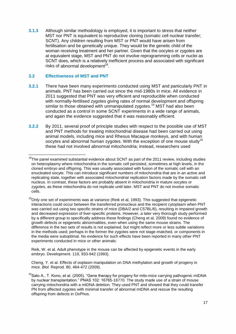
3.1.3 Although similar methodology is employed, it is important to stress that neither MST nor PNT is equivalent to reproductive cloning (somatic cell nuclear transfer; SCNT). Any children resulting from MST or PNT would have arisen from fertilisation and be genetically unique. They would be the genetic child of the woman receiving treatment and her partner. Given that the oocytes or zygotes are at equivalent stage, MST and PNT do not involve reprogramming cells or nuclei as SCNT does, which is a relatively inefficient process and associated with significant risks of abnormal development18.
3.2 Effectiveness of MST and PNT 3.2.1 There have been many experiments conducted using MST and particularly PNT in
animals. PNT has been carried out since the mid-1980s in mice. All evidence in 2011 suggested that PNT was very efficient and reproducible when conducted with normally-fertilised zygotes giving rates of normal development and offspring similar to those obtained with unmanipulated zygotes.19 MST had also been conducted as a control in some SCNT experiments in a wide range of animals, and again the evidence suggested that it was reasonably efficient.
3.2.2 By 2011, several proof of principle studies with respect to the possible use of MST
and PNT methods for treating mitochondrial disease had been carried out using animal models, including mice and Rhesus Macaque monkeys, and with human oocytes and abnormal human zygotes. With the exception of one mouse study20 these had not involved abnormal mitochondria; instead, researchers used
18
The panel examined substantial evidence about SCNT as part of the 2011 review, including studies on heteroplasmy where mitochondria in the somatic cell persisted, sometimes at high levels, in the cloned embryo and offspring. This was usually associated with fusion of the somatic cell with an enucleated oocyte. This can introduce significant numbers of mitochondria that are in an active and replicating state, together with associated mitochondrial replication factors made by the somatic cell nucleus. In contrast, these factors are probably absent in mitochondria in mature oocytes or zygotes, as these mitochondria do not replicate until later. MST and PNT do not involve somatic cells.
19Only one set of experiments was at variance (Reik et al, 1993). This suggested that epigenetic interactions could occur between the transferred pronucleus and the recipient cytoplasm when PNT was carried out using two specific strains of mice (DBA/2 and C57BL/6), resulting in impaired growth and decreased expression of liver-specific proteins. However, a later very thorough study performed by a different group to specifically address these findings (Cheng et al, 2009) found no evidence of growth defects or epigenetic abnormalities, even when using the same mouse strains. The difference in the two sets of results is not explained, but might reflect more or less subtle variations in the methods used; perhaps in the former the zygotes were not stage-matched, or components in the media were suboptimal. No evidence for such effects have been reported in many other PNT experiments conducted in mice or other animals:
Reik, W. et al. Adult phenotype in the mouse can be affected by epigenetic events in the early embryo. Development. 119, 933-942 (1993).
Cheng, Y. et al. Effects of ooplasm manipulation on DNA methylation and growth of progeny in mice. Biol. Reprod. 80, 464-472 (2009).
20Sato A., T. Kono, et al. (2005). “Gene therapy for progeny for mito-mice carrying pathogenic mtDNA by nuclear transplantation.” PNAS 102: 16765-16770. The study made use of a strain of mouse carrying mitochondria with a mtDNA deletion. They used PNT and showed that they could transfer PN from affected zygotes with minimal transfer of abnormal mtDNA and rescue the resulting offspring from defects in OxPhos.

18
substrains or subspecies, relying on the presence of different mtDNA haplotypes or haplogroups, so that sequence differences in mtDNA can be used to look at carryover and persistence of mtDNA with the spindle or pronucleus. Studies in Rhesus Macaque monkeys suggested that MST is efficient, allowing replacement of almost all mitochondria and the first set of experiments resulted in the birth of four healthy offspring showing no abnormities, then after two years (but now 5 years, see below). The rates of development to blastocyst stages and pregnancy rates in these experiments were essentially the same as for control unmanipulated embryos.
3.2.3 Studies using PNT with human zygotes were considered difficult to gauge. The
published studies then, used only abnormally-fertilised human oocytes, for example those containing three pronuclei. Embryos were reconstituted so that they had a normal complement of one maternal and one paternal pronucleus. The efficiency of blastocyst formation was about half that of control blastocysts. However, as the control blastocysts were not always good quality embryos and the PNT embryos were from abnormally fertilised oocytes, it was difficult to assess the efficiency of blastocyst formation. Nevertheless, all evidence presented to the panel (both published and unpublished) suggested that the blastocysts obtained after PNT and MST were as normal as controls, including cell numbers in the trophoblast and inner cell mass, and markers of cell type and stage.
3.2.4 At the time of the 2011 report, overall MST appeared less efficient than PNT,
which may have reflected problems in visualising the spindle and perhaps in transferring all the chromosomes. In mature metaphase II oocytes, the chromosomes are lined up attached to the spindle in preparation for the second meiotic division, rather than being enclosed in a pronuclear membrane, as is the case for PNT. Chromosomes may, therefore, be left behind, although this can be checked using fluorescent dyes that label DNA or chromatin. However, as highlighted in the 2013 report, more recent studies have made use of new polarised light microscopy techniques, and these seem to permit far more reliable visualisation and removal of the spindle, with all its associated chromosomes.
3.3 Further developments in the maternal spindle transfer technique Background 3.3.1 In a 2013 publication, MST was carried out on 65 human oocytes donated for
research (a further 33 served as controls). Although some oocytes displayed clear evidence of abnormal fertilisation (53% – determined by an irregular number of pronuclei), remaining embryos were capable of developing to blastocysts and producing embryonic stem cell lines at rates similar to controls. All five of the embryonic stem cell lines derived from zygotes predicted to have undergone normal fertilisation after MST had normal euploid karyotypes and contained exclusively donor mtDNA21,22.
21
An ES cell line was also established from a zygote that had 3 pronuclei and one polar body (3PN/1PB) instead of the normal 2PN/2PB. This had a triploid karyotype consistent with a failure to extrude the second polar body and retention of its genetic material.
22Tachibana M, et al (2013). Human embryonic stem cells derived by somatic cell nuclear transfer. Cell 6;1228–1238 2013

19
3.3.2 In 2013, a second published study also demonstrated the use of MST with human oocytes, although these were parthenogenetically activated rather than fertilised. The primary purpose of the study was to assess the degree of mitochondrial DNA carryover rather than establishing a technique for creating embryos for clinical use. MST was shown not to reduce developmental efficiency to the blastocyst stage, and genome integrity was maintained, provided that spontaneous oocyte activation was avoided through the transfer of spindle–chromosome complexes that were incompletely assembled or partially disassembled (depolymerised). The authors claimed to be able to achieve the latter by cooling the oocyte. Mitochondrial DNA transferred with the nuclear genome was initially detected at levels below 1%, decreasing in blastocysts and embryonic stem-cell lines to undetectable levels, and remained undetectable after passaging for more than one year, clonal expansion, differentiation into neurons, cardiomyocytes or pancreatic beta-cells, and after cellular reprogramming to derive iPS cells. Stem cells and differentiated cells had mitochondrial respiratory chain enzyme activities and oxygen consumption rates indistinguishable from controls. These cells were homozygous for all alleles (as they have become diploidised from an originally haploid state) and so would only give information about maternal imprinting (Paull et al, 2013)23.
2014 update 3.3.3 In 2014 the panel considered further evidence from the research group led by
Shoukrat Mitalipov at the Oregon Health and Science University. The panel was updated on the progress on the Macaques created previously as a result of MST techniques and was informed that the four male Macaques have now reached adulthood (5 years of age) and that they have shown no signs of abnormalities, but no further health outcomes follow-up has been conducted. The group is seeking to establish the fertility status of the Macaques by entering them into a breeding programme and more focussed studies looking at physiological impact will be conducted. There remains one female Macaque who is 2-3 years old (from a second set of experiments), that has not yet reached sexual maturity. The Oregon group have further explored details of the methods used for MST with human oocytes, notably to reduce the rates of abnormal fertilisation and to increase efficiency.
The panel concludes, in 2014, that good progress has been made in
developing the technique of Maternal Spindle Transfer and that researchers look likely to make good progress in further refining the technique over the short term to ensure efficacy and in establishing safety (within the constraints of in vitro systems).
3.4 Further developments in the pronuclear transfer technique Background 3.4.1 In 2011, the panel was informed of unpublished findings regarding PNT and MST
from the University of Newcastle group. Initial experiments using normally fertilised human zygotes for PNT revealed the importance of timing of the various
23
Paull, D, et al (2013) Nuclear genome transfer in human oocytes eliminates mitochondrial DNA variants. Nature 31;493(7434):632-7.

20
procedures and of matching developmental stage of the two zygotes. With optimisation, they had begun to obtain a significant proportion of manipulated embryos developing to blastocyst stages. Some zygotes resulting from PNT were successfully vitrified and further work was being carried out to improve the quality and rate of development to blastocysts and to minimise mtDNA carryover at the blastocyst stage.
3.4.2 In 2013, the panel noted that this information, together with comments from both
the other groups interviewed at this time, suggests that issues of timing may be relevant to any intended use of MST or PNT clinically since the cycles of the two egg donors will need to be synchronised. Egg retrievals will need to be carefully timed in order to be near coincident, the eggs need to be fertilised as soon as possible after collection, and for PNT the procedure needs to be carried out as soon as possible after normal fertilisation is confirmed. If there is a prolonged period of time between the two egg collections then one set of eggs may be over-mature, potentially leading to reduced development and an increase in abnormality rates. As this synchrony may not always be possible in a clinical setting, cryopreservation of eggs using vitrification was suggested as a solution. This is probably not an issue of safety, but one of efficiency, because the abnormalities are likely to be obvious and/or lead to early embryo lethality.
2014 update 3.4.3 In 2014 the panel was further updated on the progress of the Newcastle group’s
work, in both written submissions and presentations at the workshop. The panel was informed that the group had made considerable progress modifying their experiments from those using zygotes that were the product of abnormal fertilisation, which they were originally constrained to use, to those involving normal fertilisation. Because the former generally arrest at similar stages of development, irrespective of time of fertilisation, whereas the latter do not, they have established new procedures and working protocols to allow PNT to be carried out efficiently. These and other modifications to the PNT procedures now give reproducibly high rates of development to the blastocyst stage. A manuscript describing these data is currently being prepared by the group for publication. The group has also proceeded with experiments needed to demonstrate the safety and efficacy of these techniques, looking at chromosome make-up, cell numbers, markers of cell type, and mtDNA carryover. This work has identified some subtle differences in embryo development that are being investigated, but nothing has been found so far which raises concerns about safety. Pyrosequencing analysis has allowed the group to demonstrate that PNT using normally fertilised human eggs shows very low or absent levels of carryover of mitochondrial DNA – less than 2%. The group is also in the process of deriving ES cells from human embryos created by PNT.
3.4.4 The Newcastle group is also in the first phase of using MST on human eggs in
order to compare the two methods; although they explained that a limited number of human eggs are available for research, which means that it is difficult to carry out MST and PNT experiments in parallel in the same laboratory. The group is still open-minded as to which technique should be used clinically.
The panel concludes, in 2014, that good progress has been made in
developing the technique of pronuclear transfer and that researchers look likely to make further progress in refining the technique over the short term
21
to ensure efficacy and in establishing safety (within the constraints of in vitro systems).
As previously stated in the 2013 report, based on 2014 considerations the
panel still believes that there is at present insufficient evidence to choose between PNT and MST as a preferred technique.
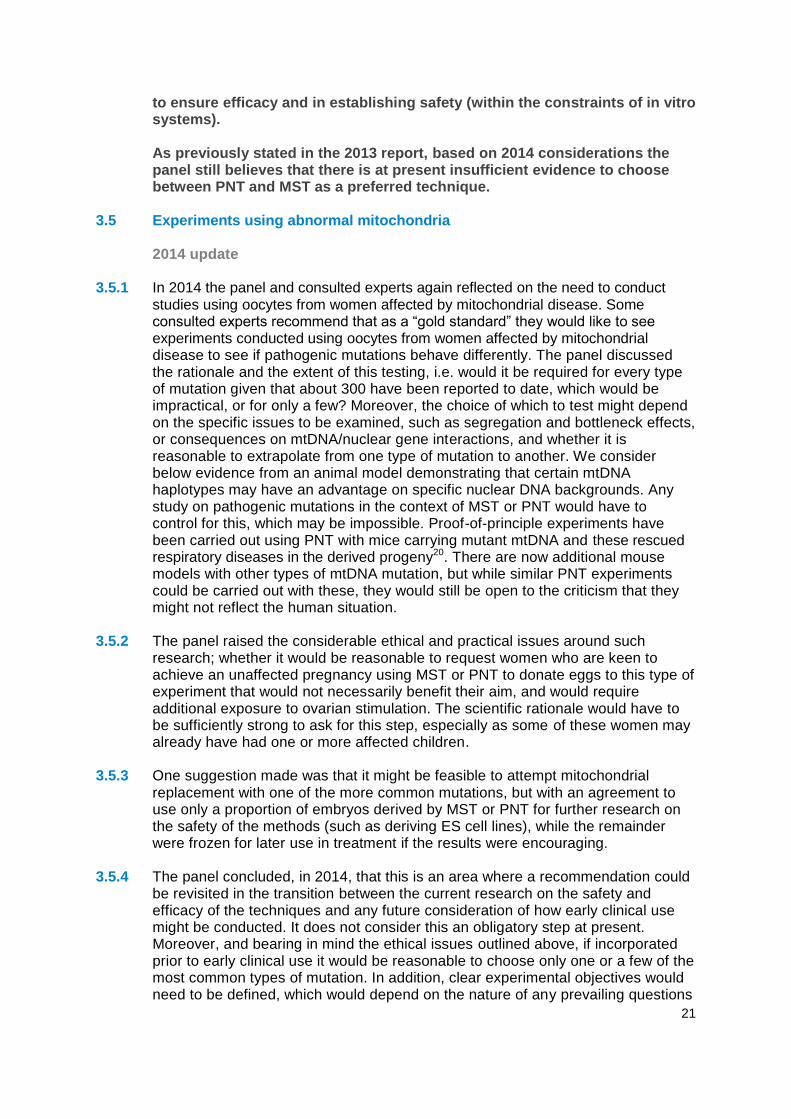
3.5 Experiments using abnormal mitochondria 2014 update 3.5.1 In 2014 the panel and consulted experts again reflected on the need to conduct
studies using oocytes from women affected by mitochondrial disease. Some consulted experts recommend that as a “gold standard” they would like to see experiments conducted using oocytes from women affected by mitochondrial disease to see if pathogenic mutations behave differently. The panel discussed the rationale and the extent of this testing, i.e. would it be required for every type of mutation given that about 300 have been reported to date, which would be impractical, or for only a few? Moreover, the choice of which to test might depend on the specific issues to be examined, such as segregation and bottleneck effects, or consequences on mtDNA/nuclear gene interactions, and whether it is reasonable to extrapolate from one type of mutation to another. We consider below evidence from an animal model demonstrating that certain mtDNA haplotypes may have an advantage on specific nuclear DNA backgrounds. Any study on pathogenic mutations in the context of MST or PNT would have to control for this, which may be impossible. Proof-of-principle experiments have been carried out using PNT with mice carrying mutant mtDNA and these rescued respiratory diseases in the derived progeny20. There are now additional mouse models with other types of mtDNA mutation, but while similar PNT experiments could be carried out with these, they would still be open to the criticism that they might not reflect the human situation.
3.5.2 The panel raised the considerable ethical and practical issues around such
research; whether it would be reasonable to request women who are keen to achieve an unaffected pregnancy using MST or PNT to donate eggs to this type of experiment that would not necessarily benefit their aim, and would require additional exposure to ovarian stimulation. The scientific rationale would have to be sufficiently strong to ask for this step, especially as some of these women may already have had one or more affected children.
3.5.3 One suggestion made was that it might be feasible to attempt mitochondrial
replacement with one of the more common mutations, but with an agreement to use only a proportion of embryos derived by MST or PNT for further research on the safety of the methods (such as deriving ES cell lines), while the remainder were frozen for later use in treatment if the results were encouraging.
3.5.4 The panel concluded, in 2014, that this is an area where a recommendation could
be revisited in the transition between the current research on the safety and efficacy of the techniques and any future consideration of how early clinical use might be conducted. It does not consider this an obligatory step at present. Moreover, and bearing in mind the ethical issues outlined above, if incorporated prior to early clinical use it would be reasonable to choose only one or a few of the most common types of mutation. In addition, clear experimental objectives would need to be defined, which would depend on the nature of any prevailing questions

22
and data concerning any potential replicative advantage attributed to the specific mutation in question.
3.6 Experiments using non-human primates
2014 update 3.6.1 The Panel received an update on MST in Macaques from Shoukrat Mitalipov
(University of Oregon) (see 3.3.3 for a summary of some of these data; other details are unpublished and thus confidential at this stage). The new data raised no issues relevant to safety; indeed, these experiments continue to provide reassurance that MST may be suitable for clinical application in humans. This prompted the panel to re-consider their decision in 2013 that it was no longer necessary to carry out PNT in a non-human primate model.
3.6.2 As stated in the 2013 report, research attempting to use PNT in Macaques had
not been successful. From unpublished data24 it appears that Macaque zygotes do not survive the PNT process well and published evidence suggests that there may be important differences between human and Macaque oocytes and early embryos; for example, different sensitivities to cryopreservation, and Macaque oocytes being less prone to abnormal activation/fertilisation following MST than human oocytes (as seen in Tachibana et al (2009)25 versus Tachibana et al (2013)35). In 2013 the panel believed that the Macaque may not be a sufficiently good model for the human in this context. While it is of course possible that further experiments using non-human primates could provide some additional useful biological information, many of the important issues around heteroplasmy with variant mtDNAs have already been addressed or at least highlighted in Macaques, rodents and human studies. Others relating to the behaviour of mutant mtDNA may be better carried out in emerging mouse models or directly using human oocytes and zygotes and assays in preimplantation embryos and ES cells derived from them. But in terms of assessing both safety and efficacy of MST and PNT the panel is concerned that the differences between Macaque and human oocytes/early zygotes will be unhelpful. Indeed, if there are critical periods of development where the human is unique, such experiments may even be misleading if carried out in animals.
3.6.3 In 2014, following further discussion with researchers, the panel uphold the view
that the use of non-human primate experiments is no longer critical and raised this for discussion at the scientific workshop on the 4th April. One expert thought that currently successful Macaque trials exploring MST techniques do not invalidate the possibility of mito-nuclear incompatibilities (discussed further at section 3.7.15) despite the use of two genetically distant subpopulations, and that the current studies have insufficient statistical power to detect mito-nuclear incompatibilities. Demonstrating that mito-nuclear interactions are not of practical concern would require a much larger sample of Macaques. An experimental design that would be statistically significant would involve >5 genotypes, with
24
Reported at a media briefing in October 2012 and reflected in a number of articles e.g. http://www.sciencenews.org/view/generic/id/346024/description/Cloning-like_method_targets_mitochondrial_diseases
25Tachibana M et al. (2009) Mitochondrial gene replacement in primate offspring and embryonic stem cells. Nature. 17;461(7262):367-72.

23
replicate observations, resulting in approximately 40 offspring in total. Trans-generational effects would require breeding and monitoring of subsequent generations.
3.6.4 Although this review is focused on the science, it is an ethical concern to carry out
experiments on animals, especially non-human primates, if these are likely to not be informative. Therefore, as stated in 2013, given that the most critical species in which to obtain results is the human, and because there are differences in the very early embryology between mammalian species, the panel also concludes that if any additional experiments on PNT and MST in other animal models reveals differences with the human, it would be not just reassuring, but important if such experiments revealed the underlying reasons, and did not merely state the problem. It is therefore the panel’s view that such experiments would be difficult to justify with respect to cost, ethical considerations and the length of time required, especially as any risks were considered to be small. The panel suggested that a more informative experimental approach would be based on the derivation of embryonic stem cell lines and subsequent differentiation of these to examine oxidative phosphorylation, gene expression levels, and other physiological parameters in distinct human lineages derived from MST/PNT embryos.
3.6.5 Shoukrat Mitalipov confirmed that his group are not pursuing PNT in Macaques.
Indeed, with respect to animal models for basic research, they are now using both MST and PNT in mice. The panel did not receive any evidence to indicate that other groups have had success with PNT in Macaques, or that anyone is attempting such experiments. The ethical arguments above therefore still stand: it would be a major undertaking to embark on a study of PNT in Macaques, or other non-human primates, when the methods need to be established, when there are clear differences in the biology of early Macaque and human embryos, and when PNT does work with human zygotes, which is of direct relevance. Furthermore, many of the questions that these experiments might address, namely issues of mtDNA carryover and its subsequent behaviour in offspring, can be addressed by studying the Macaques derived by MST. Other work, for example, studying the behaviour of mutant mtDNA after mitochondrial replacement, is best performed in a non-primate animal model where this has already been derived, namely the mouse.
In 2014 the panel therefore stands by its recommendation of 2013 that PNT
in a non-human primate model, with the demonstration that the offspring derived are normal, is not required.
3.7 Safety of MST and PNT Background 3.7.1 In 2011 an initial review of the safety of MST and PNT was discussed, based on
studies published up to March 2011 (outlined in section 4.3 of the original report). In 2013, based on the evidence submitted, the panel re-examined and commented on the following safety issues of the MST and PNT techniques: the carryover of mtDNA from the affected oocyte or zygote; the methods to prevent premature activation of oocytes or detect abnormally fertilised oocytes; the nuclear-mitochondrial interactions involved and the potential for long-lasting nuclear epigenetic modifications resulting from manipulation or altered mitochondrial states associated with mitochondrial disease. The panel did not specifically revisit previous discussions regarding the safety of reagents used to carry out the micromanipulation techniques. However, it was noted that the study by Paull et al

24
(2013)23 relied on the use of several such reagents, the combination of which might have been expected to be deleterious, yet development of the (parthenogenetically activated) embryos, and ES cell lines derived from them, was apparently normal. It was felt by the panel, and by those it interviewed in 2013, that the number of reagents and their concentration should be kept to a minimum, and should MST or PNT develop into viable clinical techniques, that the Medicines and Healthcare products Regulatory Agency (MHRA) would need to be satisfied about the provenance and clinical safety of all reagents. The areas identified in 2013 are summarised below and where applicable the 2014 update is provided.
3.7.2 mtDNA carryover: In 2013 the panel outlined that carryover of mtDNA from the
affected oocyte or zygote might be expected with both techniques because the spindle or the pronuclei are enclosed in a karyoplast during the manipulation technique, which contains a small amount of surrounding cytoplasm enclosed in cell membrane in addition to the nuclear DNA. In theory, carryover of abnormal mtDNA may be an issue if abnormal mtDNA is preferentially replicated and if there is a marked difference in segregation across tissues. However, evidence presented to the panel in 2013 continues to be reassuring that carryover after mitochondrial replacement is very low.
3.7.3 In 2013, the panel noted that a threshold of mitochondrial function is required for
normal development, and despite developmental plasticity of the embryo, impaired mitochondrial function in the embryo affects subsequent fetal and placental growth (Wakefield et al 2011)26. At this time, one study suggested that an (experimental) admixture of two normal but different mouse mtDNAs can be genetically unstable and can produce adverse physiological effects (Sharpley et al, 2012)27. These results could indicate that the differences between mtDNAs within a mammalian species may not be neutral and are suggested to explain the advantage of uniparental inheritance of mtDNA. This could be a concern for MST and PNT. However, the study used approximately equal amounts of mtDNA from two very different mouse strains, which could be considered distant subspecies. Also, another study, exploring mtDNA segregation during early embryogenesis in Macaques, produced distinctly different results - no such problems (ie. adverse physiological effects) were observed with mixtures of mtDNA from two Macaque subspecies. However, the oocytes created to be heteroplasmic (50/50) for these two types of Macaque mtDNA variants resulted in embryos exhibiting significant partitioning of the mtDNA between different blastomeres and to some extent between trophectoderm and ICM. This partitioning seems to have resulted in some of the fetuses, or ES cell lines derived from such embryos, also showing a skewed ratio (in one case about 94% of one of the mtDNA variants was present).
3.7.4 In 2013 there was no evidence of preferential selection for ‘resident’ versus ‘alien’
mtDNA, suggesting that both variants work equally well with the resident nuclear DNA, even though the mtDNA sequence of the two sub-species of Macaque are as different from each other as they are from other primate species (Lee et al,
26
Wakefield, SL. et al. (2011) “Impaired Mitochondrial Function in the Preimplantation Embryo Perturbs Fetal and Placental Development in the Mouse.” Biology of Reproduction 84, 572-580.
27Sharpley, M.S. et al. (2012) “Heteroplasmy of Mouse mtDNA is Genetically Unstable and Results in Altered Behavior and Cognition.” Cell 151: 333–343.

25
2012)28. The degree of heteroplasmy was so substantial that it could lead to homoplasmy. This could be an issue if one of the mtDNA variants is defective, with, by chance, either a beneficial or poor outcome for the individual born. However, the starting point in these experiments was about 50-50, whereas MST (or PNT) should give very low levels of carryover of mutant mtDNA, making homoplasmy for the normal mtDNA even more likely.
3.7.5 At this time, the authors also carried out MST between oocytes of the two
Macaque subspecies to explore whether this preimplantation segregation of mtDNA variants could be a problem. They first determined that isolated karyoplasts carry “bound” mtDNA (there is no evidence that it is physically bound, just closely associated) at an average level of about 0.6% of the numbers within the cytoplasm. After MST, about 68% of fertilised oocytes developed to blastocysts, confirming earlier published data from the same group (Tachibana et al, 2009)24. They then selected female blastocysts for embryo transfer, recovering two fetuses in which to survey levels of heteroplasmy. The mtDNA variant from the spindle donor oocytes was very low or undetectable in somatic tissues, suggesting a tendency towards homoplasmy. However, two out of 24 oocytes isolated from the fetal ovaries showed around 15% heteroplasmy. This difference between somatic and germ line was also evident in their earlier experiments, with segregation in oocytes appearing to be largely independent of that occurring in other tissues.
3.7.6 These findings largely support what is known about mtDNA levels and founding cell
numbers of somatic tissues and the germ line during early postimplantation development and bottleneck theories as outlined in the panel’s 2011 report. However, the observation of much earlier segregation of mtDNA, in cleavage stage embryos, is novel and contradicts evidence obtained from human embryos where blastomeres within an embryo tend to have very similar levels of heteroplasmy (as also demonstrated by Sallevelt et al11, 2013 and Treff et al, 201229 - although with a few outliers). This could suggest that there is relatively little mixing of cytoplasm after spindle (or cytoplast) transfer, such that cleavage divisions are responsible for the segregation, whereas with heteroplasmy already existing in a growing oocyte, the mtDNA variants are likely to be distributed at random.
3.7.7 Modelling the inheritance of mtDNA has led to the conclusion that for a disease with
a clinical threshold of say 60% mutant mtDNA (which is fairly typical) reducing the mutant mtDNA load to <5% with MST or PNT should dramatically reduce the chance of disease recurrence not just in the child, but in subsequent generations (Samuels et al, 2013)30. However, >5% carryover was associated with a significant chance of recurrence. Mutations with a lower clinical threshold were also likely to have a higher risk of recurrence, but reducing the amount of carryover would
28
Lee, H-S. et al. (2012) “Rapid mitochondrial DNA segregation in primate preimplantation embryos precedes somatic and germline bottleneck.” Cell Rep. 1(5): 506–515.
29Treff, N. R. et al (2012) “Blastocyst preimplantation genetic diagnosis (PGD) of a mitochondrial DNA disorder.” Fertil Steril. 98(5):1236-40.
30Samuels, D.C. et al. (2013) “Preventing the transmission of pathogenic mitochondrial DNA mutations: can we achieve long-term benefits from germ line gene transfer?” Human reproduction 28(3):554-9.

26
counteract this. If the threshold is very low, and the panel noted that there has been one report of heteroplasmy levels of less than 10% causing disease for a dominant mitochondria mutation initially detected in muscle31, then the modeling may not be adequate. Moreover, it does not take account of the possibility of preferential replication or selection of mtDNA carrying specific mutations, however, there is little evidence of this occurring.
3.7.8 Publications and discussions with researchers in 2013, indicated that PNT showed
higher level of carryover than MST (up to 2% versus 0.3%) (Tachibana et al, 201335; Paull et al, 201323; Craven et al; 201032). This may be due to differences in the geometry and volume of the transferred structures, since two pronuclei are transferred in PNT rather than one spindle associated chromosome set in MST.
3.7.9 In 2013 during the discussions, the panel was also minded to draw attention to
parallels with PGD for mtDNA mutations in terms of acceptable levels of heteroplasmy in offspring. Although the intention of such therapy is to select embryos for transfer with as low a level of mutant mtDNA as possible to avoid the birth of a child who would manifest the disease in their lifetime, issues to do with variable segregation of mutant mitochondria in their tissues and especially their gametes also apply here. Hence clear rules for acceptable levels of mtDNA heteroplasmy allowing transfer or not of an embryo should be developed for each disease by the specialist clinical team in conjunction with their patients, and follow up of such children and their offspring is strongly recommended, as the panel have recommended for offspring arising for MST and PNT.
3.7.10 In 2013, the panel concluded that any early segregation of a very low level of
mutant mtDNA is unlikely to be a problem for children born as a result of MST (or PNT). Nevertheless, they felt that it would be reassuring to verify this with human preimplantation embryos generated as a result of MST and PNT for research purposes, and in ES cells and their differentiated derivative cell types obtained from such embryos. There is a potential concern, however, for subsequent generations if a female child born after the use of these techniques had a proportion of oocytes with a significant level of heteroplasmy. This could be researched by, for example, following differentiation protocols reported to generate primordial germ cells from human ES cell in vitro. Alternatively, it may be may be sufficient to explore these ‘bottleneck’ issues by looking at ES cell sub-lines derived from single cells (‘clonal analysis’). If it turns out that there is a significant risk that a proportion of oocytes and therefore any resulting embryos from a women born after MST or PNT could be heteroplasmic, then a recommendation might be for her to make use of PGD to select for embryos homoplasmic for the normal mtDNA variant. From the data on Macaques derived by MST, if the child is female, then while some of her oocytes may have very low or undetectable levels of mutant mtDNA, it is possible that others may have a significant proportion, considerably higher than her somatic tissues. These levels may still not be sufficient to cause her children to have a problem, but subsequent generations could be affected. Although diagnostic technology may well have advanced by then, by carrying out PGD (on embryos
31
Alston CL et al. (2010). “A novel mitochondrial tRNAGlu (MTTE) gene mutation causing chronic progressive external ophthalmoplegia at low levels of heteroplasmy in muscle.”J Neurol Sci. 15;298(1-2):140-4.
32Craven L., H. A. Tuppen, et al. (2010). “Pronuclear transfer in human embryos to prevent transmission of mitochondrial DNA disease.” Nature 465(7294):82-5

27
created from oocytes of female offspring resulting from MST or PNT, who might be carriers of the mutation in some of their oocytes) it ought to be possible to select embryos for implantation that have no abnormal mitochondria. This would guarantee that subsequent generations would be free from disease.
2014 update
3.7.11 The panel agreed with its earlier conclusions on risk of mtDNA disease in
subsequent generations. Moreover, it did not support any proposal to select only male embryos for transfer after MST or PNT, even though this would avoid these issues as well as circumvent objections made by some that the methods are a form of germ line genetic alteration. Selecting only XY embryos for transfer would require PGD, an additional step that is likely to compromise early development of already manipulated embryos; moreover, it would on average immediately reduce by half the number of embryos available for transfer. This would decrease the efficiency of the techniques and make it likely that patients would have to undergo repeated cycles of treatment. Determining embryo sex after implantation would require selective termination of normal female embryos, which is unlikely to be acceptable to patients (or society). Additionally, there is already an accepted precedent for using a method of ART, which has a consequence for the next generation: when ICSI is used to overcome male infertility in patients with a Y chromosome defect, any son born as a result will carry the same defect, and ICSI will be required for him to have a child. This will be true for all subsequent generations of males. In contrast, for MST and PNT, the risks of mitochondrial disease in the next generation will be low. The subsequent use of PGD in this next generation, to select embryos with very low or no levels of abnormal mtDNA, may rid all subsequent generations of the need to have any intervention to avoid mitochondrial disease.
The panel concludes, in 2014 that no new evidence was presented in relation to this area. The panel continue to recommend that, if any uncertainty about the degree of heteroplasmy in oocytes remains when women born as a result of MST or PNT wish to have children, then either this should be examined directly in unfertilised oocytes collected from the women obtained after stimulation, and/or PGD should be carried out on fertilised embryos prior to selecting those with, no or very low, levels of abnormal mtDNA for transfer. This should minimise chances of transmission of abnormal mtDNA to subsequent generations.
3.7.12 Methods to prevent premature activation of oocytes or detect abnormally
fertilised oocytes: The proof of principle studies, outlined in section 2.2 of the 2013 report, have demonstrated that nuclear genome transfer carried out in the process of MST can lead to premature oocyte activation and abnormal fertilisation. The panel explored the measures that could be put in place to address these risks.
3.7.13 In 2013, the panel was reassured to hear that the abnormally fertilised eggs
created followed MST can easily be identified using a standard stereo-microscope by looking for normal number of pronuclei and polar bodies which have failed to extrude at the PNT stage. As part of the tests to look for normality of development, array comparative genome hybridization (CGH) was used on trophectoderm biopsies from MST derived human blastocysts. Analysis did not detect abnormalities in uniformly triploid embryos suggesting some shortcomings

28
of CGH approaches33. Some of the abnormalities associated with MST can be detected only after sperm fertilisation by sperm and some are likely to have been due to problems with oocyte ageing (Tachibana et al, 2013)35.
3.7.14 It was reported that premature oocyte activation could be prevented by partial
depolymerization of the spindle–chromosome complex through cryopreservation or cooling to room temperature, allowing normal polar body extrusion (Paull et al, 2013)23. The authors confirmed that they were satisfied that implementing a spindle chilling stage did not damage the spindle since they had not seen dispersion of the spindle, as had been suggested in a previous research paper (Pickering et al, 1990)34. The spindle came back to normal size on warming and the oocyte extruded a polar body.
In 2014 the panel received little direct new evidence in relation to this area.
However, alternative methods to prevent abnormal activation have been described in a series of recent papers reporting SCNT with human oocytes. These made use of low levels of caffeine, as a protein phosphatase inhibitor, in the medium during spindle removal and fusion of the somatic cell. In addition, it is also clear that care must be taken to prevent fusion of polar bodies back into the oocyte or zygote, as this could also lead to development of embryos with aneuploidy (Yamada et al, 2014, Chung et al, 2014 and Tachibana et al, 201321).
3.7.15 Nuclear-mitochondrial interactions: It is well known that there are functional
interactions in both directions between the nucleus and mitochondria. Moreover, it appears clear that different mtDNA haplotypes and haplogroups are associated with variations in the efficiency of OXPHOS and, that this may influence both normal and disease-associated phenotypes in humans. In 2013 the panel highlighted that a concern had been raised that there might be a failure of correct nuclear-mitochondrial interaction following MST or PNT because the donor mtDNA may be of a haplogroup different from that with which the maternal nuclear genome had been functioning. Mitochondria from separate human lineages can be classified according to similarities or differences in their DNA sequence into many different haplogroups. The more evolutionary distant the separation of two maternal lineages, the greater the differences between mitochondrial haplogroups. This is typified by comparisons between European and African mtDNA. However, in 2013 the panel maintained the view that there is no evidence for any mismatch between the nucleus and any mtDNA haplogroup, at least within a species (with the possible exception of the study by Sharpley et al (2012)27). Fifty per cent of nuclear genes are paternally inherited and are consequently ‘alien’ to the mtDNA; backcrossing can replace the nuclear DNA entirely in a few generations. Furthermore, mitochondrial disease has not been noted to be more frequent amongst mixed-race children. Tachibana et al (2013)35
33
Details relating to the shortcomings of CGH testing were provided by Mitalipov et al in supplementary information, to the panel, and are not included in the Tachibana et al 2013 published article. The panel was informed that CGH analysis of biopsied trophectoderm in blastocysts did not detect uniform triploidy. Therefore uniform triploidy was confirmed by deriving ESCs from the same blastocyst using conventional G-banding.
34Pickering SJ, et al (1990) Transient cooling to room temperature can cause irreversible disruption of the meiotic spindle in the human oocyte. Fertility and Sterility 54(1):102-108
35 Tachibana, M. et al. (2013) “Towards germline gene therapy of inherited mitochondrial diseases.”
Nature 493(7434):627-31.
29
also conducted a 3-year follow-up study on MST-derived Macaque offspring born in 2009. The two species of Macaques used in these MST experiments have distinct mitochondrial haplotypes, yet neither the mixing of mitochondria nor swopping the haplotype with respect to their nuclear genome with which it normally resides, appears to result in any adverse effects in offspring. All four (males) were healthy and had normal mitochondrial function. Moreover, there were no significant changes with age in the degree of heteroplasmy in blood and skin cell samples, which remained less than 1% from the spindle donor. However, in 2013 the panel felt that if concerns were raised, it would be possible to match mtDNA haplogroups from the egg donor and the mother.
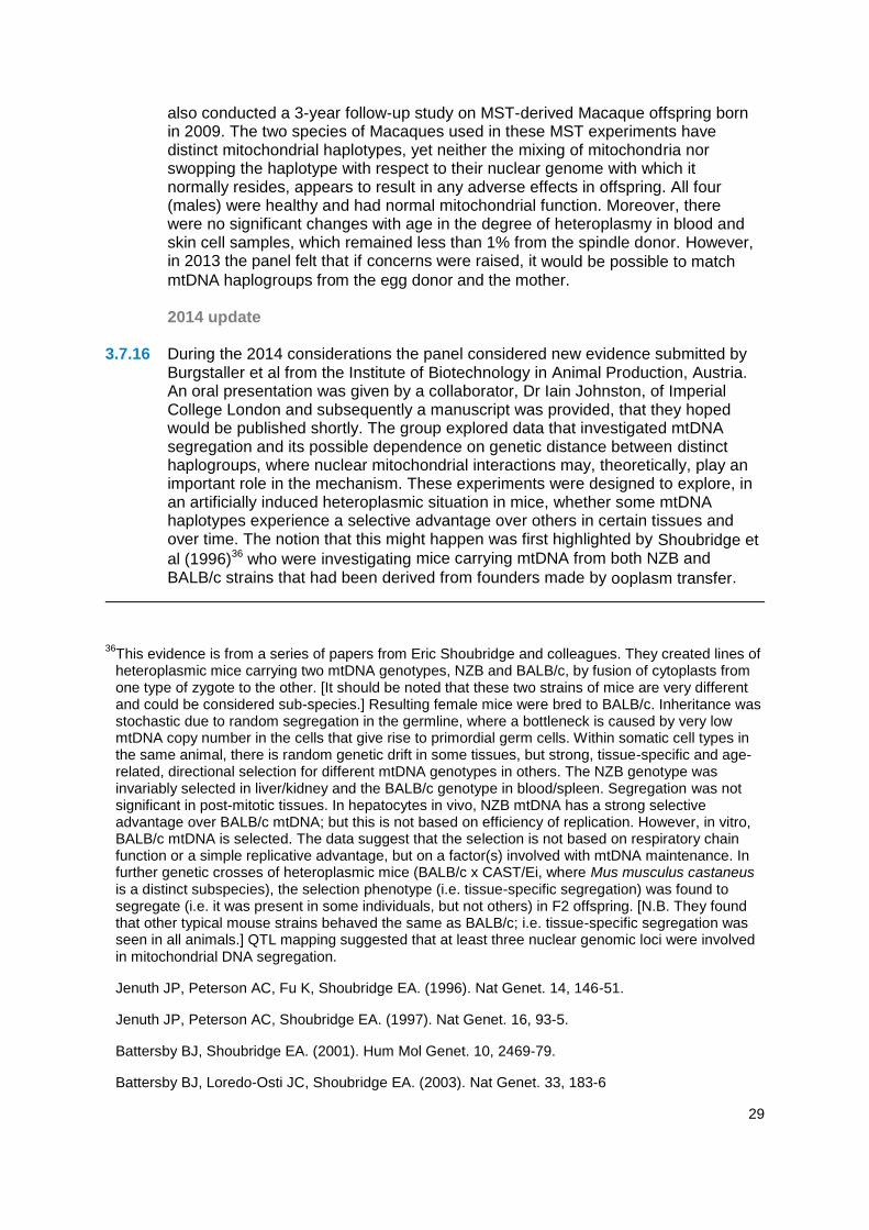
2014 update 3.7.16 During the 2014 considerations the panel considered new evidence submitted by
Burgstaller et al from the Institute of Biotechnology in Animal Production, Austria. An oral presentation was given by a collaborator, Dr Iain Johnston, of Imperial College London and subsequently a manuscript was provided, that they hoped would be published shortly. The group explored data that investigated mtDNA segregation and its possible dependence on genetic distance between distinct haplogroups, where nuclear mitochondrial interactions may, theoretically, play an important role in the mechanism. These experiments were designed to explore, in an artificially induced heteroplasmic situation in mice, whether some mtDNA haplotypes experience a selective advantage over others in certain tissues and over time. The notion that this might happen was first highlighted by Shoubridge et
al (1996)36 who were investigating mice carrying mtDNA from both NZB and
BALB/c strains that had been derived from founders made by ooplasm transfer.
36This evidence is from a series of papers from Eric Shoubridge and colleagues. They created lines of heteroplasmic mice carrying two mtDNA genotypes, NZB and BALB/c, by fusion of cytoplasts from one type of zygote to the other. [It should be noted that these two strains of mice are very different and could be considered sub-species.] Resulting female mice were bred to BALB/c. Inheritance was stochastic due to random segregation in the germline, where a bottleneck is caused by very low mtDNA copy number in the cells that give rise to primordial germ cells. Within somatic cell types in the same animal, there is random genetic drift in some tissues, but strong, tissue-specific and age-related, directional selection for different mtDNA genotypes in others. The NZB genotype was invariably selected in liver/kidney and the BALB/c genotype in blood/spleen. Segregation was not significant in post-mitotic tissues. In hepatocytes in vivo, NZB mtDNA has a strong selective advantage over BALB/c mtDNA; but this is not based on efficiency of replication. However, in vitro, BALB/c mtDNA is selected. The data suggest that the selection is not based on respiratory chain function or a simple replicative advantage, but on a factor(s) involved with mtDNA maintenance. In further genetic crosses of heteroplasmic mice (BALB/c x CAST/Ei, where Mus musculus castaneus is a distinct subspecies), the selection phenotype (i.e. tissue-specific segregation) was found to segregate (i.e. it was present in some individuals, but not others) in F2 offspring. [N.B. They found that other typical mouse strains behaved the same as BALB/c; i.e. tissue-specific segregation was seen in all animals.] QTL mapping suggested that at least three nuclear genomic loci were involved in mitochondrial DNA segregation.
Jenuth JP, Peterson AC, Fu K, Shoubridge EA. (1996). Nat Genet. 14, 146-51.
Jenuth JP, Peterson AC, Shoubridge EA. (1997). Nat Genet. 16, 93-5.
Battersby BJ, Shoubridge EA. (2001). Hum Mol Genet. 10, 2469-79.
Battersby BJ, Loredo-Osti JC, Shoubridge EA. (2003). Nat Genet. 33, 183-6

30
3.7.17 The panel also considered the separate issue of whether it is possible that an
incompatibility could arise between the nuclear and mitochondrial genomes in an embryo generated by MST or PNT. In a submission and presentation from Edward Morrow, University of Sussex, some of their group’s concerns relating to the safety of mitochondrial replacement techniques, expressed in the recent article by Reinhardt et al (2013), and in a follow-up manuscript submitted as evidence, were revisited. The presentation highlighted their views on nuclear-mtDNA interactions and the anterograde and retrograde signalling between these DNA genomes, emphasising this signalling cross-talk and the possible effects on oxidative phosphorylation, metabolic pathways and other processes that they propose might result from disrupting these interactions. Discussions concerning these two separate issues relating to mito-nuclear interactions are detailed in the box below, and the conclusions of the panel follow this.
Summary of discussions related to mito-nuclear interactions
The new experiments by Burgstaller and colleagues (2014), used a constant source of nuclear DNA and resident mtDNA (C57BL/6N), but with additional mitochondrial genomes derived from four separate wild mice populations. These were chosen to reflect the typical mtDNA variation seen across human populations. They examined heteroplasmy over time in a range of tissues in the offspring of these founder lines of mice, looking specifically for deviation from the relative proportions expected if there was no differential segregation of mtDNA haplotype. From analysing hundreds of mice, they found that a statistically significant degree of segregation occurred in some adult mouse tissues, where the extent, but not the direction of the segregation, is dependent on the degree of genetic distance between the introduced and resident mitochondrial haplotypes. The greater the genetic difference, the greater the likelihood of differential amplification of mtDNA haplotypes. The use of PNT and MST may result in a proportion of mutant mtDNA carrying over into the egg and hence the embryo that is destined to be transferred to the woman (see
section 3.3). In practice, the carryover is likely to be very small; however, Burgstaller et
al postulate that haplotype differences might lead in some cases to the preferential amplification or segregation of this originally rare mtDNA variant in some tissues, perhaps including the germ line. If the advantaged amplified variant carries the mutation, it could lead to mitochondrial disease in the child or, if female, her descendants as well. However, this latter conclusion was different from Shoubridge’s data, which suggested that any segregation in the germline was random.
Iain Johnston, on behalf of the authors, reiterated that the mutation itself is not required to contribute to differential segregation: it is essentially “hitchhiking” on the superior segregation capacity of the haplotype in which it resides. They therefore suggest that in order to mitigate this potential risk when carrying out MST or PNT, haplotype (or haplogroup) matching should be performed, for example by using, maternal relatives of the patients (i.e. those with very similar mtDNA, but who do not carry the pathological mutation) as donors. However, they highlighted that more research on segregation differences between haplotypes needs to be conducted before, for example, it could be envisaged selecting a donor with advantageous mtDNA (healthy mtDNA that will preferentially segregate). It was acknowledged that the group did not yet have any data concerning mechanisms, and so could not identify whether the mtDNA haplotype segregation observed in their model was due to a difference in mtDNA proliferation rate, or to some other mechanism

31
such as cell proliferation or competition (where cells which have by chance a higher proportion of the favoured type divided faster or survived better than the other). Although it seems likely that the skewed segregation will depend on an interaction with the nuclear genome, the panel highlighted that no attempts had been made to address this, given
that the group had introduced the variant mtDNAs into the same inbred strain nuclear
background in each case. Backcrossing these mice to another strain might also indicate whether a few or many C57BL/6N nuclear alleles were required to see the effect. Given
that humans tend to be outbred, with random segregation of alleles in nuclear DNA that are involved in interactions with mitochondria, the panel questioned the relevance of these data to children born from MST or PNT. …………………………………………………………………………………………………......
The panel also discussed the idea that mito-nuclear interactions that have co-evolved may be disrupted when using PNT and MST, referring to the literature in this area, including data from model organisms that describe a range of phenotypic effects due to mitochondrial replacement. This included the data from Edward Morrow and colleagues and that of others from the fruitfly (Drosophila melanogaster) and data reported in a published article by Innocenti et al (2011)37. They also referred to a paper by David Clancy (2008)38, which describes the original research on which these studies are based, including the derivation of the various Drosophila lines39. The presentation by Morrow, stated that animal models such as these were not considered by some to be relevant to humans, for at least three reasons:
Mitochondria replacement, like sexual reproduction, randomly shuffles mitochondrial and nuclear genomes each generation.
Genetic diversity in humans is too low to cause incompatibilities.
Incompatibilities do not occur in non-human primates.
Morrow, countered these objections with the view that MST and PNT are more likely than sexual reproduction to disrupt any mito-nuclear interactions that have co-evolved due to selective processes operating over many generations. He suggested that MST and PNT place the mtDNA with “unfamiliar” maternal and paternal nuclear genomes, rather than just with a new paternal nuclear genome as would occur with normal reproduction. If there was any mismatch between mitochondrial and nuclear genes this immediate change might lead to embryo loss, male infertility or other undesirable consequences. However,
37
Innocenti P. et al (2011) Experimental Evidence Supports a Sex-Specific Selective Sieve in Mitochondrial Genome Evolution. Science 13 332 6031 845-848
38Clancy DJ. (2008). Variation in mitochondrial genotype has substantial lifespan effects which may be modulated by nuclear background. Aging Cell. 7, 795-804. PMID: 18727704
39In 2014 Morrow et al outlined the findings of Innocenti et al’s work (2011): they obtained Drosophila melanogaster lines (the same as in Clancy 2008) in which specific “wild type” mitochondrial genomes have been exchanged with specific nuclear genomes in a single cross (by the use of “balancer chromosomes”, and isogenic donor strains which force the exchange of most of the nuclear DNA). The original inbred lines came from very different locations around the world, and have distinct mtDNA “haplotypes”, and it has been proposed that these have co-evolved with the nuclear genome in these geographically isolated populations. Innocenti et al studied several of these lines (5 genotypes, 2 sexes, 2 biological replicates, 8 individuals, and a total 160 flies) and found evidence to suggest that mtDNA type can influence nuclear gene expression in males but not females, and lead to problems of male fertility.

32
the panel and the Newcastle Group suggested that human reproduction between individuals from quite distinct ethnic/racial groups, exhibiting considerable mtDNA and nuclear DNA genetic diversity, occurs commonly without obvious detriment to offspring. Indeed, children from mixed parents tend to show increased fitness (Campbell et al 2007, Lewis et al 2010). Moreover, it takes only a few generations to effectively exchange an mtDNA haplogroup. Morrow was of the opinion that no studies have been conducted in humans to explore the consequences, if any, of the exchange of mtDNA haplogroups by reproduction; and suggested that these were unlikely to be feasible in humans. However, the panel expect that data suggestive of such effects would have become apparent in the many large scale human genome projects looking for disease associations that are currently being conducted. Morrow suggested that zygotes, with problematic interactions between mtDNA and nuclear DNA may not be viable so fail to implant in the women or are miscarried at an early stage, and that this may explain why abnormalities in children or adults due to
disrupted mito-nuclear interactions are not observed. On this basis he suggested that the
absence of evidence demonstrating mito-nuclear incompatibilities did not reduce the need for further research, but that haplogroup matching could be considered as a “potential workaround”. Morrow suggested that the risk of incompatibilities increases with greater divergence and that haplogroup matching may reduce this. However, even small genetic differences may exist between seemingly matched haplogroups and therefore greater understanding is required. The panel also discussed the idea that generating only males by MST/PNT would be preferable due to males being a genetic “dead-end” for mitochondrial transmission. Morrow highlighted that while the health of the male Macaques when adult would give a more complete picture, it would provide very little reassurance of the safety of MST/PNT more generally in humans given the low number of individuals studied. Data from Drosophila suggesting reduced male fertility after mitochondrial replacement would indicate that this might not be a suitable practice. Morrow suggested that if male Macaques did not demonstrate reduced fertility then this would provide reassuring evidence of safety. The panel also suggested that more extensive studies on Drosophila from outbred genetic backgrounds may be beneficial in determining the relevance of the fly model to humans, given that the latter are generally both outbred and where interbreeding is common. Indeed, the panel was of the opinion that many of the effects hypothesised by Morrow and his colleagues would be effectively buffered in humans by the “noise” of the likely variability in the many nuclear genes associated with mitochondrial function. The panel also referred to views of other scientists in the field, (Chinnery et al 2014, Eyre-Walker et al 2014) that also contested the concerns over MST/PNT expressed in Reinhardt et al (2013).
3.7.18 In summary, the panel considered two separate arguments for considering
haplotype/haplogroup matching. Morrow et al's concern (based on evolutionary arguments) is that some nuclear-haplotype combinations can lead to a failure of co-evolved mito-nuclear interactions with adverse consequences. They propose haplotype or at least haplogroup matching to reduce the chance of any such effect occurring. Their arguments did not address mutant mtDNA per se. Moreover, Morrow and colleagues do not address the possibility that an exchange to a different haplotype/haplogroup might, in some cases, confer advantages. The panel consider any such hypothetical problems to be very unlikely. Given, the high degree of variation in the nuclear genome that can have an impact on embryo fitness, whether this is due to effects on mitochondrial function or not, the consequences of pursuing any strategy to do with matching are likely to be
33
minimal. The fly research used inbred strains, which makes it more likely that any effects of a mismatch will be detected, whereas humans are much more outbred so that such effects are unlikely to be detectable.
3.7.19 Burgstaller et al's data raise the concern that, after MST or PNT, the now rare
mtDNA variant carrying the mutation will preferentially expand (due to some unknown mechanism) in some tissues. This would occur only if that variant possesses some "selectable" advantage in those tissues in comparison to the mtDNA variant provided by the donor oocyte. This selection could operate within (mtDNA competition) or between cells (cell competition) in the developing embryo or newborn. If this occurs, it might be deleterious for the child born from MST or PNT, and/or for subsequent generations if the child is a girl and the selection process operates in the germline. This is a different argument from that of Morrow et al, because any problem would not be due to some failure of mito-nuclear interaction, but because the advantaged mtDNA variant carries the mutation. However, the advantage conferred by the specific variant that carries the mutation, and which results in selection, might depend on mito-nuclear interactions. They proposed haplotype/haplogroup matching to remove any selectable advantage. However, it might be possible to deliberately choose a donor carrying a haplogroup that has a selectable advantage (in the context of the child's nuclear DNA), which would out-compete the variant carrying the mutation. This would be beneficial. Furthermore, as with Morrow et al, Burgstaller et al's arguments are based on an inbred animal model. The outbred nature of humans makes it much less likely that any effect on mtDNA segregation would occur or, if it did, that any reliable correlation could be made.
3.7.20 In conclusion, the panel considered the evidence presented concerning the two
issues relating to mtDNA haplotypes: (i) that mito-nucear mismatch as a result of MST or PNT might lead to unexpected adverse effects on the offspring - albeit that this does not seem to occur naturally, and (ii) possible segregation in favour of any carried-over mutant mtDNA, exacerbated by unusual nuclear mitochondrial interactions. Whilst the panel acknowledges the haplogroup matching of donors could be a possible precautionary step that might ameliorate such effects should they operate in humans, they appreciate that it may not always be possible, especially when close relatives might also be at risk of transmitting the mtDNA mutations, or when the patient has an unusual haplotype compared to the possible donor population. Further evidence is needed to understand whether and how the proposed effects might be operative.
The panel thus recommends that consideration is given to mtDNA haplogroup matching when selecting donors, although the panel considers that the risks of not doing so will be very low, and that there may be practical factors preventing it. In addition, the panel recommends that the follow-up of children born as a result of these new methods should include assessment of whether any preferential expansion of the mtDNA mutations occurs over time, and that correlations are made between the general state of health of the child and degree of haplotype mismatch. This follow-up could help to inform future decisions about matching, in addition to providing information about human mitochondrial biology.
3.7.21 Long lasting nuclear epigenetic modifications: In 2013, the panel addressed
concerns that had been expressed relating to the potential for long lasting damaging effects on development or the health of offspring due to nuclear epigenetic perturbations resulting either from MST or PNT manipulations or

34
associated with mitochondrial disease and manifest prior to manipulations. While the panel could not rule out the possibility of epigenetic alterations in either instance, there was no evidence at the time that such alterations, if they exist, have any significant or far reaching effect on development or health. A recent study reporting on MST using human oocytes included a three-year follow up on health data from non-human primates created by this procedure. This found no adverse effects (Tachibana et al, 2013)35. It remains unknown whether there are aberrations in maternally epigenetically imprinted genes in oocytes linked to mitochondrial disease. But if this were an issue, one would anticipate that it would perturb development of normally fertilised embryos and to the panel’s knowledge there was no evidence that this was the case. For example, pathologies associated with typical imprinting defects, such as Angelman or Beckwith Wiedermann syndromes, have not been noted to occur in children with mitochondrial disease. Moreover, the mitochondria in growing oocytes are in a form that suggests that they are mostly inactive; therefore, on theoretical grounds the presence of mutant mtDNA in a growing and maturing oocyte was thought likely to be of little or even no consequence to the nuclear DNA.
3.7.22 In 2014, the panel received no new evidence on epigenetic effects to alter their
conclusions reached in 2013. One of the experts the panel consulted during the 2014 review highlighted that the nuclear DNA of mothers carrying a proportion of mutant mtDNA could undergo some remodelling of genes (e.g. epigenetic modification), notably those involved in mitochondrial function, with the result that these might then compensate for the deficiency in the mutated gene on the mtDNA and thereby reduce the phenotypic consequences of the mutant mtDNA. Such epigenetic effects might also affect subsequent mito-nuclear communications in a novel way. However, to be relevant this would have to occur in the germline and particularly in oocytes, which is unlikely given the rather inactive state of mitochondria in these cells. The experts considered this to be an unknown, but of low risk.
3.7.23 The panel also considered the possibility of epigenetic or other deleterious effects
on embryos as a result of oxidative stress during the mitochondria replacement procedures. They concluded that that there is substantial evidence that mitochondria with different haplotypes vary in their tolerance to oxidative stress, however, this is likely to be more relevant in mature tissues, where mitochondrial function is critical, and less of a concern to early embryos in culture.
In 2014, therefore, the panel’s view on potential epigenetic effects remains unchanged from those expressed in previous reports. While there may be some interesting biology to explore in this area, with respect to MST and PNT any risks would appear to be very low. However, further research in this area is desirable.
3.7.24 As in 2013, the panel’s view still stands that MST and PNT have the potential
to be used for all patients with mtDNA disorders, which may make them preferential to PGD in the future. In patients with homoplasmy or high levels of heteroplasmy, these are the only techniques that would make it possible for them to have a genetically related unaffected child. Even where a proportion of embryos have levels of mutant mtDNA below the threshold known likely to lead to clinical disease, the evidence the panel reviewed in 2013, and in the original report, suggests that this does not always reflect the levels seen in offspring probably due to bottleneck effects. Moreover, if the embryos implanted after PGD are female, subsequent generations will continue to be at risk, even if

35
the levels of heteroplasmy for the mutant mtDNA are low. It might be hoped that improvement to MST or PNT might eventually lead to no, or such minimal levels of carryover that the mtDNA disease effectively is eliminated from the germline.
3.7.25 In 2013, more published work was available on MST than PNT; however further
unpublished data using PNT was presented to the panel then which was encouraging. Once an embryo begins to develop normally, the data accumulating from the two methods would appear to be very complementary. There is therefore still no reason to recommend one transfer technique over the other.
The panel agreed in 2014 that, as in previous considerations, although the
results with the two techniques continue to be promising, further experiments need to be carried out before introducing either into clinical practice to provide further reassurance about efficiency and safety.
3.7.26 As stated in 2013, the frequency of premature activation/abnormal fertilisation noted
by Tachibana et al (2013)35 is of concern, because when combined with what are probably methodological failures and the normal attrition of early human embryos, the number of normal blastocysts obtained is rather low and it might require more than one cycle to obtain a suitable embryo for transfer let alone to become a successful implantation. The cooling method used by Paull et al, (2013)23 may assist, but this has not been tested with fertilisation. An alternative approach would be to incorporate caffeine, a protein phosphatase inhibitor, into the medium during spindle removal and replacement. This approach has proved effective in SCNT experiments with human oocytes to protect the cytoplast from premature activation (Yamada et al, 2014, Chung et al, 2014 and Tachibana et al, 201324).
3.7.27 The data on PNT with normal fertilised zygotes are yet to be published, and the
panel would be reassured if this included ES cell data of a comparable type to that in Tachibana et al (2013)35 and Paull et al (2013)23. More work needs to be done to establish whether mtDNA carryover associated with the spindle in MST or with the pronuclei in PNT becomes segregated in preimplantation development in a manner that is different from naturally occurring heteroplasmy. The panel was reassured both by the actual data on carryover of variant mtDNA, and by the modelling data showing that if carryover of mutant mtDNA is <2% then it is unlikely that any resulting child will show signs of mitochondrial disease. Nevertheless, there is still a concern about segregation and bottleneck issues leading to an unacceptably high level of abnormal mitochondria in the germline of any female offspring, putting her children at risk. This can be explored with ES cell lines produced from MST and PNT embryos, preferably by deriving germ cells from them or by clonal analysis.
3.7.28 Once assessed as safe to use in clinical practice, the panel strongly
recommends that permission is sought from the parents of the children born from MST or PNT to be followed up for an extensive period, and then seek permission from the children themselves, when old enough. In the case of females, this ideally should be extended to the next generation. These recommendations should also apply to PGD for mtDNA genetic diseases.
3.7.29 Until knowledge has built up that suggests otherwise, the panel recommends
that any female born following MST or PNT should be advised, when old enough, that she may herself be at risk of having a child with a significant level of mutant mtDNA, putting her child, and if female, subsequent generations at risk of mitochondrial disease. Thus, the panel recommends

36
that any female born following MST or PST is advised that, should she wish to have children of her own, some of her oocytes are tested for the presence of abnormal mtDNA, and/or early embryos analysed by PGD in order to select for embryos free of abnormal mtDNA. This has the potential to substantially reduce or eliminate risk for subsequent generations.

37
4. Further research 4.1 At each review, the panel has reached a view that the evidence it has seen does
not suggest that these techniques are unsafe. However, this is not to say that the science in this area has stood still; far from it. Much progress has been made in the last 12 months and research in this area is growing and moving apace.
4.2 As these techniques are novel, especially as applied to human embryos, and with
relatively few data, the panel continues to recommend that additional studies be undertaken both in the basic research field to improve understanding of the biology of human mitochondria especially in development, and on translational research aimed specifically at providing further safety information on MST and PNT.
4.3 However, as stated in the 2013 report, complete reassurance will never come from
experiments conducted in animal models and with human material in vitro. Therefore, it should be accepted that there will always be some risk and unknowns associated with the use of MST or PNT in humans until it is tried in practice40.
4.4 Basic research is needed into how the mitochondrial bottleneck functions and the
critical parameters involved in the segregation of normal and any specific abnormal mitochondria amongst cell types in humans, because this is generally not well understood. For example, in the long term it may eventually be possible to influence or control replication of abnormal mtDNA in the early embryo to affect its segregation or inheritance in subsequent development41. This research may aid decisions about threshold levels when carrying out PGD, although it may be less relevant when considering the use of MST and PNT.
4.5 The panel’s view, as expressed in 2013, regarding the usefulness of the
development of embryonic stem cell lines to help understand the distribution of mitochondrial heteroplasmy after PNT still stands, since it would be critical to know whether the anticipated low level of mutant mitochondria carryover following PNT (or MST) did not change adversely during development, nor that there was preferential amplification in different tissues. This could be established by examining individual blastomeres at the morula stage and potentially by examining various tissues such as striated muscle, myocardium, neural tissue, etc; (i.e. those high energy tissues especially sensitive to mitochondrial defects), which are easily
40
Bredenoord, A. L., & Braude, P. (2010). Ethics of mitochondrial gene replacement: from bench to bedside. BMJ, 341.
41Methods have also been developed to eliminate mutant mtDNA using new genome editing techniques. These have so far only been applied to cells in culture to reduce levels of heteroplasmy, and it is unclear whether they would be useful on their own to eliminate mutant mtDNA from early embryos or mature tissues; methods to promote replication of normal mtDNA would have to be applied at the same time. It would also currently be unsafe to apply these genome-editing methods clinically due to the potential for off-target effects. Moreover, they could not be applied to oocytes or early embryos under the HFE Act, because they would constitute a form of germline modification that would require a change in primary legislation. Gammage PA, Rorbach J, Vincent AI, Rebar EJ, Minczuk M. (2014). Mitochondrially targeted ZFNs for selective degradation of pathogenic mitochondrial genomes bearing large-scale deletions or point mutations. EMBO Mol Med. 6, 458-66
Bacman SR, Williams SL, Pinto M, Peralta S, Moraes CT. (2013). Specific elimination of mutant mitochondrial genomes in patient-derived cells by mitoTALENs. Nat Med. 19, 1111-3. PMID: 23913125
38
generated from embryonic stem cells cultured from blastocysts. These experiments are required to ask if heteroplasmy that occurred as a result of MST or PNT techniques, even if it is <2%, leads to more segregation than naturally occurring heteroplasmy, Although more difficult practically, analysis of single cell (clonally)-derived embryonic stem cell sub-lines or, preferably, of primordial germ cells derived from such embryonic stem cells in order to examine levels of heteroplasmy in these cells might give an indication of next generation heteroplasmy.
4.6 The panel reviewed its 2013 recommendations and the experiments that it
considered critical to assessing the effectiveness and safety of MST and PNT techniques. This research may also inform which of the two techniques is likely to be the most appropriate for clinical use. Many of the latter experiments, whilst of potential importance for basic research and for exploring alternative methods whereby abnormal mtDNA can be selected against, will not necessarily inform the decision as to whether it is safe to proceed to clinical application of MST and PNT methods.
4.7 In 2014 the panel continues to recommend the following (minimum) set of
experiments to be undertaken and the results taken into account before MST and PNT techniques can be assessed as safe to use clinically:
MST using human oocytes that are then fertilised (not activated)
PNT using normally-fertilised human oocytes and development compared to normal ICSI-fertilised human oocytes.
4.8 The panel agreed, as in 2013, that experiments on the first of these have now been
carried out and published. It is still important for some follow-up experiments to be carried out, notably to improve efficiency if possible, and confirmatory experiments would be valuable. Experiments on the second appear to be well underway, but it will be necessary to see full details (preferably published) before any assessment is possible.
4.9 In 2014, based on concerns about carryover of mutant mitochondria the panel
continues to recommend that it is important to demonstrate the degree of heteroplasmic mosaicism in morulae42, and to provide data to address whether there was any amplification of mtDNA carried over. Therefore the following is also considered to be a critical experiment:
Studies on mosaicism in human morulae (comparing individual blastomeres) and on human embryonic stem (ES) cells (and their differentiated derivatives) derived from blastocysts, where the embryos have (i) originated from oocytes heteroplasmic for mtDNA and (ii) been created through MST and PNT using oocytes or zygotes with two different variants of mtDNA43. Although experiments are already reported on ES cells and their derivatives with MST, further
42
The stage of an embryo just prior to blastocyst formation, where it consists of 8-16 cells.
43ES cells have a low number of mitochondria that do not need to function. Differentiated cells derived from the ES cells, such as muscle, can have high numbers of mitochondria. These can be put in conditions requiring oxidative phosphorylation. It may also be possible to derive primordial germ cells in vitro to explore aspects of the mitochondrial bottleneck and whether certain abnormal mtDNA have a replication advantage.

39
corroborative experiments would be valuable to demonstrate the degree of heteroplasmic mosaicism in morulae21, and to provide data to address whether there was any amplification of mtDNA carried over.
4.10 In 2013, the panel had also recommended the following additional research to
provide useful information on mitochondrial disease and the MST and PNT techniques. The italicised text after each point outlines the panel’s revised position at 2014:
Removing the spindle or pronuclei and replacing them back into the same oocyte/zygote to better identify the impact of the manipulation technique: Given the successful development to blastocyst stages after both MST and PNT with human oocytes and zygotes, the panel no longer considers this to be necessary.
Karyotype analysis and comparative genomic hybridisation/copy number variation arrays of embryos derived from MST or PNT: this has been carried out for MST (further studies on mtDNA carryover have now been conducted in the Macaque model, as outlined above), but remains to be done after PNT, which the panel continues to recommend.
Detailed analysis of epigenetic modifications and gene expression, with a range of markers for blastocyst cell types or embryos derived from MST or PNT: this has been carried out for MST (further studies on mtDNA carryover have now been conducted in the Macaque model, as outlined above), and similar experiments on PNT-derived embryos are ongoing, but the panel continues to recommend these are completed.
MST on unfertilised human oocytes that have abnormal mtDNA and PNT on fertilised oocytes that have abnormal mtDNA : the panel considers that the scientific justification for this does not outweigh the ethical concerns about performing such experiments. Whilst it might be argued that it is useful to perform such a study, especially if any evidence arises to suggest a specific mtDNA mutation may have a replicative advantage, the panel recognises that it may be impractical to obtain sufficient numbers of oocytes or zygotes with mutant mtDNA for research.
As an alternative method for analysing the behaviour of mutant mtDNA, the use of induced pluripotent stem (iPS) cells derived from patients carrying different mtDNA mutations: the panel continues to recommend this is carried out.
Further studies on the mtDNA carryover in a non-human primate model into the possible heteroplasmy of tissues in the fetus. The possibility of carryover of even a small percentage of abnormal mtDNA, means that any females born from MST or PNT should be considered at risk of transmitting the disease to their offspring: Some relevant experiments were considered in the 2013 review notably by Lee et al, (2012) in the Macaque. On the basis of these however the panel recommended that further experiments are carried out to address this issue with human material. This recommendation still stands.
Further studies on vitrifying oocytes, karyoplasts, and zygotes in order to allow synchronisation when carrying out MST and PNT, as well as clinical

40
management of patients: The panel continues to recommend this, recognising that advances in cryopreservation are being made generally within the context of ART.
Tests for heteroplasmy should be carried out on primordial germ cells obtained from human ES cells derived from blastocysts created through MST and PNT where the oocytes had variant or abnormal mtDNA. If primordial germ cell derivation is not possible or limitations in the model undermine its utility, clonal analysis of single cell-derived human ES cells could be used. Comparisons beginning with blastocysts known to be heteroplasmic for variant or abnormal mtDNA would be informative. This recommendation still stands.

41
Annex A: Methodology of review
1. The Human Fertilisation and Embryology Authority (HFEA) was asked by the Government, in February 2014, to provide an updated view on the science to support the Department of Health’s consultation seeking views on draft regulations on the use of new techniques to prevent mothers passing serious mitochondrial diseases to their children.
2. In order to carry out this review, the HFEA convened a small panel to collate and summarise the current state of expert understanding on the efficacy and safety of pronuclear transfer and maternal spindle transfer techniques. Panel members, the majority of whom sat on the panel which produced the 2011 and 2013 review, were selected for their broad-ranging scientific and clinical expertise, and for having no direct interests in the outcome of the review.
3. Panel members are as follows:
Dr Andy Greenfield, Medical Research Council (MRC), Harwell & HFEA member
Professor Peter Braude, King’s College London
Dr Paul De Sousa, University of Edinburgh
Professor Robin Lovell-Badge, MRC National Institute for Medical Research
Professor Anneke Lucassen, University of Southampton and formerly Human Genetics Commission
4. The panel put out a call for evidence on 7 March 2014. It asked for scientific
evidence from experts in any relevant field on the safety or efficacy of pronuclear transfer and maternal spindle transfer techniques to avoid the transmission of mitochondrial disease, including published studies (which have not been submitted in previous calls for evidence or have been published since January 2013), unpublished research or statements from individuals or organisations, to be submitted by Friday 21 March 2014.
5. The call for evidence was placed on the HFEA website and sent directly to more than 41 experts in the field and to 26 professional bodies; the majority of whom had been sent the call for evidence for the original review. Recipients were invited to circulate the call to colleagues who work in the area.
6. The panel then reviewed the submitted evidence. It also spoke to the following researchers and experts for additional information and clarification, at a workshop held on 4 April 2014:
Professor Dr Patrick Chinnery, Newcastle University
Dr Mary Herbert, Newcastle University
Dr Iain Johnston, Imperial College London
Dr Shoukhrat Mitalipov, Oregon Health and Science University
Dr Edward Morrow, University of Sussex
Dr Evan Synder, US FDA Advisory Panel on Cellular, Tissue and Gene Therapies Advisory Committee
Professor Douglas Turnbull, Newcastle University
7. Further to this the panel contacted a number of researchers after the call for evidence, to gather further expertise and clarify issues. Annex B lists written evidence reviewed by the panel.

42
Annex B: Evidence reviewed 1. Statements and letters
A letter from Professor Shoukrat Mitalipov (Director, Center for Embryonic Cell and Gene Therapy Senior Scientist, Division of Reproductive and Developmental Sciences; Oregon National Primate Research Center, Oregon Health and Science University).
A joint statement from Dr. med. vet. Jörg Burgstaller (Institute of Biotechnology in Animal Production), Iain Johnston (MRC Research Fellow, Imperial College London) and Joanna Poulton (Professor of Mitochondrial Genetics Nuffield Dept Obstetrics and Gynaecology, University of Oxford)
A letter submitted by Paul S. Knoepfler (Ph.D. Associate Professor UC Davis School of Medicine).
A statement from David A. Prentice (Family Research Council, Washington).
A statement from the Council for responsible Genetics (Sheldon Krimsky, PhD Chair)
A statement from Evan Y. Snyder (Chair of the recent hearings on mitochondrial and oocyte manipulation held on 25-26 February 2014 in Washington D.C. by the United States FDA's Cellular, Tissue, and Gene Therapies Advisory Committee).
A series of articles for consideration submitted by Edward Morrow, Klaus Reinhardt, Jonci Wolff and Damian Dowling.
A statement from Dr Trevor Stammers, a Senior Lecturer in bioethics from St Mary’s University, London.
A statement from D. Joy Riley, M.D., M.A. (Executive Director, The Tennessee Center for Bioethics and Culture).
A letter from Josephine Quintavalle from Comment of Reproductive Ethics (CORE).
A statement from Sharon A. Falkenheimer, MA(Bioethcs), MD, MPH, Associate Fellow, Center for Bioethics & Human Dignity Trinity International University Deerfield, IL, USA.
A statement from independent organisation Mothers for a Human Future (http://mothersforahumanfuture.com/ (Enola G. Aird).
A statement from the Center for Genetics and Society (Executive Director) Marcy Darnovsky and 52 other ‘prominent scholars, scientists and civil society advocates from the United States, England, Scotland, Germany, Canada, Belgium, and Israel’.

43
A statement from Professor David Keefe who sits on the US FDA Advisory Panel on Cellular, Tissue and Gene Therapies Advisory Committee who recently convened to consider the safety and efficacy of PNT and MST.
2. Published articles
A slide presentation prepared by Stuart A. Newman, Ph.D., Professor of Cell Biology and Anatomy at New York Medical College, ‘that points out a number of problematic issues concerning the proposed Pronuclear Transfer (PNT) procedure and the way it is being represented to the public’. A similar discussion on the Maternal Spindle Transfer procedure being considered by the U.S. Food and Drug Administration is contained in an article by Dr. Newman in the Huffington Post http://www.huffingtonpost.com/stuart-a-newman/fda-asked-to-approve-crea_b_4809876.html.
3. Unpublished articles submitted
An in-submission article by Mitalipov S, Amato P, Parry S, Falk MJ. Limitations of preimplantation genetic diagnosis for mitochondrial DNA diseases. Cell Reports. in press at submission. Cell Reports 7:4:935–937
A statement from Doug Turnbull et al., (University of Newcastle) and copy of the in-press article: Chinnery PF, Lyndsey Craven L, Mitalipov S, Stewart JB, Herbert M, Turnbull DM. The challenges of mitochondrial replacement. PLOS Genetics. in press at submission. PLoS Genet 10(4): e1004315. e1004315
4. Published articles and reports (identified by panel members)
Ballinger SW. (2013) Beyond retrograde and anterograde signalling: mitochondrial – nuclear interactions as a means for evolutionary adaptation and contemporary disease susceptibility. Biochem Soc Trans. 2013; 41(1): 111-117.
Campbell H et al (2007) Effects of genome-wide heterozygosity on a range of biomedically relevant human quantitative traits. Hum Mol Genet. 2007 15;16(2):233-41.
Chinnery PF. et al (2014) The challenges of mitochondrial replacement. PLoS Genet. 2014 24;10(4):e1004315.
Chung YG et al (2014) Human Somatic Cell Nuclear Transfer Using Adult Cells. Cell Stem Cell. 2014 pii: S1934-5909(14)00137-4.
Clancy DJ. (2008) Variation in mitochondrial genotype has substantial lifespan effects which may be modulated by nuclear background. Ageing Cell. 2008; 7(6): 795-804.
Dowling D. (2014) Evolutionary perspectives on the links between mitochondrial genotype and disease phenotype. Biochim Biophys Acta. 2014; 1840(4): 1393-1403.
Dunham-Snary KJ. Mitochondrial genetics and obesity: evolutionary adaptation and contemporary disease susceptibility. Free Radical Biology and Medicine. 2013; 65: 1229–1237.
44
Eyre-Walker A. (2014) Mitochondrial replacement and the interaction between the nuclear and mitochondrial genome. accessed at: www.lifesci.sussex.ac.uk/home/Adam_Eyre-Walker/Website/Blog/Entries/2014/5/12_Mitochondrial_replacement_and_the_interaction_between_the_nuclear_and_mitochondrial_genomes.html
Fetterman JL, et al. (2013) Mitochondrial genetic background modulates bioenergetics and susceptibility to acute cardiac volume overload. Biochem J. 2013; 455(2): 157-167.
Gaweda-Walerych K and Zekanowski C. (2013) The impact of mitochondrial DNA and nuclear genes related to mitochondrial functioning on the risk of Parkinson's disease. Evolution. 2013; 66(10): 3189–3197.
Hill, et al. (2014) Selective propagation of functional mitochondrial DNA during oogenesis restricts the transmission of a deleterious mitochondrial variant. Nature Genetics. 2014; 46389–46392.
Horan MP and Cooper DN.(2014) The emergence of the mitochondrial genome as a partial regulator of nuclear function is providing new insights into the genetic
mechanisms underlying age‑related complex disease. Hum Genet. 2014; 133:
435–458.
Ishii T. Potential impact of human mitochondrial replacement on global policy regarding germline gene modification. Reproductive BioMedicine Online. 2014; Article (in press)
Lewis MB. Why are mixed-race people perceived as more attractive? Perception. 2010;39(1):136-8.
Ma, H et al. (2104) Transmission of mitochondrial mutations and action of purifying selection in Drosophila melanogaster. Nature Genetics. 2014; 46: 393–397.
Mitalipov, S. et al (2014). Limitations of Preimplantation Genetic Diagnosis for Mitochondrial DNA Diseases. Cell Reports 7(4);935-937.
Mitchell T, et al. (2013) Convergent mechanisms for dysregulation of mitochondrial quality control in metabolic disease: implications for mitochondrial therapeutics. Biochem Soc Trans. 2013; 41(1): 127-133.
Nepane J et al (2014) A systematic analysis of the suitability of preimplantation genetic diagnosis for mitochondrial diseases in a heteroplasmic mitochondrial mouse model. Human Reproduction 29;4; 852-859
Norcross S. (2014) How and why mitochondrial replacement gets our vote. Reproductive Medicine (online in press)
Pichaud N, et al. (2012) Naturally occurring mitochondrial DNA haplotypes exhibit metabolic differences: insight into functional properties of mitochondria. Current Genomics. 2012;14(8): 543-559(17).
Raule N, et al. (2013) The co-occurrence of mtDNA mutations on different oxidative phosphorylation subunits, not detected by haplogroup analysis, affects

45
human longevity and is population specific. Ageing Cell. 2013; 10.1111/acel.12186.
Reinhardt K. et al (2013) Mitochondrial Replacement, Evolution, and the Clinic. Science 341 20 September 2013
Sevini F. (2014) mtDNA mutations in human aging and longevity: Controversies and new perspectives opened by high-throughput technologies. Exp Gerontol. 2014; S0531-5565.
Steffann J. (2014) Data from Artificial Models of Mitochondrial DNA Disorders Are Not Always Applicable to Humans. Cell Reports 7;933–934, May 22, 2014
Yamada M et al (2014) Human oocytes reprogram adult somatic nuclei of a type 1 diabetic to diploid pluripotent stem cells. Nature. 2014 doi: 10.1038/nature13287.

46
Annex C: Summary table of recommendations for further research, made in 2011, 2013 and 2014
2011 2013 2014
Essential research
MST using human oocytes that are then fertilised (not activated).
MST using human oocytes that are then fertilised (not activated).
This has now been carried out and published, but it is still important for some follow-up experiments to be carried out, notably to improve efficiency if possible, and further corroborative experiments would be valuable.
MST using human oocytes that are then fertilised (not activated).
This has now been carried out and published, but it is still important for some follow-up experiments to be carried out, notably to improve efficiency if possible, and further corroborative experiments would be valuable.
PNT using normally-fertilised human oocytes and development compared to normal ICSI-fertilised human oocytes.
PNT using normally-fertilised human oocytes and development compared to normal ICSI-fertilised human oocytes.
Experiments comparing PNT using normally-fertilised human oocytes with normal ICSI fertilised human oocytes appear to be well underway, but their results will need assessing before they can be incorporated into future recommendations.
PNT in a non-human primate model, with the demonstration that the offspring derived are normal.
No longer recommended by the panel. No longer recommended by the panel.
Studies on mosaicism in human morulae (comparing individual blastomeres) and on human embryonic stem (ES) cells (and their differentiated derivatives) derived from
Studies on mosaicism in human morulae (comparing individual blastomeres) and on human embryonic stem (ES) cells (and their differentiated derivatives) derived from

47
blastocysts, where the embryos have (i) originated from oocytes heteroplasmic for mtDNA and (ii) been created through MST and PNT using oocytes or zygotes with two different variants of mtDNA. Although experiments are already reported on ES cells and their derivatives with MST, further corroborative experiments would be valuable to demonstrate the degree of heteroplasmic mosaicism in morulae, and to provide data to address whether there was any amplification of mtDNA carried over.
blastocysts, where the embryos have (i) originated from oocytes heteroplasmic for mtDNA and (ii) been created through MST and PNT using oocytes or zygotes with two different variants of mtDNA. Although experiments are already reported on ES cells and their derivatives with MST, further corroborative experiments would be valuable to demonstrate the degree of heteroplasmic mosaicism in morulae, and to provide data to address whether there was any amplification of mtDNA carried over.
The panel continues to recommend this is carried out.
Desirable research
Removing the spindle or pronuclei and replacing them back into the same oocyte/zygote to better identify the impact of the manipulation technique.
Removing the spindle or pronuclei and replacing them back into the same oocyte/zygote to better identify the impact of the manipulation technique.
Given the successful development to blastocyst stages after both MST and PNT with human oocytes and zygotes, the panel now considers this to be unnecessary.
Removing the spindle or pronuclei and replacing them back into the same oocyte/zygote to better identify the impact of the manipulation technique.
As in 2013, given the successful development to blastocyst stages after both MST and PNT with human oocytes and zygotes, the panel no longer considers this to be necessary.

48
Karyotype analysis and comparative genomic hybridisation/copy number variation arrays of embryos derived from MST or PNT.
Karyotype analysis and comparative genomic hybridisation/copy number variation arrays of embryos derived from MST or PNT.
This has been carried out for MST (further studies on mtDNA carryover have now been conducted in the Macaque model, as outlined above), but remain to be done after PNT, which the panel continues to recommend.
Detailed analysis of epigenetic modifications and gene expression, with a range of markers for blastocyst cell types or embryos derived from MST or PNT.
Detailed analysis of epigenetic modifications and gene expression, with a range of markers for blastocyst cell types or embryos derived from MST or PNT.
This has been carried out for MST (further studies on mtDNA carryover have now been conducted in the Macaque model, as outlined above), but remain to be done after PNT, which the panel continues to recommend.
Detailed analysis of epigenetic modifications and gene expression, with a range of markers for blastocyst cell types or embryos derived from MST or PNT.
This has been carried out for MST (further studies on mtDNA carryover have now been conducted in the Macaque model, as outlined above), and similar experiments on PNT-derived embryos are ongoing, but the panel continues to recommend these are completed.
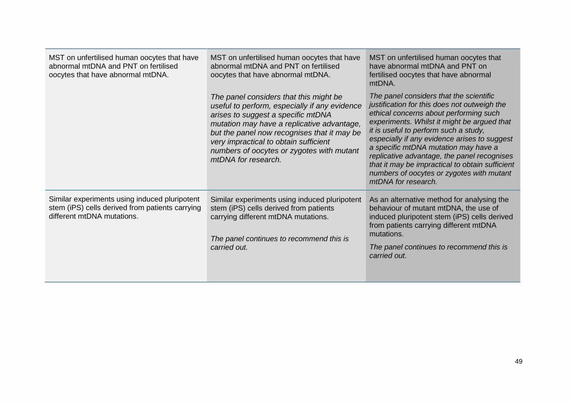
49
MST on unfertilised human oocytes that have abnormal mtDNA and PNT on fertilised oocytes that have abnormal mtDNA.
MST on unfertilised human oocytes that have abnormal mtDNA and PNT on fertilised oocytes that have abnormal mtDNA.
The panel considers that this might be useful to perform, especially if any evidence arises to suggest a specific mtDNA mutation may have a replicative advantage, but the panel now recognises that it may be very impractical to obtain sufficient numbers of oocytes or zygotes with mutant mtDNA for research.
MST on unfertilised human oocytes that have abnormal mtDNA and PNT on fertilised oocytes that have abnormal mtDNA.
The panel considers that the scientific justification for this does not outweigh the ethical concerns about performing such experiments. Whilst it might be argued that it is useful to perform such a study, especially if any evidence arises to suggest a specific mtDNA mutation may have a replicative advantage, the panel recognises that it may be impractical to obtain sufficient numbers of oocytes or zygotes with mutant mtDNA for research.
Similar experiments using induced pluripotent stem (iPS) cells derived from patients carrying different mtDNA mutations.
Similar experiments using induced pluripotent stem (iPS) cells derived from patients carrying different mtDNA mutations.
The panel continues to recommend this is carried out.
As an alternative method for analysing the behaviour of mutant mtDNA, the use of induced pluripotent stem (iPS) cells derived from patients carrying different mtDNA mutations.
The panel continues to recommend this is carried out.

50
Further studies on the mtDNA carryover in a non-human primate model into the possible heteroplasmy of tissues in the fetus. The possibility of carryover or even a small percentage of abnormal mtDNA, means that any females born from MST or PNT should be considered at risk of transmitting the disease to their offspring.
Further studies on the mtDNA carryover in a non-human primate model into the possible heteroplasmy of tissues in the fetus. The possibility of carryover or even a small percentage of abnormal mtDNA, means that any females born from MST or PNT should be considered at risk of transmitting the disease to their offspring.
Some relevant experiments have now been published on this, notably by Lee et al, (2012) in the Macaque. On the basis of these, the panel recommends that further experiments are carried out to address this issue with human material, along the lines suggested in 3.10 of the 2013 report.
Further studies on the mtDNA carryover in a non-human primate model into the possible heteroplasmy of tissues in the fetus. The possibility of carryover or even a small percentage of abnormal mtDNA, means that any females born from MST or PNT should be considered at risk of transmitting the disease to their offspring.
Some relevant experiments were considered in the 2013 review notably by Lee et al, (2012) in the Macaque. On the basis of these however the panel recommends that further experiments are carried out to address this issue with human material. This recommendation still stands.
Further studies on vitrifying zygotes created through PNT.
Further studies on vitrifying zygotes created through PNT.
The panel continues to recommend this is carried out.
Further studies on vitrifying oocytes, karyoplasts, and zygotes in order to allow synchronisation when carrying out MST and PNT, as well as clinical management of patients.
The panel continues to recommend this, recognising that advances in cryopreservation are being made generally within the context of ART.
Studies on human embryonic stem (ES) cells derived from blastocysts that are heteroplasmic for abnormal mtDNA and blastocysts created through MST and PNT
Tests for heteroplasmy should be carried out on primordial germ cells obtained from human ES cells derived from blastocysts created through MST and PNT where the
Tests for heteroplasmy should be carried out on primordial germ cells obtained from human ES cells derived from blastocysts created through MST and PNT where the

51
where oocytes had abnormal mtDNA. oocytes had variant or abnormal mtDNA. If primordial germ cell derivation is not possible or limitations in the model undermine its utility, clonal analysis of single cell-derived human ES cells could be used. Comparisons beginning with blastocysts known to be heteroplasmic for variant or abnormal mtDNA would be informative.
oocytes had variant or abnormal mtDNA. If primordial germ cell derivation is not possible or limitations in the model undermine its utility, clonal analysis of single cell-derived human ES cells could be used. Comparisons beginning with blastocysts known to be heteroplasmic for variant or abnormal mtDNA would be informative.
This recommendation still stands.

52
Annex D: Glossary
Term Definition
Activate To artificially trigger the processes that occur when an egg is fertilised
Adenosine triphosphate (ATP) Molecule which transports energy in cells. It is involved in reactions of the electron transfer chain (ETC)
Apoptosis Programmed cell death
Autophagy Where a cell begins to digest part of itself
Backcross Formally, breeding a hybrid individual with one of its parents. In this context, where a hybrid or mixed race individual breeds with one of its parental types, such as race or strain
Biopsy Procedure of obtaining cells eg, from an embryo, in order to test them
Blastocyst A 5-6 day old embryo, with both an outer trophectoderm cell layer and an inner cell mass
Blastomere A cell of an embryo prior to the blastocyst stage
Chromatin The combination of DNA and other proteins in the nucleus, which packages the chromosomes
Chromosome Structure in a nucleus that carries DNA
Chromosomal inheritance The process for passing on the DNA in the nucleus to subsequent generations
Comparative genomic hybridisation array
Screening method to detect abnormal chromosomes
Cytoplasm The jelly-like substance that fills a cell and contains the structures within it, including mitochondria
Cytoplasmic hybrids
An adult cell or nucleus that has been fused to a cell with its nucleus removed. The cells can be from different individuals or species
Cytoplasmic transfer
The technique of injecting cytoplasm from a donor egg into a recipient egg, for example one that has compromised mitochondria
De novo mutation A spontaneous new mutation (as opposed to one that is

53
inherited)
Depolymerise
To break a polymer, composed of many similar molecules (whether chemicals or proteins), into its constituent parts
Electron transfer chain (ETC) The chain of chemical reactions that produce energy in a mitochondrion
Embryogenesis The process by which an embryo forms and develops
Embryonic stem (ES) cells
Cells, derived from an embryo and cultured in a laboratory, that have the potential to form all different cell/tissue types in an animal or human and can replicate themselves indefinitely
Epigenetic
Relating to or arising from non-genetic influences on gene expression. Can result in inherited phenotypes through alteration of gene expression.
Founding cells The small number of cells that give rise to a larger number of cells/tissues later in development
Gene expression The process by which information from a gene is used in the synthesis of a functional gene product (which may or may not result in a particular phenotype)
Genetic counselling
The process that helps individuals, couples, or families to understand genetic information and how genetic findings may have an impact on them
Germ cells Any cell that will give rise to sperm or egg cells
Germline The sequence of cells that give rise to sperm or egg cells that will pass genetic information on to a child
Germline mosaicism
Also known as gonadal mosaicism, is a condition in which the precursor (germline) cells to ova and spermatazoa are a mixture (mosaic) of two or more genetically different cell lines. If the mosaicism is present in only a small proportion of cells, then it may lead to the inheritance of a condition from a parent who is unaffected by it.
Heteroplasmy
Where two or more different mtDNA types coexist in a single cell, commonly used (as in this report) where one type is abnormal, and the other normal
Homoplasmy Where all the mitochondria in a cell contain the same mtDNA, which can either be all abnormal or all normal

54
Induce pluripotent stem (iPS) cells
Adult cells that have been reprogrammed to act like embryonic stem cells
Inner cell mass
The mass of cells contained within a blastocyst stage mammalian embryo that will give rise to all cell and tissues types of the fetus
Karyoplast
A structure made up of the nuclear genetic material, surrounded by a small amount of cytoplasm, enclosed in a cell membrane, used to transfer a spindle or pronuclei during MST or PNT experiments
Karyotype The number and appearance of chromosomes in a cell
Lineage Term used to describe cells with a common ancestor
Maternal spindle transfer (MST)
Technique to transfer the nuclear genetic material from a woman’s egg into a donated egg with its nuclear genetic material removed
Meiotic division Type of cell division necessary for sexual reproduction
Metaphase II oocytes Mature oocytes or unfertilised eggs (that have undergone the first meiotic cell division)
Microfilaments Thin protein structures that contribute to the ‘skeleton’ and to the dynamics (movement) of the cytoplasm
Micromanipulation Use of physical techniques to add or remove cellular components
Microtubules
Structural components of the cytoplasm, contributing to its ‘skeleton’ and involved in many cellular processes including cell division
Mitochondria Small structures present in cells that produce the cell‟s energy (see Introduction)
Mitochondrial bottleneck
The term describing how, during the development of the embryo, the low number of mitochondria per cell and the small number of cells that go on to form each tissue type means that by chance different cell types will have different proportions of mitochondrial DNA of different types
Mitochondria carryover The proportion of mitochondria that is carried over from the original egg or zygote during MST or PNT
Mitochondrial DNA (mtDNA) The genetic material contained within the mitochondria
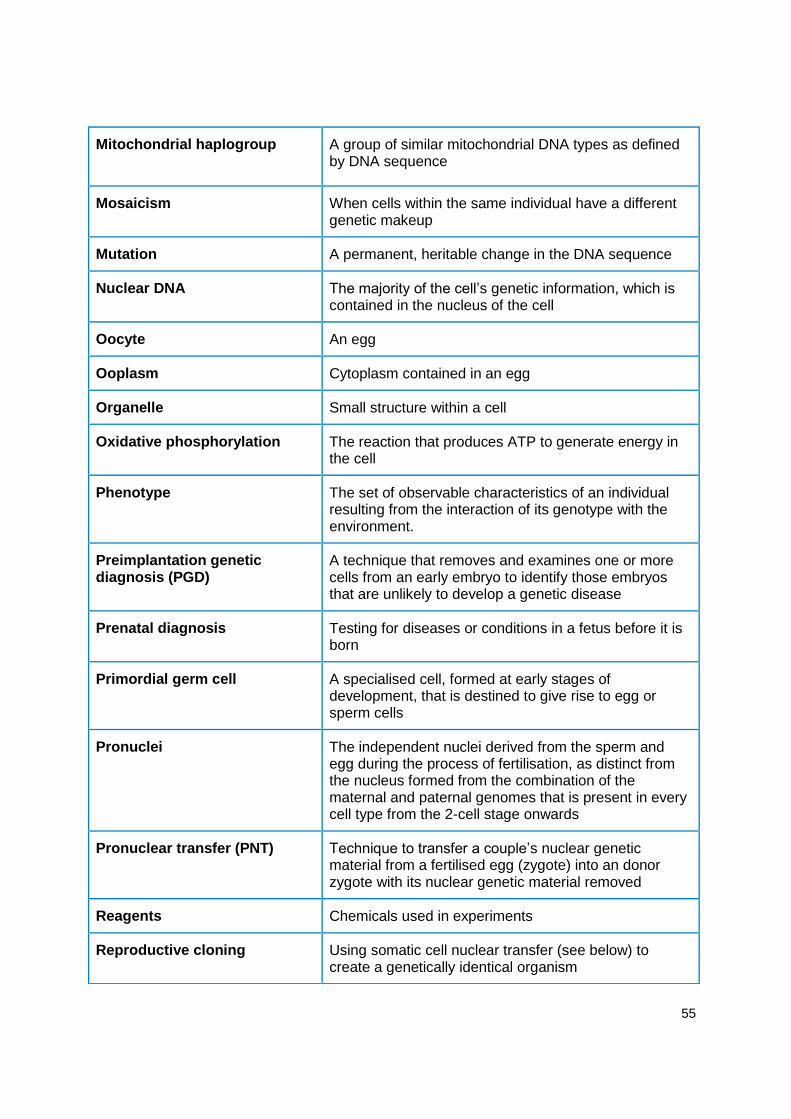
55
Mitochondrial haplogroup
A group of similar mitochondrial DNA types as defined by DNA sequence
Mosaicism When cells within the same individual have a different genetic makeup
Mutation A permanent, heritable change in the DNA sequence
Nuclear DNA The majority of the cell’s genetic information, which is contained in the nucleus of the cell
Oocyte An egg
Ooplasm Cytoplasm contained in an egg
Organelle Small structure within a cell
Oxidative phosphorylation The reaction that produces ATP to generate energy in the cell
Phenotype
The set of observable characteristics of an individual resulting from the interaction of its genotype with the environment.
Preimplantation genetic diagnosis (PGD)
A technique that removes and examines one or more cells from an early embryo to identify those embryos that are unlikely to develop a genetic disease
Prenatal diagnosis Testing for diseases or conditions in a fetus before it is born
Primordial germ cell
A specialised cell, formed at early stages of development, that is destined to give rise to egg or sperm cells
Pronuclei
The independent nuclei derived from the sperm and egg during the process of fertilisation, as distinct from the nucleus formed from the combination of the maternal and paternal genomes that is present in every cell type from the 2-cell stage onwards
Pronuclear transfer (PNT)
Technique to transfer a couple’s nuclear genetic material from a fertilised egg (zygote) into an donor zygote with its nuclear genetic material removed
Reagents Chemicals used in experiments
Reproductive cloning Using somatic cell nuclear transfer (see below) to create a genetically identical organism

56
Segregation The process that determines the genetic information present in different tissues or organisms
Selection The process by which particular entities, such as genes, or cells, are favoured
Sequencing Method for determining the order (sequence) of chemicals that make up DNA in an individual
Sex selection Selecting an embryo of a particular sex to avoid passing on a serious sex-linked condition
Single embryo transfer Transferring just one embryo into the uterus of a woman during a cycle of treatment
Somatic cell Any cell of an embryo or adult that is not a germline cell
Somatic cell nuclear transfer (SCNT)
Fusion of an adult (somatic) cell with an egg that has had its nucleus removed
Spindle A structure in a cell, made up of microtubules, that move the chromosomes when the cell divides
Syngamy The fusion of an egg and sperm
Transcription
The first part of the process of gene expression, whereby a complementary RNA-copy of a sequence of DNA is made. This can then be translated to make the protein encoded by the DNA
Transfer RNA (tRNA) RNA molecule involved in the creation of proteins encoded by DNA
Trophoblast The outer layer of cells of an embryo that will develop into the placenta
Vitrify To freeze (an egg or embryo) by plunging into liquid nitrogen
Zygote A fertilised egg