thin, millimeter scale fingernail sensors for thermal...
TRANSCRIPT
FULL PAPERwww.afm-journal.de
© 2018 WILEY-VCH Verlag GmbH & Co. KGaA, Weinheim1801380 (1 of 10)
Thin, Millimeter Scale Fingernail Sensors for Thermal Characterization of Nail Bed Tissue
Yajing Li, Yinji Ma, Chen Wei, Haiwen Luan, Shuai Xu, Mengdi Han, Hangbo Zhao, Cunman Liang, Quansan Yang, Yiyuan Yang, Kaitlyn E. Crawford, Xue Feng, Yonggang Huang,* and John A. Rogers*
Thin, flexible, body-worn technologies that allow precise, quantitative monitoring of physiological status are of broad current interest due to their potential to improve the cost and effectiveness of healthcare. Although the surface of the skin represents one of the most widely explored points of integration, recently developed millimeter scale wireless sensor platforms allow deployment on alternative surfaces of the body, such as the finger/toenails and the teeth. The work described here introduces a collection of ideas in materials science, device engineering and computational techniques that enables precise characterization of the thermal transport characteristics of the nail bed tissue from measurements on the surface of the nail. Systematic in vitro studies demonstrate the underlying measurement principles, the theoretical models for optimized sensor design and the associated experimental procedures for determining the thermal conductivity of the tissue. Measurements performed on human subjects highlight capabilities in tracking changes in perfusion of the nail bed tissues in response to various external stimuli.
DOI: 10.1002/adfm.201801380
1. Introduction
In clinical medicine, the nail unit is a well-known, useful source of informa-tion on health status and an important component of the physical exam.[1–3] Any alterations in the nature of the nail plate, the nail matrix, the hyponychium, the proximal nail fold, the lateral nail folds or the nail bed, may reflect nutri-tional, endocrine, congenital, infectious, neoplastic, traumatic, inflammatory or vascular imbalances both locally and sys-temically. The optical transparency of the nail plate allows this structure to serve as a noninvasive window into microvascular integrity through direct visualization of nail fold capillaries. As such, noninva-sive and point-of-care tools such as digital
Flexible Electronics
Dr. Y. LiDepartment of Materials Science and EngineeringCenter for Bio-Integrated ElectronicsNorthwestern UniversityEvanston, IL 60208, USADr. Y. Ma, Prof. X. FengDepartment of Engineering MechanicsCenter for Mechanics and MaterialsTsinghua UniversityBeijing 100084, ChinaC. Wei, H. LuanDepartment of Civil and Environmental Engineering, Mechanical Engineering, Materials Science and EngineeringNorthwestern UniversityEvanston, IL 60208, USADr. S. XuCenter for Bio-Integrated ElectronicsDepartment of DermatologyFeinberg School of MedicineNorthwestern UniversityEvanston, IL 60208, USADr. M. Han, Dr. H. Zhao, C. Liang, Q. Yang, Y. YangCenter for Bio-Integrated ElectronicsNorthwestern UniversityEvanston, IL 60208, USA
Prof. K. E. CrawfordDepartment of Materials Science and EngineeringBurnett School of Biomedical SciencesUniversity of Central FloridaOrlando, FL 32827, USAProf. Y. HuangDepartment of Civil and Environmental Engineering, Mechanical Engineering, Materials Science and EngineeringSkin Disease Research CenterFeinberg School of MedicineNorthwestern UniversityEvanston, IL 60208, USAE-mail: [email protected]. J. A. RogersDepartments of Materials Science and Engineering, Biomedical Engineering, Chemistry, Mechanical Engineering, Electrical Engineering and Computer Science, and Neurological SurgeryCenter for Bio-Integrated ElectronicsSimpson Querrey Institute for Nano/biotechnologyNorthwestern UniversityEvanston, IL 60208, USAE-mail: [email protected]
The ORCID identification number(s) for the author(s) of this article can be found under https://doi.org/10.1002/adfm.201801380.
Adv. Funct. Mater. 2018, 28, 1801380
www.afm-journal.dewww.advancedsciencenews.com
1801380 (2 of 10) © 2018 WILEY-VCH Verlag GmbH & Co. KGaA, Weinheim
platforms for dermoscopy and proximal nail fold capillaroscopy serve as diagnostic instruments in the evaluation of connec-tive tissue diseases (e.g., Raynaud’s phenomenon and systemic sclerosis[4]) and nail tumors.[5] Operation of these technologies and interpretation of data collected with them require, however, clinical expertise and they cannot be used for continuous moni-toring outside of a hospital or laboratory setting.
Recent work shows that advanced imaging techniques such as ultrasound, CT (computed tomography), MRI (magnetic resonance imaging), and PET (positron emission tomography) can be used to examine the dynamics and structure of tissues of the fingers and nails for diagnostic purposes. Although modern imaging technologies allow for visualization of mul-tiparameter data at multidimensional resolution, these systems are expensive and have limited utility at the point-of-care. While cutaneous skin biopsies are largely noninvasive and straight-forward, tissue sampling of the nail unit is a complex proce-dure that often requires nail avulsion, postoperative discomfort, along with risk of infection, and long-term nail dystrophy.[6,7] Thus, technologies that can derive additional information from the nail unit noninvasively at the point-of-care and/or in a continuous monitoring mode without significant cost or com-plex imaging systems could offer significant clinical value for both diagnosis and treatment management. Beyond acting as a useful source of clinical information, the nail unit also rep-resents an ideal, hard, and mechanical interface for mounting and bonding advanced device technologies. Specifically, the nail plate is a semitransparent material composed of cornified keratinocytes and keratin proteins, which imparts significant mechanical strength and resistance to envi-ronmental insult.[8] This construction enables stable coupling of sensors and devices with the human body without the risk of irritation, redness, or allergic reactions. Furthermore, the nail plate grows slowly at ≈1 to 3 mm per month, thereby allowing long-term sensing.[9]
Advanced sensing techniques developed for skin-like, or “epidermal,” electronics use precision thermal sensors and actuators to determine thermal transport properties of living tissues in a real-time, noninva-sive fashion.[10–13] These systems integrate metallic filaments with soft, thin supporting substrates to allow operation while in inti-mate contact with curvilinear skin, without irritation or sensory perception at the skin interface. Direct measurement of the body surface temperature and quantification of the thermal transport properties (i.e., thermal conductivity) associated with physiological conditions such as perfusion and hydration level are both possible, with clinical-grade accuracy. Past demonstrations focused on characterization of the upper layer of the skin,[10–13] without consideration of its natu-rally layered structure or its properties sig-nificantly below the surface. The assessment of the thermal characteristics of deep tissues is challenging and of great interest. Here,
we present a noninvasive method of exploiting nail-mounted thermal sensors to measure the thermal conductivity of the nail bed, independent of the thermal properties of the nail. Com-bined with thermal analysis techniques, the responses of such sensors provide quantitative information of the perfusion of the nailbed tissue, and of other processes that alter the thermal transport characteristics. These thin, miniaturized devices yield data of direct relevance to physiological health and offer the potential for continuous monitoring as an unusual class of wearable technology with integration of wireless power source and data communication functionality.
2. Results and Discussions
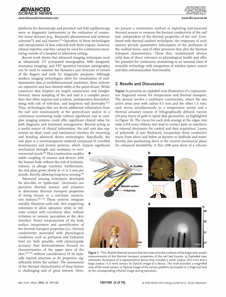
Figure 1a presents an exploded view illustration of a representa-tive fingernail sensor for temperature and thermal transport. The devices involve a multilayer construction, where the two active areas (one with radius 0.5 mm and the other 1.5 mm; each serves simultaneously as a temperature sensor and a thermal actuator) consist of lithographically defined, narrow (10 µm) traces of gold in spiral disk geometries, as highlighted in Figure 1b. The traces for each disk emerge at the edges into wide (≈0.8 mm) ribbons that lead to contact pads as interfaces to external electronics for control and data acquisition. Layers of polyimide (3 µm thickness) encapsulate these conductive traces from above and below as barriers to biofluids and water, thereby also positioning them at the neutral mechanical plane for enhanced bendability. A thin (200 µm) sheet of a silicone
Adv. Funct. Mater. 2018, 28, 1801380
Figure 1. Thin, flexible thermal sensors that laminate onto the surfaces of the fingernails enable measurements of the thermal transport properties of the nail bed tissues. a) Exploded view schematic illustration of a representative device that includes a small (radius ≈0.5 mm) and a large (radius ≈1.5 mm) sensor. b) Optical image of a device. The inset provides a magnified view of the small sensor. c) Optical image of the sensor platform laminated on a fingernail and d) the corresponding infrared image during operation.

www.afm-journal.dewww.advancedsciencenews.com
1801380 (3 of 10) © 2018 WILEY-VCH Verlag GmbH & Co. KGaA, Weinheim
elastomer serves as a mechanical support for handling and manipulation. The low modulus of this material and its tacky surface ensure intimate, conformal contact, and therefore effi-cient thermal coupling, to the nail via reversible van der Waals interactions.
The devices connect to a power supply (6220, Keithley Instru-ment) and a digital multimeter (DMM, National Instruments) to allow delivery of controlled, direct current (DC) inputs to the sensors/actuators and simultaneous measurements of their resistance. In this way, the devices serve simultaneously as thermal actuators and temperature sensors. The resulting measurement physics relies on the well-established transient plane source (TPS) method.[14] Briefly, the active element in the TPS approach delivers thermal power to the sample via Joule heating that results from application of DC current. The same device simultaneously enables time-dependent measurements of resulting changes in temperature through the temperature coefficient of resistance (TCR) of the metal. Data recorded in this manner can be combined with computational techniques to determine the intrinsic thermal transport properties, i.e., the thermal conductivity and thermal diffusivity, of the material under test. Figure 1d shows an infrared image of the devices operating in this manner on the surface of the fingernail.
The fingernail consists of a rigid plate (typically with thick-ness ≈0.5 mm that varies by only ≈50 µm from the proximal to distal end)[15–17] mechanically and thermally coupled to the underlying tissue. The nail plate is made of alpha-keratin, with
thermal conductivity between ≈0.2 and 0.4 W m−1 K−1.[18] The nail bed is made of two types of tissues: the epidermis and the deeper dermis which includes rich capillaries and glands, with thermal conductivity between 0.2 and 0.5 W m−1 K−1.[19–23] Experiments to establish the basic operating principles of the sensors and the methods to interpret data collected by them use test platforms that consist of a thin film of silicone on a thick base layer of a silicone, each with a formulation to yield thermal properties and thicknesses comparable to those of skin tissues and nail plate. The corresponding computational modeling assumes a semi-infinite substrate.
The characteristic probing depth associated with the TPS method increases with the thermal diffusivity (α) and the time for thermal actuation (t).[14,24,25] For a bilayer sample, charac-terization of the thermal properties of the bottom layer requires actuation times sufficient for heat to diffuse through the top layer. At long times, the temperature approximately saturates to a value that depends mainly on the thermal conductivity and only weakly on the thermal diffusivity.[14] Figure 2a illustrates the spatial-temporal characteristics of heat transport obtained by finite element analysis (FEA) for an actuator with radius, R = 1.5 mm and power density, q = 3 mW mm−2 on a bilayer sample with a 0.5 mm thick top layer (thermal conductivity, k1 = 0.21 W m−1 K−1; thermal diffusivity, α1 = 0.15 mm s−2) and semi-infinite bottom layer (k2 = 0.44 W m−1 K−1 and α2 = 0.15 mm s−2). At short times (0.5 and 2 s), heat transport occurs mainly in the top layer, with little increase in temperature in
Adv. Funct. Mater. 2018, 28, 1801380
Figure 2. Summary of procedures to determine the thermal conductivities of a bilayer sample. a) FEA results (quarter, cross-sectional view) for the spatial distributions of increases in temperature induced by a thermal actuator placed on the surface of a bilayer sample at several times after actuation. The parameters in the FEA are R = 1.5 mm (radius of the actuator), q = 3 mW mm−2 (thermal power from the actuator), h = 0.5 mm (thickness of the top layer), k1 = 0.21 W m−1 K−1 (thermal conductivity of the top layer) and k2 = 0.44 W m−1 K−1 (thermal conductivity of the base layer). b) Measured increases in temperature as a function of time for operation of actuators with R = 0.5 mm (q = 10 mW mm−2) and 1.5 mm (q = 3 mW mm−2). c,d) FEA results for the increases in temperature of the actuators at t = 40 s, Tss, plotted as a function of k1 and k2, c) R = 1.5 mm and q = 3 mW mm−2 and d) R = 0.5 mm and q = 10 mW mm−2. The curves in panels (c) and (d) correspond to combinations of k1 and k2 that yield a certain value of Tss. The color in these graphs corresponds to the values of Tss that result for other values k1 and k2. e) The point of intersection of the two curves in panels (c) and (d) yields k1 and k2.

www.afm-journal.dewww.advancedsciencenews.com
1801380 (4 of 10) © 2018 WILEY-VCH Verlag GmbH & Co. KGaA, Weinheim
the bottom layer. At long times (20 and 40 s), the heat passes into both layers, and the temperature increase of the actuator saturates, as expected.
For a bilayer sample with a top layer whose thickness is known, the temperature increases associated with two actua-tors that have different radii, both operated in this long time (≈40 s) regime, can be used to determine k1 and k2. Figure 2b presents the measured temperature increase (T) of the actuator on a bilayer sample with a thin top layer (0.5 mm; Ecoflex) and thick bottom layer (13 mm; Sylgard 170) as a function of thermal actuation time for each of the two actuators operated with DC current (≈100 µA) for activation at 0 s and deactivation at 40 s. Measurements involve an enclosure around the sample to reduce fluctuations in temperature induced by convective heat transfer to the room. The quasi-steady state increase in temperature, i.e., T at t = 40 s, or Tss, is 11.0 °C for the small actuator (R = 0.5 mm and q = 10 mW mm−2) and 8.4 °C for the large actuator (R = 1.5 mm and q = 3 mW mm−2). Both values fall below the threshold for damaging tissue.[26,27] Figure 2c,d
shows the corresponding temperature increases of the two actuators (radii and powers specified in Figure 2b) obtained by FEA for different k1 and k2. FEA results yield curves that define pairs of k1 and k2 that are consistent with the experimentally measured Tss for both the small and the large actuator. The point of intersection of these two curves gives k1 and k2 for the bilayer sample, i.e., k1 = 0.21 W m−1 K−1 for Ecoflex and k2 = 0.44 W m−1 K−1 for Sylgard 170 in Figure 2e. These results are consistent with the literature values for these materials.[28,29] We selected sensors with radii of 0.5 and 1.5 mm since the small (large) sensor offers greater sensitivity to properties of the top (bottom) layers, as demonstrated in Figure S6 of the Supporting Information.
Measurements on samples with top layers that have dif-ferent thicknesses further validate the measurement scheme. Figure 3a,b shows representative results (R = 0.5 mm and 1.5 mm, power q = 10 mW mm−2 and 3 mW mm−2, respec-tively) with top layer (Ecoflex) thicknesses between ≈300 and 600 µm (spatial variations of +/− 10 µm), each with the same
Adv. Funct. Mater. 2018, 28, 1801380
Figure 3. Experimental and computational results for the characterization of bilayer samples that consist of thin films (silicone, Ecoflex) with different thicknesses on a thick substrate (silicone, Sylgard 170), evaluated using sensors with radii, R, of 1.5 and 0.5 mm. a) Increase in temperature as a func-tion of time for sensors with R = 0.5 mm and b) R = 1.5 mm, with bilayer samples that have different top layer thicknesses, with activation at 0 s and deactivation at 40 s. c) Increase in temperature measured with the two sensors as a function of time for repeated measurements on a sample with top layer thickness of 0.5 mm. d) Analysis of error and uncertainty in the parameters extracted from the data, determined by FEA. Each curve represents the measured value of Tss shifted by ±δTss. The inset shows a magnified view of Tss of each curve in panel (c). The experimental variations in Tss lead to values of δTss that are generally less than 0.1 °C. The points of intersection of these pairs of curves define the thermal conductivity values and their uncertainties, k1,2 ± Δk1,2.

www.afm-journal.dewww.advancedsciencenews.com
1801380 (5 of 10) © 2018 WILEY-VCH Verlag GmbH & Co. KGaA, Weinheim
type of bottom layer (Sylgard 170). Quasi-steady state temper-atures, Tss, analyzed using the scheme described previously yield values for the thermal conductivity. The results show consistent results, independent of the top layer thickness, i.e., k1 = 0.21 W m−1 K−1, k2 = 0.42 W m−1 K−1 for h = 310 µm; k1 = 0.21 W m−1 K−1, k2 = 0.42 W m−1 K−1 for h = 410 µm; k1 = 0.21 W m−1 K−1, k2 = 0.44 W m−1 K−1 for h = 500 µm; k1 = 0.21 Wm−1K−1, k2 = 0.44 W m−1 K−1 for h = 600 µm. Figure 3c and the inset in 3d show that the repeatability for Tss is ≈0.1 °C, roughly comparable to fluctuations in the ambient tempera-ture (see Figure S2 of the Supporting Information). This value defines uncertainties in the extracted thermal conductivities, as summarized in Figure 3d. These uncertainties are consistent with the variations in values observed across samples with dif-ferent top layer thicknesses, i.e., k1 = 0.21 ± 0.01 W m−1 K−1 and k2 = 0.44 ± 0.04 W m−1 K−1.
In many applications, the properties of the nail bed tissues are more important than those of the nail because they vary depending on physiological state. Measurements of changes in the thermal properties of the system are likely to be domi-nated by those of the tissue, as opposed to the nail. Studies of the sensitivity of the measurement to the bottom layer provide insights in this context. Here, the samples consist of bilayer structures with a fixed top layer (0.3 mm thick, Ecoflex) and various bottom layers (Ecoflex, Sylgard 567, Sylgard 170, and Sylgard 164), as summarized in Figure 4a,b. For both sensors, the Ecoflex/Ecoflex case yields values of Tss that are larger than those of Ecoflex/Sylgard567, Ecoflex/Slygard170, and Ecoflex/Sylgard164. The trends follow the thermal conductivities of the bottom materials (k = 0.21 ± 0.01 W m−1 K−1 for Ecoflex, k = 0.3 ± 0.02 W m−1 K−1 for Sylgard 567, k = 0.44 ± 0.04 W m−1 K−1 for Sylgard 170, and k = 0.66 ± 0.01 W m−1 K−1 for Sylgard 164).
Figure 4c presents a plot of Tss as a function of k2 for R = 1.5 mm, q = 3 mW mm−2, for two values of k1. The curves clearly depend on k1, which suggests that k2 cannot be deter-mined by using just one sensor/actuator if k1 is unknown. Nev-ertheless, for physiological monitoring based on measurement of the nail and nail bed tissue, the change in k1 can be assumed to be much smaller than the change in k2. If Δk2 and ΔTss are the change in k2 and Tss, respectively, then we can plot ΔTss/Tss as a function of Δk2/k2 for several values of k1 and k2, as in Figure 4d,e. Remarkably, the results show that this relationship is only weakly dependent of k1 (Figure 4d) and k2 (Figure 4e) over this physiologically relevant range. As a result, Δk2/k2 can be determined directly from ΔTss/Tss, independent of the value of k1 and k2. This conclusion is clearly supported by experi-ments, as shown in Figure 4f, for different top-layer materials (Ecoflex and Sylgard567) and bottom-layer materials (Ecoflex, Sylgard567, Sylgard170, and Sylgard164). Here, the actuator radius and heating time are the same as those in the FEA in Figure 4c–e.
These studies establish a baseline of understanding that allows interpretation of data from these types of sensors used on human subjects (healthy female, age 29; left middle fingernail with nail thickness of 0.42 ± 0.01 mm measured with a caliper). Figure 5a shows measurement results from a single subject across 8 d of three repeated measurements performed in the afternoon on each day. To reduce the rates of convective heat transfer, measurements involve a piece
of plastic foam to enclose the hand of the subject without touching the fingernail, as shown in Figure S5 of the sup-porting information. The thermal conductivity of the nail plate varies from 0.27 to 0.29 W m−1 K−1 over the obser-vation period, in a narrow range consistent with values measured using other techniques.[18] The thermal conduc-tivity of the nail bed varies more significantly, from 0.43 to 0.52 W mm−2, as might be expected due to normal variations in hydration and surface blood flow, both of which can affect the thermal conductivity.[30,31] For instance, previous reports suggest that variations in blood flow can induce changes in thermal conductivity from 0.25 W m−1 K−1 (null blood flow) to 1 W m−1 K−1 (vasodilation).[32] These variations likely reflect the physiological changes, as opposed to variations that result from changes in the environment or the sensor response. Results in the Supporting Information show that repeated measurements on silicone samples over the course of 7 d reveal that variations in Tss are ≈0.1 °C, consistent with previ-ously reported fluctuations in temperature.
Perfusion behaviors affect the distributions of temperature in living systems, with important purposes in thermoregu-lation. As such, perfusion is an important index for clinical procedures such as the treatment of tumors.[33] Abnormalities of peripheral microcirculation can play a central role in sys-temic sclerosis (SSc). Previous studies[10,11,21,34,35] indicate that the thermal conductivity of tissues can be strongly affected by micro and macrovascular blood flow. Figure 5b–f illustrates the temporal evolution of the thermal conductivity of the nail bed tissue as a result of changes in blood flow associated with a local, pressure induced occlusion of the blood vessels in the middle left finger of the subject (female, age 29). The meas-urements used a sensor with R = 1.5 mm laminated onto the center of the nail plate for continuous thermal characteriza-tion during and after the occlusion. Figure 5c presents the change in temperature of the nail plate of the left middle finger recorded by an infrared camera at the beginning of the period of occlusion. The results show that during the occlusion, the temperature decreases monotonically by >4 °C in the first 2 min followed by further reductions but with a reduced rate in the subsequent 7 min. The color of the finger turns gray and the pinkish tone of the tissue under the nail plate fades into pale shades. Releasing the occlusion causes an increase and overshoot of the temperature by 9 °C within 100 s, coin-cident with an increase in blood flow above the initial value and a change in the color of the finger to red. Although the rapid, time dependent variations in temperature and blood flow frustrate precise analysis of the measurements, approxi-mate values of the thermal conductivity of the nail bed tissue can be deduced with k1 fixed to the average value of the meas-urement in Figure 5a. The thermal conductivity of the skin adjacent to the nail plate is measured with the sensor directly mounted on top of the skin, as indicated in the optical image in Figure 5c. Figure 5e shows that the conductivity decreases with occlusion, from 0.5 ± 0.03 to 0.44 ± 0.02 W m−1 K−1, cor-responding to 14% change independent of the specific value of k1 as in Figure 5f. Similar changes occur in the adjacent skin. Overall, changes in thermal conductivity track those in temper-ature, as expected in the case that the blood flow affects both temperature and thermal transport. The perfusion resulted
Adv. Funct. Mater. 2018, 28, 1801380

www.afm-journal.dewww.advancedsciencenews.com
1801380 (6 of 10) © 2018 WILEY-VCH Verlag GmbH & Co. KGaA, Weinheim
in changes in thermal conductivity, as further studied on two subjects (subject 1, previous female; subject 2, male at age 26, nail thickness 0.49 ± 0.01 mm, k1 = 0.26 ± 0.01 W m−1 K−1) before and after exercise. Thermal conductivity was measured for both subjects at rest before exercise (stationary bike for 15 min) and after, following a rest of 10 min. Figure 5f shows
that the thermal conductivity of the nailbed tissue increases after exercise for both subjects. The male subject shows an increase that is larger than that of the female subject, likely cor-responding to elevated blood flow.[36]
A second demonstration of measurements on human subjects involves aspects related to thermoregulation.
Adv. Funct. Mater. 2018, 28, 1801380
Figure 4. Thermal characterization with a focus on the thermal conductivity of the bottom layer. a) Increases in temperature as a function of time for actuators with R = 0.5 mm and b) 1.5 mm on bilayer samples of Ecoflex (top layer, thickness = 0.3 mm) on bottom layers of different materials (Ecoflex, Sylgard567, Sylgard170, Sylgard164). c) FEA results for Tss as a function of k2 for different values k1. d) ΔTss/Tss as a function of Δk2/k2 with different k1. e) ΔTss/Tss as a function of Δk2/k2 with different k2. f) Comparison of ΔTss/Tss as a function of Δk2/k2 from FEA and experimental results.

www.afm-journal.dewww.advancedsciencenews.com
1801380 (7 of 10) © 2018 WILEY-VCH Verlag GmbH & Co. KGaA, Weinheim
Specifically, changes in the surrounding temperature can alter blood flow in deep tissues.[37] Here, studies involve two healthy subjects (previous female, and another male at age 30, with nail thickness of 0.51 ± 0.01 mm), each with their left middle finger placed on ice bag for 10 min. Procedures summarized in Figure 4a define the thermal conductivity of the nail plate (knail = 0.28 ± 0.03 W m−1 K−1 for female and knail = 0.26 ± 0.02 W m−1 K−1 for male); this value is assumed to remain constant. Figure 6a–e, presents measurement results and analysis of data from the female subject. During
the cooling period, the thermal conductivity of nail bed tissue decreases by ≈12%, likely a result of vasoconstriction induced by cooling. Removing the ice bag, and exercising the finger (rubbing and warm hand wash) for 6 min prepare the sub-ject for a second set of measurements. During this process, the thermal conductivity of the nailbed recovers to a value ≈7% below the initial state after 6 min and only ≈2% below after 8 min. The results obtained from the male subject in Figure 6b,d,f show similar trends, but with a higher thermal conductivity and a larger change ≈30% compared to the female
Adv. Funct. Mater. 2018, 28, 1801380
Figure 5. Results of thermal characterization studies on volunteer subjects. a) Thermal conductivity of the nail plate and nail bed tissue measured at room temperature each day for eight consecutive days. b) Time-dependent changes in temperature during before (<0 s), during (between 0 and 40 s), and after (>40 s) activation of a sensor with R = 1.5 mm on the left middle finger. c) Temperature of the fingernail of a subject determined using an infrared camera during and after occlusion of blood flow. d) Time-dependent changes in the thermal conductivity of the nail bed tissue and adjacent skin during and after occlusion. Measurement locations on the fingernail and adjacent skin are indicated by red and black circle in the inset in panel (c). e) Relative change in thermal conductivity Δknailbed/knailbed of the nail bed tissue. f) Thermal conductivity changes of subject 1 and 2 before and after exercise.

www.afm-journal.dewww.advancedsciencenews.com
1801380 (8 of 10) © 2018 WILEY-VCH Verlag GmbH & Co. KGaA, Weinheim
subject. After exercise, the male subject shows an increase of thermal conductivity of the nailbed tissue, the value reaches 5% below the one measured at the room temperature at 6 min and 2% above at 8 min.
3. Conclusion
The results presented here establish a general set of materials, device structures, measurement approaches, and analysis techniques for noninvasive characterization of the thermal
properties of systems consisting of a thin layer of material on top of a semi-infinite substrate, specifically modeled after the nail/nailbed structures of the human body. Measurements on a range of synthetic analogs to fingernails highlight the key considerations and define the optimized modes of analysis. Evaluations on human subjects illustrate possibilities for tracking changes in perfusion in the nailbed tissue via meas-urements from the surface of the nail plate. Future work will explore advanced sensor layouts to enhance the measurement precision and capabilities in depth profiling of the transport characteristics. Addition of other types of sensors, such as
Adv. Funct. Mater. 2018, 28, 1801380
Figure 6. Studies of changes in thermal transport characteristics of the nailbed tissue associated with cooling the finger. a,b) Temperature responses associated with operation of a sensor with R = 1.5 mm measured on subject 1 (female, 29) and subject 2 (male, 30). c,d) Fitted thermal conductivity of the nailbed with knail fixed to the minimum, mean, and maximum value of the thermal conductivity of the fingernail for each subject. e,f) Relative change in thermal conductivity, Δknailbed/knailbed, during and after cooling for each subject. The error bar corresponds to the deviation of the Δknailbed/knailbed calculated using values of knail in panels (c) and (d).

www.afm-journal.dewww.advancedsciencenews.com
1801380 (9 of 10) © 2018 WILEY-VCH Verlag GmbH & Co. KGaA, Weinheim
optical devices for determining blood oxygenation and cap-turing photoplethysograms, will yield multimodal platforms for tracking physiological health status. These and other opportunities, taken in the context of the advantages of the nail plate as a point of device integration with the body, sug-gest many fruitful directions for further research and engi-neering development.
4. Experimental SectionDevice Fabrication: Fabrication and design details are in the
Supporting Information. Briefly, the process starts with spin casting a thin sacrificial layer of (poly)methyl-methacrylate (PMMA Microchem, Westborough, MA) on a clean silicon wafer. A film of polyimide (PI 2545, Parlin, NJ, 3 µm thick) spin-cast and cured on the top of this layer forms the bottom side of the encapsulation. A bilayer of Cr (10 nm)/Au (100 nm) deposited on top of the polyimide by electron beam evaporation and then patterned by photolithography and wet etching forms the conducting traces for the devices. The use of wide interconnect lines minimizes their resistances. A second layer of PI layer formed by spin casting and curing yields the top encapsulation. Photolithography and etching the PI defines the outline of the sensor. Immersing the wafer in acetone removes the underlying PMMA, thereby releasing the sensors from the wafer. Retrieval using a polyvinyl alcohol (PVA)-based water soluble tape (3M, Minneapolis, MN) followed by deposition of a thin layer of SiO2 facilitates adhesive bonding to a thin (50 µm) silicone-based substrate (Ecoflex, Smooth-On Inc., Macungie, PA). Removing the PVA by immersion in warm water completes the fabrication.
Measurement Scheme: The sensors connect with a flexible cable to a custom printed circuit board as an interface to the measurement hardware. A precision DC current source (Keithley 6220, USA) supplies a constant current to the sensors and a DMM (National Instrument, USA) records the voltages. Instrument control and Data acquisition are performed using a custom computer program (LabVIEW, National Instruments, USA) via a GPIB-USB interface. Consent was obtained from the human subjects participating in the experiments.
Supporting InformationSupporting Information is available from the Wiley Online Library or from the author.
AcknowledgementsY.L. and Y.M. contributed equally to this work. Y.M. and X.F. acknowledge the support from the National Basic Research Program of China (Grant No. 2015CB351900) and the National Natural Science Foundation of China (Grant Nos. 11402135 and 11320101001). Y.H. acknowledges the support from NSF (Grant Nos. 1400169, 1534120, and 1635443) and NIH (Grant No. R01EB019337). This work utilized Northwestern University Micro/Nano Fabrication Facility (NUFAB), which was partially supported by Soft and Hybrid Nanotechnology Experimental (SHyNE) Resource (Grant No. NSF ECCS-1542205), the Materials Research Science and Engineering Center (Grant No. DMR-1720139), the State of Illinois, and Northwestern University.
Conflict of InterestThe authors declare no conflict of interest.
Keywordsfingernail devices, flexible electronics, noninvasive biomedical applications, perfusion tracking, thermal sensors
Received: February 22, 2018Revised: April 25, 2018
Published online: June 3, 2018
[1] M. N. Zaiac, A. Walker, Clin. Dermatol. 2013, 31, 627.[2] K. N. Shah, A. I. Rubin, Curr. Probl. Pediatr. Adolesc. Health Care
2012, 42, 204.[3] R. S. Fawcett, S. Linford, D. L. Stulberg, Am. Fam. Physician 2004,
69, 1417.[4] M. Cutolo, C. Pizzorni, M. E. Secchi, A. Sulli, Best Pract. Res. Clin.
Rheumatol. 2008, 22, 1093.[5] L. Thomas, E. G. Zook, E. Haneke, J.-L. Drapé, R. Baran, J. F. Kreusch,
in Baran & Dawber’s Diseases of the Nails and their Management (Eds: R. Baran, D. A. R. de Berker, M. Holzberg, L. Thomas), Wiley-Blackwell, Hoboken, NJ, USA 2012, pp. 637–743.
[6] C. Grover, S. Bansal, Indian Dermatol. Online J. 2018, 9, 3.[7] T. E. Rohrer, B. Leslie, D. J. Grande, J. Dermatol. Surg. Oncol. 1994,
20, 19.[8] R. H. Rice, Y. Xia, R. J. Alvarado, B. S. Phinney, J. Proteome Res.
2010, 9, 6752.[9] S. Yaemsiri, N. Hou, M. M. Slining, K. He, J. Eur. Acad. Dermatol.
Venereol. 2010, 24, 420.[10] R. C. Webb, Y. Ma, S. Krishnan, Y. Li, S. Yoon, X. Guo, X. Feng,
Y. Shi, M. Seidel, N. H. Cho, J. Kurniawan, J. Ahad, N. Sheth, J. Kim, J. G. T. Vi, T. Darlington, K. Chang, W. Huang, J. Ayers, A. Gruebele, R. M. Pielak, M. J. Slepian, Y. Huang, A. M. Gorbach, J. A. Rogers, Sci. Adv. 2015, 1, e1500701.
[11] R. C. Webb, R. M. Pielak, P. Bastien, J. Ayers, J. Niittynen, J. Kurniawan, M. Manco, A. Lin, N. H. Cho, V. Malyrchuk, G. Balooch, J. A. Rogers, PloS One 2015, 10, e0118131.
[12] R. C. Webb, A. P. Bonifas, A. Behnaz, Y. Zhang, K. J. Yu, H. Cheng, M. Shi, Z. Bian, Z. Liu, Y.-S. Kim, W.-H. Yeo, J. S. Park, J. Song, Y. Li, Y. Huang, A. M. Gorbach, J. A. Rogers, Nat. Mater. 2013, 12, 938.
[13] S. Amendola, G. Bovesecchi, P. Coppa, G. Marrocco, in 2016 IEEE International Symposium on Antennas and Propagation (APSURSI), Fajardo, Puerto Rico, USA 2016, pp. 461–462.
[14] S. E. Gustafsson, Rev. Sci. Instrum. 1991, 62, 797.[15] M. Johnson, S. Shuster, Br. J. Dermatol. 1994, 130, 195.[16] J. B. Hamilton, H. Terada, G. E. Mestler, J. Gerontol. 1955, 10, 401.[17] U. Wollina, M. Berger, K. Karte, Skin Res. Technol. 2001, 7, 60.[18] D. T. Dias, A. Steimacher, A. C. Bento, A. M. Neto, M. L. Baesso,
Photochem. Photobiol. 2007, 83, 1144.[19] T. E. Cooper, G. J. Trezek, Aerosp. Med. 1971, 42, 24.[20] T. A. Balasubramaniam, H. F. Bowman, J. Biomech. Eng. 1977, 99,
148.[21] W. J. B. M. van de Staak, A. J. M. Brakkee, H. E. de Rijke-Herweijer,
J. Invest. Dermatol. 1968, 51, 149.[22] A. Chanmugam, A. Bhargava, C. Herman, Int. Mech. Eng. Congr.
Expo. 2012, 2, 717.[23] A. M. Stoll, J. Invest. Dermatol. 1977, 69, 328.[24] A. Sizov, D. Cederkrantz, L. Salmi, A. Rosén, L. Jacobson,
S. E. Gustafsson, M. Gustavsson, Rev. Sci. Instrum. 2016, 87, 74901.[25] Thermal Conductivity: Theory, Properties, and Applications (Ed:
T. M. Tritt), Physics of Solids and Liquids, Springer, New York, NY, US 2004.
[26] A. R. Moritz, F. C. Henriques, Am. J. Pathol. 1947, 23, 695.[27] J. P. Bull, J. C. Lawrence, Fire Mater. 1979, 3, 100.
Adv. Funct. Mater. 2018, 28, 1801380

www.afm-journal.dewww.advancedsciencenews.com
1801380 (10 of 10) © 2018 WILEY-VCH Verlag GmbH & Co. KGaA, Weinheim
[28] Dow Corning Sylgard170 Silicone Elastomer Product Information, Dow Corning, 2017, https://consumer.dow.com/documents/en-us/productdatasheet/11/11-31/11-3181-sylgard-170-silicone-elastomer.pdf?iframe=true.
[29] L. Tian, Y. Li, R. C. Webb, S. Krishnan, Z. Bian, J. Song, X. Ning, K. Crawford, J. Kurniawan, A. Bonifas, J. Ma, Y. Liu, X. Xie, J. Chen, Y. Liu, Z. Shi, T. Wu, R. Ning, D. Li, S. Sinha, D. G. Cahill, Y. Huang, J. A. Rogers, Adv. Funct. Mater. 2017, 27, 1701282.
[30] T. H. Benzinger, A. W. Pratt, C. Kitzinger, Proc. Natl. Acad. Sci. USA 1961, 47, 730.
[31] R. Refinetti, Exp. Physiol. 2003, 88, 423.[32] A. Dittmar, T. Pauchard, G. Delhomme, E. Vernet-Maury, Sens. Actu-
ators, B 1992, 7, 327.[33] M. Salcman, E. Moriyama, H. J. Elsner, H. Rossman, R. A. Gettleman,
G. Neuberth, G. Corradino, J. Neurosurg. 1989, 70, 592.[34] J. Grayson, J. Physiol. 1952, 118, 54.[35] R. K. Jain, F. H. Grantham, P. M. Gullino, J. Natl. Cancer Inst. 1979,
62, 927.[36] J. Bangsbo, Y. Hellsten, Acta Physiol. Scand. 1998, 162, 305.[37] H. Barcroft, O. G. Edholm, J. Physiol. 1943, 102, 5.
Adv. Funct. Mater. 2018, 28, 1801380