thesis. €04. hm degree of m. 5. michigan state universe
TRANSCRIPT
BOVINE MAMMARY INTRACESTERNAL
YEMP-EE RA'WRES
Thesis. €04. Hm Degree of M. 5.
MICHIGAN STATE UNIVERSE“
Harish Chandra Joshi
1962

THESIS
‘l
.0.‘...V.
n...vilI'1'rl
III.
I\

ABSTRACT
BOVINE MAMMARY INTRACISTERNAL TEMPERATURES
by Harish Chandra Joshi
In this work, the mammary intracisternal temperatures
of l non-lactating and 8 lactating cows were determined and
evaluated as a possible adjunct to the diagnosis of mastitis.
Intracisternal temperatures of 109 clinically normal
quarters were recorded with a Tri-R electronic thermometer’.
Rectal and room temperatures were also observed. Rectal
temperatures were taken with a clinical thermometer (Cary)*‘,
and the room temperature with a mercury thermometer.
Experiments were performed to determine the most
effective chemical for sterilization of the probe of the
Tri-R electronic thermometer. Four disinfectants were
tested: Liquid Germicidal Detergent"*, Clenesco Liquid 75****,
ethyl alcohol 70% and Novadine***‘. ‘Superior results were
obtained with Novadine at a strength of 1:200 (1 part of
Novadine and 199 parts of water), which killed Staphylococcus-
aureus as a test organism in less than 2 minutes. Various

Harish Chandra Joshi
diagnostic tests were also performed to the milk; California
lastitis Test (GMT), leukocyte counts, and bacteriological
examination.
The intracisternal temperatures were found to vary
from 91.5 F to 96.6 F in different normal quarters of the
cows.
Intracisternal temperatures were also recorded in a
cow with a natural infection of staphylococcic mastitis, and
the intracisternal temperature of that quarter was found to
be significantly higher than the other quarters.
The quarters of a cow were infused with 2% NaCl
solution to produce an acute inflammation of the udder. Pre-
and post-infusion intracisternal temperatures were recorded.
Twelve hours after 20 ml. of 2% NaCl solution were infused
the intracisternal temperature raised to 5.1 F and the rectal
temperature to 1.5 F above the pre-infusion level, and the
leukocyte counts were also high. The rectal temperature
returned to the normal pre-infusion level after 24 hours,
however, the intracisternal temperature did not return until
after 72 hours. The bacteriological examination of the milk
sample of this cow revealed non-hemolytic Staphylococcus
aureus before the infusion, but 12 hours later a hemolytic
Staphylococcus aureus was recovered. Within 24 hours this

Harish Chandra Joshi
organism again became non-hemolytic. This change in the
pattern of staphyloccic microorganisms in the production of
hemolysin could not be explained. It might be possible that
the severity of irritation produced this change.
Intracisternal temperatures were also registered when
the quarters of a cow were infused with a 24 hour broth
culture of non—hemolytic and coagulase-negative Staphylococcus
aureus. Five hours after 5 m1. of this broth culture were
infused the intracisternal temperature was elevated to 7.0 F
and the rectal temperature to 4.9 F above the pre-infusion
level. The rectal temperature returned to the pre—infusion
level after 57 hours but the intracisternal temperature
remained above normal for 12 days.
From the results it appears that determination of
intracisternal temperatures might be a possible adjunct to
the diagnosis of udder irritation in the early stage of
infection.
' Tri—R instruments, Jamaica 35, N. Y.
‘* Cary instruments Ltd., 44 Whitehall St., N. Y.
*“ Parke, Davis & Company, Detroit, Mich.
*‘*' Cowles & Company, Cleveland, Ohio.
BOVINE MAMMARY INTRACISTERNAL TEMPERATURES
By
Harish Chandra Joshi
A THESIS
Submitted to
Michigan State University
in partial fulfillment of the requirements
for the degree of
MASTER OF SCIENCE
Department of Surgery and Medicine
1962
ACKNOWLEDGMENTS
I am deeply grateful to Dr. A. R. Drury, for his
generous help and advice, which I received during this
work. His intense interest and devotion to the research
will always be memorable.
I express my appreciation to Dr. G. H. Conner, for
his wholehearted cooperation and affection. His valuable
suggestions are framed in the form of this thesis.
To Dr. W. O. Brinker, Head of the Department of
Surgery and Medicine, I express my gratitude, for his keen
interest in this project and the facilities provided.
I would also like to express my thanks to
Mrs. Carolyn Easter, who assisted me in all my laboratory
work.
lastly, I express my heart felt thanks to the
department of Surgery and Medicine.

Respectfully dedicated to——
C. S. Bryan, the late Dean of the College
of Veterinary Medicine, Michigan State
University, for his intense and vast work
in the field of mastitis

TABLE OF CONTENTS
Chapter a Page
I. INTRODUCTION . . . . . . . . . . . . . . . . . 1
11. REVIEW OF LITERATURE . . . . . . . . . . . . . 4
1. Evaluation of disinfectants . . . . . . . 4
2. Inflammation . . . . . . . . . . . . . . . 7
3. Staphylococcic mastitis . . . . . . . . . 10
III. MATERIALS AND METHODS . . . . . . . . . . . . 14
1. Evaluation of disinfectants . . . . . . . 14
2. Thermometers used . . . . . . . . . . . . 16
a. Cary thermometer . . . . . . . . . . . 16
b. Tri-R thermometer . . . . . . . . . . 17
c. A standard laboratory mercury
thermometer (centigrade) . . . . . . . l7
3. Measurement of intracisternal temperature 17
4. Tests applied to milk . . . . . . . . . . 18
a. California Mastitis Test (CMT) . . . . 18
b. Direct leukocyte count . . . . . . . . 18
c. Hemolysis test . . . . . . . . . . . . 19
d. Coagulase test . . . . . . . . . . . . 19

Chapter
IV.
VI.
VII.
VIII.
IX.
5. Artificially induced mastitis
a. 2% NaCl solution
b. Staphylococcus aureus .
RESULTS .
1. Evaluation of disinfectants
2. Thermometers used
3. Intracisternal temperatures
b. Cows with mastitis
DISCUSSION
1. Selection of disinfectants
a. Ethyl alcohol
b. Clenesco Liquid 75 Cleaner
0. Liquid Germicidal Detergent
d. Novadine
Clinically normal cows
(LCD)
2. Normal intracisternal temperature .
3. Intracisternal temperatures of cows with
mastitis
SUMMARY .
CONCLUSIONS
REFERENCES
APPENDIX
Page
19
19
19
21
21
21
22
22
23
41
41
42
43
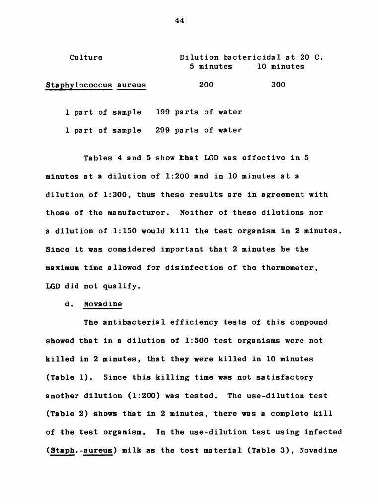
43
44
45
47
57
59
60
67

LIST OF FIGURES
Figure Page
1. "Cary" thermometer . . . . . . . . . . . . . ..... 50
2. Tri—R thermometer . . . . . . . . . . . . . . . . 51
3. Leukocyte count after infusion of 20 m1. of 2%
NaCl solution in left rear quarter: cow 591 . . . 52
4. Intracisternal temperature after infusion of 20 m1.
of 2% NaCl solution in left rear quarter: cow 591 53
5. Leukocyte count after infusion of 10 ml. of 2%
NaCl solution in right rear quarter: cow 591 . . 54
6. Intracisternal temperature after infusion of
10 ml. of 2% NaCl solution in right rear quarter:
cow 591 . . . . . . . . . . . . . . . . . . . . . 55
7. Rectal temperature following the intracisternal
infusion of 2% NaCl solution (20 m1. and 10 ml.
in left rear and right rear quarters respectively):
cow 591 O O O 0 O 0 O O O O O 0 O O 0 O O O O O O 56

LIST OF TABLES
Table Page
1. USE-DILUTION TEST ON NOVADINE (1:500)
TEST ORGANISM - STAPHYLOCOCCUS AUREUS . . . . . . 26
2. USE-DILUTION TEST ON NOVADINE (1:200)
TEST ORGANISM - STAPHYLOCOCCUS AUREUS . . . . . . 27
3. USE-DILUTION TEST ON NOVADINE (1:200)
TEST MATERIAL - MILK SAMPLE . . . . . . . . . . . 28
4. USE-DILUTION TEST 0N LIQUID GERMICIDAL DETERGENT
(1:300) TEST ORGANISM - STAPHYLOCOCCUS AUREUS . . 29
5. USE-DILUTION TEST ON LIQUID GERMICIDAL DETERGENT
(1:200) TEST ORGANISM - STAPHYLOCOCCUS AUREUS . . 30
6. USE-DILUTION TEST ON LIQUID GERMICIDAL DETERGENT
(1:150) TEST ORGANISM - STAPHTLOCOCCUS AUREUS . . 31
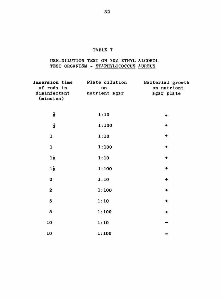
7. USE-DILUTION TEST ON 70% ETHYL ALCOHOL
TEST ORGANISM - STAPHYLOCOCCUS AUREUS . . . . . . 32
8. USE-DILUTION TEST ON CLENESCO LIQUID 75 CLEANER
(1:300) TEST ORGANISM — STAPHYLOCOCCUS AUREUS . . 33
9. TEMPERATURE READINGS OF DIFFERENT THERMOMETERS
(F) RECORDED IN HOT WATER BATH . . . . . . . . . 34

Table Page
10. TEMPERATURE AND MILK EXAMINATION DATA OF
CLINICALLY NORMAL COWS . . . . . . . . . . . . . 35
ll. TEMPERATURE AND MILK EXAMINATION DATA OF COW 591
WITH ACUTE NON-SYSTEMIC MASTITIS (RF QUARTER) . 36
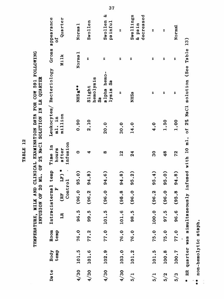
12. TEMPERATURE, MILK AND CLINICAL EXAMINATION DATA
FOR COW 591 FOLLOWING INFUSION OF 20 ML. OF 2%
NaCl SOLUTION IN LR QUARTER . . . . . . . . . . 37
13. TEMPERATURE, MILK AND CLINICAL EXAMINATION DATA
FOR COW 591 FOLLOWING INFUSION OF 10 ML. OF 2%
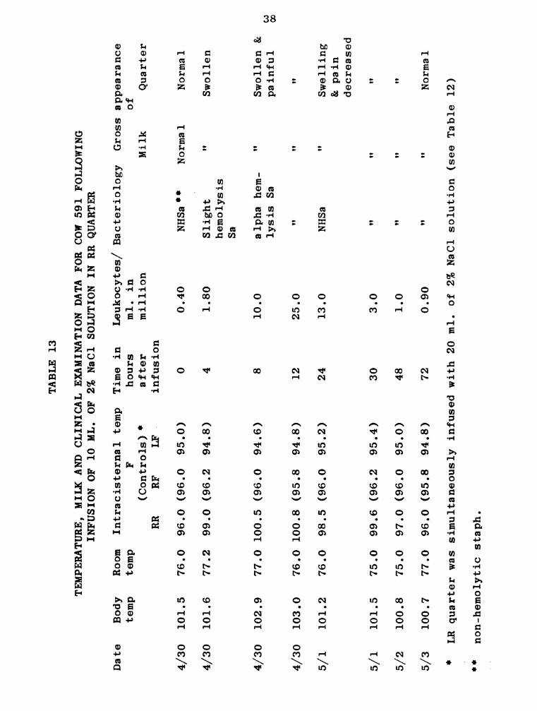
NaCl SOLUTION IN RR QUARTER . . . . . . . . . . 38
14. TEMPERATURE, MILK AND CLINICAL EXAMINATION DATA
FOR COW 591 FOLLOWING INFUSION 0F 5 ML. 0F 24
HOUR BROTH CULTURE OF NON-HEMOLYTIC
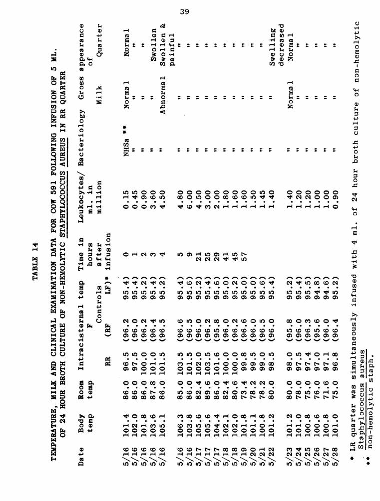
STAPHYLOCOCCUS AUREUS IN RR QUARTER . . . . . . 39
15. TEMPERATURE, MILK AND CLINICAL EXAMINATION DATA
FOR COW 591 FOLLOWING INFUSION OF 4 ML. OF 24
HOUR BROTH CULTURE OF NON-HEMOLYTIC
STAPHYLOCOCCUS AUREUS IN LR QUARTER . . . . . . 40
16. TEMPERATURE AND MILK EXAMINATION DATA OF
CLINICALLY NORMAL cows . . . .1. . . . . . . . . 68
I. INTRODUCTION
For many years mastitis has been a serious economic
problem to the dairy industry. Mastitis not only causes a
loss in milk yield, but may be responsible for the loss of
valuable animals either through death or complete cessation
of production. Before 1939, mastitis caused by streptococcic
microorganisms was the most predominant and troublesome type
in the United States. Since that time staphylococcic micro—
organisms have become increasingly more prevalent as the
etiological agents of mastitis. These organisms have an
omnipresent distribution and include both pathogenic and non-
pathogenic varieties.
One of the reasons that mastitis has continued to
be prevalent is the lack of early and adequate diagnostic
procedure. According to Merchant and Packer (1952) the
diagnosis of mastitis caused by bacteria was made by Nocard
and Mollereu in 1884 when they described a streptococci
isolated from the milk of a sick cow. Prescott and Breed
(1910) described a method for determination of leukocytes
and epithelial cells in milk associated with an abnormal

milk. The basic principle of leukocyte determination is
still used today although modifications of the original
method continue to be made. One disadvantage to this test
is that it is strictly a laboratory procedure. Schalm and
Noorlander (1957) developed a macroscopic field test based
on the presence of leukocytes. This test which they termed
the California Mastitis Test (CMT) gives evidence of a
precipitate or gel formation when the fore-milk has a cell
count over 100,000 cells per ml. or when "strippings" have
a cell count in excess of 200,000 cells per ml. Since this
reaction only indicates the presence of leukocytes, bacter-
iological tests are necessary for a complete diagnosis.
Early diagnosis of bovine mastitis is essential to
enable efficient curative treatment before there is permanent
damage to the secreting tissues. In this work, an attempt
was made to determine if intracisternal temperature might be
of value in the early detection of mastitis. Intracisternal
temperatures of bovine mammary glands were measured with a
Tri-R electronic thermometer*. The intracisternal tempera—
tures of cows with mastitis, some of which occurred naturally,
and some of which was induced by the intramammary infusion of
either 2% NaCl solution or Staphylococcus aureus organisms.
* ‘Tri-R Instrument, Jamaica 35, N. Y.
Concurrent with the temperature studies, the following dia—
gnostic tests were performed on milk samples: California
Mastitis Test, hemolysin production on blood agar, coagulase
production, leukocyte counts, and bacterial examination.

II. REVIEW OF LITERATURE
A survey of the literature available in the Michigan
State University library failed to reveal any material on the
determination of intracisternal temperatures in the diagnosis
of udder infections.
In view of the complete lack of literature on intra-
cisternal temperatures it was thought that a brief review of
certain phases related to the carrying out of this experimental
work would be helpful. The following subject areas are
reviewed:
1. Evaluation of disinfectants.
2. Inflammation.
3. Staphylococcic mastitis.
1. Evaluation of disinfectants
Before attempts to take intracisternal temperatures
were carried out a method of disinfecting the thermometer
was necessary.
Reddish (1957) in his text book makes mention of
several noteworthy facts pertaining to methods of testing
and evaluating disinfectants and are included among the

following statements. Buchhaltz, as early as 1875 developed
a method for evaluating the effectiveness of antiseptics.
He used tobacco-infusion as a culture medium and observed
the effectiveness of many antiseptics. However, Robert Koch
(1881) compared the germicidal value of various disinfectants
upon a culture of microorganisms. He extended his experimenta—
tion to include the impregnation of silk threads with anthrax
spores and then exposed them to solutions of chemicals for
different intervals of time. After the exposure, the silk
threads were washed with water and placed into nutrient broth
and observed for bacterial growth. Geppert in 1889 repeated
the work of Koch, but he found that mercuric chloride solution
(1:1,000) failed to kill anthrax spores within that period
which was reported by Koch. In 1897, Kronig and Paul devised
a method for determining the germicidal value of a disinfectant.
They modified the method of Koch and instead of using silk
threads substituted garnets. During the experimentation they
observed that the relative value of disinfectants is dependent
upon various conditions such as temperature, number and species
of microorganisms, and also the absence of organic matter.
Rideal and walker reported a more reliable method, whereby
the effect of a disinfectant was compared with phenol. This
method is generally spoken of as the "Phenol Coefficient
Method” for testing the effectiveness of antiseptics and
disinfectants.
Anderson and McClintic (1911) modified the method
of Rideal and Walker, and the resulting procedures became
known as the Hygienic laboratory Method. This method was
used widely in the United States for many years.
In the year 1931, an official method for evaluating
antiseptics and disinfectants based on the method of Rideal
and Walker was prescribed with very few changes, and is
known today as the Food and Drug Administration (FDA) Method.
This method is the standard phenol coefficient test by which
many antiseptics and disinfectants are tested. At the
present time the official method employed in the evaluation
of disinfectants is known as the "A.O.A.C. Phenol Coefficient
Method"‘.
.Mallmann and Hanes (1945) proposed a method for
evaluating antiseptics and disinfectants under all practical
applications which is called the "Use-Dilution Method".
Leavit (1946) worked out the comparative study of the use-
dilution method and the FDA Method. In a subsequent study,
Mallnan and Leavit (1948) reported that the use-dilution
method is more satisfactory than the older procedures.
‘ Association of Official Agricultural Chemists.
Literature concerning disinfectants used in this study
was not available in the case of Novadine and Clenesco
Liquid 75.
Liquid Germicidal Detergent (LGD) was demonstrated
by Bryan et al. (1948) to be effective as an udder dis-
infectant.
Gershenfeld et a1. (1951) reported on the steriliza-
tion of clinical thermometers. During experimentation they
contaminated segments of the thermometers (about two inches
in length) with bacterial cultures and reported that 70%
ethyl alcohol killed staphylococci in 10 minutes.
2. Inflammation
Menkin (1950) has given us a classical report on the
basic concepts of inflammation. The following information
is taken from his textbook. A rise of temperature is one of
the most dramatic manifestations of inflammation of_1iving
tissue, and has been known for a long time. Even Hippocrates
frequently used this observation in the diagnosis of diseases,
and regarded inflammation as being closely associated with
fever. Galen discussed the subject of inflammation and
thought it to be a common disease. For the first time the
phenomenon of inflammation was described as a local fever.
This idea of Galen was expanded by Hunter who reported that

the phenomenon of inflammation was the reaction to any injury
caused therein and regarded the inflammatory process as a
defensive reaction. Virchow reported that there is a swell-
ing of parenchymatous structures when injured. Cohnheim also
analyzed that the process of inflammation was due to some
disturbed local physiology, and he reported that in inflamma—
tory reactions there was increased permeability of the
capillary wall, and an outward migration of leukocytes.
Wolf (1923) studied the phenomenon of chemotaxis and
reported that histamine was strongly chemotactic. Dixon and
McCutcheon (1935) reported that polymorphonuclear leukocytes
were strongly attracted by staphylococcic organisms. Moon
(1935) reported that the cells of normal tissues contained
histamine in a non-diffusible form, and when the cells of
the body were injured histamine was liberated locally in a
diffusible form. Adjacent capillaries then become dilated
and there was an increase in the permeability of the capillary
endothelium resulting in transudation of plasma which produced
edema. Grant and Wood (1928) reported that histamine had no
appreciable power to cause emigration of leukocytes from the
vessels.
Menkin (1937) concluded that the migration of poly—
morphonuclear leukocytes in inflammation was related to the
liberation of an active substance "leukotaxine" which was
chemotactic to leukocytes. He further reported in the year
1943 that the important aspect of the inflammatory reaction
was to localize or to fix the irritating material.
Extensive work has been done on the subject of
inflammation, but this literature will be limited to the
extent to which it is pertinent. Runnels (1946) reported
the etiological agents of inflammation to be irritants such
as physical injuries, chemical irritants, bacteria, and other
parasites of lower animal life.
Menkin et a1. (1937) reported that the cytological
picture in an inflamed area was dependent upon the pH of the
injured part. They further reported that hydrogen ion con-
centration was the factor conditioning the cellular pattern.
The polymorphonuclear leukocytes were most predominant in an
acute inflammation when the pH of the medium was seven or
more. When the macrophages were predominent then this stage
was called a chronic inflammation. The polymorphonuclear
leukocytes contain an intracellular enzyme which was active
in an alkaline medium. They have also shown that the poly-
morphonuclear leukocytes were incapable of surviving in an
acid medium and the survival of leukocytes was determined
by the hydrogen ion concentration.

10
3. Staphylococcic mastitis
Staphylococcic microorganisms are widespread in nature
and found on the skin of animals and particularly on the
mucous membranes of the nose and mouth, and become: a part of
the udder flora in the bovine.
Evans (1916) obtained "micrococci" from 58.8% of the
milk samples drawn from the udders which they regarded as
normal. Klimmer (1930) in Germany investigated the occurrence
of mastitis of an acute type due to staphylococci in dairy
cows. Little and Foley (1935) found 9 acute cases of bovine
mastitis due to staphylococci. Gwatkin et al. (1936) observed
143 cows affected with mastitis, and found that 30 cases were
due to Staphylococcus aureus. Minett (1937) worked on the
incidence of Staphylococcus aureus mastitis in five dairy
herds. He demonstrated that out of 415 cows, 145 were found
to shed staphylococcic microorganisms in the milk and the
percentage of infection due to staphylococcic microorganisms
was 34.9%. Little and Plastridge (1946) observed that 5.9%
of the milk samples contained staphylococci associated with
1,000,000 or more leukocytes per ml. of milk, and an additional
4.8% contained staphylococci associated with leukocytes from
500,000 to 1,000,000 per m1. of milk. Packer (1952) reported,
during his six years of study, that 70% of the milk samples

11
contained staphylococci. Schalm (1953) reported that from
25.6 to 75% of the dairy cows he explained had micrococci
(25.6% of the herd in July 1949, to 75% in June 1950). During
six years of study (1947 to 1952) on one dairy herd the
average incidence (percentage of cows) of shedding of
staphylococci increased from year to year as follows: 43, 49,
47, 64, 57, and 62. Both Schalm (1953) and Packer (1952)
concluded that the incidence was correlated with increasing
lactation age.
Carpenter (1922) introduced a 24 hour broth culture
of Staphylococcus aureus organisms isolated from a case of
mastitis into the udders of 2 cows and 3 heifers. For 4 cows
he reported a severe type of mastitis with systemic involve—
ment. In 1 cow there was no external evidence of inflammation
of the udder. Parshel (1934) introduced Staphylococcus
aureus into the quarters of 2 cows, and reported that the
quarters became swollen and hot. The reaction was of a mild
nature and subsided after a few days. Little and Foley (1935)
injected staphylococcic organisms through the teat meatus on
nine different occasions. They used approximately 800
staphylococci for the first injection and 2,500 staphylococci
‘were injected later. Acute mastitis was produced after the
last injection of 2,500 organisms. They further observed

12
that there was swelling and congestion of the quarter, and
the quarter was painful on manipulation. Minett (1937)
exposed quarters two times with 1 m1. of a 24 hour broth
culture of Staphylococcus aureus and established infection
which lasted for a few days. Miller and Heishman (1943) in-
fused each of the udders of 8 cows with increasing numbers
of staphylococcic organisms ranging from 700 to 30,000.
Observations after 25 exposures revealed that 22 quarters
were infected. Schalm (1944) produced gangrenous mastitis
in two lactating cows by the intramammary infusion of 5 ml.
of a pure broth culture of Staphylococcus aureus. Using the
exudate from natural cases of mastitis or the Staphylococcus
aureus organisms isolated therefrom, he produced transistory
mastitis in two dry cows. Slanetz and Bartley (1953) produced
an acute type of mastitis with large numbers of organisms
and observed that there was a wide variation in response of
mammary glands to inoculation of strains of staphylococcic
(Jrganisms. After the injection of large numbers of phage
tuype 80/81 (staphylococcic organisms) into the udders of two
cows Drury et al. (1961) demonstrated the continual shedding
of staphylococcic organisms in milk for four months. Nat
(1962) introduced 16 x 102 to 28 x 102 staphylococcic
Organisms into 12 quarters and observed acute mastitis in one

l3
quarter. When he infused 38 x 103 to 57 x 104 staphyloccic
organisms in addition to mild trauma, he observed an acute
mastitis in 2 of 12 quarters. For the third experiment he
injected 1 x 10¹⁰ staphylococcic organisms into 13 quarters
and observed acute mastitis in 4 quarters.

III. MATERIALS AND METHODS
1. Evaluation of disinfectants
Staphylococcus aureus was the test organism used to
determine the efficacy of the products that might be used to
disinfect the thermometers. The organism was taken from a.
stock culture of Staphylococcus aureus, transferred to a
nutrient agar slant, incubated for 48 hours at 37 C., and
then streaked on blood agar plates (5% of bovine blood), and
again incubated for 24 hours. Suitable colonies were trans-
ferred to nutrient broth tubes and,after 24 hours incubation,
were used for the testing procedure. Six glass rods (3 x 1/4
inches in size) were used to simulate thermometers. Metal
rods of the same length and size as the glass rods in the
form of nails were also used.
Disinfectants tested
The following four disinfectants were tested:
Novadine"I which is a suspension of nonyl phenoxy ethanol-
iodine complex (providing 1.75% available iodine; Liquid
* Cowles and Co., Cleveland, Ohio
14

15
Germicidal Detergent’ which is a suspension of high molecular
weight alkylamine hydrochlorides containing phemerol chloride
as its active ingredient; Clenesco Liquid 75"is specifically
designed for all food plant cleaning operations where the
solution is applied by hand. It is a high foaming,mildly
alkaline cleaner; and 70% ethyl alcohol.
Employing the use-dilution method (Mallmann 1945)
the glass and metal rods were dipped into the broth culture
for a period of 15 minutes and then removed from the broth
and carefully laid on sterile filter paper in a closed petri
dish for a 30 minutes drying period at room temperature.
Care was taken not to roll while drying. At the end of the
drying period these rods were dipped into the test dis-
infectant for the following periods of time: 0.5, 1.0 and
1.5 minutes. In the event that 1.5 minutes did not kill the
organisms, the test was repeated using 2, 5, and 10 minute
periods of time. After removal from the disinfectant the
rods were rinsed in sterile water for one minute to remove
excess disinfectant thus preventing further bacteriostatic
effect. Rods were then dipped into tubes containing sterile
nutrient broth and were shaken vigorously to remove organisms
'9 Parke, Davis and Co., Detroit 1, Mich.
Iii Cowles and Co., Cleveland, Ohio

16
adhering to the rods. Suitable dilutions were then placed
on nutrient agar to measure quantitatively the extent of kill.
The count for the bacterial growth was performed after 24
hours of incubation at 37 C.
Controls were run in the same manner, except that
they were not dipped into the tubes containing disinfectant.
Glass and metal rods were also dipped in milk from
a cow known to be shedding staphylococci in her milk. The
testing with milk was performed with the disinfectant that
gave superior results with the test organisms as had been
determined previously. The presence or the absence of
bacterial growth was determined by the use of blood agar
media (5% of bovine blood).
2. Thermometers used
The following thermometers were obtained and used
according to the manufacturer's instructions. Before use,
the thermometers were checked for accuracy by partial
immersion in a hot water bath. The "Cary" thermometer that
compared most favorably with the readings on the Tri-R and
mercury thermometers was used for determining rectal
temperatures.
a. ”Cary" thermometer*
The "Cary" thermometer (Fig. l) is an unbreakable
* Cary Instruments, Limited, 44 Whitehall St., New York

17
metal dial thermometer, which is free from glass and mercury
and can be used with complete safety. This thermometer was
used only for the measurement of rectal temperatures since
it was found to be too large to insert into the teat meati
of some cows used in this experiment.
b. Tri-R thermometer"
This thermometer (Fig. 2) is a battery operated model
supplied with a built—in long life mercury cell designed to
provide a useful operating life exceeding 4,000 hours.
c. A standard laboratory mercury thermometer (centigrade)
This was used to determine room temperatures.
3. Measurement of intracisternal temperature.
Eight lactating and one non-lactating Holstein cows,
ranging in age from 4 to 8 years were used for the recording
of intracisternal temperatures. Some of these cows had a
history of mastitis. All of the cows had teats of approxi-
mately equal diameter and length. Two of the cows were
housed and fed in the MSU clinic; the others were housed
and fed at the University Dairy barn. Animals were milked
twice a day with milking machines.
The intracisternal temperatures were taken between
9 and 10 A.M., after the morning milking, and between 2 and
‘5 Tri-R Instruments Co., Jamaica 35, New York.
18
3 P.M., just before the evening milking. Since the
temperatures were taken after and before the regular milking
periods, the effect of the let-down phenomenon could be
evaluated. At the time of measuring the intracisternal
temperatures, cows were not provided with any kind of feed,
nor were they excited by the presence of visitors. A line
etched on the electronic thermometer 5 cm. from the point
served as a marker to insure uniformity in depth of each
insertion. The probe was disinfected for 2 minutes by
immersing in Novadine solution (1:200) before each insertion.
At the time intracisternal temperatures were taken,
the room temperatures were determined.
4. Tests applied to milk.
a. California Mastitis Test (CMT)
The procedure followed was that of Schalm and
Noorlander (1957) as modified by Drury, et al. (1961).
b. Direct leukocyte count
Prescott and Breed (1910) investigated a method for
determining the number of leukocytes in milk. This technique
was used with the modification (Bryan, 1941) that instead
of measuring the milk by a graduated pipette, a closed
platinum loop which delivered a known amount of milk was
used for spreading the milk on the slide.

19
c. Hemolysis test
Hemolysis was observed on blood (5% bovine) agar
plates and was scored according to the method of Slanetz
and Bartley (1953).
d. Coagualse test.
This test was performed according to the procedures
outlined in the Difco Manual‘.
5. Artificially induced mastitis.
Intracisternal and rectal temperatures were taken
prior to and following the intramammary infusions of agents
to induce mastitis in a cow. Temperatures were also determined
at the time of sample collections. The following agents were
used to produce mastitis:
a. 2% NaCl solution
Ten ml. of the saline solution were infused into the
right rear quarter and twenty ml. into the left rear quarter.
Milk samples were aseptically collected for bacteriological
examination after each 4 hours on the first day and at 12 hour
intervals for the next day and again after 24 hours.
b. Staphylococcus aureus
After an interval of 13 days, the above mentioned
cow was used for this phase of the work. Four ml. of a 24
' Difco Manual: Difco laboratories, Inc., Detroit 1, Michigan.
9th edition. 1953.
20
hour broth culture of non-hemolytic and coagulase-negative
Staphylococcus aureus organisms were infused into the left
rear quarter and 5 m1. of the same culture were infused into
the right rear quarter. Milk samples were taken at hourly
intervals for the first 5 hours of the first day and at 4
hour intervals on the second and third day. Subsequently
one sample was collected on each of the next 9 days.
IV. RESULTS
1. Evaluation of disinfectants
The results on the evaluation of the four disinfectants
tested are presented in Tables 1 through 8. As may be noted,
Novadine was the most efficient disinfectant. Using test
organisms it was found to be effective at a dilution of 1:200
in two minutes (Table 2) but not effective at 1:500 in 5
minutes (Table 1). In Table 3 results indicate that Novadine
(1:200) was also effective in two minutes when infected
(Staphylococcus aureus) milk was the test sample. Practical
application of this use of Novadine (1:200) indicated that
Staphylococcus aureus were killed on the thermometer probe
following its use in taking intracisternal temperatures.
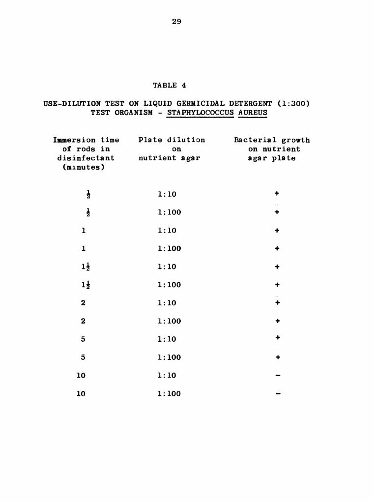
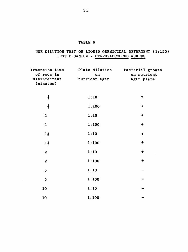
The remaining disinfectants (LGD, 70% ethyl alcohol,“
and Clenesco Liquid 75) tested were ineffective in two minutes
as indicated in Tables 4 through 8.
2. Thermometers used
The comparison of the readings of the thermometers
placed in hot water is shown in Table 9 and indicates that
the Tri-R and mercury thermometer agreed closely. The
21
22
variation was 0.0 to 0.3 F. Of the four Cary thermometers
tested, number 97517 was in closest agreement to the other
thermometers used. Here the variation was 0.1 to 0.3 F.
3. Intracisternal temperatures
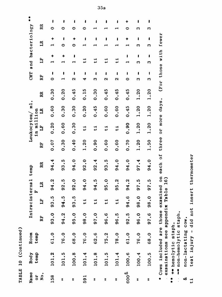
a. Clinically normal cows
The intracisternal temperatures of 5 clinically
normal cows, each of which were determined on three or more
days, are presented in Table 10. In this table milk examina-
tion data, body and room temperatures are also found, for each
day the intracisternal temperatures were taken.
For cow 573 (Table 10) the greatest variation among
the four intracisternal temperatures determined for a single
day was 5.0 F (RF 95.0 F to RR 100.0 F). During the five
days that this cow's intracisternal temperatures were taken,
the highest temperature was 100.0 F and the lowest was 93.0 F.
The cow named Daya (Table 10) showed an intracisternal
temperature variation among the four quarters on a single day
of 2.0 F (95 F to 97 F). The highest intracisternal tempera-
ture throughout the seven days on which temperatures were
determined was 97 F and the lowest was 94.2 F.
Cow 600 (Table 10) was not lactating during the time
of this experiment. For a single day, the greatest variation
among the four intracisternal temperatures was 2.1 F. The
23
highest and lowest intracisternal temperature noted in three
days was 98.0 F and 92.5 F respectively.
Cow 158 (Table 10), on a given day, had a maximum
intracisternal temperature variation of 3.0 F among the four
quarters. The highest and lowest intracisternal temperatures
determined in three days was 95.0 F and 92.0 F respectively.
For cow 591 (Table 10) the greatest variation among
the four intracisternal temperatures determined for a single
day was 6 F. During the four days that observations were
made the highest intracisternal temperature was 98.0 F and
the lowest was 92.0 F.
Intracisternal temperature data on four cows that
were not examined for more than two days is presented in the
appendix. These intracisternal temperatures compare favor-
ably with those in Table 10.
b. Cows with mastitis
The intracisternal temperature of the RF quarter of
cow 591 (Table 11) during an acute flare-up of non-systemic
mastitis was from two to three degrees higher than the highest
temperature of the non-mastitic quarters. Milk from the RF
quarter also had leukocyte counts appreciably higher than
milk from the other three quarters. The CMT score for the
24
milk from the RF quarter was likewise higher than for the
other quarters.
Intracisternal temperatures and supplementary data
for cow 591 in which mastitis was artificially induced by
infusing 2% NaCl solution are presented in Tables 12 and 13
and Figures 3, 4, 5, 6, and 7.
At the peak of the inflammatory reaction as judged
by leukocyte counts (Table 12) of 20, 30, and 14 millions per
ml. of milk at 8, 12, and 24 hours respectively, following
infusion of 20 ml. of 2% NaCl solution, the intracisternal
temperature ranged from 101.6 to 98.5 F. These temperatures
were from 2.5 to 6.8 degrees higher than intracisternal
temperatures of the control quarters. Accompanying this
period the infused quarter was swollen and painful. The 8
and 12 hour rectal temperatures as taken from Table 12 are
also at peak level.
When 10 m1. of 2% NaCl solution were infused into
the RR quarter of cow 591 (concurrently with the infusion of
20 ml. of the same solution into the LR quarter) results
(Table 13 and Figures 5, 6, and 7) were similar to those
mentioned following the infusion of 20 m1. of NaCl solution
(Table 12 and Figures 3, 4, and 7). The major difference is
an indication of a less severe inflammatory reaction as

25
judged by lower leukocyte counts and not as much increase
in the intracisternal temperature.
When mastitis was produced by the intramammary infusion
of 5 m1. of a 24 hour broth culture of non-hemolytic
Staphylococcus aureus organisms into the RR quarter of
cow 591 (Table 14), there were marked increases in the intra-
cisternal temperature of the infected quarter. From the
second to the 45th post infusion hour the temperature was
100 F or higher and on two occasions went as high as 103.5 F.
The highest intracisternal temperature of the control
quarters during this time was 96.6 F. Concurrent with the
high temperature of the infected quarter were an elevation
in rectal temperature to 106.3 F, and increase in the milk
leukocyte count to 6 million per ml., painful swelling of
the infused quarter, and grossly abnormal milk.
When 4 ml. of the same broth culture as used in the
RR quarter were concurrently injected into the LR quarter
results (Table 15) were similar to those presented in
Table 14. The increases in the intracisternal temperature
and leukocyte count were of the same magnitude but the
duration of temperatures over 100 F was 26 hours as compared
to 43 hours for the RR quarter.

26
TABLE 1
USE—DILUTION TEST ON NOVADINE (1:500)
TEST ORGANISM — STAPHYLOCOCCUS AUREUS
Immersion time Plate dilution Bacterial growth
of rods in on on nutrient
disinfectant nutrient agar agar plate
(minutes)
5 1:10 +
i 1:100 +
1 1:10 +
l 1:100 +
1% 1:10 +
I; 1:100 +
2 1:10 +
2 1:100 +
5 1:10 +
5 1:100 4
10 1:10 -
10 1:100 -

27
TABLE 2
USE-DILUTION TEST ON NOVADINE (1:200)
TEST ORGANISM - STAPHYLOCOCCUS AUREUS
Immersion time Plate dilution Bacterial growth
of rods in on on nutrient
disinfectant nutrient agar agar plate
(minutes)
1 1:10 +
l 1:100 +
2 1:10 -
2 1:100 -
5 1:10 -
5 1:100 -

28
TABLE 3
USE-DILUTION TEST ON NOVADINE (1:200)
TEST MATERIAL — MILK SAMPLE"I
Immersion time Plate dilution Bacterial growth
of rods in - on on nutrient
disinfectant nutrient agar agar plate
(minutes)
1 1:10 +
l 1:100 +
2 1:10 -
2 1:100 -
5 1:10 -
5 1:100 -
’ Milk from a cow shedding Staphylococcus aureus.
29
TABLE 4
USE-DILUTION TEST ON LIQUID GERMICIDAL DETERGENT (1:300)
TEST ORGANISM - STAPHYLOCOCCUS AUREUS
Immersion time Plate dilution Bacterial growth
of rods in on on nutrient
disinfectant nutrient agar agar plate
(minutes)
i 1:10 t
§ 1:100 f
1 1:10 +
1 1:100 +
1% 1:10 +
1% 1:100 +
2 1:10 4
2 1:100 +
5 1:10 +
5 1:100 4
10 1:10 -
10 1:100 -

30
TABLE 5
USE—DILUTION TEST ON LIQUID GERMICIDAL DETERGENT (1:200)
TEST ORGANISM - STAPHYLOCOCCUS AUREUS
Immersion time Plate dilution Bacterial growth
of rods in on on nutrient
disinfectant nutrient agar agar plate
(minutes)
i 1:10 +
1; 1:100 +
1 1:10 +
1 1:100 +
It 1:10 +
1% 1:100 +
2 1:10 +
2 1:100 +
5 1:10 “
5 1:100 -
10 1:10 -
10 1:100 -
31
TABLE 6
USE-DILUTION TEST ON LIQUID GERMICIDAL DETERGENT (1:150)
TEST ORGANISM - STAPHYLOCOCCUS AUREUS
Immersion time Plate dilution Bacterial growth
of rods in on on nutrient
disinfectant nutrient agar agar plate
(minutes)
; 1:10 +
g 1:100 +
1 1:10 +
1 1:100 +
1; 1:10 4-
1% 1:100 +
2 1:10 +
2 1:100 +
5 1:10 -
5 1:100 -
10 1:10 -
10 1:100 -
32
TABLE 7
USE—DILUTION TEST ON 70% ETHYL ALCOHOL
TEST ORGANISM - STAPHYLOCOCCUS AUREUS
Immersion time Plate dilution
of rods in on
disinfectant nutrient agar
(minutes)
A 1:10
fi 1:100
1 1:10
1 1:100
1% 1:10
1; 1:100
2 1:10
2 1:100
5 1:10
5 1:100
10 1:10
10 1:100
Bacterial growth
on nutrient
agar plate
33
TABLE 8
USE-DILUTION TEST ON CLENESCO LIQUID 75 CLEANER (1:300)
TEST ORGANISM - STAPHYLOCOCCUS AUREUS
Immersion time Plate dilution
of rods in on
disinfectant nutrient agar
(minutes)
i 1:10
5 1:100
1 1:10
1 1:100
19,; 1:10
15 1 100
2 1:10
2 1:100
5 1:10
5 1:100
10 1:10
10 1:100
Bacterial growth
on nutrient
agar plate
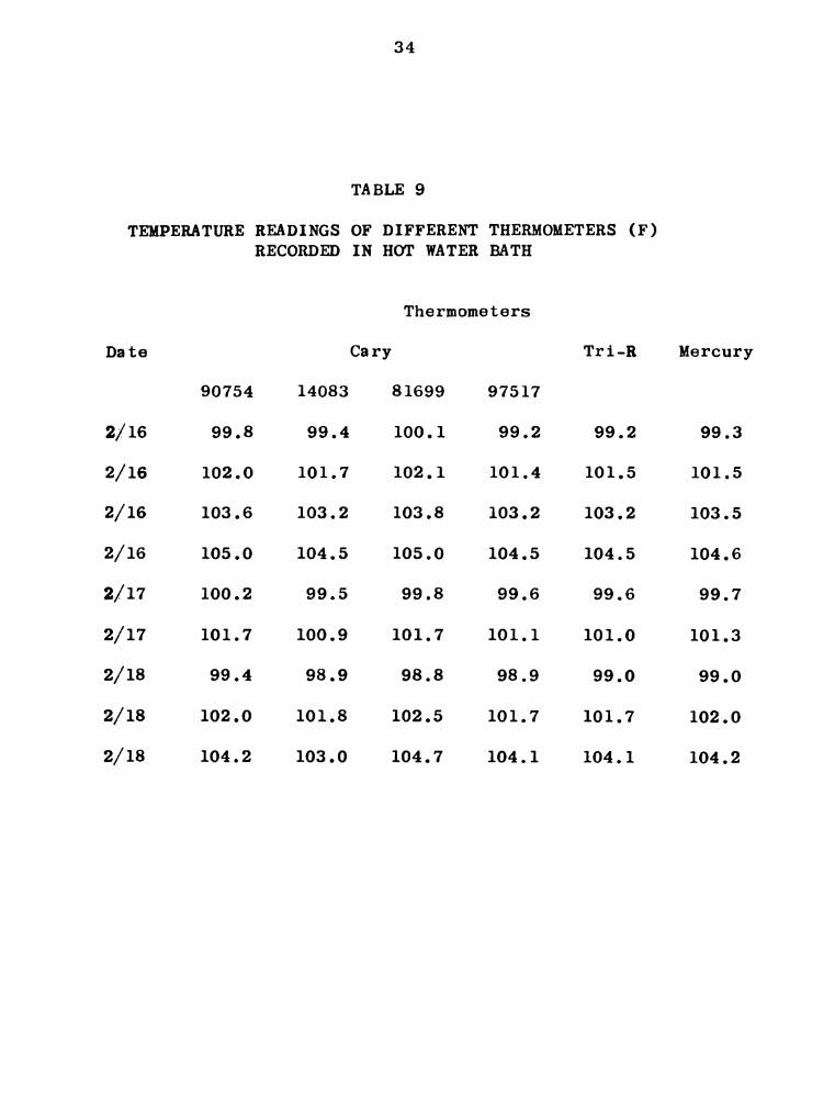
TEMPERATURE READINGS OF DIFFERENT THERMOMETERS (F)
RECORDED IN HOT WATER BATH
Date
2/16
2/16
2/16
2/16
2/17
2/17
2/18
2/18
2/18
90754
99.8
102.0
103.6
105.0
100.2
101.7
99.4
102.0
104.2
14083
99.4
101.7
103.2
104.5
99.5
100.9
98.9
101.8
103.0
34
TABLE 9
Cary
Thermometers
81699 97517
100.1 99.2
102.1 101.4
103.8 103.2
105.0 104.5
99.8 99.6
101.7 101.1
98.8 98.9
102.5 101.7
104.7 104.1
Tri-R
99.2
101.5
103.2
104.5
99.6
101.0
99.0
101.7
104.1
Mercury
99.3
101.5
103.5
104.6
99.7
101.3
99.0
102.0
104.2

Name
or
No.
573
N N N H
Daya
H H H H '1
Body
temp
99.5
100.7
103.2
100.5
100.8
101.2
101.2
101.2
100.9
101.2
101.3
100.8
TABLE
10
TEMPERATUREAND
MILK
EXAMINATION
DATA
OF
CLINICALLY
NORMALCOWS"
Room
temp
53.6
61.0
61.0
76.0
68.0
66.2
66.2
66.2
77.0
62.6
76.0
68.0
Intracisternal
temp
RF
94.0
93.5
95.0
93.6
95.5
96.0
95.4
95.2.
ti
ti
ti
95.5
LF
94.0
93.0
99.0
93.5
98.0
95.5
94.8
94.2
96.5
96.0
95.0
95.0
F
LR
93.6
93.4
96.5
96.0
96.0
94.3
95.0
94.3
95.6
96.0
96.4
95.0
RR
95.0
93.3
100.0
93.5
96.25
94.4
95.5
94.4
96.0
96.6
97.0
96.0
Leukocytes/
ml.
inmillion
RF
0.60
0.60
0.90
0.90
0.90
ti
ti
ti
0.30
LF
0.90
0.90
0.30
0.90
1.20
0.60
LR
0.45
0.90
1.20
0.45
1.20
0.30
0.45
0.60
0.90
0.90
0.30
0.90
1.20
CMT
and
bacteriology"
RF
1+
1+
1+
1+
ti
ti
ti
LF
LR
l-+
1.-
Continued
RR
2+
35
TABLE
Name
or
No.
158
N "
591
H " "
6003c
"
*Cows
included
are
those
examined
on
each
of
three
10
(Continued)
Body
temp
101.2
101.5
100.8
101.4
101.8
101.5
101.4
100.6
100.4
100.5
Room
temp
61.0
76.0
68.0
76.0
62.6
75.2
78.0
61.0
76.0
68.0
Intracisternal
temp
RF
93.0
94.2
95.0
98.0
97.0
96.6
96.5
92.5
96.0
97.6
LF
93.5
94.5
93.5
ti
ti
ti
ti
94.6
98.0
98.0
F
LR
94.2
92.5
92.0
94.0
94.5
95.0
95.2
94.2
97.5
97.5
RR
94.4
93.5
94.0
92.0
92.4
93.5
94.0
94.0
97.4
94.0
Leukocytes/
ml.
RF
inmillion
LF
ti
ti
LR
0.45
1.20
1.20
RR
0.45
0.45
1.20
1.20
CMT
RF
and
bacteriology
LF
ti
ti
ti
ti
or
more
days.
(For
those
with
examinations
see
appendix
Table
16)
n:
+-hemolytic
staph.
-non-hemolytic
staph.
&
ti
non-lactating
cow.
teat
injury
-did
not
insert
thermometer
LR
1+
fewer
0.
35a

TABLE
11
TEMPERATUREAND
MILK
EXAMINATION
DATA
0F
COW
591AFFECTED
WITHACUTE
NON-SYSTEMIC
MASTITIS
(RF
QUARTER)
Leukocytes/
ml.
inmillion
Date
Body
CMT
and
bacteriology"
temp
Room
Intracisternal
temp.
temp
F
4/20
4/22
4/23
4/24
4/25
4/26
4/27
4/28
102.0
101.8
101.5
101.6
101.5
101.5
101.6
101.2
63.0
68.0
71.0
75.0
75.0
78.0
78.5
78.0
RF
98.0
97.8
97.6
97.4
97.7
97.5
97.5
97.0
LF
ti
ti
ti
ti
ti
ti
ti
ti
LR
94.5
94.2
93.0
94.0
94.0
94.2
94.5
94.5
RR
92.4
92.6
94.0
95.0
95.0
94.8
95.0
95.0
f-
anon-hemolytic,
coagulase
negative
staph.
ti
-teat
injury
-did
not
insert
thermometer.
RF
1.20
1.20
1.20
1.30
1.50
1.40
1.50
1.20
LF
0.60
0.60
0.45
0.60
0.90
0.60
LR
RF
4
LF
2
LR
1
RR
0
36
Date
4/30
4/30
4/30
4/30
5/1
5/1
5/2
5/3
Body
temp
101.5
101.6
102.9
103.0
101.2
101.5
100.8
100.7
Room
temp
76.0
77.2
77.0
76.0
76.0
75.0
75.0
77.0
-
Intracisternal
temp
Time
in
LR
96.5
99.5
101.5
101.6
98.5
100.0
97.5
96.6
(RF
F
LF)‘
Control
(96.0
(96.2
(96.0
(95.8
(96.0
(96.2
(96.0
(95.8
95.0)
94.8)
94.6)
94.8)
95.2)
95.4)
95.0)
94.8)
TABLE
12
hours
after
infusion
0
12
24
30
48
72
0.90
2.10
20.0
30.0
14.0
4.0
1.50
1.00
NHSa“
Slight
hemolysis
Sa
alpha
hemo-
lysis
Sa
NHSa
TEMPERATURE,
MILKAND
CLINICAL
EXAMINATION
DATA
FOR
COW
591
FOLLOWING
INFUSION
OF
20
ML.
OF
2%
NaCl
SOLUTION
IN
LRQUARTER
Milk
Normal
H N
Leukocytes/
Bacteriology
Gross
appearance
ml.
in
million
of
Quarter
Normal
Swollen
Swollen
&
painful
H
Swellings
&pain
decreased
H
Normal
‘RR
quarter
was
simultaneously
infused
with
10
ml.
of
2%
NaCl
solution
(See
Table
13)
**
non-hemolytic
staph.
37
Date
4/30
4/30
4/30
4/30
5/1
5/1
5/2
5/3
a
##
Body
temp
101.5
101.6
102.9
103.0
101.2
101.5
100.8
100.7
TABLE
13
TEMPERATURE,
MILKAND
CLINICAL
EXAMINATION
DATA
FOR
COW
591
FOLLOWING
INFUSION
OF
10
ML.
OF
2%
NaCl
SOLUTION
INRR
QUARTER
Room
temp
RR
76.0
96.0
77.2
99.0
77.0
100.5
76.0
100.8
76.0
98.5
75.0
99.6
75.0
97.0
77.0
96.0
non-hemolytic
staph.
(Controls)
“
RF
(96.0
(96.2
(96.0
(95.8
(96.0
(96.2
(96.0
(95.8
LF.
95.0)
94.8)
94.6)
94.8)
95.2)
95.4)
95.0)
94.8)
Intracisternal
temp
Time
in
hours
after
ml.
mill
infusion
0 4
12
24
30
48
72
LR
quarter
was
simultaneously
infused
with
0.40
1.80
10.0
25.0
13.0
3.0
1.0
in
ion
Milk
NHSa"
Normal
Slight
"
hemolysis
Sa
alpha
hem-
"
lysis
Sa
NHSa
"
NH
0.90
NH
20
m1.
Leukocytes/
Bacteriology
Gross
appearance
Quarter
Normal
Swollen
Swollen
&
painful
N
Swelling
&pain
decreased
H
Normal
of
2%
NaCl
solution
(see
Table
12)
00
oo
TABLE
14
TEMPERATURE,
MILKAND
CLINICAL
EXAMINATION
DATA
FOR
COW
591
FOLLOWING
INFUSION
OF
5ML.
OF
24
HOUR
BROTH
CULTURE
OF
NON-HEMOLYTIC
STAPHYLOCOCCUS
AUREUS
IN
RRQUARTER
Date
Body
temp
Room
Intracisternal
temp
Time
in
Leukocytes/
Bacteriology
Gross
appearance
temp
Fhours
ml.
in
of
Controls
after
million
Milk
LF)‘
infusion
Quarter
RR
(RF
5/16
5/16
5/16
5/16
5/16
5/16
5/16
5/17
5/17
5/17
5/18
5/18
5/19
5/20
5/21
5/22
5/23
5/24
5/25
5/26
5/27
5/28
*LR
it
101.4
102.0
101.8
103.6
105.1
106.3
103.8
105.6
105.6
104.4
102.1
102.0
101.8
101.1
100.8
101.2
101.2
101.0
100.8
100.6
100.8
101.0
quarter
was
simultaneously
infused
with
4ml.
Staphylococcus
aureus
non-hemolytic
staph.
86.0
86.0
86.0
87.8
86.0
85.0
86.0
82.4
89.6
86.0
82.4
80.6
73.4
78.2
78.2
80.0
80.0
78.0
75.0
76.0
71.6
75.0
96.5
97.5
100.0
101.0
101.5
103.5
101.5
102.0
103.5
101.6
100.0
100.0
99.8
99.5
99.0
98.5
98.0
97.5
97.4
97.0
97.1
96.8
(96.2
(96.0
(96.2
(96.4
(96.5
(96.6
(96.5
(96.0
(96.2
(95.8
(96.0
(96.2
(96.6
(96.0
(96.5
(96.0
(95.8
(96.0
(96.3
(95.6
(96.0
(96.4
95.4)
95.4)
95.2)
95.4)
95.2)
95.4)
95.6)
95.2)
95.4)
95.6)
95.0)
95.2)
95.0)
95.0)
95.6)
95.4)
95.2)
95.4)
95.5)
94.8)
94.6)
95.2)
OHNO’DV‘ lam?"
25
29
41
45
57
NHSa
*‘
N H H 'I
1!
H V!
N H H H H H H N N H H H
Normal
H
Abnormal
N N N H H H
Normal
H N H
Normal
N
Swollen
Swollen
&
painful
N H N N H N H H H
Swelling
decreased
Normal
N H H H
of
24
hour
broth
culture
of
non-hemolytic
39

’TABLE
15
TEMPERATURE,
MILKAND
CLINICAL
EXAMINATION
DATA
FOR
COW
591
FOLLOWING
INFUSION
OF
4ML.
Date
5/16
5/16
5/16
5/16
5/16
5/16
5/16
5/17
5/17
5/17
5/18
5/18
5/19
5/20
5/21
5/22
5/23
5/24
5/25
5/26
5/27
5/28
'RR
OF
24
HOUR
BROTH
CULTURE
OF
NON-HEMOLYTIC
STAPHYLOCOCCUSAUREUS
IN
LRQUARTER
Body
temp
101.4
102.0
101.8
103.6
105.1
106.3
103.8
105.6
105.6
104.4
102.1
102.0
101.8
101.1
100.8
101.2
101.2
101.0
100.8
100.6
100.8
101.0
Room
temp
86.0
86.0
86.0
87.8
86.0
85.0
86.0
82.4
89.6
86.0
82.4
80.0
73.4
78.2
78.2
80.0
80.0
78.0
75.0
76.0
71.6
75.0
Intracisternal
temp
Time
in
LR
96.0
97.5
99.8
100.5
101.5
103.5
101.0
101.5
102.0
101.0
99.5
99.2
99.0
98.5
98.0
97.8
97.0
96.6
96.5
96.4
96.2
95.8
.Staphylococcus
aureus
“non—hemolytic
staph.
F Controls
(RF
(96.2
(96.0
(96.2
(96.4
(96.5
(96.6
(96.5
(96.0
(96.2
(95.8
(96.0
(96.2
(96.6
(96.0
(96.5
(96.0
(95.8
(96.0
(96.3
(95.6
(96.0
(96.4
LF)‘
95.4)
95.4)
95.2)
95.4)
95.2)
95.4)
95.6)
95.2)
95.4)
95.6)
95.0)
95.2)
95.0)
95.0)
95.6)
95.4)
95.2)
95.4)
95.5)
94.8)
94.6)
95.2)
hours
after
infusion
OHNM‘U‘ IOGH
25
29
41
45
57
quarter
was
simultaneously
infused
with
5ml.
0.075
0.30
0.75
2.70
3.90
4.50
5.40
NHSa**
N H N H H H i!
N N H
of
24
hour
broth
culture
Milk
Normal
N H N H H H H
of
Leukocytes/
Bacteriology
Gross
appearance
ml.
in
million
Quarter
Normal
" H
Swollen
Swollen
&
painful
H H H N H H H H H
Swelling
decreased
Normal
9'
H I!
N
of
non-hemolytic
pk
O
V. DISCUSSION
1. Selection of disinfectant
In considering the selection of the disinfectant for
thermometer sterilization, the following list of properties
were considered:
Bacterial activity
An ideal disinfectant or antiseptic should have a
wide range of killing power. In this work Staphylococcus
aureus was selected as the test organism because of its pre-
dominence in most cases of mastitis. However the final
criterion on the effectiveness of a disinfectant was con-
sidered under conditions of practical application.
Availability and cost
The disinfectant chosen for this purpose should be
readily available and relatively inexpensive.
Toxic effect on tissues
Toxic effect of the evaluated antiseptic should be
considered when a selection is to be made. In the opinion
of the author every chemical agent is somewhat toxic to
animal tissue, however no work was done in this connection.
41

42
Only the gross appearance of the quarter was noted for any
abnormality following insertion of chemically sterilized
thermometer, and none could be detected.
Speed of action
The speed of action or rate of kill is also an
important factor when selecting an antiseptic or the dis-
infectant. The action of the antiseptic or the disinfectant
should be rapid, so that little time is required for
effective sterilization.
Stability
A good disinfectant should not lose its effectiveness
when stored for reasonable lengths of time at ordinary room
or barn temperature.
a. Ethyl alcohol
Force and Kerr (1920) reported that clinical
thermometers were best sterilized by chemical disinfectants
and an exposure of 4 minutes was found to be necessary with
50% alcohol to secure adequate disinfection of oral
thermometers. Price (1950) indicated that an exposure of
5 minutes to either 70 or 80% ethyl alcohol was necessary
to kill the Mh_pyogenes var. aureus. Reddish (1956)
reported that 70% alcohol was satisfactory for the
disinfection of clinical thermometers. Immersion of the

43
thermometers in 70% alcohol for 10 minutes was sufficient
for this purpose.
During this work 70% ethyl alcohol was tested as
one of the disinfectants. However, it would not kill
Staphylococcus aureus, the test organism, in 5 minutes, but
did kill effectively in 10 minutes (Table 7). Since economy
of time was considered important in this work a ten minute
sterilization time was considered impractical.
b. Clenesco Liquid 75 Cleaner
Actually this product is specifically designed for
food plant cleaning operations. When tested for use in this
work it did not give any promising results. It would not
kill the test organism at the suitable dilution in 10 minutes
(Table 8).
c. Liquid Germicidal Detergent (LGD)
Bryan and associates (1948) reported that this
product was an efficient udder disinfectant and would destroy
mastitis producing organisms that might be on the external
surface of the teats and udder.
The results of the research conducted by the Parke,
Davis and Company on Liquid Germicidal Detergent using
Staphylococcus aureus as one of the test organisms are shown
as follows:
44
Culture Dilution bactericidal at 20 C.
5 minutes 10 minutes
Staphylococcus aureus 200 300
1 part of sample 199 parts of water
1 part of sample 299 parts of water
Tables 4 and 5 show that LGD was effective in 5
minutes at a dilution of 1:200 and in 10 minutes at a
dilution of 1:300, thus these results are in agreement with
those of the manufacturer. Neither of these dilutions nor
a dilution of 1:150 would kill the test organism in 2 minutes.
Since it was considered important that 2 minutes be the
maximum time allowed for disinfection of the thermometer,
LGD did not qualify.
d. Novadine
The antibacterial efficiency tests of this compound
showed that in a dilution of 1:500 test organisms were not
killed in 2 minutes, that they were killed in 10 minutes
(Table 1). Since this killing time was not satisfactory
another dilution (1:200) was tested. The use-dilution test
(Table 2) shows that in 2 minutes, there was a complete kill
of the test organism. In the use-dilution test using infected
(Staph.-aureus) milk as the test material (Table 3), Novadine

45
was also found to be effective in 2 minutes at a dilution of
1:200.
Novadine was the disinfectant of choice for thermometer
sterilization because in addition to fulfilling the properties
previously discussed, it was found to be effective within
2 minutes in a concentration that did not cause noticeable
irritation.
2. Normal intracisternal temperature
The mammary intracisternal temperatures of 9 clinically
normal cows (Table 10 and 16) varied from 91.5 F to 100.0 F.
A total of 109 intracisternal temperatures were determined
on these cows throughout a period of two months. Intra-
cisternal temperatures were taken on as many as seven
different days in the case of the cow named Daya.
Room temperatures encountered during this study did
not seem to have any notable effect on the intracisternal
temperatures (Table 10).
With one exception rectal temperatures did not
exceed 101.8 F. In the case of cow 573 (Table 10, line 3)
the rectal temperature was 103.2 F. At this time there was
also a leukocyte count of 1,200,000 per m1. of milk from the
RR quarter and a CMT score of 3. In view of the fact that
this cow's body temperature was slightly elevated and there

46
was a high leukocyte count in the RR quaater, it is felt that
a subclinical case of mastitis was present and was possibly
responsible for the high intracisternal temperature of 100.0 F
in the RR quaater. If this cow was considered to have
mastitis and if the determinations for this day are excluded
from the table, the highest intracisternal temperature
recorded for normal cows was 98.0 F (Table 10, lines 5, 14,
15, and 19).
Cow 600 was non-lactating at the time of these
observations (Table 10). The first observations were made
shortly after she had finished her lactation period (had not
been milked for five days). When the second and third
observations were made the animal had been non—lactating for
9 and 13 days respectively. According to Schalm et a1.
(1957) high milk leukocyte counts are present when cows are
between consecutive lactation periods. Table 10 shows this
to be true for cow 600. Accompanying the high milk body cell
count CMT scores of 3. It is noteworthy that intracisternal
temperatures tend to be slightly higher during non-lactating
periods.
Cow 591 (Table 10), on the first day, had a milk
leukocyte count of 1,200,000 per ml. of milk, in the RF
Quarter accompanied by a CMT score of 4. Although not

47
clinically evident, it is felt that mastitis was present in
this Quarter and that the intracisternal temperature of 98 F
was the result of the inflammation.
If the two cases of probable mastitis (cow 573, line 3;
and cow 591, line 19) and cow 600 during the non-lactating
period excluded from Table 10, the intracisternal temperature
for mastitis-free, lactating cows are found to vary from
91.5 F to 96.6 F.
3. Intracisternal temperatures of cows with mastitis
Cow 591 was observed during an acute attack of non-
systemic mastitis in the RF quarter. In Table 11 can be seen
the intracisternal temperatures for this quarter in comparison
to the other two quarters. The leukocyte counts of the milk
from this quarter ranged from 1,200,000 to 1,500,000 per ml.,
the CMT score was consistently 4, and the intracisternal
temperatures were from 2 to 3 degrees (F) higher than the non-
inflamed quarters.
When mastitis was artificially induced by the injection
of 2% NaCl solution or Staphylococcus aureus organism, the
inflamed quarters had intracisternal temperatures considerably
higher than the control quarters: of the two amounts of 2% NaCl
solution, the 20 ml. injection resulted in slightly higher
intracisternal temperatures than the 10 m1. injection

48
(Tables 12 and 13). Both quarters had intracisternal
temperatures 2 to 5 degrees F higher than temperatures of
the control quarters. Following each of the NaCl infusions,
it was noted that the non-hemolytic Staphylococcus aureus
which was present in the quarters became hemolytic within
four hours after the infusion (Tables 12 and 13). The
organism continued to be hemolytic for 12 hours after which
time it again became non-hemolytic.
Intracisternal temperatures of quarters injected
with 4 m1. and 5 m1. of 24 hour broth culture of non-hemolytic
Staphylococcus aureus organisms (Tables 14 and 15) followed
similar patterns to that observed when NaCl solution was
injected. However, in the case of the Staphylococcus aureus
injections the intracisternal temperatures of the injected
quarters became higher and there was more evidence of a
systemic reaction evidenced by rectal temperatures up to
106.3 F. It is interesting to note that the injection of
the Staphylococcus organisms did not result in milk leukocyte
counts (Tables 14 and 15) as high as were recorded following
injection of the NaCl solution (Tables 12 and 13; Figures 3
and 5). The 5 m1. injection of organisms (Table 14)
resulted in elevated intracisternal temperatures for a longer

49
period (approximately 43 hours) than when a four m1. injection
was given (approximately 26 hours; Table 15).

FIGURE 1
"Cary" thermometer
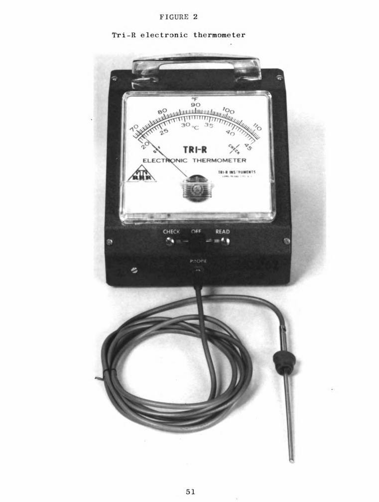
FIGURE 2
Tri-R electronic thermometer
c /
TR "R /("
NIC THERMOME YER
III I DIS “ll-fl“
51

52
FIGURE 3
Leukocyte count after infusion. of 20 ml. of
2% NaCl solution in left rear quarter: cow 591
30r-
28r-
26L
24-
22”
Leukocytes
inMillions
IOi-
1 1 1 1 l l l 1 DJ
0 8 I6 24 32 4O 48 56 64 72
Time in Hours
* Infusion was made 0 hour after immediately making the
1 anknnvfn nnnnf

53
FIGURE 4
Intracisternal temperature after infusion‘ of
20 m1. of 2% NaCl solution in left rear
quarter: cow 591
I02—
IOI
IOO
u.—” 99
L
2
E 98Q)
‘e‘,3 97
B
E 962.
.20
S5 95 F
s'
94 _
93-
92..
l l l J l l l J l
0 8 I6 24 32 4O 48 56 64 72
Time in Hours
‘ Infusion was made at 0 hour immediately after taking the
intracisternal temperature.

54
FIGURE 5
Leukocyte count after infusion"of 10 m1. of
2% NaCl solution in right rear quarter: cow 591
so—
23—
26—-
24—
22—
20—
l8"
Leukocytes
inMillions
3
I
.o.. t
FF.
0 I l l l l 1 l 1 #1
0.8 I6 24 32 4o 43 56 64 72
Time in Hours
‘ Infusion was made at 0 hour after immediately making the
leukocyte count.

55
FIGURE 6
Intracisternal temperature after infusion‘ of
10 ml. of 2% NaCl solution in right rear
quarter: cow 591
I02 -
IOI ~
”30 b
h;
m 99 _
‘-
2
§98~
Q
E
.2 97
B
596
33
.33
0
'g 95 _
E
94_
93 _
92 —
I I 1 I l l 1 1 J O 8 I6 24 32 40 48 56 64 72
Time in Hours
* Infusion was made at 0 hour immediately after taking
intracisternal temperature.
56
FIGURE 7
Rectal temperature following the intracisternal
infusion"of 2% NaCl solution (20 and 10 ml. in
left rear-and right rear quarters respectively):
cow 591
I04 p
u;
3 ICE» -k
2
E :02 -Q)
Q.
E
,2 no: —
E:UIIOCI-
6‘3
J I I I I I I I I
O 8 IS 24 32 4O 48 56 64 72
Time in Hours
* Infusion was made at 0 hour immediately after taking
the rectal temperature.

VI. SUMMARY
For this work, it was found that of the four
disinfectants tested, Novadine was the most satisfactory
for sterilization of the thermometer used for the
determination of mammary intracisternal temperatures.
The mammary intracisternal temperatures of 9 clinically
normal dairy cows were determined by the use of an electronic
thermometer (Tri—R). From repeated observations over a
period of four months it was found that intracisternal
temperatures of mastitis-free, lactating cows varied from
91.5 to 96.6 F. In one lactating cow, intracisternal
temperatures ranged from 92.5 to 98.0 F.
Collaborative data collected during this study
include: rectal temperature, room temperature, California
Mastitis Test, milk leukocyte counts, and bacteriological
examination of milk samples.
The intracisternal temperatures of one cow with
naturally occuring mastitis in the RF quarter were 2 to 3
degrees (F) higher than the non-inflamed quarters.
57

58
Acute mastitis was produced by intramammary injections
of 2% NaCl solution and also by injections of non-hemolytic
Staphylococcus aureus organisms. Pre- and post-injection
intracisternal temperatures were taken. When the 2% NaCl
solution was the mastitis inducing agent intracisternal
temperatures of the inflamed quarters were 2 to 5 degrees
(I) higher than the pre-injection or control quarter
temperature. When Staphylococcus aureus was the mastitis
inducing agent intracisternal temperatures of the inflamed
quarters were 3 to 7 degrees F higher than pre—injection or
control quarter temperatures.
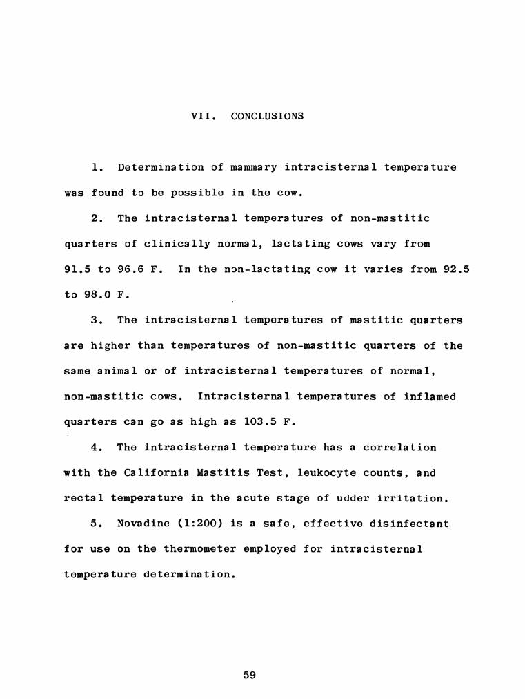
VII. CONCLUSIONS
1. Determination of mammary intracisternal temperature
was found to be possible in the cow.
2. The intracisternal temperatures of non-mastitic
quarters of clinically normal, lactating cows vary from
91.5 to 96.6 F. In the non—lactating cow it varies from 92.5
to 98.0 F.
3. The intracisternal temperatures of mastitic quarters
are higher than temperatures of non—mastitic quarters of the
same animal or of intracisternal temperatures of normal,
non-mastitic cows. Intracisternal temperatures of inflamed
quarters can go as high as 103.5 F.
A 4. The intracisternal temperature has a correlation
with the California Mastitis Test, leukocyte counts, and
rectal temperature in the acute stage of udder irritation.
5. Novadine (1:200) is a safe, effective disinfectant
for use on the thermometer employed for intracisternal
temperature determination.
59

VIII. REFERENCES
Andberg, W. G., and Weirether, F. J. "The effect of
chemotherapeutic agents on the normal bovine
mammary glad 1. The effect of Novoxil."
Am. J. Vet. Res., 4, (1943) 134-142.
Anderson, J. F., and McClintic, T. B. "A method for
bacteriological standardization of disinfectants."
J. Inf. Dis., 8, (1911) 1—26.
Blackburn, P. S., Laing, C. M., and Malcolm, D. F.
"A comparison of the diagnostic value of the total
and differential cell counts of bovine milk."
J. Dairy Res., 8, (1955) 37-42.
Bryan, C. S. "The microscopic diagnosis of infectious
mastitis. Vet. Med., 30 (1935) 149—155.
Bryan, C. S. "The microscopic detection of bacterial
defects of milk." Vet. Med., 36 (1941) 415-419.
Bryan, C. S. "Dairy Bacteriology and Public Health." Burges
Publishing Co., Minneapolis, Minn., 1948.
Bryan, C. S., Brinker, W. 0., Sales, E. K., Young, F. W.,
Grafton, T. S., and Hutton, J. P. "A Liquid
Germicidal Detergent in Veterinary Surgery."
Vet. Med., 8 (1948) 324-329.
Bryan, C. S. and Brown, R. "Six months of daily mastitis
testing of an infected and non-infected cows."
Mich. Agr. Expt. Sta. Quarterly Bull., 32 (1950)
322-327.
Cappell, D. F. Muirs Text Book of Pathology. 7th ed.
Edward Arnold (publishing) Ltd., London, 1958.
Carpenter, C. M. "Experimental Production of Bovine Mastitis
with Streptococci and Other Bacteria." J. Inf. Dis.,
31 (1922) 1-9. '
60
61
Carpenter, C. M. "The bacterial content of milk or
inflammatory exudates from bovine mastitis."
J.A.V.M.A., 67 (1925) 317-323.
Chapman, G. H. "The coagulation of plasma by Staph.
Chapman, G. H., Berens, C. "The differentiation of
pathogenic staphylococci from nonpathogenic types."
Chapman, G. H., Berens, C., Peters, A. and Curcio, L.
"Coagulase and hemolysin tests as measures of the
pathogenicity of staphylococci." J. Bact., 28,
(1934) 343-363.
Cowan, S. T. "The classification of staphylococci by
precipitation and biological reactions." J. Path.
and Bact., 46 (1938) 31-45.
Derbyshire, J. B. "The pathology of experimental staph.
mastitis in the goat." J. Comp. Path. and Therap.,
68 (1958) 449-454.
Deubler, M. J. and Cole, E. J. "Studies on hicrococcal
mastitis in individual herd." Vet. Med., 51 (1956)
111-113.
Diggs, L. W., Dorothsturn and Ann Bell. "The morphology of
blood cells". Abbott laboratories, N. Chicago, IAl.
(1954).
Drury, A. R. "Evaluation of Neomysin Sulphate in the treat-
ment of bovine mastitis." M.S. Thesis, Mich. State
University (1952).
Drury, A. R. "Practical aspects of diagnosing mastitis."
M. S. U. Vet., 12 (1952) 104-108.
Drury, A. R., and Reed, G. L. "A herd irritation index using
the CMT". Vet. Med., 56 (1961) 147-152.
Drury, A. R., and Sanclemente, C. L. "Response of the bovine
udder to artificial introduction of Staphylococcus
aureus Phage Type 80/81, and subsequent treatment
with various drugs." Am. J. Vet. Res., 14 (1962)
262-266.
62
Dukes, H. H. "The physiology of domestic animals." Comstock
Publishing Co. Inc., Ithaca, N. Y. (1942).
Du Bois, F. Eugene. "Fever and the regulation of body
temperature." Charles C. Thomas, publisher,
Springfield, Ill. (1948).
Edwards, S. J., and Rippon, John E. "The characters and
distribution of certain staphylococci, pathogenic
for the bovine udder." J. Comp. Path. and Therap.,
67 (1957) 111-125.
Elek, S. D., and Leavy, E. "Distribution of hemolysin in
pathogenic and nonpathogenic staphylococci." J. Path.
and Bact., 62 (1950) 541-554.
Evans, A. C. "The bacteria of milk freshly drawn from normal
udders." J. Inf. Dis., 18 (1916) 437-476.
Fairbrother, R. W. "Coagulase production as a criterion for
the classification of staphylococci." J. Path. and
Bact., 50 (1940) 83-88.
Filion, P. R. "Pathology of mastitis." Canadian Comp. Med.,
12 (1948) 301-305.
Force, J. N., and Kerr, W. J. "The efficient disinfection
of hospital, clinical thermometers." Modern Hospital,
15 (1920) 150-158.
Gershenfeld, L., Greene, A., and Witlin, B. "Disinfection of
clinical thermometers." Journal of American Pharm.
Ass., 40 (1951) 457-460.
Gibbons, W. J. "The histophthology of mastitis, A preliminary
report." Cornell Vet., 28 (1938) 240-249.
Grant, R. T., and Wood, J. B. "Histamine and Leukocytes."
J. Path. and Bact., 31 (1928) 1-7.
Gwatkin, R., and Hadwen, S. "Staphylococci with infections
of the bovine udder." Can. J. Pub. Health, 27 (1936)
391-400.
Hanes, M. E. "The Use Dilution Method of testing Disinfectants."
M. S. Thesis, Michigan State College (1945).
63
Howard, Flory. "General pathology." 2nd ed., W. B. Saunders
Publishing Co., Phil. (1958).
Jasmin, G. Robert A. "The mechanism of inflammation. An
international symposium." ACTA Inc. Medical
publishers, Montreal, Canada (1953).
Johnston, T. "Anatomical and experimental study of the teat
of cow with particular reference to streptococcic
mastitis." J. Comp. Path., 51 (1938) 69-77. -
Jones, F. S. "Studies on bovine mastitis. III. Infection
of the udder with micrococci and other micro-
organisms." J. Expt. Med., 28 (1918) 721-733.
Jose, Britto Figueiredo. "A comparison of the California
Mastitis Test with the other commonly employed
diagnostic tests." M.S. Thesis, Mich. State
University (1957).
Klimmer, M. "Acute Infections Mastitis." 11th International
Vet. Congress Reports, 3 (1930) 422-440.
Leavitt, A. H. "Comparative study of the use-dilution
method and the F.D.A. Phenol Coefficient Method of
testing Veterinary Disinfectants." M.S. Thesis,
Mich. State University (1946).
Liquid Germicidal Detergent. Parke, Davis and Co., Detroit,
Mich.
Little, R. B., and Jones, F. S. "Correlation between certain
properties of milk and the type of inflammation in
acute mastitis." J.A.V.M.A., 82 (1933) 818-825.
Little, R. B., and Foley, E. J. "Staphylococci associated
with mastitis." J.A.V.M.A., 87 (1935) 637-649.
Little, R. B., and Lpastridge, W. N. "Bovine Mastitis." lst
ed., McGraw Hill Book Co., Inc., N. Y. (1946).
Malik, B. S. "Studies on Staphylococci with particular
reference to strains of bovine udder." M.V.Sc.
Thesis, Agra University (1959).
Mallmann, W. L. "Evaluation of Disinfectants." Soap and
Sanitary Chemicals (1944).
64
Mallmann, W. L., and Hanes, M. "The Use-Dilution Method of
Testing Disinfectants." J. Bact., 49 (1945) 526.
Mallmann, W. L., and Leavitt, A. H. "A Comparative Study of
the Use-Dilution Method and the F.D.A. Phenol
Coefficient Method Applied to Some Veterinary
Disinfectants." Am. J. Vet. Res., 9 (1948) 104-108.
McCulloch, E. C. "Disinfection and Sterilization." 2nd ed.,
Lea and Febiger, Phi1., (1945).
McDiarmid, A. "A carriage of Staphylococcus aureus in the
milk and on the teats of normal cows." Vet. Rec.,
59 (1947) 11-12.
McDonald, 1., and White, F. "Some observations on an out-
break of staphylococcic mastitis in a herd." J. Comp.
Path. Ther., 71 (1961) 159-170.
Menkin, Valy. "Mechanism of inflammation." Arch. of Path.,
24 (1937) 65—81.
Menkin, Valy. "Chemical basis of injury in inflammation."
Arch. of Path., 36 (1943) 269-288.
Menkin, Valy. "Dynamics of inflammation." The Macmillan Co.
(1940).
Menkin, Valy. "Newer concepts of inflammation." lst ed.,
Charles C. Thomas, Publisher, Sprinfield, Ill. (1950).
Menkin, Valy, and Warner, R. C. "Studies on inflammation.
XIII. Carbohydrate Metabolism, Local Acidosis, and
the Cytological Picture in Inflammation." Am. J.
Path., 13 (1937) 25-41.
Miller, W. T., and Heishman, J. O. "Staphylococcic mastitis.
1. Artificial exposure of the bovine udder with
Staphylococcus aureus." Am. J. Vet. Res., 4 (1943)
318-324.
Minett, F. C. "Studies on bovine mastitis. XII. Mastitis
due to staphylococci." J. Comp. Path. and Therap.,
50 (1937 A) 101-121.
65
Minett, F. C. "Staphylococcus antitoxin in the blood and
milk of cows and other animals." J. Comp. Path. and
Therap., 50 (1937 B) 173-190.
Price, P. B. "Reevaluation of ethyl alcohol as a germicide."
Arch. Surg., 60 (1950) 492-502.
Reddish, G. F. "Antiseptics in the hospital pharmacy."
Am. Society of Hospital Pharmacists, 13 (1956) 545-556.
Reddish, G. F. "Antiseptics, disinfectants, fungicide, and
chemical and physical sterilization." 2nd ed., Lea
and Febiger Phil., (1957).
Reid, W. B., and Wilson, J. B. "A study of staphylococci
associated with the bovine udder." Am. J. Vet. Res.,
20 (1959) 825-831.
Ruehle, G.L.A., and Brewer, C. M. "U. 8. Food and Drug
Administration Method of Testing Antiseptics and
Disinfectants." U. S. Dept. Agr. Circ., 198 (1931).
Runnels, R. A. "Animal Pathology." 4th ed., Iowa State
College Press,Ames, Iowa (1946).
Schalm, O. W. "Gangrenous mastitis." Vet. Med., 39 (1944)
279-284.
Schalm, O. W. "Bovine mastitis, and its control program in
California." Canadian Vet. J., 3 (1962) 90-92.
Schalm, O. W., and Woods, G. M. "Micrococcus pyogenes in
bovine milk. 1. Identification." Am. J. Vet. Res.,
14 (1953) 530-533.
Schalm, O. W., Woods, G. M. "Micrococcus pyogenes in bovine
milk. 2. Relationship of shedding characteristics
to occurrence of clinical mastitis." Am. J. Vet.
Res., 14 (1953) 534-538.
Schalm, O. W., and Noorlander, D. 0. "Experiments and
observations leading to development of California
Mastitis Test." J.A.V.M.A., 130 (1957) 199-204.
66
Slanetz, L. W., and Bartley, C. H. "The diagnosis of
staphylococcal mastitis, with special reference to
characteristics of mastitis staphylococci." J. Inf.
Dis., 92 (1953) 139-151.
Solomon, J. J., Sanclemente, C. L., and Drury, A. R.
"Quantitative coagulase activity and Phage patterns
of strains of Staphylococcus aureus isolated from
cows in Michigan." Am. J. Vet. Res., 91 (1961)
975-981.
Standard Methods for the Examination of Dairy Products.
11th ed., American Public Health Association, Inc.,
New York 19, N. Y. (1960).
Velilla, D. M., Faber, J. E., and Pelczer, Jr., M. J. "A
technique for the isolation of coagulase producing
staph. from the milk in bovine mastitis." Am. J.
Vet. Res., 8 (1947) 275-279.
Wolf, E. P. "Experimental studies on inflammation. 11.
Experimental Chemical Inflammation in Vivo." J.
Exptl. Med., 37 (1923) 511-524.
Zebovitz, E., Evans, B., and Niven, C. F. "Tellurite Glycine
Agar--A selective plating medium for the quantitative
detection of coagulase positive staphylococci." J.
Bact., 70 (1955) 686-690.
Zinn, R. D., Anderson, C. R., and Skaggs, J. W. 'Staphylococcic
infections in cattle." J.A.V.M.A., 138 (1961) 382-386.

IX. APPENDIX

TABLE
16
TEMPERATUREAND
MILK
EXAMINATION
DATA
OF
CLINICALLY
NORMAL
COWS
Name
Body
Room
Intracisternal
temp
Leukocytes/
m1.
CMT
and
bacteriology
‘
or
temp
temp
Finmillion
NO.
RF
LF
LR
RR
RF
LF
LR
RR
RF
LF
LR
RR
575
101.0
53.6
95.0
95.0
94.1
94.5
0.15
0.60
.0.10
0.30
2-
2«-
1—
2+
"101.6
61.0
95.0
94.2
96.0
95.0
0.15
0.60
0.60
0.90
2«-
1.-
2i
2+
73
101.4
53.6
93.0
93.0
94.3
93.0
0.60
0.30
0.45
0.07
1-
l.-
2+
1-
"101.2
61.0
94.0
94.0
94.0
94.5
0.20
0.20
0.15
0.20
2..
2..
2+
2-
657
100.8
61.0
93.50
92.0
91.5
92.3
0.20
0.20
0.15
0.30
l.+
l.+
1+
l+
"100.8
76.0
94.4
92.4
92.4
93.0
0.45
0.45
0.30
0.60
1-+
1.+
1+
1+
174
99.2
68.0
94.0
95.0
94.6
96.0
0.20
0.20
0.30
0.45
O-b
0‘+
0+
0+
‘+
ahemolytic
staph.
“-
anon-hemolytic
staph.
68

'5” 3 o '63
