theroleofsyk/card9-coupledc-typelectinreceptorsin
TRANSCRIPT

Hindawi Publishing CorporationClinical and Developmental ImmunologyVolume 2010, Article ID 567571, 9 pagesdoi:10.1155/2010/567571
Review Article
The Role of Syk/CARD9-Coupled C-Type Lectin Receptors inImmunity to Mycobacterium tuberculosis Infections
Mohlopheni Jackson Marakalala,1 Lisa M. Graham,1 and Gordon D. Brown2
1 Institute of Infectious Diseases and Molecular Medicine, Division of Immunology, CLS, University of Cape Town,Cape Town 7925, South Africa
2 Aberdeen Fungal Group, Section of Immunology and Infection, Division of Applied Medicine, Institute of Medical Sciences,University of Aberdeen, Aberdeen AB25 2ZD, UK
Correspondence should be addressed to Gordon D. Brown, [email protected]
Received 1 November 2010; Accepted 16 December 2010
Academic Editor: Taro Kawai
Copyright © 2010 Mohlopheni Jackson Marakalala et al. This is an open access article distributed under the Creative CommonsAttribution License, which permits unrestricted use, distribution, and reproduction in any medium, provided the original work isproperly cited.
There is increasing interest in understanding the mechanisms underlying the interactions that occur between Mycobacteriumtuberculosis and host innate immune cells. These cells express pattern recognition receptors (PRRs) which recognise mycobacterialpathogen-associated molecular patterns (PAMPs) and which can influence the host immune response to the infection. Althoughmany of the PRRs appear to be redundant in the control of M. tuberculosis infection in vivo, recent discoveries have revealed a key,nonredundant, role of the Syk/CARD9 signalling pathway in antimycobacterial immunity. Here we review these discoveries, aswell as recent data investigating the role of the Syk/CARD9-coupled PRRs that have been implicated in mycobacterial recognition,including Dectin-1 and Mincle.
1. Introduction
Despite over a century of research, tuberculosis (TB) remainsthe deadliest bacterial infection causing about 1.6 milliondeaths per year worldwide [1]. In most cases of TBinfection, the immune response is successful in containingMycobacterium tuberculosis (MTB), which then remains in adormant, nonreplicative state, termed latency. This clinicallatency often extends for the lifetime of the individual butreactivation of the infection can occur due to perturbationsof the immune system [2]. An estimated one-third of theworld’s population are latently infected with MTB, andabout 10% of these people are likely to become sick withactive TB in their lifetime. The emergence of new resistantstrains as well as coinfection with HIV also provides morechallenges in the treatment of TB, making it a globalproblem that needs urgent attention [1]. Thus developmentof new antitubercular drugs and a more effective vaccine isa priority; both of which depend, in large part, on obtaininga better understanding of interaction between the host andthis pathogen.
The immune system of humans and other mammalsis comprised of two interrelated components, the innateand adaptive response, both of which are required for theresolution of the infection. The innate immune system, inparticular, consists of phagocytes such as macrophages anddendritic cells which are involved in the initial capture of thebacteria, as well as the presentation of microbial antigens.Serving as first line of defence, these innate cells expressa variety of evolutionarily conserved pattern recognitionreceptors (PRRs), which are involved in the recognition ofpathogen-associated molecular patterns (PAMPs) [3].
One of the first events after the inhaled mycobacteriahave reached the alveolar space is its recognition by alveolarmacrophage PRRs. The role of PRRs, such as the toll-like receptors (TLRs) and NOD-like receptors (NLRs), inantimycobacterial immunity has been a focus of considerableinterest [4, 5]. TLR-2, TLR-4, for example, were shown toplay a role in the long-term control of M. tuberculosis [4].Furthermore, TLR-9 was reported to be important in theinduction of protective responses, and in cooperation withTLR-2, to mediate resistance to MTB infection [6]. Studies
2 Clinical and Developmental Immunology
using MyD88-deficient mice also demonstrated a role of thisdownstream adaptor in the acute control of MTB infection[7]. However, recent data have shown that MyD88 andTLR-2, -4, and -9 are dispensable for the induction of Tcell-mediated adaptive immunity to MTB infection [8, 9].TLR-2, -4, and -9-deficient mice were also not impaired intheir macrophage effector mechanisms and displayed normalcontrol of MTB replication [8]. Thus, the TLRs, even incombination, appear to be redundant for the control of MTBinfection.
Nucleotide-binding oligomerization domain proteins(NODs) are modular cytoplasmic proteins that recognisecomponents of peptidoglycan [4]. NOD2 is the wideststudied NLR in the MTB infections and has been shown tosynergize with TLR-2 for the production of proinflammatorycytokines in response to MTB. Gandotra et al. have reportedthat NOD2-deficient macrophages and DCs were impairedin the production of proinflammatory cytokines in responseto live MTB. However this NLR seems to be dispensablein the in vivo control of the infection, as NOD2-deficientand the wild-type control mice show similar levels ofsusceptibility when infected with virulent MTB [10]. Morework is needed to understand the role of other NLRs inantimycobacterial infections.
Other PRRs have also been implicated in the immuneresponse to M. tuberculosis, including the mannose receptor(MR), complement receptor 3 (CR3), dendritic cell-specificintercellular adhesion molecule 3 (ICAM-3)-grabbing non-integrin (DC-SIGN), class A scavenger receptor, mannosebinding lectin (MBL), and surfactant protein A (Sp-A) [11,12]. Nonetheless, each of these receptors also seem to befunctionally redundant, as it has been shown that singledeficiencies in PRRs such as Scavenger receptor A, CD36,MARCO, mannose receptor, and SIGNR1 do not impair thecontrol of M. tuberculosis in vivo [13]. Thus, we and othershave speculated that mycobacteria are likely to simultane-ously engage multiple receptors and that deficiency of onereceptor can be compensated by another [5, 13]. However,recent discoveries suggest that the Syk/CARD9 pathway playsa nonredundant role in antimycobacterial immunity, andtwo C-type lectin receptors (CLRs) recognising mycobacteriathat utilise this pathway have been identified. Here we reviewour current understanding of the Syk/CARD9 pathway, aswell as the two CLRs (macrophage-inducible C-type lectin(Mincle) and Dectin-1), with particular emphasis on theirroles in antimycobacterial immunity.
2. CARD9 (Caspase RecruitmentDomain-Containing Protein 9): An AdaptorMolecule Involved in ITAM-CoupledReceptor Signalling
CARD9 is an adaptor molecule that contains N-terminal cas-pase recruitment domain (CARD) and a C-terminal coiled-coil region that is important for protein oligomerization[14]. Structurally, this adaptor is related to CARMA (CARDMAGUK) family of proteins, which include CARMA1(CARD11), CARMA2 (CARD10), and CARMA3 (CARD13)
[15]. Originally discovered through a database search ofCARD-containing proteins, CARD9 is expressed in varioustissues including the lung, liver, spleen, placenta, peripheralblood leukocytes, and bone marrow. CARD9 is not expressedin lymph nodes, T cells, or B cells, but is abundantlyexpressed in innate myeloid cells including dendritic cellsand macrophages [14, 16].
This adaptor protein operates downstream of immun-oreceptor tyrosine-based activation motif-(ITAM-) associ-ated PRRs and cooperates with B cell lymphoma 10 (Bcl10)and the paracaspase MALT1 to form a trimolecular complexthat transduces signalling to the canonical NFκB pathway[15]. CARD9 was initially identified as a key transducer ofDectin-1 signalling [17], but more recently this signallingpathway has also been shown to be important for myeloidcell activation via DAP12- and FcRγ-associated receptors[18].
Several murine studies have demonstrated that CARD9plays an essential role in protection against fungal andbacterial pathogens, including C. albicans and Listeria mono-cytogenes [16, 17]. A recent study by Glocker et al. hasdescribed a CARD9 mutation in humans that is associatedwith an increased susceptibility to chronic mucocutaneouscandidiasis [19]. Interestingly, these patients had signifi-cantly reduced numbers of Th17 cells [19], and murinestudies have shown that signalling through the CARD9pathway, from receptors including Dectin-1 (see below)and Dectin-2, is important for the development of thesetypes of adaptive responses [20–22]. Indeed, LeibundGut-Landmann et al. have shown that CARD9 is indispensablefor the induction of Th17 responses in mice infected withC. albicans [20]. Poeck et al. have recently shown that RIG-Iengages CARD9-Bcl10 signalling pathway for the productionof IL-1β in response to RNA viruses [23]. Thus CARD9 is acentral molecule that transduces signals from multiple PRRsto induce immunity to various pathogens.
3. CARD9 and Antimycobacterial Immunity
The essential role of CARD9 control of pulmonary tuber-culosis, at least in mice, was recently demonstrated byDorhoi et al. [24]. These authors found that Card9−/−
mice succumbed rapidly to aerosol infection with M.tuberculosis H37Rv, and, compared with their littermatecontrols, the CARD9-deficient mice had significantly higherpulmonary bacillary burdens and more profound tissuedamage, characterized by acute pneumonia with acceleratedaccumulation of inflammatory cells. There was also evidenceof secondary necrosis in the Card9−/− lung tissue, suggestinga correlation between cell death in lungs of these mice andtheir susceptibility to tuberculosis infection [24].
To investigate the effect of CARD9 deficiency in theactivation of antigen presenting cells (APCs), Dorhoi et al.infected bone-marrow-derived macrophages (BMMø) withMTB H37Rv and analyzed various innate responses. Boththe WT and Card9−/− BMMø produced similar levels ofnitric oxide (NO), and the adaptor deficiency did not havean effect on internalization or killing of MTB after IFN-γ

Clinical and Developmental Immunology 3
activation. The authors also demonstrated that the clearanceof apoptotic cells was also not impaired in Card9−/− BMMø.Furthermore, apoptotic cell death occurred at similar fre-quency in both the WT and Card9−/− cells [24]. However,infected Card9−/− BMMø had significant reductions in thesynthesis of proinflammatory cytokines, TNF, IL-1β, and IL-6, and produced less IL-12 and CCL5, when compared tothe WT cells. The MTB-infected Card9−/− BMDCs were alsoimpaired in their ability to synthesise TNF and IL-1β. Thesedata indicated that CARD9 is critical for activation of innateimmune cells following the recognition of MTB [24].
To characterize the PRRs involved in APC activation,Dorhoi et al. stimulated BMMø and DCs with MTB-derivedPAMPs, including manLam, peptidoglycan (PG), mycolyl-arabinogalactan peptidoglycan (mAGP), and mycobacterialcord factor. The stimulation with manLam and cord factorled to cellular activation, suggesting possible involvementof MR and Mincle [24, 25] (see below). The stimulationof wild-type BMMø and DCs with mAGP triggered therelease of TNF and IL-10, which were abrogated in theCARD9-deficient cells. These results suggested the involve-ment of nucleotide-binding oligomerization domain protein2 (NOD2) in the CARD9-mediated innate response toMTB [24], as mAGP-induced cytokine production has beenpreviously shown to partially depend on this receptor [10].Furthermore, the authors showed that the treatment ofwild-type BMMø with laminarin (a β-glucan antagonistof Dectin-1) and piceatannol (a Syk kinase inhibitor)specifically blocked TNF and IL-6 production, suggestingthat signalling through the Dectin-1/Syk pathway was alsoimportant in the induction of MTB-triggered proinflam-matory responses [24]. Thus, the authors speculated thatduring MTB recognition CARD9 is likely to converge signalsfrom a number of PRRs to mediate innate responses to thispathogen [24].
To characterize the role of CARD9 in adaptive cellresponses, Dorhoi et al. measured various T cell populationsand the recruitment of leukocytes to the lungs followinginfection with MTB. At day 14 postinfection, there wereno differences between WT and Card9−/− mice in therecruitment of T cell subpopulations to the lung. Also,the ratio of CD4+/CD8+ cells in the wild-type and theknockout mice were similar at both days 14 and 28 afterinfection. Furthermore, there were similar frequencies ofCD8+ T cells and FoxP3+ T lymphocytes in both groupsof mice [24]. Thus, CARD9 deficiency does not affect Tcell responses in MTB infection. In addition to T cells, theauthors showed that there was no difference in the frequencyof lung macrophages, DCs, as well as the expression of CD86(an APC-expressed maturation marker) [24].
There was, however, a substantial accumulation of neu-trophils in the lungs of CARD9-deficient mice comparedto the wild-type control mice. The accelerated recruitmentof the polymorphonuclear neutrophils (PMNs) to the lungcorresponded to the disease progression, implicating thesecells in the susceptibility of Card9−/− mice to MTB infec-tion [24]. In an endeavour to determine what triggeredthe neutrophilic inflammation during the infection, theauthors measured chemokine and cytokine levels in serum
and lung homogenates. Intriguingly, MCP-1, KC, and G-CSF were significantly higher in the Card9−/− mice. Inaddition, elevated concentrations of granulocyte-derivedmyeloperoxidase (MPO) were detected in sera of thesemice compared to the wild type control animals. Thus, theauthors conclude that the systemic inflammatory diseasethat leads to tissue damage emanates from G-CSF-mediatedaugmented granulocyte differentiation and the KC-drivenaccelerated recruitment of the neutrophils to the lung [24].Rescue experiments performed by either neutralizing G-CSF or depleting PMN with anti-Gr-1 monoclonal antibodyresulted in the reduction of lung inflammation and pro-longed survival. Furthermore, the authors also demonstratedthat the CARD9-deficient PMNs lost their capacity toproduce IL-10 upon MTB challenge and thus failed to fine-tune the level of inflammation in the lung [24]. Collectively,the data by Dorhoi et al. demonstrates an essential role ofCARD9 signalling in the control of MTB infection [24].
In another recent study, Werninghaus et al. reportedthat the mycobacterial PAMP, trehalose-6,6-dimycolate(TDM, cord factor), and its synthetic analogue trehalose-6,6-dibehenate (TDB) activate antigen presenting cells,macrophages, and DCs, via the Syk-CARD9 pathway toinduce innate responses such as NO production and therelease of cytokines such as TNF, IL-1β, and IL-6 [25]. TDMwas first identified in 1950 by Bloch as a glycolipid thatwas responsible for cording in tubercle bacilli [26], andsix decades later this glycolipid remains one of the widelystudied components of mycobacterial cell membrane mainlydue to its ability to activate the innate immune system inmammals [27]. Werninghaus et al. showed in vivo that theseglycolipids could be used as adjuvants to drive combined Th1and Th17 T cell responses to an MTB subunit vaccine in aCARD9-dependent manner [25]. These responses were alsoFcRγ-dependent and subsequently identified to be mediatedthrough Mincle (see below).
4. Dectin-1 Structure, Expression,and Signalling
Dectin-1 is a glycosylated type II transmembrane receptorpossessing a single extracellular C-type lectin-like domain(CTLD) and a cytoplasmic signalling domain which isinvolved in cellular activation [28, 29]. This receptor wasfirst discovered through subtractive cDNA cloning frommurine dendritic cells (DCs) [30] and later identified andcharacterized in humans [31, 32]. In both of these organisms,alternative splicing of Dectin-1 results in two major isoformsand a number of minor isoforms, with the major onesdiffering in the presence or absence of the stalk region[31, 33]. Dectin-1 is predominantly expressed on myeloidcells, including dendritic cells, monocytes/macrophages, andneutrophils, as well as a subset of T cells [34]. This PRRhas been shown to be expressed at high levels at portalsof pathogen entry, including the lung [34, 35]. Dectin-1is a major β-glucan receptor on leukocytes [36] and canmediate a variety of cellular responses including endocytosis,phagocytosis, the respiratory burst, DC maturation, and the

4 Clinical and Developmental Immunology
Y YP
Syk
Src Src
CARD9
MALT1
Bcl10
MyD88
IRA
K4
TRAF-6
B-glucans/mycobacterial ligands
TL
R2
Dectin-1
Syk
Mincle
P
Mycobacterial cord factor
Th1/Th17 immunity
CD4+ T cell
APC
P Y
Y P
TDM/TDB
FcRγ
ITAM
NFκB
Inflammatory cytokine responses
TNF-α, IL-6, IL-12
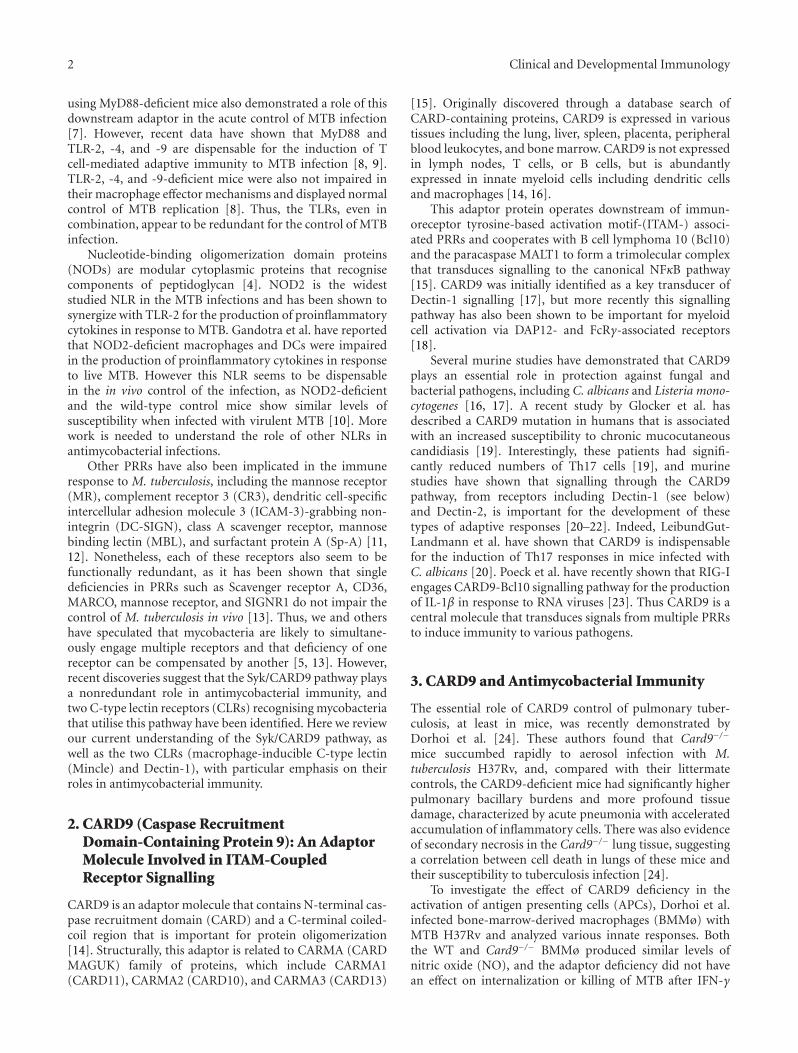
Figure 1: Mincle- and Dectin-1-mediated signalling in response to mycobacteria. Upon recognition of their mycobacterial ligands, Dectin-1and Mincle signal via the Syk/CARD9 pathway for the production of inflammatory cytokines and the induction of Th1 and Th17 responses.There is also evidence that Dectin-1 signalling can cooperate with TLR2 to induce protective immunity.
induction of cytokine/chemokine production [28, 29]. Likethe TLRs, Dectin-1 can directly induce the development ofadaptive immunity, particularly Th17 and Th1 responses[20, 37].
Increasing data from in vivo studies, although notentirely consistent, have demonstrated an important rolefor Dectin-1 in antifungal immunity [38, 39]. This receptorhas been shown to mediate recognition of several fungalpathogens and to play an essential role in mouse models ofinfection with Candida, Aspergillus, and Pneumocystis [40–43]. In humans, deficiencies in Dectin-1 and in CARD9 haverecently been linked to susceptibility to fungal infections[19, 44].
Dectin-1 signalling is mediated by the ITAM-like motifalso known as hemITAM. The ITAM motifs typically containan amino sequence comprising of a duplicate YxxL/I motif(YxxL/Ix6−12YxxL/I), where Y is tyrosine, L is Leucine,I is Isoleucine, and x denotes any amino acid [45, 46].Upon ligand engagement, the ITAM-like motif of Dectin-1becomes tyrosine phosphorylated by Src kinases, providinga docking site for Syk kinase, which links the Dectin-1 signalling to NF-κB activation via the CARD9-Bcl10-MALT1 complex [17, 18]. In addition to the Syk-pathwayDectin-1 can signal via a second pathway mediated by theserine-threonine kinase Raf-1, which integrates with the Sykpathway at the point of NF-κB activation [37]. Furthermore,Dectin-1 can also trigger the activation of NFAT, whichregulates the induction of early growth response (Egr) family
transcription factors, Egr-2 and Egr-3 [47]. This activationhas been shown to occur through a pathway that requiresphospholipase C-γ2 (PLC-γ2) [48]. Signalling from Dectin-1 also interacts with pathways induced by other receptors,such as the TLRs, resulting in the synergistic induction ofcytokines including TNF, IL-10, and IL-23 [49–51].
5. Dectin-1 Recognition of Mycobacteria
Although the involvement of Dectin-1 in antifungal immu-nity has been extensively characterized [29], the role ofthis receptor in the control of mycobacterial infectionsremains poorly understood. Studies from a number oflaboratories have implicated the role of Dectin-1 in responseto mycobacterial infections, although the ligand involved isstill unknown.
In the initial study, Yadav and Schorey demonstratedthat blocking Dectin-1 function with monoclonal antibodiesinhibited the production of TNF-α by bone-marrow-derivedmurine macrophages (BMMø) infected with M. smegmatis[52]. Cytokine production was also impaired in the TLR2-deficient macrophages, indicating the requirement of thistoll-like receptor for the Dectin-1-facilitated proinflamma-tory responses. Dectin-1 was also shown to be required forthe production of RANTES, IL-6, and G-CSF in BMMøinfected with nonpathogenic strains such as attenuated M.bovis and avirulent M. tuberculosis H37Ra (see Figure 1).

Clinical and Developmental Immunology 5
Interestingly, Dectin-1 was not required for the inductionof macrophage proinflammatory responses upon infectionwith virulent M. tuberculosis strain, H37Rv [52]. The authorsargued that the minimal macrophage proinflammatoryresponse induced by the virulent mycobacterium might bedue to limited engagement of Dectin-1 by this strain [52].However, in other studies (see below) Dectin-1 was found tointeract with virulent mycobacteria.
Rothfuchs et al. subsequently showed that Dectin-1is important for IL-12p40 production in murine splenicdendritic cells (SpDCs) infected with MTB [53]. The authorsdemonstrated that blockade of Dectin-1 with laminarinresulted in the reduction of IL-12p40 production by thesecells. In addition, the SpDCs from Dectin-1−/−chimeric micedisplayed reduced levels of IL-12p40 when compared to thecells from the WT chimeras following stimulation with liveMTB or M. bovis. Furthermore, the production of IL-12p40by the MTB-stimulated WT SpDCs was impaired by theinhibition of Syk kinase with piceatannol [53]. However, inthese experiments TLR2 deficiency had no effect. Rothfuchset al. further showed that a soluble Dectin-1-Fc fusionprotein [54] could bind to M. bovis BCG in a laminarin-inhibitable manner [53].
Using the nontuberculous mycobacterium, M. abscessus,Shin et al. demonstrated that the interaction of Dectin-1 andTLR2 is required for the efficient murine macrophage uptakeof this organism and subsequent Syk activation leading tothe production of TNF-α, IL-12p40, and IL-6 [55] (seeFigure 1). The observations on TLR2-Dectin-1 cooperationare in agreement with the work by Yadav and Schorey [52],but differ with that of Rothfuchs et al. [53]; however, itshould be noted that each of these studies focussed ondifferent cell types and mycobacterial species and/or strains.Shin et al. also demonstrated the importance of Dectin-1/Sykpathway in the production of ROS by BMMø infected withM. abscessus [55].
More recently, the role of Dectin-1 in MTB-inducedinnate immune responses has been examined in humancells. Lee et al. have shown that the expression of Dectin-1 mRNA and protein in A549 airway epithelial cells wasMTB-inducible in a TLR2-dependent manner. These authorsalso demonstrated the role of Dectin-1 in the production ofROS, proinflammatory cytokines, and antimicrobial activityduring the intracellular growth of MTB in these cells [56].
In another study, Van de Veerdonk et al. showed thatDectin-1 and TLR4 are the key receptors for the induction ofM. tuberculosis-triggered IL-17A responses in human PBMCs[57]. Furthermore, these authors demonstrated that theblockade of IL-1β signalling with IL-1RA resulted in thesignificant decrease of IL-17A production by the PBMCsinfected with MTB. van de Veerdonk et al. concluded thatthe endogenous IL-1 pathway plays a central role in theinduction of the MTB-induced Th17 response in humanPBMCs [57].
Using human monocyte-derived DCs, Zenaro et al. haveshown that stimulation of these cells with MTB leads tothe Dectin-1-dependent production of TNF-α, IL-6, IL-1β,and IL-23, enabling DCs to “instruct” CD4+ lymphocytes toproduce IL-17 and IFN-γ. The authors further investigated
the effect of other receptors on these Dectin-1-dependentresponses and demonstrated that stimulation of the DCswith biglycan and ManLAM (ligands of DC-SIGN and MR,resp.) increased MTB-triggered T-cell production of IFN-γbut resulted in the reduced amounts of IL-17. The authorsconcluded that Dectin-1 interaction with MTB-stimulatedDCs induces Th1 and Th17 responses and that the DC-SIGNand MR engagement with DCs blocks the Dectin-1-triggeredmechanisms, therefore inhibiting Th17 and favouring Th1responses [58].
To explore the role of this receptor in response to M.tuberculosis in vivo, we characterized the effect of Dectin-1-deficiency in mice following aerosol infection with M.tuberculosis H37Rv (MJM and GDB, paper in press). Wefound a significant and reproducible reduction (∼0.5 log)in the pulmonary bacilli burdens in the Dectin-1-deficientmice compared to the wild-type animals, but this did notcorrelate with any changes in pulmonary pathology, cytokineproduction, and ability to resist infection. Thus these datasuggest that Dectin-1 plays a minor role in antimycobacterialimmunity in vivo, at least in murine models.
More work is still required to understand the exactrole of this receptor in antimycobacterial immunity, andin particular, the mycobacterial ligand(s) recognised by thisreceptor. Mycobacteria do not possess β-glucans, so it is likelythat Dectin-1 recognises a novel ligand on these pathogens.The role of Dectin-1 in human mycobacterial infections alsowarrants further investigation.
6. Mincle: An ITAM-Coupled FcRγ-DependentSignalling Receptor
Mincle is a type II C-type lectin belonging to the “Dectin-2 cluster” of receptors [59, 60]. Also known as Clec4e andClecsf9, Mincle was first identified by Matsumoto et al. as aprotein whose expression was induced by lipopolysaccharide(LPS) [61]. The gene expression of Mincle was also shownto be induced in peritoneal macrophages by several proin-flammatory cytokines, including IFN-γ, TNF-α, and IL-6[61]. However, Mincle expression was demonstrated to beseverely impaired in response to these stimuli in the NF-IL-6-deficient macrophages indicating that Mincle is a transcrip-tional target for NF-IL-6 [61]. Like other receptors in theDectin-2 cluster, particularly Dectin-2 and DCAR, Mincle isexpressed predominantly on cells of myeloid lineage, includ-ing dendritic cells and macrophages [59, 61]. This receptoris also expressed on B cells as well as on microglia in thebrain [59, 62]. Structurally, Mincle contains a single extra-cellular carbohydrate recognition domain, a transmembranedomain, and a short cytoplasmic domain without a knownsignalling motif [61]. However, typical of the Dectin-2 clusterof receptors, Mincle possesses a positively charged residuenear the transmembrane region through which it associateswith the adaptor FcRγ to trigger intracellular signalling viaSyk/CARD9 pathway [63, 64]. A report by Yamasaki et al.has demonstrated that cross-linking Mincle on the surfaceof peritoneal macrophages resulted in the production ofproinflammatory cytokines which were abrogated in FcRγ-deficient cells as well as CARD9-deficient cells [64].

6 Clinical and Developmental Immunology
Mincle can act as a receptor for both endogenous andexogenous ligands, having been shown to recognise PAMPsfrom a number of fungal pathogens, including C. albicans,Saccharomyces cerevisiae, and Malassezia species [65–67].Specifically, Wells et al. reported that Mincle recognises C.albicans and that although this receptor did not act as aphagocytic receptor, it played a role in TNFα productionin RAW264.7 and BMMøs in response to this fungus.Furthermore, they demonstrated that Mincle-deficient micedisplayed a significantly greater fungal burden in the kidneyscompared to wild-type mice during systemic candidiasis[66]. Bugarcic et al. further demonstrated that recombinantCRDs of both human and mouse Mincle could recognise C.albicans [65].
In contrast, Yamasaki et al. reported that C. albicanswas not recognised by Mincle using a cell-based reportersystem, but rather that the receptor specifically recognisedMalassezia species (a pathogen associated with skin diseasessuch as tinea versicolor, folliculitis, and atopic dermatitis)[67]. However, the strains of Candida screened were differentfrom those reported by Wells et al. [66], and Yamasakiand coworkers have suggested that Mincle may distinguishbetween different strains of C. albicans [67]. Recognition ofMalassezia by Mincle was shown to require the mannosebinding EPN-motif in the receptor’s CRD, as well as Ca2+,and α-mannose was identified as the ligand of this receptor[67]. Stimulation of BMMø with Malassezia resulted in theproduction of MIP-2, KC, IL-10, and TNF-α, which wassignificantly reduced in the Mincle-deficient macrophages[67]. Additionally, intraperitoneal injection of this fungusresulted in impaired IL-6 and TNFα production and neu-trophil infiltration in Mincle-deficient mice compared towild-type mice [67]
In addition to the role in fungal recognition, Yamasakiet al. have demonstrated that Mincle senses necrosis. Usingan NFAT-GFP-based reporter system the authors found thatGFP expression was greatly increased in the presence ofnecrotic cells, as well as with supernatants from necrotic cellsand lysates generated from normal cells. Mutation of theEPN motif in the CRD did not reduce recognition of thedead cells, suggesting that Mincle recognised the endogenousligand in a carbohydrate independent manner. The authorssubsequently identified spliceosome-associated protein 130(SAP130), a preformed soluble factor released upon necrosis,as a ligand for this receptor. Stimulation of peritonealmacrophages or Mincle expressing T cell hybridomas withSAP130 resulted in MIP-2 and IL-2 production, respec-tively. In vivo, the authors demonstrated that inflammatoryresponses induced by this PRR promoted the infiltration ofneutrophils to the site of necrosis, induced either by wholebody irradiation or by peritoneal injection of necrotic cells[64].
7. Mincle-Mediated Immunity to Mycobacteria
In addition to fungi, Mincle can recognise MTB, an activitywhich requires the EPN motif within the carbohydraterecognition domain of this receptor [63]. To identify the
specific ligand for this receptor in mycobacteria, Ishikawaet al. analyzed various components of M. smegmatis lipidextracts using chromatographic techniques. These screeningexperiments revealed TDM as the mycobacterial ligandfor Mincle. TDM is made up of a trehalose moiety andtwo mycolate chains [68], components which could notindividually activate Mincle-expressing cells, indicating thata combination of both the sugar and the lipid moieties ofthis glycolipid are important for its interaction with Mincle[63]. Another independent report by Schoenen et al. hasconfirmed Mincle as the major receptor of TDM [69].
As discussed above, the mycobacterial cord factor TDMand its synthetic analogue TDB can activate the Syk-CARD9-Bcl10 pathway and induce antimycobacterial immunity[25]. Mincle was identified as the main receptor initiatingthis signalling pathway and driving the TDM/TDB-inducedantimycobacterial immunity [63, 69]. Loss of Mincle resultedin greatly reduced production of inflammatory cytokines andnitric oxide in vitro [63, 69]. Further in vitro analysis alsodemonstrated that the TDM/TDB-induced expression of G-CSF and IL-1β in BMMø was abrogated in the FcRγ−/−
cells, confirming the involvement of the FcRγ adaptorchain in Mincle-mediated signalling [69]. In vivo, Mincle-deficient and FcRγ-deficient mice had decreased TDM-induced IL-6 and TNF production in the sera [69]. Theseresults indicate the critical roles of Mincle and FcRγ inthe innate immune responses induced by the mycobacterialglycolipids.
To investigate the role of Mincle in driving TDB-induced cellular immunity, Schoenen et al. analysed T cellresponses in the draining lymph nodes of wild-type andMincle-deficient mice following vaccination with the subunitvaccine H1 (an MTB fusion protein of Ag85B and ESAT-6). Compared to the wild-type mice, which produced robustamounts of IFN-γ and IL-17, there were significantly reducedT cell numbers in the lymph nodes of Mincle-deficientmice when TDB was used as an adjuvant, indicating thatMincle is responsible for the Th1 and Th17 responsesinduced by this glycolipid [69]. The T cell responses werealso FcRγ dependent [69]. Thus, the Mincle-FcRγ signallingpathway is crucial in the induction of Th1 and Th17protective immunity that is triggered by the mycobacterialglycolipids in this setting (see Figure 1). Collectively, thesedata suggest that Mincle is a key receptor involved in theinduction of the CARD9 signalling pathway in response toMTB.
8. SIGNR3 in Host Response toM. tuberculosis Infection
Other Syk-dependent receptors have been implicated in thecontrol of M. tuberculosis infection. Recently a study byTanne et al. reported that SIGNR3, a murine homologueof DC-SIGN, that signals via intracellular hemITAM isimportant for early immune response to M. tuberculosisinfection [70]. The authors demonstrated that SIGNR3 candirectly interact with live M. tuberculosis and promote theability of fibroblasts to mediate endocytosis and to take

Clinical and Developmental Immunology 7
up ManLAM [70]. To investigate the effects of SIGNR3deficiency on host resistance to mycobacterial infection,the authors infected SIGNR3 knockout and wild-type micewith M. tuberculosis and compared their ability to controlthe infection. Although the SIGNR3-deficient mice hadsignificantly higher amounts of the lung bacilli, their abilityto mount Th1, Th2, Tc1, and Th17 responses was similar tothe wild-type mice. In addition, there were no differencesin the formation of granulomatous lesions and the survivalpatterns between the SIGNR3-deficient and the control mice[70]. These results indicated that SIGNR3 is important inearly host resistance to M. tuberculosis but redundant forthe establishment of long-term defence against the infection[70].
Tanne et al. also demonstrated that stimulation of theSIGNR3-expressing macrophages with either ManLAM orlive M. tuberculosis resulted in the production of highamounts of IL-6 and TNF in comparison with the controlcells [70]. Specific inhibition of Syk by piceatannol abolishedSIGNR3 signalling and the cytokine production in a dosedependent manner, allowing the authors to conclude thatthe SIGNR3-mediated production of IL-6 and TNF occurredin a Syk-dependent manner [70]. Conclusively, the work byTanne et al. provided evidence that the Syk-coupled CLRSIGNR3 plays a role in early host resistance to M. tuberculosisin mice [70].
Because of the differences between SIGNR3 and therelated human molecule, DC-SIGN, it is difficult to speculateon the implication of the current murine SIGNR3 data inthe human infections. DC-SIGN utilizes a different signallingmechanism from the mouse homologue; it does not requireSyk kinase for signalling [71], but its interaction withManLAM leads to the activation of serine/threonine kinaseRaf-1 [72]. In vitro work has shown that DC-SIGN canbe exploited by M. tuberculosis to evade the host immunesurveillance [73]. Furthermore, a clinical study investigatingeffects of polymorphisms in the gene of DC-SIGN hassuggested that decreased level of DC-SIGN is associated withincreased protection against TB [74].
9. Concluding Remarks
Many PRRs that recognise mycobacteria have been identi-fied, but those studied to date appear to be largely redundantwhen examined in vivo. The identification of the Mincleand the CARD9 pathway provides the first evidence foran essential nonredundant signalling that is crucial forantimycobacterial immunity. However, more work is neededto determine if Mincle promotes protective or nonprotectiveresponses during Mtb infection in vivo. Furthermore, giventhe number of PRRs that can activate the Syk/CARD9pathway it is likely that there are other receptors feedinginto this pathway. Special attention should be given to FcRγ-associated PRRs, such as Dectin-2 which also recognise MTB[75]. Identifying these receptors, and understanding theirroles during infection, will be critical if we wish to use thisinformation for the development of novel adjuvants and/orvaccines.
Conflict of Interests
The authors declare no conflict of interests.
Acknowledgments
This work was supported in part by the National ResearchFoundation, Medical Research Council of South Africa, theUniversity of Cape Town, and the Wellcome Trust. M. J.Marakalala is a Sydney Brenner Postdoctoral Fellow at theUniversity of Cape Town.
References
[1] WHO, “Tuberculosis,” Fact Sheet 104, WHO, Geneva, Switzer-land, 2010.
[2] J. L. Flynn and J. Chan, “Tuberculosis: latency and reactiva-tion,” Infection and Immunity, vol. 69, no. 7, pp. 4195–4201,2001.
[3] C. A. Janeway and R. Medzhitov, “Innate immune recogni-tion,” Annual Review of Immunology, vol. 20, pp. 197–216,2002.
[4] E. K. Jo, “Mycobacterial interaction with innate receptors:TLRs, C-type lectins, and NLRs,” Current Opinion in InfectiousDiseases, vol. 21, no. 3, pp. 279–286, 2008.
[5] G. Schafer, M. Jacobs, R. J. Wilkinson, and G. D. Brown,“Non-opsonic recognition of mycobacterium tuberculosis byphagocytes,” Journal of Innate Immunity, vol. 1, no. 3, pp. 231–243, 2009.
[6] A. Bafica, C. A. Scanga, C. G. Feng, C. Leifer, A. Cheever,and A. Sher, “TLR9 regulates Th1 responses and cooperateswith TLR2 in mediating optimal resistance to Mycobacteriumtuberculosis,” Journal of Experimental Medicine, vol. 202, no.12, pp. 1715–1724, 2005.
[7] C. M. Fremond, V. Yeremeev, D. M. Nicolle, M. Jacobs, V. F.Quesniaux, and B. Ryffel, “Fatal Mycobacterium tuberculosisinfection despite adaptive immune response in the absence ofMyD88,” Journal of Clinical Investigation, vol. 114, no. 12, pp.1790–1799, 2004.
[8] C. Holscher, N. Reiling, U. E. Schaible et al., “Containment ofaerogenic Mycobacterium tuberculosis infection in mice doesnot require MyD88 adaptor function for TLR2, -4 and -9,”European Journal of Immunology, vol. 38, no. 3, pp. 680–694,2008.
[9] N. Reiling, S. Ehlers, and C. Holscher, “MyDths and un-TOLLed truths: sensor, instructive and effector immunity totuberculosis,” Immunology Letters, vol. 116, no. 1, pp. 15–23,2008.
[10] S. Gandotra, S. Jang, P. J. Murray, P. Salgame, and S.Ehrt, “Nucleotide-binding oligomerization domain protein 2-deficient mice control infection with Mycobacterium tubercu-losis,” Infection and Immunity, vol. 75, no. 11, pp. 5127–5134,2007.
[11] W. R. Berrington and T. R. Hawn, “Mycobacterium tuber-culosis, macrophages, and the innate immune response: doescommon variation matter?” Immunological Reviews, vol. 219,no. 1, pp. 167–186, 2007.
[12] D. S. Korbel, B. E. Schneider, and U. E. Schaible, “Innateimmunity in tuberculosis: myths and truth,” Microbes andInfection, vol. 10, no. 9, pp. 995–1004, 2008.
[13] N. Court, V. Vasseur, R. Vacher et al., “Partial redundancyof the pattern recognition receptors, scavenger receptors, and

8 Clinical and Developmental Immunology
C-type lectins for the long-term control of Mycobacteriumtuberculosis infection,” Journal of Immunology, vol. 184, no.12, pp. 7057–7070, 2010.
[14] J. Bertin, Y. Guo, L. Wang et al., “CARD9 is a novel caspaserecruitment domain-containing protein that interacts withBCL10/CLAP and activates NF-κB,” Journal of BiologicalChemistry, vol. 275, no. 52, pp. 41082–41086, 2000.
[15] J. Ruland, “CARD9 signaling in the innate immune response,”Annals of the New York Academy of Sciences, vol. 1143, pp. 35–44, 2008.
[16] Y. M. S. Hsu, Y. Zhang, Y. You et al., “The adaptor proteinCARD9 is required for innate immune responses to intracellu-lar pathogens,” Nature Immunology, vol. 8, no. 2, pp. 198–205,2007.
[17] O. Gross, A. Gewies, K. Finger et al., “Card9 controls a non-TLR signalling pathway for innate anti-fungal immunity,”Nature, vol. 442, no. 7103, pp. 651–656, 2006.
[18] H. Hara, C. Ishihara, A. Takeuchi et al., “The adaptor proteinCARD9 is essential for the activation of myeloid cells throughITAM-associated and Toll-like receptors,” Nature Immunology,vol. 8, no. 6, pp. 619–629, 2007.
[19] E. O. Glocker, A. Hennigs, M. Nabavi et al., “A homozygousCARD9 mutation in a family with susceptibility to fungalinfections,” New England Journal of Medicine, vol. 361, no. 18,pp. 1727–1735, 2009.
[20] S. LeibundGut-Landmann, O. Groß, M. J. Robinson et al.,“Syk- and CARD9-dependent coupling of innate immunity tothe induction of T helper cells that produce interleukin 17,”Nature Immunology, vol. 8, no. 6, pp. 630–638, 2007.
[21] M. J. Robinson, F. Osorio, M. Rosas et al., “Dectin-2is a Syk-coupled pattern recognition receptor crucial forTh17 responses to fungal infection,” Journal of ExperimentalMedicine, vol. 206, no. 9, pp. 2037–2051, 2009.
[22] S. Saijo, S. Ikeda, K. Yamabe et al., “Dectin-2 recognition of α-mannans and induction of Th17 cell differentiation is essentialfor host defense against candida albicans,” Immunity, vol. 32,no. 5, pp. 681–691, 2010.
[23] H. Poeck, M. Bscheider, O. Gross et al., “Recognition of RNAvirus by RIG-I results in activation of CARD9 and inflam-masome signaling for interleukin 1β production,” NatureImmunology, vol. 11, no. 1, pp. 63–69, 2010.
[24] A. Dorhoi, C. Desel, V. Yeremeev et al., “The adaptormolecule CARD9 is essential for tuberculosis control,” Journalof Experimental Medicine, vol. 207, no. 4, pp. 777–792, 2010.
[25] K. Werninghaus, A. Babiak, O. Groß et al., “Adjuvanticityof a synthetic cord factor analogue for subunit Mycobac-terium tuberculosis vaccination requires FclRγ-Syk- Card9-dependent innate immune activation,” Journal of ExperimentalMedicine, vol. 206, no. 1, pp. 89–97, 2009.
[26] H. Bloch, “Studies on the virulence of tubercle bacilli; therelationship of the physiological state of the organisms to theirpathogenicity,” The Journal of experimental medicine, vol. 92,no. 6, pp. 507–526, 1950.
[27] R. L. Hunter, M. R. Olsen, C. Jagannath, and J. K. Actor,“Multiple roles of cord factor in the pathogenesis of primary,secondary, and cavitary tuberculosis, including a reviseddescription of the pathology of secondary disease,” Annals ofClinical and Laboratory Science, vol. 36, no. 4, pp. 371–386,2006.
[28] G. D. Brown, “Dectin-1 : a signalling non-TLR pattern-recognition receptor,” Nature Reviews Immunology, vol. 6, no.1, pp. 33–43, 2006.
[29] D. M. Reid, N. A. Gow, and G. D. Brown, “Pattern recognition:recent insights from Dectin-1,” Current Opinion in Immunol-ogy, vol. 21, no. 1, pp. 30–37, 2009.
[30] K. Ariizumi, G. L. Shen, S. Shikano et al., “Identificationof a novel, dendritic cell-associated molecule, dectin-1, bysubtractive cDNA cloning,” Journal of Biological Chemistry,vol. 275, no. 26, pp. 20157–20167, 2000.
[31] J. A. Willment, S. Gordon, and G. D. Brown, “Characterizationof the human β-glucan receptor and its alternatively splicedisoforms,” Journal of Biological Chemistry, vol. 276, no. 47, pp.43818–43823, 2001.
[32] K. Yokota, A. Takashima, P. R. Bergstresser, and K. Ariizumi,“Identification of a human homologue of the dendritic cell-associated C-type lectin-1, dectin-1,” Gene, vol. 272, no. 1-2,pp. 51–60, 2001.
[33] S. E. M. Heinsbroek, P. R. Taylor, M. Rosas et al., “Expres-sion of functionally different dectin-1 isoforms by murinemacrophages,” Journal of Immunology, vol. 176, no. 9, pp.5513–5518, 2006.
[34] P. R. Taylor, G. D. Brown, D. M. Reid et al., “The β-glucanreceptor, dectin-1, is predominantly expressed on the surfaceof cells of the monocyte/macrophage and neutrophil lineages,”Journal of Immunology, vol. 169, no. 7, pp. 3876–3882, 2002.
[35] D. M. Reid, M. Montoya, P. R. Taylor et al., “Expressionof the β-glucan receptor, Dectin-1, on murine leukocytes insitu correlates with its function in pathogen recognition andreveals potential roles in leukocyte interactions,” Journal ofLeukocyte Biology, vol. 76, no. 1, pp. 86–94, 2004.
[36] G. D. Brown and S. Gordon, “Immune recognition. A newreceptor for beta-glucans,” Nature, vol. 413, pp. 36–37, 2001.
[37] S. I. Gringhuis, J. den Dunnen, M. Litjens et al., “Dectin-1 directs T helper cell differentiation by controlling non-canonical NF-κB activation through Raf-1 and Syk,” NatureImmunology, vol. 10, no. 2, pp. 203–213, 2009.
[38] M. Kimberg and G. D. Brown, “Dectin-1 and its role inantifungal immunity,” Medical Mycology, vol. 46, no. 7, pp.631–636, 2008.
[39] M. J. Marakalala, A. M. Kerrigan, and G. D. Brown, “Dectin-1: a role in antifungal defense andconsequences of geneticpolymorphisms in humans,” Mammalian Genome. In press.
[40] K. M. Dennehy and G. D. Brown, “The role of the β-glucanreceptor Dectin-1 in control of fungal infection,” Journal ofLeukocyte Biology, vol. 82, no. 2, pp. 253–258, 2007.
[41] S. Saijo, N. Fujikado, T. Furuta et al., “Dectin-1 is requiredfor host defense against Pneumocystis carinii but not againstCandida albicans,” Nature Immunology, vol. 8, no. 1, pp. 39–46, 2007.
[42] C. Steele, R. R. Rapaka, A. Metz et al., “The beta-glucan recep-tor dectin-1 recognizes specific morphologies of Aspergillusfumigatus,” PLoS Pathogens, vol. 1, no. 4, article e42, 2005.
[43] P. R. Taylor, S. V. Tsoni, J. A. Willment et al., “Dectin-1is required for β-glucan recognition and control of fungalinfection,” Nature Immunology, vol. 8, no. 1, pp. 31–38, 2007.
[44] B. Ferwerda, G. Ferwerda, T. S. Plantinga et al., “Humandectin-1 deficiency and mucocutaneous fungal infections,”New England Journal of Medicine, vol. 361, no. 18, pp. 1760–1767, 2009.
[45] A. M. Kerrigan and G. D. Brown, “Syk-coupled C-type lectinreceptors that mediate cellular activation via single tyrosinebased activation motifs,” Immunological Reviews, vol. 234, no.1, pp. 335–352, 2010.

Clinical and Developmental Immunology 9
[46] D. M. Underhill and H. S. Goodridge, “The many faces ofITAMs,” Trends in Immunology, vol. 28, no. 2, pp. 66–73, 2007.
[47] H. S. Goodridge, R. M. Simmons, and D. M. Underhill,“Dectin-1 stimulation by Candida albicans yeast or zymosantriggers NFAT activation in macrophages and dendritic cells,”Journal of Immunology, vol. 178, no. 5, pp. 3107–3115, 2007.
[48] I. Tassi, M. Cella, I. Castro, S. Gilfillan, W. N. Khan, andM. Colonna, “Requirement of phospholipase C-γ2 (PLCγ2)for dectin-1-induced antigen presentation and induction ofT1/T17 polarization,” European Journal of Immunology, vol.39, no. 5, pp. 1369–1378, 2009.
[49] K. M. Dennehy, G. Ferwerda, I. Faro-Trindade et al., “Sykkinase is required for collaborative cytokine productioninduced through Dectin-1 and Toll-like receptors,” EuropeanJournal of Immunology, vol. 38, no. 2, pp. 500–506, 2008.
[50] K. M. Dennehy, J. A. Willment, D. L. Williams, and G. D.Brown, “Reciprocal regulation of IL-23 and IL-12 followingco-activation of dectin-1 and TLR signaling pathways,” Euro-pean Journal of Immunology, vol. 39, no. 5, pp. 1379–1386,2009.
[51] G. Ferwerda, F. Meyer-Wentrup, B. J. Kullberg, M. G. Netea,and G. J. Adema, “Dectin-1 synergizes with TLR2 and TLR4for cytokine production in human primary monocytes andmacrophages,” Cellular Microbiology, vol. 10, no. 10, pp. 2058–2066, 2008.
[52] M. Yadav and J. S. Schorey, “The β-glucan receptor dectin-1 functions together with TLR2 to mediate macrophageactivation by mycobacteria,” Blood, vol. 108, no. 9, pp. 3168–3175, 2006.
[53] A. G. Rothfuchs, A. Bafica, C. G. Feng et al., “Dectin-1 inter-action with Mycobacterium tuberculosis leads to enhancedIL-12p40 production by splenic dendritic cells,” Journal ofImmunology, vol. 179, no. 6, pp. 3463–3471, 2007.
[54] L. M. Graham, S. V. Tsoni, J. A. Willment et al., “SolubleDectin-1 as a tool to detect β-glucans,” Journal of Immunolog-ical Methods, vol. 314, no. 1-2, pp. 164–169, 2006.
[55] D. M. Shin, C. S. Yang, J. M. Yuk et al., “Mycobacteriumabscessus activates the macrophage innate immune responsevia a physical and functional interaction between TLR2 anddectin-1,” Cellular Microbiology, vol. 10, no. 8, pp. 1608–1621,2008.
[56] H. M. Lee, J. M. Yuk, D. M. Shin, and E. K. Jo, “Dectin-1 isinducible and plays an essential role for mycobacteria-inducedinnate immune responses in airway epithelial cells,” Journal ofClinical Immunology, vol. 29, no. 6, pp. 795–805, 2009.
[57] F. L. Van De Veerdonk, A. C. Teirlinck, J. Kleinnijenhuis etal., “Mycobacterium tuberculosis induces IL-17A responsesthrough TLR4 and dectin-1 and is critically dependent onendogenous IL-1,” Journal of Leukocyte Biology, vol. 88, no. 2,pp. 227–232, 2010.
[58] E. Zenaro, M. Donini, and S. Dusi, “Induction of Th1/Th17immune response by Mycobacterium tuberculosis: role ofdectin-1, mannose receptor, and DC-SIGN,” Journal of Leuko-cyte Biology, vol. 86, no. 6, pp. 1393–1401, 2009.
[59] L. M. Flornes, Y. T. Bryceson, A. Spurkland, J. C. Lorentzen, E.Dissen, and S. Fossum, “Identification of lectin-like receptorsexpressed by antigen presenting cells and neutrophils and theirmapping to a novel gene complex,” Immunogenetics, vol. 56,no. 7, pp. 506–517, 2004.
[60] L. M. Graham and G. D. Brown, “The Dectin-2 family of C-type lectins in immunity and homeostasis,” Cytokine, vol. 48,no. 1-2, pp. 148–155, 2009.
[61] M. Matsumoto, T. Tanaka, T. Kaisho et al., “A novel LPS-inducible C-type lectin is a transcriptional target of NF- IL6in macrophages,” Journal of Immunology, vol. 163, no. 9, pp.5039–5048, 1999.
[62] C. S. McKimmie, D. Roy, T. Forster, and J. K. Fazakerley,“Innate immune response gene expression profiles of N9microglia are pathogen-type specific,” Journal of Neuroim-munology, vol. 175, no. 1-2, pp. 128–141, 2006.
[63] E. Ishikawa, T. Ishikawa, Y. S. Morita et al., “Direct recognitionof the mycobacterial glycolipid, trehalose dimycolate, by C-type lectin Mincle,” Journal of Experimental Medicine, vol. 206,no. 13, pp. 2879–2888, 2009.
[64] S. Yamasaki, E. Ishikawa, M. Sakuma, H. Hara, K. Ogata, andT. Saito, “Mincle is an ITAM-coupled activating receptor thatsenses damaged cells,” Nature Immunology, vol. 9, no. 10, pp.1179–1188, 2008.
[65] A. Bugarcic, K. Hitchens, A. G. Beckhouse, C. A. Wells, R. B.Ashman, and H. Blanchard, “Human and mouse macrophage-inducible C-type lectin (Mincle) bind Candida albicans,”Glycobiology, vol. 18, no. 9, pp. 679–685, 2008.
[66] C. A. Wells, J. A. J. Salvage, X. Li et al., “The macrophage-inducible C-type lectin, mincle, is an essential component ofthe innate immune response to candida albicans,” Journal ofImmunology, vol. 180, no. 11, pp. 7404–7413, 2008.
[67] S. Yamasaki, M. Matsumoto, O. Takeuchi et al., “C-typelectin Mincle is an activating receptor for pathogenic fungus,Malassezia,” Proceedings of the National Academy of Sciences ofthe United States of America, vol. 106, no. 6, pp. 1897–1902,2009.
[68] H. Noll, H. Bloch, J. Asselineau, and E. Lederer, “The chemicalstructure of the cord factor of Mycobacterium tuberculosis,”Biochimica et Biophysica Acta, vol. 20, pp. 299–309, 1956.
[69] H. Schoenen, B. Bodendorfer, K. Hitchens et al., “CuttingEdge: mincle is essential for recognition and adjuvanticityof the mycobacterial cord factor and its synthetic analogtrehalose-dibehenate,” Journal of Immunology, vol. 184, no. 6,pp. 2756–2760, 2010.
[70] A. Tanne, B. O. Ma, F. Boudou et al., “A murine DC-SIGNhomologue contributes to early host defense against Mycobac-terium tuberculosis,” Journal of Experimental Medicine, vol.206, no. 10, pp. 2205–2220, 2009.
[71] G. L. J. Fuller, J. A. E. Williams, M. G. Tomlinson et al., “TheC-type lectin receptors CLEC-2 and Dectin-1, but not DC-SIGN, signal via a novel YXXL-dependent signaling cascade,”Journal of Biological Chemistry, vol. 282, no. 17, pp. 12397–12409, 2007.
[72] J. Den Dunnen, S. I. Gringhuis, and T. B. H. Geijtenbeek,“Innate signaling by the C-type lectin DC-SIGN dictatesimmune responses,” Cancer Immunology, Immunotherapy, vol.58, no. 7, pp. 1149–1157, 2009.
[73] T. B. H. Geijtenbeek, S. J. Van Vliet, E. A. Koppel et al.,“Mycobacteria target DC-SIGN to suppress dendritic cellfunction,” Journal of Experimental Medicine, vol. 197, no. 1,pp. 7–17, 2003.
[74] F. O. Vannberg, S. J. Chapman, C. C. Khor et al., “CD209genetic polymorphism and tuberculosis disease,” PLoS ONE,vol. 3, no. 1, Article ID e1388, 2008.
[75] E. P. McGreal, M. Rosas, G. D. Brown et al., “Thecarbohydrate-recognition domain of Dectin-2 is a C-typelectin with specificity for high mannose,” Glycobiology, vol. 16,no. 5, pp. 422–430, 2006.

Submit your manuscripts athttp://www.hindawi.com
Stem CellsInternational
Hindawi Publishing Corporationhttp://www.hindawi.com Volume 2014
Hindawi Publishing Corporationhttp://www.hindawi.com Volume 2014
MEDIATORSINFLAMMATION
of
Hindawi Publishing Corporationhttp://www.hindawi.com Volume 2014
Behavioural Neurology
EndocrinologyInternational Journal of
Hindawi Publishing Corporationhttp://www.hindawi.com Volume 2014
Hindawi Publishing Corporationhttp://www.hindawi.com Volume 2014
Disease Markers
Hindawi Publishing Corporationhttp://www.hindawi.com Volume 2014
BioMed Research International
OncologyJournal of
Hindawi Publishing Corporationhttp://www.hindawi.com Volume 2014
Hindawi Publishing Corporationhttp://www.hindawi.com Volume 2014
Oxidative Medicine and Cellular Longevity
Hindawi Publishing Corporationhttp://www.hindawi.com Volume 2014
PPAR Research
The Scientific World JournalHindawi Publishing Corporation http://www.hindawi.com Volume 2014
Immunology ResearchHindawi Publishing Corporationhttp://www.hindawi.com Volume 2014
Journal of
ObesityJournal of
Hindawi Publishing Corporationhttp://www.hindawi.com Volume 2014
Hindawi Publishing Corporationhttp://www.hindawi.com Volume 2014
Computational and Mathematical Methods in Medicine
OphthalmologyJournal of
Hindawi Publishing Corporationhttp://www.hindawi.com Volume 2014
Diabetes ResearchJournal of
Hindawi Publishing Corporationhttp://www.hindawi.com Volume 2014
Hindawi Publishing Corporationhttp://www.hindawi.com Volume 2014
Research and TreatmentAIDS
Hindawi Publishing Corporationhttp://www.hindawi.com Volume 2014
Gastroenterology Research and Practice
Hindawi Publishing Corporationhttp://www.hindawi.com Volume 2014
Parkinson’s Disease
Evidence-Based Complementary and Alternative Medicine
Volume 2014Hindawi Publishing Corporationhttp://www.hindawi.com