thermal effects of prenatal doppler ultrasound on …
TRANSCRIPT

THERMAL EFFECTS OF PRENATAL DOPPLER
ULTRASOUND ON NEWBORNS OF ORYCTOLAGUS
CUNICULUS
BY
UMI NADRAH BINTI AMRAN
A thesis submitted in fulfilment of the requirement for the
Master of Health Sciences (Medical Imaging)
Kulliyyah of Allied Health Sciences
International Islamic University Malaysia
MARCH 2020

1
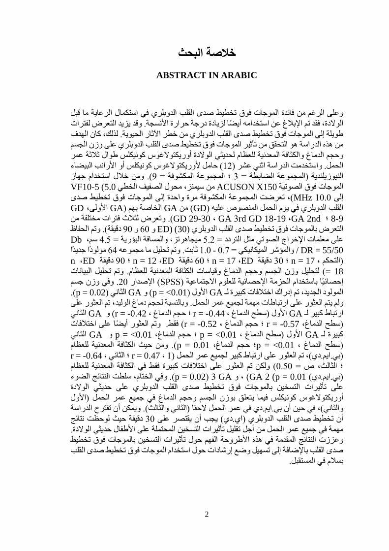
ABSTRACT
Despite the usefulness of Doppler ultrasound in complementing prenatal care, its usage
had also been reported to increase the temperature of tissues. Prolonged exposure to
Doppler ultrasound may increase the risk of bioeffects. Therefore, the aim of this study
was to investigate the influence of Doppler ultrasound on the body weight, brain size
and bone mineral density (BMD) of newborn Oryctolagus cuniculus throughout three
gestational ages (GA). The study utilised twelve (12) pregnant Oryctolagus cuniculus
or New Zealand white rabbits (NZWR) (Control Group = 3; Exposed Group = 9). By
utilizing Siemens Model ACUSON X150 ultrasound machine, linear array transducer
VF10-5 (5.0 to 10.0 MHz) the exposed group was exposed once to Doppler ultrasound
at a stipulated gestational day (GD) of their GAs (1st GA, GD 8-9; 2nd GA, GD 18-19;
3rd GA, GD 29-30). They were exposed to three different Doppler ultrasound exposure
durations (ED) (30, 60 and 90 minutes). Acoustic output parameters such as frequency
= 5.2 MHz, focal distance = 4.5 cm, Db/DR = 55/50 and mechanical index = 0.7 - 1.0
were kept constant. A total of 64 newborns (control, n = 17; 30 minutes ED, n = 17; 60
minutes ED, n = 12; 90 minutes ED, n = 18) were analysed for body weight, brain size
and BMD measurements. The data were statistically analysed using Statistical Package
for Social Sciences (SPSS) version 20. In the newborn’s body weight, significant
differences were perceived for 1st GA (p = <0.01) and 2nd GA (p = 0.02). No significant
correlations were found for all GAs. For the newborn’s brain size, significant
correlation was found for 1st GA (brain surface, r = -0.44; brain volume, r = -0.42) and
2nd GA (brain surface, r = -0.57; brain volume, r = -0.52) only. Significant differences
were also found for 1st GA (brain surface, p = <0.01; brain volume, p = <0.01 and 2nd
GA (brain surface, p = <0.01; brain volume, p = 0.01). In terms of BMD, significant
correlation was found for all GAs (1st, r = 0.47; 2nd, r = -0.64; 3rd, r = 0.50) but significant
differences were only found in BMD for 2nd GA (p = 0.01), and 3rd GA (p = 0.02). In
conclusion, the results highlighted the heating effects of Doppler ultrasound on
Oryctolagus cuniculus newborns with respect to body weight and brain size in early
GAs (1st & 2nd), whereas BMD in later GAs (2nd & 3rd). The study could suggest that
Doppler ultrasound ED should be limited to 30 minutes as significant results were
observed in all GAs in order to reduce the potential heating effects on the newborns.
The findings presented in this thesis fortified the understanding on the heating effects
of Doppler ultrasound besides facilitating the establishment of guidelines on using
Doppler ultrasound safely in the future.
2
خلاصة البحث
ABSTRACT IN ARABIC
بلق ما الرعاية استكمال في تخطيط صدى القلب الدوبلري فوق الموجات فائدة من الرغم وعلى
فتراتل التعرض يزيد وقد. الأنسجة حرارة درجة لزيادة أيضًا استخدامه عن الإبلاغ تم فقد الولادة،
لهدفا كان الحيوية. لذلك، الآثار خطر تخطيط صدى القلب الدوبلري من فوق الموجات إلى طويلة
لجسما وزن على تخطيط صدى القلب الدوبلري فوق الموجات تأثير من التحقق هو الدراسة هذه من
عمر ثةثلا الولادة أوريكتولاغوس كونيكلس طوال لحديثي للعظام المعدنية والكثافة الدماغ وحجم
البيضاء الأرانب كونيكلس أو لأوريكتولاغوس حامل( 12) عشر اثني راسةالد الحمل. واستخدمت
جهاز استخدام خلال ومن (.9= المكشوفة المجموعة ؛ 3= الضابطة المجموعة) النيوزيلندية
VF10-5 (5.0 الخطي الصفيف من سيمنز، محول ACUSON X150الصوتية فوق الموجات
تخطيط صدى فوق الموجات إلى واحدة مرة المكشوفة المجموعة ، تعرضت(MHz 10.0 إلى
GD الأولى، GA) بهم الخاصة GA من( GD) عليه المنصوص الحمل يوم في القلب الدوبلري
من مختلفة فترات لثلاث وتعرض (.GA 2nd ، GD 18-19 rd3 GA ، GD 29-30 ؛ 8-9
الحفاظ وتم(. دقيقة 90 و 60 و 30) (ED) تخطيط صدى القلب الدوبلري فوق بالموجات التعرض
Db سم، 4.5= البؤرية والمسافة ميجاهرتز، 5.2= التردد مثل الصوتي الإخراج معلمات على
/ DR = 55/50 جديداً مولوداً 64 مجموعه ما تحليل وتم .ثابت 1.0 - 0.7= الميكانيكي والمؤشر
ED ، n دقيقة 90 ؛ ED ، n = 12 دقيقة 60 ؛ ED ، n = 17 دقيقة 30 ؛ n = 17 ، التحكم)
ياناتالب تحليل وتم .للعظام المعدنية وقياسات الكثافة الدماغ وحجم الجسم وزن لتحليل( 18 =
جسم وزن وفي. 20 الإصدار( SPSS) الاجتماعية للعلوم الإحصائية الحزمة باستخدام إحصائياً
(.p = 0.02) الثاني GA و( p = <0.01) الأول GA لـ كبيرة اختلافات إدراك تم الجديد، المولود
على ورالعث تم الوليد، دماغ لحجم لجميع عمر الحمل. وبالنسبة مهمة ارتباطات على العثور يتم ولم
الثاني GA و( r = -0.42 ، الدماغ حجم ؛ r = -0.44 ، الدماغ سطح) الأول GA لـ كبير ارتباط
اختلافات على أيضًا العثور فقط. وتم (r = -0.52 ، الدماغ حجم ؛ r = -0.57 الدماغ، سطح)
الثاني GA و p = <0.01 الدماغ، حجم ؛ p = <0.01 ، الدماغ سطح) الأول GA لـ كبيرة
للعظام المعدنية ومن حيث الكثافة (.p = 0.01 الدماغ، حجم ؛p = <0.01 ، الدماغ سطح)
r = -0.64 ، الثاني ؛ r = 0.47 ، 1)لجميع عمر الحمل كبير ارتباط على العثور )بي.ايم.دي(، تم
عظام لل المعدنية الكثافة في فقط كبيرة اختلافات على العثور تم ولكن( 0.50= ص الثالث، ؛
الضوء النتائج سلطت الختام، . وفيGA 3 (p = 0.02) و ،GA 2 (p = 0.01 ) )بي.ايم.دي(
ة الولاد حديثي على تخطيط صدى القلب الدوبلري فوق بالموجات التسخين تأثيرات على
في جميع عمر الحمل )الأول الدماغ وحجم الجسم بوزن يتعلق أوريكتولاغوس كونيكلس فيما
لدراسةا تقترح أن أن بي.ايم.دي في عمر الحمل لاحقا )الثاني والثالث(. ويمكن حين والثاني(، في
نتائج لوحظت حيث دقيقة 30 على يقتصر أن أن تخطيط صدى القلب الدوبلري )اي.دي( يجب
لولادة. ا حديثي الأطفال على المحتملة التسخين تأثيرات تقليل أجل جميع عمر الحمل من في مهمة
يط تخط فوق بالموجات التسخين تأثيرات حول الفهم الأطروحة هذه في المقدمة النتائج وعززت
لقلب اتخطيط صدى فوق الموجات استخدام حول إرشادات وضع تسهيل إلى صدى القلب بالإضافة
.المستقبل في بسلام

3
APPROVAL PAGE
I certify that I have supervised and read this study and that in my opinion, it conforms
to acceptable standards of scholarly presentation and is fully adequate, in scope and
quality, as a thesis for the Master of Health Sciences (Medical Imaging)
…………………………………..
Asst. Prof. Dr. Farah Wahida
Ahmad Zaiki
Supervisor
…………………………………..
Assoc. Prof. Dr. Hj. Sulaiman Md
Dom
Co-Supervisor
I certify that I have read this study and that in my opinion it conforms to acceptable
standards of scholarly presentation and is fully adequate, in scope and quality, as a thesis
for the Master of Health Sciences (Medical Imaging)
…………………………………..
Assoc. Prof. Dr. Sayed Inayatullah
Shah
Internal Examiner

4
…………………………………..
Asst. Prof. Dr. Noor Shafini
Mohamad
External Examiner
This thesis was submitted to the Department of Diagnostic Imaging and Radiotherapy
and is accepted as a fulfilment of the requirement for the Master of Health Sciences
(Medical Imaging)
…………………………………..
Assoc. Prof. Dr. Sayed Inayatullah
Shah
Head, Department of Diagnostic
Imaging and Radiotherapy
This thesis was submitted to the Kulliyyah of Allied Health Sciences and is accepted as
a fulfilment of the requirement for the Master of Health Sciences (Medical Imaging)
…………………………………..
Prof. Dr. Suzanah Abdul Rahman
Dean, Kulliyyah of Allied of
Health Sciences

5
DECLARATION
I hereby declare that this thesis is the result of my own investigations, except where
otherwise stated. I also declare that it has not been previously or concurrently submitted
as a whole for any other degrees at IIUM or other institutions.
Umi Nadrah binti Amran
Signature........................................................... Date.........................................
6
INTERNATIONAL ISLAMIC UNIVERSITY MALAYSIA
DECLARATION OF COPYRIGHT AND AFFIRMATION OF
FAIR USE OF UNPUBLISHED RESEARCH
THERMAL EFFECTS OF PRENATAL DOPPLER ULTRASOUND
ON NEWBORNS OF ORYCTOLAGUS CUNICULUS
I declare that the copyright holders of this thesis are jointly owned by the student
and IIUM.
Copyright © 2020 Umi Nadrah binti Amran and International Islamic University Malaysia. All
rights reserved.
No part of this unpublished research may be reproduced, stored in a retrieval system,
or transmitted, in any form or by any means, electronic, mechanical, photocopying,
recording or otherwise without prior written permission of the copyright holder
except as provided below
1. Any material contained in or derived from this unpublished research
may be used by others in their writing with due acknowledgement.
2. IIUM or its library will have the right to make and transmit copies (print
or electronic) for institutional and academic purposes.
3. The IIUM library will have the right to make, store in a retrieved system
and supply copies of this unpublished research if requested by other
universities and research libraries.
By signing this form, I acknowledged that I have read and understand the IIUM
Intellectual Property Right and Commercialization policy.
Affirmed by Umi Nadrah binti Amran
……..…………………….. ………………………..
Signature Date

7
DEDICATIONS
I dedicated this thesis to myself for being brave, patience and strong enough to complete
this unexpected master’s journey. This is for the sacrifices, tears, blood, and sweat that
accompany me all along.
8
ACKNOWLEDGEMENTS
Bismillahirrahmannirrahim. In the name of Allah the most Benevolent and the most
Merciful. First and foremost, praise to Allah S. W. T, with His blessings, I able to finish
this study after a few ups and downs throughout my master’s journey. I would like to
express my countless gratitude to my supervisor, Asst. Prof. Dr. Farah Wahida Ahmad
Zaiki and co-supervisor, Assoc. Prof. Dr. Hj. Sulaiman Md Dom for their advice,
beneficial critics, patience, and kindness through this whole time guiding me without
fail.
I shall thank all the lecturers, staffs from Diagnostic Imaging and Radiotherapy
Department (DDIR), as well as other lecturers and staffs from Kulliyyah Allied Health
Sciences (KAHS) IIUM, Department of Orthopaedics from Jalan Hospital Campus
(JHC) and Centre of Medical Imaging, Faculty of Health Sciences, UiTM Puncak Alam
for their unconditional assistance and suggestions. Not forgotten to my lab partner,
Nadzirah Mohamad Radzi and fellow friends who walk by my sides, together holding
me up, always reminding me to stay strong and never once failed to offer help whenever
I need to have one.
Lastly, my special appreciation would be to my both parents, Amran bin Zakaria and
Siti Rohana binti Asmuni, my fiancé, Engku Fathul Shah Nor Haqim bin E Shahrunizad
and my families for always believe and being thoughtful for every single thing I have
done to complete this journey. I would not be able to put my two legs at where I stand
today if there are no prayers from them. Thank you for always have faith in this one
human, unwearyingly waited for her to complete her long study journey and for every
penny you spent for her educational fees and well-being. May each and every single
person who involves in this journey, directly or indirectly, is granted with His blessings
for this life and hereafter.
Thank you.
Umi Nadrah binti Amran

9
TABLE OF CONTENTS
Abstract ....................................................................................................... 1
Abstract in Arabic .................................................................................................. 2
Approval Page ....................................................................................................... 3
Declaration ....................................................................................................... 5
Copyright ....................................................................................................... 6
Dedications ....................................................................................................... 7
Acknowledgements ................................................................................................ 8
Table of Contents ................................................................................................... 9
List of Tables ....................................................................................................... 12
List of Figures ....................................................................................................... 14
List of Equations .................................................................................................... 15
List of Symbols / SI Units ...................................................................................... 16
List of Abbreviations ............................................................................................. 18
CHAPTER ONE: INTRODUCTION .................................................................. 20
Background of the Study ........................................................................ 20
Statement of the Problem........................................................................ 22
Purpose of the Study ............................................................................... 24
Research Objectives................................................................................ 24
Research Questions ................................................................................. 25
Significance of the Study ........................................................................ 25
Limitations of the Study ......................................................................... 26
Outline of the Thesis ............................................................................... 30
Chapter Summary ................................................................................... 31
CHAPTER TWO: LITERATURE REVIEW .................................................... 32
Introduction............................................................................................. 32
A Brief History on Human Embryology ................................................ 32
Oryctolagus cuniculus ............................................................................ 35
The Similarity of Features Between Human and Oryctolagus
Cuniculus ................................................................................................ 36
Micro-Computed Tomography (Micro-CT) ........................................... 37
Ultrasound Physics ................................................................................. 39
2.6.1 Sound Waves................................................................................. 39
2.6.2 Relationship between Acoustic Variables ..................................... 41
2.6.3 Transducer ..................................................................................... 43
Interaction of Ultrasound with Tissue .................................................... 44
2.7.1 Attenuation of Ultrasound in Tissue ............................................. 45
2.7.1.1 Absorption ........................................................................ 45
2.7.1.2 Reflection.......................................................................... 46
2.7.1.3 Scattering .......................................................................... 46
2.7.1.4 Refraction ......................................................................... 47
2.7.1.5 Divergence of Beam ......................................................... 47
2.7.2 Mechanism of Ultrasound Interaction with Tissue ....................... 49
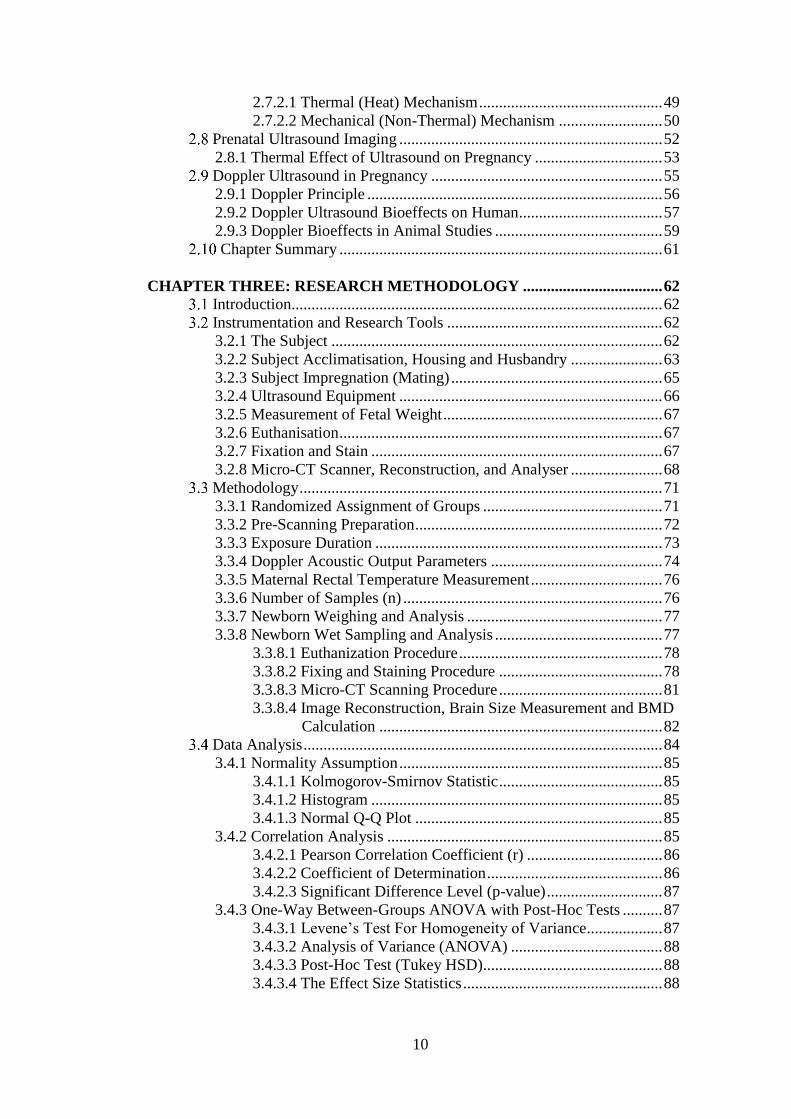
10
2.7.2.1 Thermal (Heat) Mechanism .............................................. 49
2.7.2.2 Mechanical (Non-Thermal) Mechanism .......................... 50
Prenatal Ultrasound Imaging .................................................................. 52
2.8.1 Thermal Effect of Ultrasound on Pregnancy ................................ 53
Doppler Ultrasound in Pregnancy .......................................................... 55
2.9.1 Doppler Principle .......................................................................... 56
2.9.2 Doppler Ultrasound Bioeffects on Human .................................... 57
2.9.3 Doppler Bioeffects in Animal Studies .......................................... 59
Chapter Summary ................................................................................. 61
CHAPTER THREE: RESEARCH METHODOLOGY ................................... 62
Introduction............................................................................................. 62
Instrumentation and Research Tools ...................................................... 62
3.2.1 The Subject ................................................................................... 62
3.2.2 Subject Acclimatisation, Housing and Husbandry ....................... 63
3.2.3 Subject Impregnation (Mating) ..................................................... 65
3.2.4 Ultrasound Equipment .................................................................. 66
3.2.5 Measurement of Fetal Weight ....................................................... 67
3.2.6 Euthanisation ................................................................................. 67
3.2.7 Fixation and Stain ......................................................................... 67
3.2.8 Micro-CT Scanner, Reconstruction, and Analyser ....................... 68
Methodology ........................................................................................... 71
3.3.1 Randomized Assignment of Groups ............................................. 71
3.3.2 Pre-Scanning Preparation .............................................................. 72
3.3.3 Exposure Duration ........................................................................ 73
3.3.4 Doppler Acoustic Output Parameters ........................................... 74
3.3.5 Maternal Rectal Temperature Measurement ................................. 76
3.3.6 Number of Samples (n) ................................................................. 76
3.3.7 Newborn Weighing and Analysis ................................................. 77
3.3.8 Newborn Wet Sampling and Analysis .......................................... 77
3.3.8.1 Euthanization Procedure ................................................... 78
3.3.8.2 Fixing and Staining Procedure ......................................... 78
3.3.8.3 Micro-CT Scanning Procedure ......................................... 81
3.3.8.4 Image Reconstruction, Brain Size Measurement and BMD
Calculation ....................................................................... 82
Data Analysis .......................................................................................... 84
3.4.1 Normality Assumption .................................................................. 85
3.4.1.1 Kolmogorov-Smirnov Statistic ......................................... 85
3.4.1.2 Histogram ......................................................................... 85
3.4.1.3 Normal Q-Q Plot .............................................................. 85
3.4.2 Correlation Analysis ..................................................................... 85
3.4.2.1 Pearson Correlation Coefficient (r) .................................. 86
3.4.2.2 Coefficient of Determination ............................................ 86
3.4.2.3 Significant Difference Level (p-value) ............................. 87
3.4.3 One-Way Between-Groups ANOVA with Post-Hoc Tests .......... 87
3.4.3.1 Levene’s Test For Homogeneity of Variance ................... 87
3.4.3.2 Analysis of Variance (ANOVA) ...................................... 88
3.4.3.3 Post-Hoc Test (Tukey HSD)............................................. 88
3.4.3.4 The Effect Size Statistics .................................................. 88

11
3.4.4 Kruskal-Wallis Test With Mann-Whitney U Test ........................ 89
Flow Chart of the Study .......................................................................... 89
Chapter Summary ................................................................................... 91
CHAPTER FOUR: RESULTS ............................................................................ 92
Introduction............................................................................................. 92
Maternal Temperature Results ................................................................ 92
Part I: Newborns Body Weight Analysis................................................ 93
4.3.1 Normality Assumption .................................................................. 93
4.3.2 Descriptive Analysis ..................................................................... 94
4.3.3 Correlation Analysis ..................................................................... 95
4.3.4 One-Way ANOVA with Post-Hoc Test ........................................ 97
Part II: Newborns Brain Size Analysis ................................................... 100
4.4.1 Normality Assumption .................................................................. 100
4.4.2 Descriptive Analysis ..................................................................... 102
4.4.2.1 Newborns Brain Surface Area .......................................... 102
4.4.2.2 Newborns Brain Volume .................................................. 103
4.4.3 Correlation Analysis ..................................................................... 104
4.4.3.1 Newborns Brain Surface Area .......................................... 104
4.4.3.2 Newborns Brain Volume .................................................. 105
4.4.4 One-Way ANOVA with Post-Hoc Test and Kruskal-Wallis
With Mann-Whitney U Test ......................................................... 107
4.4.4.1 Newborns Brain Surface Area .......................................... 107
4.4.4.2 Newborns Brain Volume .................................................. 110
Part III: Newborns Bone Mineral Density (BMD) Analysis .................. 113
4.5.1 Normality Assumption .................................................................. 113
4.5.2 Descriptive Analysis ..................................................................... 114
4.5.3 Correlation Analysis ..................................................................... 115
4.5.4 One-Way ANOVA with Post-Hoc Test and Kruskal-Wallis
with Mann-Whitney U Test .......................................................... 116
Chapter Summary ................................................................................... 120
CHAPTER FIVE: DISCUSSION AND CONCLUSION .................................. 121
Introduction............................................................................................. 121
Maternal Temperature ............................................................................ 121
Part I: Newborns Body Weight............................................................... 122
Part II: Newborns Brain Size .................................................................. 126
Part III: Newborns Bone Mineral Density (BMD) ................................. 130
Conclusion and Implications .................................................................. 133
Future Recommendation ......................................................................... 135
Chapter Summary ................................................................................... 136
REFERENCES ....................................................................................................... 137
APPENDIX A: STATISTICAL RESULTS ............................................................ 146
APPENDIX B: ETHICAL CLEARANCE BY IIUM I-ACUC .............................. 166
APPENDIX C: PUBLISHED ARTICLES / MANUSCRIPTS /
PROCEEDINGS / ABSTRACTS ................................................. 167
APPENDIX D: CERTIFICATES ............................................................................ 180

12
LIST OF TABLES
Table 2.1 Acoustic variables 41
Table 2.2 Acoustic output during the ultrasound studies 58
Table 3.1 10% buffered formalin recipe 68
Table 3.2 Skyscan 1176 technical specification 69
Table 3.3 Acoustic parameters 75
Table 3.4 Number of samples (n) used in the study 77
Table 3.5 Micro-CT scanning parameters for brain 82
Table 3.6 Micro-CT scanning parameters for femur 82
Table 4.1 Maternal rectal temperature range and mean 93
Table 4.2 Normality assumption for newborns body weight 94
Table 4.3 Correlation between newborns body weight and exposure durations
at different gestational ages 96
Table 4.4 Summary of ANOVA and Post-Hoc test on newborns body weight 98
Table 4.5 Normality assumption for newborns brain surface area 100
Table 4.6 Normality assumption for newborns brain volume 101
Table 4.7 Correlation between newborns brain surface area and exposure
durations at different gestational ages 104
Table 4.8 Correlation between newborns brain volume and exposure durations
at different gestational ages 105
Table 4.9 Summary of ANOVA and Post-Hoc test on newborns brain surface
area 108
Table 4.10 Summary of ANOVA and Post-Hoc test on newborns brain volume 110
Table 4.11 Summary of Kruskal-Wallis and Mann-Whitney U test on
newborns brain volume 111
Table 4.12 Normality assumption for newborns left femur BMD 113

13
Table 4.13 Correlation between newborns left femur BMD and exposure
durations at different gestational ages 115
Table 4.14 Summary of ANOVA and Post-Hoc test for newborns left femur
BMD 117
Table 4.15 Summary of Kruskal-Wallis and Mann-Whitney U test for
newborns left femur BMD 118

14
LIST OF FIGURES
Figure 2.1 Human prenatal development 34
Figure 2.2 Categories of sound 40
Figure 2.3 Phase of sound waves 42
Figure 3.1 NZWR in an iron steel cage 63
Figure 3.2 Skyscan 1176 Micro-CT 70
Figure 3.3 Randomized assignment of groups 72
Figure 3.4 Ultrasound images produced using selected acoustic output 75
Figure 3.5 Wet samples in 10% buffered formalin 79
Figure 3.6 Wet sampling and analysis workflow 80
Figure 3.7 Placement of wet sample inside the Micro-CT scanner 81
Figure 3.8 Reconstruction of the newborn’s brain and left femur using NRecon
software 83
Figure 3.9 Measurement of newborn’s brain size and BMD of left femur using
CTan software 84
Figure 3.10 Research workflow 90
Figure 4.1 Mean comparison of newborns body weight and exposure durations
at different gestational ages 95
Figure 4.2 Mean comparison of newborns brain surface area and exposure
durations at different gestational ages 102
Figure 4.3 Mean/median comparison of newborns brain volume and exposure
durations at different gestational ages 103
Figure 4.4 Mean comparison of newborns left femur BMD and exposure
durations at different gestational ages 114

15
LIST OF EQUATIONS
Equation 2.1 Period versus frequency 42
Equation 2.2 Wavelength equation 43
Equation 2.3 Acoustic impedence 48
Equation 3.1 Dilution equation 68
Equation 3.2 Coefficient of determination 87
Equation 3.3 The effect size 88
16
LIST OF SYMBOLS / SI UNITS
% Percent
” Inch
± Precision of an approximation
° Degree
µm Micrometre
µm2 Micrometre squared
µm2 Square micrometre
µm3 Cube micrometre
µm3 Micrometre cubed
µs Microsecond
A Amplitude
℃ Degree Celsius
c Velocity
cm Centimetre
df Doppler shift
f Frequency
g Gram
g.cm3 Gram per cubic centimetre
h Hour
Hz Hertz
kg Kilogram
kV Kilovoltage
M Mean value
m/s Metre per second
17
M1 Molarity of concentrated solution
M2 Molarity of diluted solution
Md Median value
MHz Megahertz
ml Millilitre
ml/kg Millilitre per kilogram
mm Millimetre
ms Millisecond
mW/cm Milliwatt per centimetre
n Number of samples
Na2HPO4 Diphasic sodium phosphate
NaCL Sodium chloride
p P-value
r Pearson correlation coefficient
T Period (time)
U Mann-Whitney U value
V1 Volume of concentrated solution
V2 Volume of diluted solution
W/kg Watt per kilogram
z Z-value
λ Wavelength
𝑥̅ Mean

18
LIST OF ABBREVIATIONS
2D Two dimensional
3D Three dimensional
3Rs Replacement, reduction and refinement
AIUM American Institute of Ultrasound in Medicine
ALARA As low as reasonably achievable
ANOVA Analysis of variance
BMD Bone mineral density
B-mode Brightness mode
BMUS British Medical Ultrasound Safety
CE Conformité Européene / European Conformity
CPU Central Processing Unit
CT Computed tomography
CTAn Computed tomography analyser
ED Exposure duration
FDA Food and Drug Administration
GA Gestational age
GD Gestational day
I-ACUC Animal Care and Use Committee
IBM International Business Machine Corporation
IIUM International Islamic University Malaysia
IPCC Intergovernmental Panel on Climate Change
ISPTA Spatial-pulse-temporal-average intensities
KAHS Kulliyyah of Allied Health Sciences
LCD Liquid Crystal Display

19
MI Mechanical index
Micro-CT Micro-computed tomography
M-mode Motion mode
MRI Magnetic resonance imaging
MRM Magnetic resonance microscopy
mRNA Messenger-ribonucleic acid
NIR Non-ionizing radiation
NRecon NReconstruction software
NZWR New Zealand white rabbit
PET Positron emission tomography
PTA Phosphotungstic acid
ROI Region of interest
SAR Specific absorption rate
SD Standard deviation
SPECT Single photon emission computed tomography
SPSS Statistical Package for Social Sciences
TI Thermal index
TPC Tenderness, pamper and care
UiTM Universiti Teknologi MARA
20
CHAPTER ONE
INTRODUCTION
BACKGROUND OF THE STUDY
Ultrasound has been long known as the safest imaging modality as it involves no
ionising radiation and is used frequently in prenatal care for decades. Doppler
ultrasound plays a role in the obstetrics and gynaecology field to serve as a
complementary mode in a standard prenatal scan (Chau, 2002). It aids in investigating
foetus blood flow in expectant mothers’ wombs, which is usually for those who come
with pregnancy complications (Alfirevic, Stampalija, & Dowswell, 2017; Schellpfeffer,
2013).
After the invention of ultrasound in the late 1950s, it has continued to develop
throughout these decades (Chau, 2002). Today, Doppler ultrasound has been
commercially applied by private companies and healthcare institution for prenatal care.
Doppler ultrasound’s technology advancement has improved the service of standard
prenatal ultrasound scanning. The practicality of Doppler effects by the motion or
direction of blood flow helps practitioners to evaluate and estimate blood circulation
abnormalities of the foetus in a better view (Oglat et al., 2018).
21
As of current practice, only an expectant mother with high potential for
complication gets the privilege of having Doppler ultrasound to check on her foetus
(Hill, 2016). As reported by Hill (2016), the percentage of stillborn rate can be reduced
if Doppler ultrasound is implemented as one of the standard prenatal scanning
procedure. In an interview conducted, a mother who had lost her unborn child believed
that her child could be saved if Doppler ultrasound has been made as a standard practice
in the health institution. She said Doppler ultrasound should be done on every pregnant
woman since standard prenatal ultrasound could not give any information whether the
foetus had gotten enough oxygen, nutrients and blood supply from the placenta to grow
healthy in the womb.
In contrary to the conventional two dimensional (2D) ultrasound, the Doppler
ultrasound beam is focused at only one point. This may lead to heat accumulation in the
area, thus increasing the temperature. Therefore, concerns arise regarding the safety of
Doppler ultrasound mode’s prolonged use on the foetus. It is proven that temperature
elevations on both mother and foetus contributed to numerous adverse outcomes
(Strand, Barnett, & Tong, 2011). These include low birth weight, spontaneous abortion,
stillbirth and premature contraction (Goldenberg, Culhane, Iams, & Romero, 2008;
Salvesen et al., 2011).
The possible harm of ultrasound including Doppler is uncertain since it is well-
known to be the safest imaging modalities among others (Barnett & Maulik, 2001).
Nonetheless, ultrasound still has quite a number of possibilities to cause heat (thermal)
and mechanical (non-thermal) effects. The bioeffect risk increases as the technology of
Doppler ultrasound advances through the decades. It has been reported that potential
cavitation can happen when Doppler is used together with three dimensional (3D)

22
ultrasound (Pooh et al., 2016). Other literature has also stated that the Doppler’s
acoustic outputs are relatively sufficient to result in obvious biological effects when
maximum operating settings are used (Barnett & Maulik, 2001).
STATEMENT OF THE PROBLEM
Years before the trend set, USA Today (2004) has reported that the American Institute
of Ultrasound in Medicine (AIUM) notifies expecting parents about possible harms in
having an unregulated ultrasound for entertainment purposes (“Parents ignoring FDA
warning against prenatal portraits,” 2004). Even though there are no confirmed
biological effects from the regulated prenatal ultrasound, unregulated prenatal
ultrasound takes longer time and uses more energy compared to regulated ones. Food
and Drug Administration (FDA) has made a statement concerning the unknown long-
term effects of tissue heating by frequent visits and prolonged examination time
(Pawlowski, 2014; Romm, 2014). Therefore, experts have stated that ultrasound using
Doppler should only be done on expectant mothers when there is a medical purpose to
perform it. If it is performed on an expectant mother without any diagnostic purpose, it
begs the question of whether it is safe and justified for the foetus.
In recent years, there are increasing interests in exploring the bioeffects of
Doppler ultrasound. Previous studies have reported several results on Doppler
ultrasound bioeffects in various animals. Jia et al. (2005) have found that the
insonification fetal group has a higher significant difference in myocardial apoptosis
compared to the control group. Later in 2009, the finding of Schneider-Kolsky et al.
(2009) confirmed that exposure to Doppler ultrasound may result in an impairment
towards a mammal’s cognitive function. They found significant memory impairment
23
after 2 days post-hatch following the Doppler exposure to the foetal chick for several
minutes on day 19 of the incubation period.
In 2011, a study on the effect of pulse Doppler examination on ductus venosus
in rat foetuses showed a positive result where there was a positive linear correlation
between Doppler exposure time and apoptotic activities of exposed liver tissues
(Pellicer et al., 2011). Helmy, Bader, Koch, Tiringer, and Kollmann (2015) have
undertaken an in-vitro study in measuring the thermal output of Doppler ultrasound.
The energy output of the ultrasound transducer was investigated using the water bath
model. In the study, they found that the activation of Doppler ultrasound in water bath
increased the temperature of water in one minute. Thus, they come into a conclusion
that Doppler ultrasound can induce a thermal effect to the foetuses, especially in early
pregnancy.
In spite of several new findings of the bioeffects of the Doppler ultrasound,
several other studies have been done to investigate the heating effects of prenatal
ultrasound without using Doppler mode as well. In 2013, a study of fluctuations in
haematological analysis and foetal weight was statistically found a significant
difference in the newborn of Oryctolagus cuniculus after being exposed to prenatal
ultrasound (Zaiki, Dom, Razak, & Hassan, 2013; Zaiki & Dom, 2014). Zaiki and Dom
(2016) later found the heating effect during prenatal scanning does interfere with the
foetal neuro-development. In 2016, Isa and Dom (2016) found that the lowermost
Oryctolagus cuniculus growth is recorded when they are exposed to 2D ultrasound for
90 minutes long at the 2nd gestational age.