therapy of skin diseases - download.e-bookshelf.de
TRANSCRIPT

Therapy of Skin Diseases

Thomas KriegDavid R. Bickers Yoshiki Miyachi (Eds.)
Therapy of Skin Diseases
A Worldwide Perspective on Therapeutic Approaches and Their Molecular Basis

ISBN: 978-3-540-78813-3 e-ISBN: 978-3-540-78814-0
DOI: 10.1007/978-3-540-78814-0
Springer Heidelberg Dordrecht London New York
Library of Congress Control Number: 2009933266
© Springer-Verlag Berlin Heidelberg 2010
This work is subject to copyright. All rights are reserved, whether the whole or part of the material is concerned, specifi cally the rights of translation, reprinting, reuse of illustrations, recitation, broadcasting, reproduction on microfi lm or in any other way, and storage in data banks. Duplication of this publication or parts thereof is permitted only under the provisions of the German Copyright Law of September 9, 1965, in its current version, and permission for use must always be obtained from Springer. Violations are liable to prosecution under the German Copyright Law.
The use of general descriptive names, registered names, trademarks, etc. in this publication does not imply, even in the absence of a specifi c statement, that such names are exempt from the relevant protective laws and regulations and therefore free for general use.
Product liability: The publishers cannot guarantee the accuracy of any information about dosage and appli-cation contained in this book. In every individual case the user must check such information by consulting the relevant literature.
Cover design: eStudio Calamar, Figueres/Berlin
Printed on acid-free paper
Springer is part of Springer Science+Business Media (www.springer.com)
Prof. Thomas KriegUniversitätsklinikum KölnKlinik und Poliklinik fürDermatologie und VenerologieKerpener Str. 6250924 Kö[email protected]
Prof. David R. BickersColumbia University Medical CenterDept. Dermatology12th Floor, Herbert Irving Pavilion161 Fort Washington Ave.New York NY [email protected]
Prof. Yoshiki MiyachiKyoto UniversityGraduate School of MedicineDept. Dermatology &Cutaneous Sciences54 Kawahara-choShogoin, Sakyo-kuKyoto 606-8507 [email protected]

v
Treatment of skin diseases has changed remarkably during the last decade. This is largely the result of a better understanding of the molecular and cellular basis of many skin diseases. Thus, novel targets have been identifi ed and specifi c drugs devel-oped which directly interfere with or alter the disease processes. This change is read-ily apparent when considering the novel agents available for psoriasis and atopic dermatitis, and also for viral and other infectious skin diseases. Interestingly, many of these new agents are administered systemically either orally or by subcutaneous injection. And yet, as with all forms of drug therapy, these highly effi cacious agents can also be associated with severe side effects and drug-induced toxicity. Accordingly, the dermatologist must be aware of the medical status of the patient as well as all other medications that are being prescribed concomitantly. Careful monitoring of the risk:benefi t ratio is always critical.
Simultaneously, with the rapid development of novel therapeutics, there has been a major evolution in the clinical practice of dermatology with considerable variations across different areas of the world. In European countries, the discipline is relatively broad, in some countries including allergy and phlebology, as well as dermatologic surgery and dermatologic oncology. In European countries, patients with skin disease are often treated as in-patients by dermatologists, whereas in the United States, this occurs only rarely and the patients are admitted to beds assigned to Internal Medicine and dermatologists consult on their management. Asian dermatology has been pro-foundly affected by both European and American dermatology and the selection of therapeutic agents often refl ects those infl uences.
The result of these developments is that regional differences are commonplace in the treatment of skin diseases, some correlating with the dermatologic features of patients from diverse ethnic backgrounds, others relating to variations in the different health care systems and/or the medical education and the awareness of dermatolo-gists regarding particular treatment options. In the age of Internet, novel therapies are instantly available and potentially applicable to patients globally.
This book was conceived to address these changes and it has two major aims. First, it summarizes novel therapeutic procedures that are based on understanding the pathophysiology of skin diseases. Second, it aims to bring together in one place the variability of treatment modalities employed in the practice of dermatology around the world in Asia, Europe, and the USA. Every effort has been made to assure that all chapters indicate global variations either in the occurrence or the expression of skin
Preface

vi Preface
diseases and their treatment. All manuscripts have been reviewed carefully by experts familiar with the practice of dermatology in Asia, Europe, and the USA. It is our hope that this book will prove to be a valuable reference tool for dermatologists everywhere.
Köln, Germany Thomas KriegNew York, USA David R. BickersKyoto, Japan Yoshiki Miyachi

vii
We owe special acknowledgement to the cooperation ofWalter Burgdorf, MDTraubinger Strasse 54A, 82327 Tutzing, [email protected] Fujita, MD, PhDAssociate Professor, Department of Dermatology, University of Colorado Denver, SOM, Mail Stop 8127, RC-1 South 4th fl ., 12801 E 17th Avenue, Aurora, CO 80045, USA, [email protected]
Acknowledgements

ix
Part I Introduction . . . . . . . . . . . . . . . . . . . . . . . . . . . . . . . . . . . . . . . . . . . . 1
1.1 Biology of the Skin . . . . . . . . . . . . . . . . . . . . . . . . . . . . . . . . . . . . . . . . . 3Beate Eckes, Thomas Krieg, and Carien M. Niessen
1.2 Immune Mechanisms. . . . . . . . . . . . . . . . . . . . . . . . . . . . . . . . . . . . . . . 15Thomas Schwarz and Stefan Beissert
1.3 General Pharmacology . . . . . . . . . . . . . . . . . . . . . . . . . . . . . . . . . . . . . 21David R. Bickers
1.4 Immunomodulation in Dermatology . . . . . . . . . . . . . . . . . . . . . . . . . . 29Rebecca G. Pomerantz, Thomas S. Kupper, and Abrar A. Qureshi
1.5 Basic Principles of Genetics and Gene Therapy . . . . . . . . . . . . . . . . . 39Liv Kraemer and Angela M. Christiano
1.6 Percutaneous Absorption and Principles of Corneotherapy/Skin Care . . . . . . . . . . . . . . . . . . . . . . . . . . . . . . . . . . . 57Hachiro Tagami
1.7 Principles of Systemic Therapy . . . . . . . . . . . . . . . . . . . . . . . . . . . . . . 63Lindy P. Fox
1.8 Retinoid Pharmacology . . . . . . . . . . . . . . . . . . . . . . . . . . . . . . . . . . . . . 77Jens M. Baron
1.9 Ultraviolet (UV) A and (UV) B Phototherapy . . . . . . . . . . . . . . . . . . . 87Akimichi Morita
1.10 Laser Therapy . . . . . . . . . . . . . . . . . . . . . . . . . . . . . . . . . . . . . . . . . . . . 93David J. Goldberg
1.11 Photodynamic Therapy . . . . . . . . . . . . . . . . . . . . . . . . . . . . . . . . . . . . . 105Yoshiki Tokura and Shin-ichi Moriwaki
1.12 Dermatologic Surgery . . . . . . . . . . . . . . . . . . . . . . . . . . . . . . . . . . . . . . 113Murad Alam
Contents

x Contents
1.13 Neurophysiology of Itch. . . . . . . . . . . . . . . . . . . . . . . . . . . . . . . . . . . . . 121Akihiko Ikoma
Part II Infectious Diseases . . . . . . . . . . . . . . . . . . . . . . . . . . . . . . . . . . . . . . 127
2.1 Bacterial and Mycobacterial Infections . . . . . . . . . . . . . . . . . . . . . . . . 129Nicole French and Robert L. Modlin
2.2 Fungal Infection . . . . . . . . . . . . . . . . . . . . . . . . . . . . . . . . . . . . . . . . . . . 149Takashi Mochizuki
2.3 Viral Infections . . . . . . . . . . . . . . . . . . . . . . . . . . . . . . . . . . . . . . . . . . . . 157Annabelle Lozano, Anita Arora, Natalia Mendoza, Vandana Madkan, and Stephen K. Tyring
2.4 Sexually Transmitted Diseases (STDs). . . . . . . . . . . . . . . . . . . . . . . . . 165Anja Potthoff, Heinrich Rasokat, and Norbert H. Brockmeyer
2.5 Human Immunodefi ciency Virus (HIV) . . . . . . . . . . . . . . . . . . . . . . . 173Anja Potthoff, Heinrich Rasokat, and Norbert H. Brockmeyer
2.6 Ectoparasitic and Protozoan Diseases . . . . . . . . . . . . . . . . . . . . . . . . . 181Dirk M. Elston
Part III Papulosquamous Dermatoses . . . . . . . . . . . . . . . . . . . . . . . . . . . . 191
3.1 Psoriasis . . . . . . . . . . . . . . . . . . . . . . . . . . . . . . . . . . . . . . . . . . . . . . . . . 193Hajime Iizuka
3.2 Parapsoriasis and Related Disorders . . . . . . . . . . . . . . . . . . . . . . . . . . 207Peter C. M. van de Kerkhof
3.3 Lichen Planus . . . . . . . . . . . . . . . . . . . . . . . . . . . . . . . . . . . . . . . . . . . . . 213Tetsuo Shiohara, Yoshiko Mizukawa, and Yoko Kano
Part IV Atopic Dermatitis and Related Diseases . . . . . . . . . . . . . . . . . . . . 223
4.1 Atopic Dermatitis . . . . . . . . . . . . . . . . . . . . . . . . . . . . . . . . . . . . . . . . . . 225Andreas Wollenberg and Thomas Bieber
4.2 Pruritus . . . . . . . . . . . . . . . . . . . . . . . . . . . . . . . . . . . . . . . . . . . . . . . . . . 235Sonja Ständer and Thomas A. Luger
4.3 Urticaria . . . . . . . . . . . . . . . . . . . . . . . . . . . . . . . . . . . . . . . . . . . . . . . . . 247Michihiro Hide
4.4 Mastocytosis . . . . . . . . . . . . . . . . . . . . . . . . . . . . . . . . . . . . . . . . . . . . . . 263Naotomo Kambe, Akane Tanaka, and Yoshiki Miyachi

Contents xi
Part V Allergic Reactions and Hypersensitive Diseases . . . . . . . . . . . . . . 273
5.1 Allergic Contact Dermatitis . . . . . . . . . . . . . . . . . . . . . . . . . . . . . . . . . 275Cecilia Svedman and Magnus Bruze
5.2 Photosensitivity Diseases . . . . . . . . . . . . . . . . . . . . . . . . . . . . . . . . . . . . 285Taskeshi Horio
5.3 Drug Reactions . . . . . . . . . . . . . . . . . . . . . . . . . . . . . . . . . . . . . . . . . . . . 297Hans F. Merk and Daniela Höller Obrigkeit
5.4 Hypersensitivity Syndrome Reaction . . . . . . . . . . . . . . . . . . . . . . . . . . 321Sandra R. Knowles and Neil H. Shear
5.5 Eosinophilic Dermatoses . . . . . . . . . . . . . . . . . . . . . . . . . . . . . . . . . . . . 327Ichiro Katayama and Hiroyuki Murota
5.6 Neutrophilic Dermatoses . . . . . . . . . . . . . . . . . . . . . . . . . . . . . . . . . . . . 337Tadashi Terui
5.7 Skin Manifestations in Rheumatologic Disorders. . . . . . . . . . . . . . . . 349Manabu Fujimoto and Kazuhiko Takehara
Part VI Acne and Rosacea . . . . . . . . . . . . . . . . . . . . . . . . . . . . . . . . . . . . . . 357
6.1 Acne and Its Variants . . . . . . . . . . . . . . . . . . . . . . . . . . . . . . . . . . . . . . 359Christos C. Zouboulis and Mohamed Badawy Abdel-Naser
6.2 Rosacea and Related Diseases. . . . . . . . . . . . . . . . . . . . . . . . . . . . . . . . 375Mohamed Badawy Abdel-Naser and Christos C. Zouboulis
Part VII Autoimmune Diseases . . . . . . . . . . . . . . . . . . . . . . . . . . . . . . . . . . 387
7.1 Acquired Bullous Disease . . . . . . . . . . . . . . . . . . . . . . . . . . . . . . . . . . . 389Akiko Tanikawa and Masayuki Amagai
7.2 Connective Tissue Diseases . . . . . . . . . . . . . . . . . . . . . . . . . . . . . . . . . . 407Minoru Hasegawa and Shinichi Sato
7.3 Cutaneous Vasculitis . . . . . . . . . . . . . . . . . . . . . . . . . . . . . . . . . . . . . . . 427Nicolas Hunzelmann
7.4 Graft-Versus-Host Disease . . . . . . . . . . . . . . . . . . . . . . . . . . . . . . . . . . 433Robert Knobler, Michal Kouba, and David Pohlreich
7.5 Vitiligo . . . . . . . . . . . . . . . . . . . . . . . . . . . . . . . . . . . . . . . . . . . . . . . . . . . 443Philippe Bahadoran and Jean-Paul Ortonne

xii Contents
7.6 Therapy of Noninfectious Granulomatous Diseases . . . . . . . . . . . . . . 459Franco Rongioletti and Alfredo Rebora
Part VIII Metabolic Diseases . . . . . . . . . . . . . . . . . . . . . . . . . . . . . . . . . . . . 467
8.1 The Porphyrias . . . . . . . . . . . . . . . . . . . . . . . . . . . . . . . . . . . . . . . . . . . . 469Jorge Frank
8.2 Deposition Diseases . . . . . . . . . . . . . . . . . . . . . . . . . . . . . . . . . . . . . . . . 487Takahiro Hamada
Part IX Cosmetic Dermatology . . . . . . . . . . . . . . . . . . . . . . . . . . . . . . . . . . 497
9.1 Hair Diseases (Alopecia Areata and Androgenetic Alopecia) . . . . . . 499Satoshi Itami and Shigeki Inui
9.2 Nail Diseases . . . . . . . . . . . . . . . . . . . . . . . . . . . . . . . . . . . . . . . . . . . . . . 509Maurice J. Dahdah and Richard K. Scher
9.3 Hyperhidrosis . . . . . . . . . . . . . . . . . . . . . . . . . . . . . . . . . . . . . . . . . . . . . 517Robyn D. Siperstein and Robert A. Schwartz
9.4 Disorders of Pigmentation. . . . . . . . . . . . . . . . . . . . . . . . . . . . . . . . . . . 525Yoko Funasaka
9.5 Cosmetic Surgery . . . . . . . . . . . . . . . . . . . . . . . . . . . . . . . . . . . . . . . . . . 539Murad Alam
Part X Inherited Diseases . . . . . . . . . . . . . . . . . . . . . . . . . . . . . . . . . . . . . . . 547
10.1 Inherited Bullous Diseases . . . . . . . . . . . . . . . . . . . . . . . . . . . . . . . . . . 549Leena Bruckner-Tuderman and Cristina Has
10.2 Inherited Keratinocyte Diseases (Ichthyosis and Related Disorders) . . . . . . . . . . . . . . . . . . . . . . . . . . . 561Akemi Ishida-Yamamoto
10.3 Immunodefi ciency Disorders . . . . . . . . . . . . . . . . . . . . . . . . . . . . . . . . 575Giuseppe Micali, Dennis P. West, and Amy S. Paller
10.4 Disorders of DNA Repair . . . . . . . . . . . . . . . . . . . . . . . . . . . . . . . . . . . 589Shinichi Moriwaki and Kenneth H. Kraemer
Part XI Benign and Malignant Tumors . . . . . . . . . . . . . . . . . . . . . . . . . . . 597
11.1 Nonmelanoma Skin Cancer . . . . . . . . . . . . . . . . . . . . . . . . . . . . . . . . . 599Alexander G. Marneros and David R. Bickers
11.2 Malignant Melanoma. . . . . . . . . . . . . . . . . . . . . . . . . . . . . . . . . . . . . . . 621Toshiaki Saida

Contents xiii
11.3 Treatment of Cutaneous Lymphomas . . . . . . . . . . . . . . . . . . . . . . . . . 633Chalid Assaf and Wolfram Sterry
11.4 Vascular Malformations . . . . . . . . . . . . . . . . . . . . . . . . . . . . . . . . . . . . 643Maria C. Garzon and Philip M. Meyers
11.5 Rare Malignancies of the Skin . . . . . . . . . . . . . . . . . . . . . . . . . . . . . . . 659Bernhard Zelger and Oliver Bechter
Part XII Miscellaneous Disorders . . . . . . . . . . . . . . . . . . . . . . . . . . . . . . . . 675
12.1 Diseases of Pregnancy and Their Management . . . . . . . . . . . . . . . . . 677George Kroumpouzos and Lisa M. Cohen
12.2 Pediatric Dermatology . . . . . . . . . . . . . . . . . . . . . . . . . . . . . . . . . . . . . . 693Alain Taïeb, Franck Boralevi, and Christine Labrèze
12.3 Aging and Photoaging of the Skin . . . . . . . . . . . . . . . . . . . . . . . . . . . . 705Laure Rittié, Gary J. Fisher, and John J. Voorhees
12.4 Occupational Dermatoses . . . . . . . . . . . . . . . . . . . . . . . . . . . . . . . . . . . 717S. Mark Wilkinson and Pieter-Jan Coenraads
12.5 Wound Healing. . . . . . . . . . . . . . . . . . . . . . . . . . . . . . . . . . . . . . . . . . . . 735Sabine A. Eming
Subject Index . . . . . . . . . . . . . . . . . . . . . . . . . . . . . . . . . . . . . . . . . . . . . . . . . . 753

xv
Mohamed Badawy Abdel-Naser, MD Departments of Dermatology, Venereology, Allergology and Immunology, Dessau Medical Center, Auenweg 38, 06847 Dessau, Germany, [email protected]
Murad Alam, MD Northwestern University Dermatology, Clinical Trials Unit, 676 N St Clair, Suite 1600, Chicago, IL 60611, [email protected]
Masayuki Amagai, MD, PhD Department of Dermatology, Keio University, School of Medicine, 35 Shinanomachi, Shinjuku-ku, Tokyo 160-8582, [email protected]
Anita Arora, MD 6655 Travis, Suite 120, Houston, TX 77030, [email protected]
Chalid Assaf, MD Department of Dermatology, Helios Clinics Krefeld, Lutherplatz 40, 47805 Krefeld, [email protected]
Philippe Bahadoran, MD Service de Dermatologie, Hôpital l’Archet 2, CHU de Nice, Route St. Antoine Gnestière, 06202 Nice Cedex 3, [email protected]
Jens Malte Baron, MD Department of Dermatology and Allergology, University Hospital RWTH Aachen, Pauwelsstrasse 30, 52074 Aachen, [email protected]
Oliver Bechter, MD Department of Internal Medicine, Innsbruck Medical University, Anichstrasse 35, 6020 Innsbruck, [email protected]
Stefan Beissert, MD, PhD Department of Dermatology, University of Münster, Von Esmarchstrasse 58, 48149 Münster, [email protected]
David R. Bickers, MD New York Presbyterian Hospital, 161 Fort Washington Avenue, 12th fl oor, New York, NY 10032, [email protected]
Thomas Bieber, MD, PhD Department of Dermatology and Allergy, University of Bonn, Sigmund-Freud-Strasse 25, 53105 Bonn, [email protected]
Contributors
xvi Contributors
Franck Boralevi, MD Hôpital Pellegrin-Enfants, CHU de Bordeaux, Place Amélie Raba-Léon, 33076 Bordeaux Cedex, [email protected]
Norbert H. Brockmeyer, MD Department of Dermatology and Allergology, Ruhr Unversity Bochum, Gudrunstrabe 56, 44791 Bochum, [email protected]
Leena Bruckner-Tuderman, MD Department of Dermatology, University Medical Center Freiburg, Hauptstrabe 7, 79104 Freiburg, [email protected]
Magnus Bruze, MD Department of Occupational and Environmental Dermatology, Malmö University Hospital, UMAS, Ing 73 (Entrance 45), S-205 02 Malmö, [email protected]
Angela M. Christiano, PhD Department of Dermatology and Genetics and Development, Columbia University, College of Physicians and Surgeons, 630 West 168th Street VC15 204A, New York, NY 10032, [email protected]
Pieter-Jan Coenraads, MD Occupational and Environmental Dermatology Unit, University Medical Center Groningen, Hanzeplein 1, 9700 RB GroningenThe [email protected]
Lisa M. Cohen, MD Cohen Dermatopathology, 320 Needham Street, Suite 200, Newton, MA 02464, [email protected]
Maurice J. Dahdah, MD Department of Dermatology, 161 Fort Washington Avenue, IP 12th fl oor, New York, NY 10032, [email protected]
Beate Eckes, PhD Department of Dermatology, University of Cologne, Kerpener Strabe 62, 50937 Cologne, [email protected]
Dirk M. Elston, MD Department of Dermatology, Geisinger Medical Center, 100 North Academy Avenue, Danville, PA 17821, [email protected]
Sabine A. Eming, MD Department of Dermatology, University of Cologne, Kerpener Strabe 62, 50937 Cologne, [email protected]
Gary J. Fisher, PhD 6447 Med Sci I, 1150 W Medical Center Drive, Ann Arbor, MI 48109-0609, [email protected]
Lindy P. Fox, MD Department of Dermatology, University of California, San Francisco, 1701 Divisadero, Box 0316, San Francisco, CA 94143, [email protected]

Contributors xvii
Jorge Frank, MD, PhD Maastricht University, Center for Molecular Dermatology, University Medical Center Maastricht, P. Debyelaan 25, 6202 AZ Maastricht, The [email protected]
Nicole French, PhD 11963 Walnut Lane, Apt 5, Los Angeles, CA 90025, [email protected]
Manabu Fujimoto, MD Department of Dermatology, Kanazawa University, Graduate School of Medical Science, 13-1 Takaramachi, KanazawaIshikawa 920-8641, Japan [email protected]
Yoko Funasaka, MD Department of Clinical Molecular Medicine, Division of Dermatology, Kobe University School of Medicine, 7-5-1 Kusunoki-cho Chuo-ku, Kobe 650-0017, [email protected]
Maria C. Garzon, MD Department of Dermatology, Columbia University, College of Physicians and Surgeons, Herbert Irving Pavillion, 12th Floor, 161 Fort Washington Avenue, New York, NY 10032, [email protected]
David J. Goldberg, MD Clinical Professor of Dermatology, The Galleria, 115 E. 57th Street, Suite 10, New York, NY 10022, [email protected]
Laser Research, Mount Sinai School of Medicine, New York, NY, USA
Takahiro Hamada, MD Department of Dermatology, Kurume University School of Medicine, 67 Asahimachi, Kurume, Fukuoka 830-0011, [email protected]
Cristina Has, MD Department of Dermatology, University Medical Center Freiburg, Hauptstrabe 7, 79104 Freiburg, [email protected]
Minoru Hasegawa, MD, PhD Department of Dermatology, Kanazawa University, Graduate School of Medical Science, 13-1, Takara-machi, Kanazawa, Ishikawa 920-8641, [email protected]
Michihiro Hide, MD, PhD Department of Dermatology, Division of Molecular Medical Science, Graduate School of Biomedical Sciences Hiroshima University, 1-2-3 Kasumi, Minami-ku, Hiroshima 734-8551, [email protected]
Taskeshi Horio, MD Department of Dermatology, Kansai Medical University, Fumizono 10-15, Moriguchi, Osaka 570-8507, [email protected]
Nicolas Hunzelmann, MD Department of Dermatology, University of Cologne, Kerpener Strabe 62, 50937 Köln, [email protected]

xviii Contributors
Hajime Iizuka, MD Department of Dermatology, Asahikawa Medical College, 2-1-1-1 Higashi Midorigaoka, Asahikawa-Shi, Hokkaido 078-8510, [email protected]
Akihiko Ikoma, MD, PhD Department of Dermatology, University of California, San Francisco, 513 Parnassus Avenue, Room S-1268, San Francisco, CA 94143-0660, [email protected]
Shigeki Inui, MD Department of Regenerative Dermatology, Graduate School of Medicine, Osaka University, 2-2 (G5), Yamadaoka, Suita-shi, Osaka 565-0871, [email protected]
Akemi Ishida-Yamamoto, MD Department of Dermatology, Asahikawa Medical College, Midorigaoka-Higashi 2-1-1-1, Asahikawa 078-8510, [email protected]
Satoshi Itami, MD, PhD Department of Regenerative Dermatology, Graduate School of Medicine, Osaka University, 2-2 (G5), Yamadaoka, Suita-shi, Osaka 565-0871, [email protected]
Naotomo Kambe, MD, PhD Department of Dermatology, Kyoto University Graduate School of Medicine, 54 Kawahara-cho, Shogoin, Sakyo-ku, Kyoto 606-8507, [email protected]
Yoko Kano, MD Department of Dermatology, Kyorin University School of Medicine, 6-20-2 Shinkawa, Mitaka, Tokyo 181-8611, Japan
Ichiro Katayama, MD, PhD Department of Dermatology, Integrated Medicine, Graduate School of Medicine, Osaka University, Suita-shi, Osaka 565-0871, [email protected]
Robert Knobler, MD Department of Dermatology, University of Vienna Medical School, Währinger Gürtel 18-20, 1090 Vienna, [email protected]
Sandra R. Knowles, RPH, BSc Phm Drug Safety Pharmacist, Sunnybrook and Women’s HSC, 2075 Bayview Avenue, Room EG03, Toronto, ON, Canada M4N [email protected]
Michal Kouba, MD Institute of Hematology and Blood Transfusion, Charles University Prague, U Nemocnice 1, 12820, Prague 2, Czech [email protected]
Liv Kraemer, MD, PhD Department of Dermatology, Columbia University, New York Presbyterian Hospital, 161 Fort Washington Avenue, 12th Floor, New York, NY 10032, USA

Contributors xix
Kenneth H. Kraemer, MD DNA Repair Section, Basic Research Laboratory, National Cancer Institute, Building 37, Room 4002, Bethesda, MD 20892, [email protected]
Thomas Krieg, MD Department of Dermatology, University of Cologne, Kerpener Strabe 62, 50937 Cologne, [email protected]
George Kroumpouzos, MD, PhD, FAAD 9 Hawthorne Place, Suite 6D, Boston, MA 02114, [email protected]
Thomas S. Kupper, MD Department of Dermatology, Brigham and Women’s Hospital, 221 Longwood Avenue, Boston, MA 02115, [email protected]
Christine Labrèze, MD Hôpital Pellegrin-Enfants, CHU de Bordeaux, Place Amélie Raba-Léon, 33076 Bordeaux Cedex, Francechristine.labrè[email protected]
Annabelle Lozano, BS 7675 Phoenix Dr. #509, Houston, TX 77030, [email protected]
Thomas A. Luger, MD Department of Dermatology, Clinical Neurodermatology, Ludwig-Boltzmann Institute for Cell Biology and Immunobiology of the Skin, University of Münster, Von-Esmarch-Strabe 58, 48149 Münster, [email protected]
Vandana Madkan, MD 6655 Travis, Suite 120, Houston, TX 77030, [email protected]
Alexander G. Marneros, MD Department of Dermatology, The Irving Pavillion, Columbia Presbyterian Medical Center, 161 Fort Washington Avenue, 12th fl oor, New York, NY 10032, [email protected]
Natalia Mendoza, MD, MSc 6655 Travis, Suite 120, Houston, TX 77030, [email protected]
Hans F. Merk, MD Department of Dermatology, RWTH Aachen, Pauwelsstrabe 30, 52074 Aachen, [email protected]
Philip M. Meyers, MD Columbia and Cornell University Medical Centers, Neurological Institute, 710 West 168th Street, New York, NY 10032, [email protected]
Giuseppe Micali, MD Department of Dermatology, University of Catania, Piazza S. Agata La Vetere 6, 95124 Catania, [email protected]
Yoshiki Miyachi, MD, PhD Department of Dermatology, Kyoto University Graduate School of Medicine, 54 Kawahara-cho, Shogoin, Sakyo-ku, Kyoto 606-8507, [email protected]

xx Contributors
Yoshiko Mizukawa, MD Department of Dermatology, Kyorin University School of Medicine, 6-20-2 Shinkawa, Mitaka, Tokyo 181-8611, Japan
Robert L. Modlin, MD Department of Dermatology, 52-121, UCLA School of Medicine, 10833 Le Conte Avenue, Los Angeles, CA 90024, [email protected]
Takashi Mochizuki, MD, PhD Department of Dermatology, Kanazawa Medical University, Daigaku 1-1, Uchinada, Kahoku, Ishikawa 920-0293, [email protected]
Akimichi Morita, MD, PhD Department of Geriatric and Environmental Dermatology, Nagoya City University Graduate School of Medical Sciences, 1-Kawasumi, Mizuho-cho, Mizuho-ku, Nagoya 467-8601, [email protected]
Shin-ichi Moriwaki, MD Department of Dermatology, Osaka Medical College, 2 – 7 Daigaku-cho, Takatsuki 569-8686, [email protected]
Carien M. Niessen, PhD Department of Dermatology, University of Cologne, Kerpener Strabe 62, 50937 Cologne, [email protected]
Daniela Höller Obrigkeit, MD Department of Dermatology, RWTH Aachen, Pauwelsstrabe 30, 52074 Aachen, [email protected]
Jean-Paul Ortonne, MD Service de Dermatologie, PC-Médicaux-Niveau 0, Hôpital l’Archet 2, CHU de Nice, Route St., Antoine Gnestière, 06202 Nice Cedex 3, [email protected]
Amy S. Paller, MD Department of Dermatology, Northwestern University, Feinberg School of Medicine, 676 N. St. Clair Street, Suite 1600, Chicago, IL 60611, [email protected]
David Pohlreich, MD 1st Department of Medicine, Charles University Prague, U Nemocnice 1, 12820, Prague 2, Czech [email protected]
Rebecca G. Pomerantz, MD Department of Dermatology, University of Pittsburgh School of Medicine, BSTWR 1032, 3550 Terrace Street, Pittsburgh, PA 15261, [email protected]
Anja Potthoff, MD Department of Dermatology and Allergology, Ruhr University Bochum, Gudrunstrabe 56, 44791 Bochum, [email protected]
Abrar A. Qureshi, MD Department of Dermatology, Brigham and Women’s Hospital, 221 Longwood Avenue, Boston, MA, USA

Contributors xxi
Heinrich Rasokat, MD Department of Dermatology, University of Cologne, Kerpener Strabe 62, 50937 Cologne, [email protected]
Alfredo Rebora, MD Section of Dermatology, D, SEM, University of Genova, Viale Benedetto XV 7, I-16132 Genova, [email protected]
Laure Rittié, PhD University of Michigan Medical School, 1301 E. Catherine, Ann Arbor, MI 48109-0609, [email protected]
Franco Rongioletti, MD Section of Dermatology, D, SEM, University of Genova, Viale Benedetto XV 7, I-16132 Genova, [email protected]
Toshiaki Saida, MD, PhD Department of Dermatology, Shinshu University School of Medicine, 3-1-1 Asahi, Matsumoto 390-8621, [email protected]
Shinichi Sato, MD, PhD Department of Dermatology, Nagasaki University, Graduate School of Biomedical Sciences, 1-7-1 Sakamoto, Nagasaki 852-8501, [email protected]
Richard K. Scher, MD, FACP Department of Dermatology, 161 Fort Washington Avenue, IP 12th fl oor, New York, NY 10032, [email protected]
Thomas Schwarz, MD Department of Dermatology, University Kiel, Schittenhelmstrabe 7, 24105 Kiel, [email protected]
Robert A. Schwartz, MD MPH Dermatology, UMDNJ-New Jersey Medical School, 185 South Orange Avenue, Newark, NJ 07103-2714, [email protected]
Neil H. Shear, MD Department of Dermatology, University of Toronto, 2075 Bayview Avenue, Room M1737, Toronto, ON, M4N 3MS, [email protected]
Tetsuo Shiohara, MD Department of Dermatology, Kyorin University School of Medicine, 6-20-2 Shinkawa, Mitaka, Tokyo 181-8611, [email protected]
Robyn D. Siperstein, MD 10151 Enterprise Center Blvd, Suite 108, Boynton Beach, FL 33437, [email protected]
347 N New River Dr E, Apt 2811, Fort Lauderdale, FL 33301, USA
Sonja Ständer, MD Department of Dermatology, Clinical Neurodermatology, Ludwig-Boltzmann Institute for Cell Biology and Immunobiology of the Skin, University of Münster, Von-Esmarch-Strabe 58, 48149 Münster, [email protected]

xxii Contributors
Wolfram Sterry, MD Department of Dermatology and Allergy, Charité University Medicine Berlin, Charitéplatz 1, 10117 Berlin, [email protected]
Cecilia Svedman, MD Department of Occupational and Environmental Dermatology, Malmö University Hospital, UMAS, Ing 73 (Entrance 45), S-205 02 Malmö, [email protected]
Hachiro Tagami, MD, PhD Tohoku University School of Medicine, 3-27-1 Kaigamori, Aoba-ku, Sendai 981-0942, [email protected]
Kazuhiko Takehara, MD, PhD Department of Dermatology, Kanazawa University, Graduate School of Medical Science, 13-1 Takaramachi, Kanazawa, Ishikawa 920-8641, [email protected]
Alain Taieb, MD Hôpital Pellegrin-Enfants, CHU de Bordeaux, Place Amélie Raba-Léon, 33076 Bordeaux Cedex, [email protected]
Akane Tanaka, DVM, PhD Laboratory of Veterinary Molecular Pathology and Therapeutics, Division of Animal Life Science, Graduate School, Tokyo University of Agriculture and Technology, 3-5-8 Saiwai-cho, Fuchu, Tokyo 183-8509, [email protected]
Akiko Tanikawa, MD, PhD Department of Dermatology, Keio University, School of Medicine, 35 Shinanomachi, Shinjuku-ku, Tokyo 160-8582, [email protected]
Tadashi Terui, MD Department of Dermatology, Nihon University School of Medicine, 30-1 Oyaguchi-Kamimachi, Itabashi-ku, Tokyo 173-8610, [email protected]
Yoshiki Tokura, MD Department of Dermatology, University of Occupational and Environmental Health, 1-1 Iseigaoka, Yhatanishi-ku, Kitakyushu 807-8555, [email protected]
Stephen K. Tyring, MD, PhD, MBA 6655 Travis, Suite 120, Houston, TX 77030, [email protected]
Peter C.M. van de Kerkhof, MD Department of Dermatology, University Hospital Nijmegen, Postbus 9101, 6500 HB Nijmegen, The Netherlands, [email protected]
John J. Voorhees, MD 1910 Taubman Center, 1500 East Medical Center Dr., Ann Arbor, MI 48109-0314, [email protected]

Contributors xxiii
Dennis P. West, PhD Department of Dermatology, Northwestern University, Feinberg School of Medicine, 676 N. St. Clair Street, Suite 1600, Chicago, IL 60611, [email protected]
S. Mark Wilkinson, MD Dermatology Department, Leeds Teaching Hospitals NHS Trust, Great George Street, Leeds, West Yorkshire LS1 3EX, [email protected]
Andreas Wollenberg, MD Department of Dermatology and Allergy, Ludwig-Maximilian University, Frauenlobstrabe 9-11, 80337 Munich, [email protected]
Bernhard Zelger, MD, MSc Department of Dermatology and Venereology, Innsbruck Medical University, Anichstrabe 35, 6020 Innsbruck, [email protected]
Christos C. Zouboulis, MD Departments of Dermatology, Venereology, Allergology and Immunology, Dessau Medical Center, Auenweg 38, 06847 Dessau, [email protected]

Part
IntroductionI

3T. Krieg et al. (eds.), Therapy of Skin Diseases, DOI: 10.1007/978-3-540-78814-0_1.1, © Springer-Verlag Berlin Heidelberg 2010
The skin is the largest organ of the body and crucial for terrestrial life by providing a sturdy barrier toward the outside world. This barrier protects the organism from dehydration and prevents microbes and damaging agents from entering. The skin is challenged on a daily basis by a range of external insults, such as changes in temperature, UV light, and bacteria to thermal and mechanical injuries. Since in most cases, the skin is able to handle challenges without the occurrence of overt disease, this organ, by nature, must be an extremely versatile and dynamic tissue.
During evolution, the skin has gained a number of structural and functional features that allow it to react in an adequate manner to those external signals and inju-ries. Most importantly, the skin has developed ultra-structurally defi ned subcompartments that topically restrict external and internal damage.
The skin is composed of an epithelial and a mesen-chymal compartment, the epidermis and the dermis,
which are connected by a highly specialized extracel-lular matrix structure, and the basement membrane hjkfgkzone (Fig. 1.1.1 ). The dermis is resting on the subcutis, a fat layer that connects it to the fascia and the underly-ing muscles. These two compartments communicate extensively in various ways and at different levels, and this is crucial to establish, maintain, and restore homeo-stasis. During skin morphogenesis, this reciprocal interaction also determines the formation of the epi-dermal appendages, such as hair follicles, sweat glands, sebaceous glands, and nails, all structures that are required for normal skin function. Strong regional dif-ferences exist in the thickness and differentiation sta-tus of dermis and/or epidermis and the distribution of skin appendages. These variations are ontogenetically determined and form the basis for differential skin function required in various anatomical areas.
This chapter provides a general overview of, and introduction to, the cellular composition of the skin, the most important functions of the different cell types, and their particular contribution to the multifunctional skin barrier. Although it emphasizes several aspects more than others, this chapter does not aim at going extensively into details, and for the most part, uses citations of excellent and comprehensive reviews to refer to the reader who is interested to learn more on particular subjects. In addi-tion, many chapters of this book will discuss the different aspects discussed here in light of skin disease.
1.1.1 Cellular Composition of the Epidermis
The epidermis and its appendages, hair follicles, seba-ceous, and sweat glands form the physical barrier of the organism of the outside world. As a barrier, it
Biology of the Skin
Beate Eckes , Thomas Krieg , and Carien M. Niessen
B. Eckes (�) Department of Dermatology, Center for Molecular Medicine Cologne , University of Cologne , Kerpener Strabe 62 , 50937 Cologne , Germany e-mail: [email protected]
1.1
Key Features
Skin cell types and their function › Epidermal differentiation › Skin stem cell biology › Skin homeostasis › ECM and its function for skin biology › Epidermal-dermal communication ›

4 B. Eckes et al.
serves several important functions, both physical and immunological, which are refl ected in the cell types and differentiation status that make up the epidermis.
Keratinocytes are the most predominant cell type in the epidermis and form the cornerstone of its overall structure and function. Epidermal keratinocytes bal-ance lifelong self-renewal with a spatiotemporally strictly regulated terminal differentiation program, which ultimately leads to the formation of a dead, cornifi ed, and water impermeable cell layer [1, 2] . This differentiation program generates four functionally different layers, each of which is characterized by a specifi c expression repertoire of intracellular and cell surface associated proteins (Fig. 1.1.1 ): (a) the basal layer or stratum basale consists of undifferentiated, proliferating cells. (b) the spinous layer or stratum spinosum contains the cells that have withdrawn from the cell cycle, migrated up from the basal layer while committing to differentiation. These cells also have switched keratins to synthesize a mechanically more stable keratin network. (c) The granular layer or stra-tum granulosum, dedicated to producing the majority of proteins, lipids, and enzymes for formation of the
stratum corneum and (d) the stratum corneum, which is also known as the cornifi ed layer, consists of corneo-cytes, composed of an insoluble cross-linked protein structure, the cornifi ed envelope that serves as a scaf-fold for specialized lipids that form the intercellular lamina, thereby providing the epidermis with a water-impermeable barrier. Ultimately, this cornifi ed layer is sloughed off in an only partially understood process called desquamation. The epidermal terminal differen-tiation program is a form of a programmed cell death that relates to the process of apoptosis, but is funda-mentally different in key elements: e.g., cells are not phagocytosed and no activation of classical caspases occurs [3, 4] .
Different populations of stem and progenitor cells located in the basal layer of interfollicular epidermis (IFE) and in specifi c areas of hair follicles guarantee constant self-renewal under steady state conditions and suffi cient plasticity for the fast replacement of lost tissue in case of injury. Morphogenetic signal pathways, such as Wnts, BMPs/TGF- b , Notch and Hedgehogs, control the determination, renewal and maintenance of these stem cells [5– 10] . The fl exible
stratumcorneumstratumgranulosum
stratumspinosum
stratumbasale
basementmembrane
dermis
T cell
melanocyte
Merkel cell
Langerhans cell
mast cell
macrophage
fibroblast
filaggrinloricrin
keratin 2e
keratin 1, 10
keratin 5, 14
Fig. 1.1.1 Schematic representation of human skin. This simplifi ed model depicts the major cell types present in the skin under homeostatic conditions. Their presence and number vary depending on the anatomical location of the skin. Note that hair follicles and other append-ages are not represented
51.1 Biology of the Skin
balance between self-renewal and terminal differen-tiation is determined by the variable conditions of the extracellular environment.
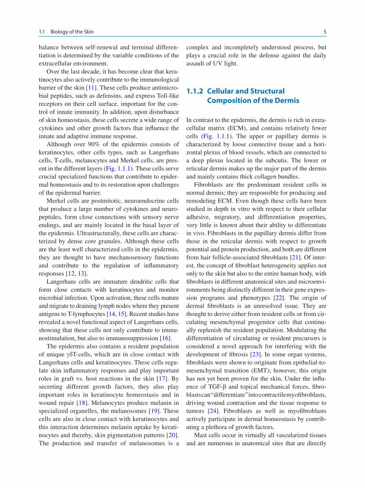
Over the last decade, it has become clear that kera-tinocytes also actively contribute to the immunological barrier of the skin [11] . These cells produce antimicro-bial peptides, such as defensins, and express Toll-like receptors on their cell surface, important for the con-trol of innate immunity. In addition, upon disturbance of skin homeostasis, these cells secrete a wide range of cytokines and other growth factors that infl uence the innate and adaptive immune response.
Although over 90% of the epidermis consists of keratinocytes, other cells types, such as Langerhans cells, T-cells, melanocytes and Merkel cells, are pres-ent in the different layers (Fig. 1.1.1 ). These cells serve crucial specialized functions that contribute to epider-mal homeostasis and to its restoration upon challenges of the epidermal barrier.
Merkel cells are postmitotic, neuroendocrine cells that produce a large number of cytokines and neuro-peptides, form close connections with sensory nerve endings, and are mainly located in the basal layer of the epidermis. Ultrastructurally, these cells are charac-terized by dense core granules. Although these cells are the least well characterized cells in the epidermis, they are thought to have mechanosensory functions and contribute to the regulation of infl ammatory responses [12, 13] .
Langerhans cells are immature dendritic cells that form close contacts with keratinocytes and monitor microbial infection. Upon activation, these cells mature and migrate to draining lymph nodes where they present antigens to T-lymphocytes [14, 15] . Recent studies have revealed a novel functional aspect of Langerhans cells, showing that these cells not only contribute to immu-nostimulation, but also to immunosuppression [16] .
The epidermis also contains a resident population of unique g d T-cells, which are in close contact with Langerhans cells and keratinocytes. These cells regu-late skin infl ammatory responses and play important roles in graft vs. host reactions in the skin [17] . By secreting different growth factors, they also play important roles in keratinocyte homeostasis and in wound repair [18] . Melanocytes produce melanin in specialized organelles, the melanosomes [19] . These cells are also in close contact with keratinocytes and this interaction determines melanin uptake by kerati-nocytes and thereby, skin pigmentation patterns [20] . The production and transfer of melanosomes is a
complex and incompletely understood process, but plays a crucial role in the defense against the daily assault of UV light.
1.1.2 Cellular and Structural Composition of the Dermis
In contrast to the epidermis, the dermis is rich in extra-cellular matrix (ECM), and contains relatively fewer cells (Fig. 1.1.1 ). The upper or papillary dermis is characterized by loose connective tissue and a hori-zontal plexus of blood vessels, which are connected to a deep plexus located in the subcutis. The lower or reticular dermis makes up the major part of the dermis and mainly contains thick collagen bundles.
Fibroblasts are the predominant resident cells in normal dermis; they are responsible for producing and remodeling ECM. Even though these cells have been studied in depth in vitro with respect to their cellular adhesive, migratory, and differentiation properties, very little is known about their ability to differentiate in vivo. Fibroblasts in the papillary dermis differ from those in the reticular dermis with respect to growth potential and protein production, and both are different from hair follicle-associated fi broblasts [21] . Of inter-est, the concept of fi broblast heterogeneity applies not only to the skin but also to the entire human body, with fi broblasts in different anatomical sites and microenvi-ronments being distinctly different in their gene expres-sion programs and phenotypes [22] . The origin of dermal fi broblasts is an unresolved issue. They are thought to derive either from resident cells or from cir-culating mesenchymal progenitor cells that continu-ally replenish the resident population. Modulating the differentiation of circulating or resident precursors is considered a novel approach for interfering with the development of fi brosis [23] . In some organ systems, fi broblasts were shown to originate from epithelial-to-mesenchymal transition (EMT); however, this origin has not yet been proven for the skin. Under the infl u-ence of TGF- b and topical mechanical forces, fi bro-blasts can “differentiate” into contractile myofi broblasts, driving wound contraction and the tissue response to tumors [24] . Fibroblasts as well as myofi broblasts actively participate in dermal homeostasis by contrib-uting a plethora of growth factors.
Mast cells occur in virtually all vascularized tissues and are numerous in anatomical sites that are directly

6 B. Eckes et al.
exposed to the environment and easily identifi ed by the presence of prominent cytoplasmic granules [25] . In the skin, they are frequently associated with blood vessels and appendages. Mast cells constitute an important cell type of the innate immune system and play an important role in infl ammation and tissue remodeling. Their acti-vation mainly occurs via the high affi nity IgE receptor (Fc e RI) or by contact with pathogens. Activated mast cells release an array of mediators e.g., histamine, pro-teases, and lipid metabolites, thereby causing extensive vasodilation, urticae, and itching, and are a rich source of growth factors. Although many released substances act as pro-infl ammatory mediators, mast cells also seem to have immunosuppressive and anti-infl ammatory roles through the release of IL-10 and TGF- b .
Other important constituents of the dermis are blood vessels and a lymphatic system, which are closely inter-connected. The cutaneous microcirculation is organized as two horizontal plexuses, the upper one at the level of dermal papillae and the lower one at the dermal-subcuta-neous junction. These are joined by paired ascending arterioles and descending venules. Microvascular endothelial cells supply nutrients to the skin and are essential for wound repair and the growth of tumors, and they regulate heat loss and temperature control. Depending on the size of the blood vessel and its loca-tion within the dermis, the endothelial tube is sur-rounded by up to several layers of smooth muscle cells or pericytes and by an outer basement membrane [26] . Endothelial cells and smooth muscle cells/pericytes form tight intercellular junctions with interdigitating processes, which together with the basement membrane control the distribution of biologically active molecules, mediators, or bioactive ECM fragments. Microvascular endothelial cells express a number of adhesion mole-cules for platelets and leukocytes to safeguard hemosta-sis and the transmigration of infl ammatory and precursor cells from the circulation into the skin.
Much less is known about the lymphatic system, which drains protein-rich fl uid from the extracellular space and transports immune cells from the skin to regional lymph nodes [27] . Lymph capillaries are lined by endothelial cells and are highly permeable due to the lack of a continuous basement membrane. The main difference between vascular and lymphatic endothelial cells in normal adult skin is the presence of VEGF receptor-1 or -2 on vascular endothelial cells, respond-ing to the VEGF-A isoform, and VEGF receptor-3 on lymphatic endothelial cells, responding to VEGF-C. During tissue repair and tumor vascularization, this
distinction is less clear and novel markers for the lym-phatic system will help in the analysis.
The major part of the dermis is the connective tissue, composed of structural proteins and nonstructural ele-ments produced predominantly by fi broblasts. This ECM provides structural support, organization and ori-entation to tissues. The structural elements are com-posed of collagens, elastin, fi brillins, fi bronectin and other high molecular weight glycoproteins, which are members of smaller or larger protein families. They are large modular molecules assembled from a limited set of modules or domains, which have biological activity on their own. Most ECM genes have arisen by duplica-tion of genes already present in ancestral organisms. The ECM proteins are embedded in a so-called ground substance of proteoglycans, which supply hydration and elasticity. Interaction between the different macromole-cules builds a large macromolecular network [28– 30] .
One of the less appreciated, but not less important, functions of the ECM is the retention of growth fac-tors such as TGF- b [31, 32] . Adequate stimulation or proteolytic activity can liberate the mediators and topi-cally restrict their activity; this quick adaptive response is critical, for example, in infl ammation.
Apart from exerting biological functions such as promoting migration or proliferation as entire ECM proteins, fragments cleaved off from them have gained attention for their own and have distinct properties, which may differ from the parental molecule [33] . One classic example is endostatin with antiangiogenic activity, which is cleaved off from the basement mem-brane collagen XVIII.
Probably, the largest ECM protein family is that of the collagens (Table 1.1.1 ), which exist in 28 different
Table 1.1.1 Collagen types in the skin
Dermis
Fibril-forming collagens I, III, V
FACITS (fi bril-associated collagen with interrupted triple helix)
XII, XIV, XVI
Microfi brillar collagen VI
Basement membrane collagens
Ubiquitous collagens IV, XVIII
Anchoring fi bril collagen VII
Anchoring fi lament collagen XVII
Endothelial basement membranes VIII
Epidermal/transmembrane collagens XIII, XVII, XXIII

71.1 Biology of the Skin
types [34] . All have a similar structure with a charac-teristic triple helix, which can vary considerably in length. The fi bril-forming collagens I, III and V make up most of the net weight of the dermis and represent the principle tensile element. The interstitial connec-tive tissue also harbors the microfi brillar collagen VI and the fi bril-associated collagen XIV. Collagen IV is a network forming molecule that is an essential con-stituent of the dermo-epidermal basement membrane. Collagen VII is the molecular component of the anchoring fi brils, which connect the basement mem-brane to the dermis.
Next to secreted collagens, there are unusual, trans-membrane, collagens [35] . Interestingly, several of these unusual collagens are subject to ectodomain shedding by metalloproteases, resulting in the release of the extracellular domain. These collagens can thus exist in two functionally potentially very different pro-tein forms. Although the functional signifi cance is unclear currently, this mechanism allows cells to switch rapidly from a cell surface (adhesive) receptor to a secreted form that can serve as an ECM compo-nent. One of the best studied examples is collagen XVII, which is an important cellular component of hemidesmosomes in epidermal keratinocytes. It was initially discovered as one of auto-immune antigens in bullous pemphigoid, hence its alternative name BP180. Recently, a novel variant of collagen VI [36] was reported as potential molecular target of atopic derma-titis [37] .
1.1.3 Basement Membranes
The epidermis and dermis cooperate in the formation of a highly specialized ECM structure, the basement mem-brane zone (BM), which physically separates these two compartments. The BM zone consists of a highly com-plex network of interconnecting ECM proteins, the key components being collagen IV, laminin, nidrogen and proteolycans [38– 40] . The skin basement membrane zone is characterized by auxiliary structures, the anchor-ing complexes, which consist of adhesion structures called hemidesmosomes (see below), anchoring fi la-ments and anchoring fi brils. The anchoring fi laments are mainly made up of Laminin 5, the major laminin isoform present in basement membranes of the skin and a crucial adhesive substrate for basal keratinocytes. Laminin-5 physically links the epidermis to collagen
VII, the molecular constituent of the anchoring fi brils, which form the mechanical connection of the base-ment membrane to the underlying dermis. The impor-tance of the anchoring complex in the maintenance of skin integrity is underscored by skin blistering diseases that are either caused by genetic mutations in one of these protein constituents of the basement membrane or by the production of auto-antibodies against several components [41– 43] .
1.1.4 Cell–Matrix and Cell–Cell Adhesion in the Skin
Intercellular and cell–matrix adhesion are crucial for cellular communication and play important roles in skin homeostasis and in the response to skin chal-lenges. Cell adhesion is mediated by a large variety of cell adhesion receptors that can be subdivided into sev-eral different families. The most prominent of these are the integrin family of cell–matrix and cell–cell recep-tors, the cadherin superfamily of intercellular adhesion receptors, the IgG family of cell–cell and cell–matrix receptors, the selectins and the proteoglycan receptor family. Upon adhesion, most of these receptors cluster into specialized junctional structures that are associ-ated with the cytoskeleton (Fig. 1.1.2 ). These struc-tures not only have important adhesive functions but also provide the cell with spatial landmarks important for localized signaling. Indeed, for most adhesion receptors, it is now clear that they not only connect cells to their environment but also, by connecting to signaling molecules, can communicate signals from the cell to its environment (so called inside-out signal-ing), and from the environment to the cell (outside-in signaling) [44] .
Intercellular junctions are most prominent in kerati-nocytes and endothelial cells (Fig. 1.1.2 ). However, dermal fi broblasts do form gap junctions and special-ized forms of adherens junctions, which can be established over relatively long distances. Intercellular Junction formation is also dynamically regulated upon activation of dermal fi broblasts. In addition, intercel-lular junctions are crucial for dermal vascular integrity. Dynamic intercellular adhesion also plays a crucial role in the interaction of immune and infl ammatory cells with other cell types when skin integrity is per-turbed. Four different types of intercellular junctions characterize the epidermis:

8 B. Eckes et al.
1. Desmosomes consist of desmosomal cadherins that are linked to the keratin fi lament system through specialized cytoskeletal adapator proteins, such as plakoglobin, plakophilins and the plakin desmoplakin. In the skin, desmosomes are not only found in the epidermis but also in vascular endothe-lia, where they form an intermixed structure with adherens junctions, called syndesmosomes. Although desmosomes show an ultrastructurally similar appearance throughout the epidermis, they have distinct molecular compositions that depend on the differentiation status of the keratinocytes, and most likely contribute specifi c functions [45] . For example, corneocytes are connected by a specialized variant, the corneodesmosome. Their importance for epidermal integrity is underscored by the existence of genetic and auto-immune skin blistering diseases, which are characterized by mutations in, or antibodies against,desmosomal components [46] . Next to their importance in pro-viding mechanical strength to epidermal intercel-lular cohesion, novel functions have emerged for
desmosomal components in the regulation of dif-ferentiation, survival, and growth.
2. Tight junctions form size and ion specifi c barriers in epithelia and vascular endothelial cells. In the epidermis, functional tight junctions are present in the granular layer. Tight junctions consist of two different four transmembrane spanning protein families, the occludins and claudins that link to actin via several different linker molecules e.g., the scaffolding proteins ZO-1/2. Differential expres-sion of claudins provides tight junctions found at different sites with their size and ion specifi city and, thereby, determine the tightness of the epithelial and vascular barrier [47, 48] . For keratinizing epi-thelia, it was originally thought that the secretion and deposition of a cross-linked protein–lipid bar-rier obviated the need for a tight junction barrier in such tissues, even though tight junctional proteins were identifi ed in the epidermis. The fi rst functional evidence that a tight junction component is required for barrier function in epidermis came from clau-din-1 knockout mice, which showed severe water
gap
junction
hemidesmosomejunction
focaladhesion
tight
junction
found onkeratinocytesendothelial cells
keratinocytes
keratinocytesendothelial cells
keratinocytesmelanocytesendothelial cellsfibroblastsinflammatory cells?
keratinocytesfibroblastsendothelial cellsmelanocytes?inflammatory cells?
keratinocytesfibroblastsendothelial cells
actin
actinIF
IF
adherens
junction
desmosome
junction
Fig. 1.1.2 Schematic representation of the different types of cell–matrix and cell–cell junctions

91.1 Biology of the Skin
loss due to impaired barrier function of the stratum granulosum. Subsequently, a dense network of strands resembling tight junctions was shown to be present in the stratum granulosum of human epider-mis. It is now widely accepted that tight junctions form a critical part of the barrier in the upper viable layer of stratifying epithelia [47] . Improper func-tion of tight junctions most likely contributes to human skin diseases, since claudin-1 mutations have been found to be associated with a rare form of ichthyosis [46] .
3. Adherens junctions link cellular adhesive contacts, mediated by classical cadherins and the IgG sub-family of nectins, to the actin cytoskeleton. Both adhesion molecules can form a dynamic link to the cytoskeleton by interacting with actin binding proteins and with regulators of actin dynamics [49] . Their importance of adhesion in the skin is underscored by the recent observation that auto-antibodies to E-cadherin results in skin blistering diseases [50] . In addition, cell contacts of keratino-cytes with T-cells, Langerhans cells and melano-cytes are mediated by E-cadherin [51] . Dynamic differential classical cadherin expression in the basal layer also contributes to epidermal and hair follicle morphogenesis [52] . This is further empha-sized by the observation that mutations in P-cadherin underlie hair disorders as well as rare forms of ecto-dermal dysplasia [53, 54] . In mice, it was shown that epidermal E-cadherin is crucial for functional tight junctions in the granular layer, thereby regu-lating epidermal barrier function [47] .
4. Gap junctions are crucial structures for intercellular communication by forming pores that allow the passage and exchange of small molecules between adjacent cells [55] . Connexin proteins constitute the molecular basis of the pores. Their importance for skin function is demonstrated by connexin muta-tions that underlie a number of inherited skin related diseases, including Vohlwinkel syndrome and ich-tyosis, and palmoplantar keratoderma related enti-ties [46, 56] .
Cell–matrix adhesion structures consist of integrin based hemidesmosomes and focal adhesions (Fig. 1.1.2 ). Hemidesmosomes anchor epidermal keratinocytes fi rmly to the underlying basement membrane and are absent in other skin cell types. Focal adhesion like struc-tures can be observed both in the dermis and epidermis.
Integrins are heterodimeric adhesion receptors, consist-ing of an a and b subunit, the composition of which determines ligand specifi city. Integrins are found on almost all cell types of the body where they function not only as cell–cell or cell–matrix adhesion receptors, but also communicate signals from the inside to the outside of the cell and vice versa. As such, they perform crucial functions in the regulation of connective tissue metabo-lism, epithelial morphogenesis and homeostasis, vascu-lar function, and immune system. They form the most important group of cell–matrix receptors in the skin.
Hemidesmosomes, or half desmosomes, resemble desmosomes at the ultrastructural and functional level, in that they show a similar organization, and by connecting to keratin fi laments, are crucial structures for mechanical stability. Nevertheless, their molecular composition is very different, consisting of the integrin a 6 b 4 and the previously mentioned collagen XVII (formerly known as BPAG1) as adhesion receptors and several cytoskeletal linker molecules of the plakin family, such as plectin and BP230 [57] . The b 4 subunit is unique among the integrin b sununits because of its long cytoplasmic domain that, unlike the other actin-linked b subunits, links a 6 b 4 to intermediate fi laments. Hemidesmosomes are crucial for the integrity of the skin, since mutations have been found in each of its known components, all of which lead to skin blistering diseases [42, 58] .
The b 1 and a v integrin subfamilies provide the scaf-fold of focal adhesions, which recruit a variety of cytoskeletal and signaling proteins, the most prominent ones being talin, vinculin, kindlins, the focal adhesion kinase (FAK), and integrin linked kinase (ILK) [59] . These structures are crucial for skin homeostasis, since they contribute to a wide variety of functions on the dif-ferent skin cells. Many of these are important for skin homeostasis, as underscored by the loss of b 1-integrins in the epidermis of mice, resulting not only in the for-mation of microblisters, but also in proliferative defects and skin infl ammation [60] . Other functions become more important when the skin is challenged. For exam-ple, a 2 integrins regulate vascularization during wound healing [61] . Although focal contacts as a structure have not been identifi ed in the skin in vivo, related structures are most likely important, as emphasized by the mutations in different focal contact components that underlie skin blistering related diseases [46] .
A recently emerging theme is that adhesive junc-tions may not only be crucial for tissue integrity and serve as clustering sites for signaling molecules, but

10 B. Eckes et al.
may also regulate communication with the nucleus at two different levels. First, it is now clear that many of the cytoskeletal linker proteins associated with adhe-sive junctions can also translocate to the nucleus where they regulate transcription [44, 62] . In addition, several cytoskeletal linker proteins also interact with compo-nents of the nuclear matrix, thereby potentially linking cell adhesion to nuclear positioning and shape changes, which can affect general transcriptional activity [63] .
1.1.5 Molecular Basis of the Epidermal Barrier
The physical epidermal barrier is built up by two phys-ically separated compartments: the tight junctions present in the uppermost viable layer, the stratum granulosum and the stratum corneum, which consists of a lipid and protein component, often referred to as “brick and mortar” [64, 65] . Tight junctions and the stratum corneum may cooperate in the formation of a functional barrier in stratifying epithelia. For example, overexpression of claudin-6 in the upper layers of the epidermis or epidermal deletion of the membrane anchored serine protease (CAP)1/Prss8 induced bar-rier defects that involved alterations in both tight junc-tions and stratum corneum. Although the underlying mechanisms are unknown, they may involve the coor-dinated regulation of both barriers by signal molecules such as IKK1 and retinoic acid receptor signaling [66] . In simple epithelia, tight junctions form a fence, thereby separating the apical membrane domain from the basolateral membrane domain. Since formation of the stratum corneum depends on the fusion of lamellar bodies and keratohyalin granules with plasma mem-branes at the transition between stratum granulosum and stratum corneum layers, it is tempting to speculate that the specifi c occurrence of tight junctions in the stratum granulosum regulates targeting of protein and lipid vesicles directly towards the “apically localized” stratum corneum (reviewed in [47] ).
The importance of the formation and maintenance of the physical barrier for the skin and, thus, the organ-ism is perhaps best illustrated by the fact that struc-tural barrier proteins, mostly keratins and fi laggrin, make up 80% of the total proteins of the epidermis. Indeed, the ultimate goal of the lifelong renewal and differentiation process described earlier is dedicated to
formation of the barrier. Keratins represent a large intermediate fi lament subfamily of about 50 proteins, divided into two classes, the acidic type I and the basic type II monomers. Dimers formed by one type I and one type II protein form the basic building blocks that subsequently assemble in higher order intermediate fi laments. Because of their unique, spatial and differ-entiation dependent expression pattern, keratins serve as excellent markers for the different compartments of the skin (Fig. 1.1.1 ), for regional skin pattern and for alterations in skin homeostasis [67, 68] .
For example, the initial differentiation step involves a switching of keratin expression from K5/K14 to K1/K10 in the suprabasal layers of the IFE. Others like K15 or K19 are used as markers to identify the local-ization of the bulge region, which contains the stem cell compartment of the hair follicle. Similarly, specialized hair follicle keratins contribute specifi c strengthening and barrier characteristics to each of the hair follicle differentiation layers. Moreover, specialized keratins, such as keratin 9, provide additional mechanical and barrier strength to skin on body sites prone to daily shear stress and “wear and tear,” such as palms or soles. In addition, alterations in keratins can indicate a patho-logical state of the skin. For example, K6/K16, K17, and K19 are normally confi ned to the hair follicle, but strongly upregulated in the IFE upon disturbance of skin homeostasis.
The importance of site specifi c expression of keratins is best refl ected in the identifi cation of mutations in, until now, 19 keratins in skin related diseases, most of which are associated with skin blistering [69] . These keratin related diseases not only emphasize their crucial importance in providing regional and site specifi c mechanical strength to epithelia, but also provide intrigu-ing hints for other keratin related functions independent of structure. Keratin mutations identifi ed in both mice and human are associated with pigment defects, albeit the underlying mechanisms by which keratins regulate epithelial pigmentation patterns are mostly unclear. Studies in mice have also uncovered roles of keratins in determining the onset of apoptosis crucial for hair folli-cle cycling and in the regulation of protein synthesis and cell size. Recently, a fascinating link has been estab-lished between focal adhesion formation and keratin fi lament assembly, suggesting that the different adhe-sion structures and their associated cytoskeletal net-works communicate directly to provide mechanical strength to cells (reviewed in Gu et al., 2007). The plakin

111.1 Biology of the Skin
family of cytoskeletal binding proteins may perform key functions in these processes, since they can interact with both actin and intermediate fi laments [63] .
Keratins form the core components of the corneo-cytes, the anucleate cells of the stratum corneum [70] . This requires bundling of the keratin fi laments, in which the late differentiation protein fi laggrin plays an important role in the bundling of keratins and in the formation of the cornifi ed envelope that forms the outer layer of the corneocytes. At the late steps of cornifi ca-tion, fi laggrin is processed into free hygroscopic amino acids that act as the natural moisturizing factors of the skin. Indeed, mutations in fi laggrin underlie ichthyosis variants and are also associated with atopic dermatitis, indicating its crucial importance not only in stratum corneum formation, but also in skin hydration [71] . Filaggrin is initially produced in the granular layer as a huge precursor, profi laggrin, which aggregate to form the characteristic keratohyalin granules of this layer. The cornifi ed envelope consists of a dense network of proteins, mostly loricrin, involucrin and cornifi n, which are tightly cross-linked to each other by enzymes such as transglutaminases [3] . Specialized desmosomes, so-called corneodesmosomes, connect corneocytes. A crucial step of desquamation is the proteolytic cleav-age of these corneodesmosomes. The intercellular space between corneocytes is fi lled by the lipid lamel-lae, a specialized structure of lipids crucial for epider-mal water barrier function [72, 73] .
An important aspect of cornifi cation and the subse-quent process of desquamation is the spatiotemporal activation and inhibition of proteases, cross linkers and lipid enzymes [74] . Although not well understood, these complex processes are balanced by inhibitors and activators of these enzymes and are at least partially regulated by gradients in pH and Ca 2 + -concentrations. The importance of proper spatiotemporal activation is stressed by diseases caused either by inappropriate activation or inhibition of these different enzymes due to e.g., lack of inhibitors or activators [75, 76] .
1.1.6 Cellular Communication Within the Skin
In recent years, it has become increasingly clear that the different skin cell types have a profound functional infl uence on each other, and that an extensive cellular
cross-talk regulates cell proliferation, differentiation, and coordinates the cellular and immunological responses to environmental challenges. Data generated in the recent past have resulted in a change of paradigm in understanding skin homeostasis and substantial number of skin diseases. It is now clear that keratino-cytes and fi broblasts not only represent the scaffold of the epidermis and dermis, but are also actively involved in the regulation of e.g., the innate and adaptive immune system [77] . They do so by secreting a large number of different mediators to communicate with endothelial cells and with infl ammatory cells during wounding or disease conditions. These include pro-infl ammatory cytokines, such as IL-1, IL10, and IL-6, antibacterial peptides, and growth factors such as VEGF or TGF- b . In turn, the activity of these cells and their extracellu-lar mediators profoundly infl uence the differentiation status of the keratinocytes and fi broblasts, thereby determining their response when the multifunctional skin barrier is challenged [78, 79] . An important mod-ulatory role in skin communication is provided by the ECM that not only serves as an important structural scaffold but also, by interacting with cells and many of the cytokines and growth factors, alters their functional activity. Together, these new fi ndings and insights have led to the realization that the primary cause of skin dis-eases associated with barrier dysfunction and infl am-mation can reside in keratinocytes and fi broblasts with a secondary contribution of classical infl ammatory cell types, activated through cellular communication. In addition, such primary defects in the skin can affect homeostasis of other organs, resulting in associated diseases in these organs.
1.1.7 Concluding Remarks
The last decade has brought a tremendous progress in the cell biology of the skin and thereby contributed to a better understanding of human skin diseases, as many of the examples mentioned in the following chapters exemplify. What many of these studies clearly revealed, regardless of whether one is looking from a fi broblast, endothelial cell, a keratinocyte, or immune cell point of view, is the extensive communication that occurs between cells and compartments of the skin. These new insights and fi ndings can now be used to identify the primary and secondary events that are still unknown