therapeutic outcomes of patients with multifocal papillary...
TRANSCRIPT
Research ArticleTherapeutic Outcomes of Patients with Multifocal PapillaryThyroid Microcarcinomas and Larger Tumors
Soh-Ching Ng,1 Sheng-Fong Kuo,1 Szu-Tah Chen,2 Chuen Hsueh,3 Bie-Yu Huang,1 andJen-Der Lin2
1Division of Endocrinology and Metabolism, Department of Internal Medicine, Chang Gung Memorial Hospital, Keelung, Taiwan2Division of Endocrinology and Metabolism, Department of Internal Medicine, Chang Gung Memorial Hospital and Chang GungUniversity, Keelung, Taiwan3Department of Pathology, Chang Gung Memorial Hospital and Chang Gung University, Keelung, Taiwan
Correspondence should be addressed to Jen-Der Lin; [email protected]
Received 20 December 2016; Revised 2 March 2017; Accepted 9 April 2017; Published 31 May 2017
Academic Editor: Thomas J. Fahey
Copyright © 2017 Soh-Ching Ng et al. This is an open access article distributed under the Creative Commons AttributionLicense, which permits unrestricted use, distribution, and reproduction in any medium, provided the original work isproperly cited.
A retrospective review of 626 patients with multifocal papillary thyroid carcinoma (PTC) including 147 patients (23.5%) withmultifocal papillary thyroid microcarcinoma (PTMC) from a total of 2,536 patients with PTC who visited the Chang GungMedical Center in Linkou, Taiwan, was performed. A comparison of the clinical features between 626 multifocal and 1,910solitary PTC cases showed that patients in the multifocal PTC group were older and had a smaller mean tumor size, amore advanced tumor-node-metastasis (TNM) stage, and a higher percentage of nonremission status compared to patientsin the solitary PTC group. Of the 626 patients with multifocal PTC, the group with larger tumors showed a moreadvanced TNM stage, a higher percentage of lymph node metastasis and soft tissue invasion, and a higher nonremissionrate compared to the multifocal PTMC group. Of the 626 patients with multifocal PTC, 25 patients (4%) died during amean follow-up period of 7.1± 5.3 years. Kaplan-Meier survival curves showed a significantly lower survival rate associatedwith multifocal PTMC compared to that with solitary PTMC.
1. Introduction
Multifocal papillary thyroid carcinoma (PTC) may present asmicrocarcinoma (≤1.0 cm) or larger tumors in two or moreindividual locations within the thyroid gland. An increasein the incidence of papillary thyroid microcarcinoma(PTMC) and multifocal PTC has been reported in the recentdecade [1–3]. Intrathyroid multifocal PTMC is categorized asa low-risk group in recent American Thyroid Association(ATA) guidelines [4]. In contrast, multifocal PTMC withextrathyroidal extensions is categorized as an intermediate-risk group. Bilateral multifocal PTMC has a worse prognosisthan unilateral PTMC [5, 6]. Multifocal PTC may be diag-nosed as “incidental” in the final histopathology examinationor “nonincidental” if it is diagnosed before or during thyroidsurgery [7, 8]. For both incidental and nonincidental PTCs,
prognostic factors of these patients are important to deter-mine whether a second surgery for complete total thyroidec-tomy is necessary. Postoperative radioiodine (131I) remnantablation and other imaging techniques are important foridentifying patients with a high risk of incidental or noninci-dental PTC [9]. However, data are lacking concerning thelong-term therapeutic outcomes of patients with multifocalPTMC and multifocal PTC with larger tumors in order toprovide appropriate therapeutic modalities for these patientgroups. Analyses of long-term follow-up results after multi-ple modality treatments are important and may provide bet-ter therapeutic strategies for the treatment of patients withmultifocal PTC.
The aims of this study were to investigate the long-termfollow-up outcomes of patients after different therapeuticstrategies for the treatment of multifocal PTC including
HindawiInternational Journal of EndocrinologyVolume 2017, Article ID 4208178, 8 pageshttps://doi.org/10.1155/2017/4208178

PTMC and larger tumors. We analyzed different therapeuticstrategies including surgical methods, 131I remnant ablation,131I treatment, imaging studies, and external radiotherapy.Various therapeutic strategies and risk factors for cancermortality and recurrence in different patient groups wereanalyzed.
2. Materials and Methods
We selected 3265 patients with PTC from a total of 4062patients with thyroid cancer who had undergone thyroid sur-gery between 1977 and 2013 at the Chang Gung MedicalCenter in Linkou, Taiwan. Patients who did not undergo afollow-up for over 1 year and who underwent the initial thy-roid surgery at a different hospital and patients without dataregarding tumor sizes were excluded from our analyses(Figure 1). All patients had undergone primary thyroidsurgery and a long-term clinical follow-up at Chang GungMedical Center. Frozen tissue specimens from 525 patientswere obtained during surgery and evaluated by pathologists.Initially, each specimen was assessed macroscopically toidentify the most significant nodules and the most invasivetumors and to plan the surgical dissection of the surgicalsample. Intraoperative examinations of the frozen sectionwere performed on the cut surface of the thyroid nodulesincluding the interface between the nodule and adjacent thy-roid tissue. A total thyroidectomy was performed on patientswith PTC with extrathyroidal extensions and lymph nodemetastasis. Patients with PTMC and an absence of extrathyr-oidal invasion as diagnosed by postoperative histopathologyunderwent follow-up care if they had undergone a subtotalthyroidectomy. Subtotal thyroidectomy was defined asremoving more than 50% of the entire gland on both lobes.One hundred and forty patients received secondary thyroidoperations for a complete thyroidectomy after PTC was con-firmed by histology.
A total of 2536 PTC patients were enrolled in this study,including 626 patients with multifocal PTC and 147 patientswith multifocal PTMC. In contrast, there were 1910 patientswith solitary PTC including 420 (22.0%) patients with papil-lary microcarcinomas. After thyroid surgery, patients werestaged using the Union for International Cancer Controltumor-node-metastasis (TNM) criteria (6th edition) [10].All thyroid carcinoma tissues were pathologically classifiedaccording to the World Health Organization criteria [11].PTMC was defined as the largest tumor diameter ≤ 1 cm inthe final histological slides. Cases were defined as multifocalif two or more isolated foci of PTC were found.
In our center, patients with PTC at high or intermediaterisk were recommended to undergo thyroid 131I remnantablation 4 to 6 weeks after thyroidectomy [4]. The doseof 131I ablation for most patients was 30–100mCi (1.1–3.7GBq). One week after 131I administration, a whole-bodyscan (WBS) was performed using a dual-head gamma cam-era (Siemens Medical Solutions USA Inc., USA). Thyroidscintigraphy was performed using a pinhole collimator witha 4mm aperture placed 7 cm above the neck for a total of50,000 counts for 30 minutes. Levothyroxine treatment wasinitiated to decrease the levels of thyroid-stimulatinghormone without inducing clinical thyrotoxicosis. If 131Iuptake extended beyond the thyroid bed, patients wereclassified as having residual disease or metastasis unlessproven to be a false-positive result. Higher therapeutic dosesof 3.7–7.4GBq (100–200mCi) were administered to thesepatients. Patients receiving doses exceeding 1.1GBq wereisolated at hospital admission. A WBS was performed 2weeks after administering higher therapeutic doses of 131I.Neck ultrasonography was performed 6–12 months afterthyroidectomy to exclude the possibility of local recurrence.
At the end of 2014, patients were categorized intofour groups: thyroid cancer mortality, nonremission,remission, and disease-free. The remission group consistedof patients with negative 131I WBS results and no evidence
Total thyroid cancer patients n = 4,062 (1977–2013)
PTC (n = 3265, 80.4%)Excluded:(i) Lost to follow-up, n = 469(ii) 1st operation outside hospital (n = 260)
Multifocal PTC (n = 626)Female (n = 499, 79.7%)Male (n = 127, 20.3%)
Macro-PTC (n = 464, 76.5%)
Mortality (n = 4, 0.6%) Mortality (n = 21, 3.3%)
PTC (n = 2536)
Solitary PTC (n = 1910)Female (n = 1497, 78.4%)Male (n = 413, 21.6%)
Macro-PTC (n = 1490, 78.0%)
Mortality (n = 2, 0.1%) Mortality (n = 82, 4.3%)
PTMC(n = 147, 23.5%)
PTMC(n = 420, 22.0%)
Figure 1: Distribution of papillary thyroid carcinoma patients in the current study. PTC: papillary thyroid cancer; PTMC: papillarythyroid microcarcinoma.
2 International Journal of Endocrinology
of local or distant metastasis upon noninvasive examina-tion. Disease-free patients were defined as patients inremission with undetectable levels of stimulated thyroglob-ulin (Tg) and undetectable Tg antibodies at the finalfollow-up appointment.
The Chang Gung Medical Foundation InstitutionalReview Board approved this study (104-3901B); the require-ment for informed consent was waived because of the retro-spective nature of the study.
In our hospital, serum Tg levels were measured usingan immunoradiometric assay kit (CIS Bio International,Gif-sur-Yvette, France) before the end of 2014. The detectionlimit of the Tg kit was 0.5 ng/mL. The functional sensitivity ofthis assay, as assessed in our laboratory, was 1.2 ng/mL. Tgantibody levels were measured using a competitive radio-immunoassay (Biocode, Liège, Belgium) with an analyticalsensitivity of 6 IU/mL.
Unpaired t-tests were used to compare continuous databetween groups. Categorical data were compared usingchi-square or Fisher’s exact tests for small data sets.Cancer-related mortality was calculated, and the follow-upperiod was determined from the date of diagnosis to the date
of cancer-related mortality of the last survivor undergoingfollow-up care. Survival rates were calculated using theKaplan-Meier method and compared using a log-rank test[12]. A multivariate Cox proportional hazard regressionmodel was used to estimate the mortality risk. All statisticalanalyses were performed using the SPSS software, version17.0 (SPSS Inc., Chicago, IL, USA). p values < 0 05 were con-sidered statistically significant in all tests.
3. Results
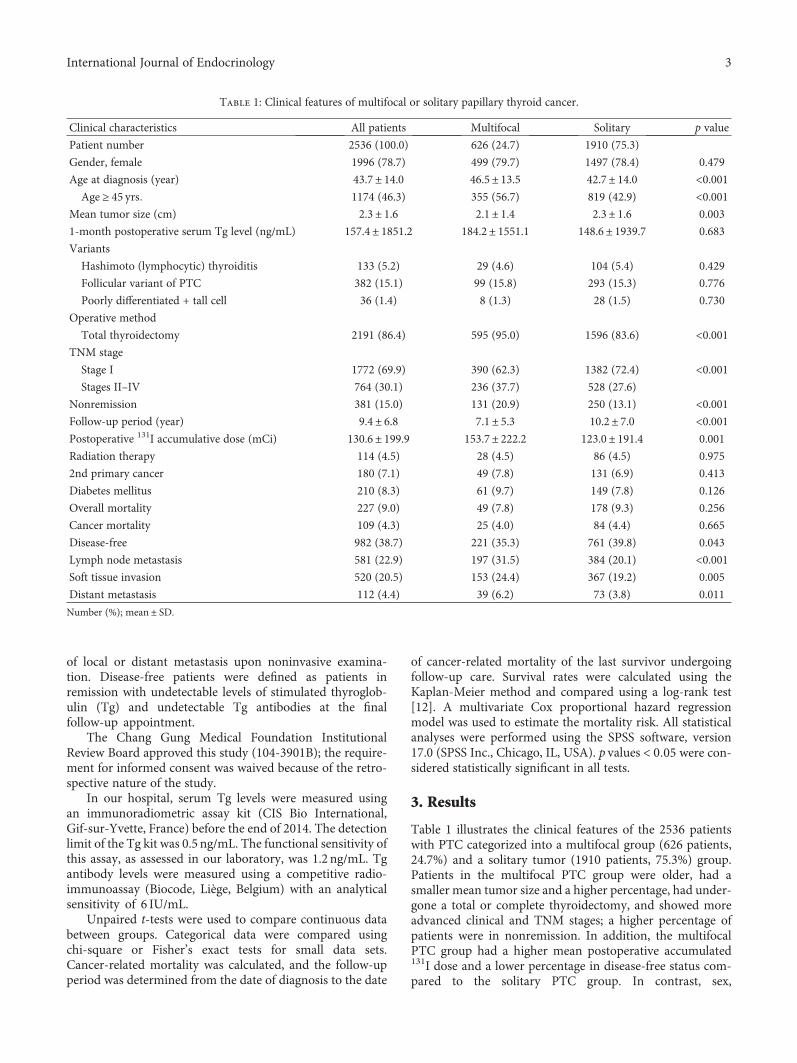
Table 1 illustrates the clinical features of the 2536 patientswith PTC categorized into a multifocal group (626 patients,24.7%) and a solitary tumor (1910 patients, 75.3%) group.Patients in the multifocal PTC group were older, had asmaller mean tumor size and a higher percentage, had under-gone a total or complete thyroidectomy, and showed moreadvanced clinical and TNM stages; a higher percentage ofpatients were in nonremission. In addition, the multifocalPTC group had a higher mean postoperative accumulated131I dose and a lower percentage in disease-free status com-pared to the solitary PTC group. In contrast, sex,
Table 1: Clinical features of multifocal or solitary papillary thyroid cancer.
Clinical characteristics All patients Multifocal Solitary p value
Patient number 2536 (100.0) 626 (24.7) 1910 (75.3)
Gender, female 1996 (78.7) 499 (79.7) 1497 (78.4) 0.479
Age at diagnosis (year) 43.7± 14.0 46.5± 13.5 42.7± 14.0 <0.001Age ≥ 45 yrs 1174 (46.3) 355 (56.7) 819 (42.9) <0.001
Mean tumor size (cm) 2.3± 1.6 2.1± 1.4 2.3± 1.6 0.003
1-month postoperative serum Tg level (ng/mL) 157.4± 1851.2 184.2± 1551.1 148.6± 1939.7 0.683
Variants
Hashimoto (lymphocytic) thyroiditis 133 (5.2) 29 (4.6) 104 (5.4) 0.429
Follicular variant of PTC 382 (15.1) 99 (15.8) 293 (15.3) 0.776
Poorly differentiated + tall cell 36 (1.4) 8 (1.3) 28 (1.5) 0.730
Operative method
Total thyroidectomy 2191 (86.4) 595 (95.0) 1596 (83.6) <0.001TNM stage
Stage I 1772 (69.9) 390 (62.3) 1382 (72.4) <0.001Stages II–IV 764 (30.1) 236 (37.7) 528 (27.6)
Nonremission 381 (15.0) 131 (20.9) 250 (13.1) <0.001Follow-up period (year) 9.4± 6.8 7.1± 5.3 10.2± 7.0 <0.001Postoperative 131I accumulative dose (mCi) 130.6± 199.9 153.7± 222.2 123.0± 191.4 0.001
Radiation therapy 114 (4.5) 28 (4.5) 86 (4.5) 0.975
2nd primary cancer 180 (7.1) 49 (7.8) 131 (6.9) 0.413
Diabetes mellitus 210 (8.3) 61 (9.7) 149 (7.8) 0.126
Overall mortality 227 (9.0) 49 (7.8) 178 (9.3) 0.256
Cancer mortality 109 (4.3) 25 (4.0) 84 (4.4) 0.665
Disease-free 982 (38.7) 221 (35.3) 761 (39.8) 0.043
Lymph node metastasis 581 (22.9) 197 (31.5) 384 (20.1) <0.001Soft tissue invasion 520 (20.5) 153 (24.4) 367 (19.2) 0.005
Distant metastasis 112 (4.4) 39 (6.2) 73 (3.8) 0.011
Number (%); mean ± SD.
3International Journal of Endocrinology

postoperative serum Tg levels, histological variants of PTC,thyroid cancer-specific mortality, and total mortality didnot differ significantly between these two groups.
Of the 626 patients with multifocal PTC, 147 (23.5%)patients had tumors of less than or equal to 1.0 cm. Thegroup with larger tumors had a significantly higher percent-age of patients that had undergone a total thyroidectomy,had a more advanced TNM stage, had a higher percentageof lymph node metastasis and soft tissue invasion, and hada higher nonremission rate than the patient group with mul-tifocal PTMC (Table 2). Age, sex, disease-specific mortality,overall mortality, and disease-free status did not differ signif-icantly between these groups. The follicular variant of PTCwas observed in 15.8% of all multifocal PTC diagnoses. Thelarger tumor group had a higher percentage of follicular var-iant PTC compared to the microcarcinoma group (17.7%versus 9.5%; p = 0 017).
Of the 626 patients, 31 (5.0%) underwent subtotal orlobectomy. Most of these patients presented with smalltumor sizes and a less advanced TNM stage. There weresingle patients with lymph node metastasis, soft tissueinvasion, and distant metastasis. Due to old age or
advanced local invasion, three patients did not undergo acomplete thyroidectomy. After the mean follow-up periodof 7.1 years, the nonremission rate, disease-specific mortality,and overall mortality did not differ significantly betweenpatients that had undergone a total thyroidectomy andpatients who had undergone less aggressive surgical treat-ments. Of the 626 patients, 335 patients had clinical stage 1disease, without lymph node metastasis, soft tissue invasion,or distant metastasis. At the end of the follow-up period,there was no disease-specific mortality in these 335 patients;however, there were nine cases of nonthyroid cancer mortal-ity. In addition, 17 of the 335 patients presented with recur-rent disease after a thyroidectomy including 10 patientswith lymph node metastasis and 2 patients with thyroidbed soft tissue recurrence.
After the mean follow-up period of 7.1± 5.3 years,131 (20.9%) patients were diagnosed as having a nonre-mission status. Table 3 shows the clinical characteristicsof patients in remission and nonremission. The nonre-mission group was predominantly male and had largertumor sizes, higher postoperative levels of serum Tg, amore advanced TNM stage, higher disease-specific and
Table 2: Clinical features of multifocal papillary thyroid cancer in different tumor sizes.
Clinical characteristics All patients Tumor size ≤ 1 0 cm Tumor size > 1 0 cm p value
Patient number 626 (100.0) 147 (23.5) 479 (76.5)
Gender, female 499 (79.7) 116 (78.9) 383 (80.0) 0.783
Age at diagnosis (year) 46.5± 13.5 47.1± 11.4 46.3± 14.1 0.541
Age ≥ 45 yrs 355 (56.7) 90 (61.2) 265 (55.3) 0.207
1-month postoperative serum Tg level (ng/mL) 184.2± 1551.1 296.5± 2725.5 150.3± 941.6 0.331
Operative method
Total thyroidectomy 595 (95.0) 130 (88.4) 465 (97.1) <0.001TNM stage
Stage I 390 (62.3) 122 (83.0) 268 (55.9) <0.001Stages II–IV 236 (37.7) 25 (17.0) 211 (44.1)
Nonremission 131 (20.9) 18 (12.2) 113 (23.6) 0.003
Residual 83 (13.3) 13 (8.8) 70 (14.6) 0.071
Relapsed 48 (7.7) 5 (3.4) 43 (9.0) 0.026
Follow-up period (year) 7.1± 5.3 6.6± 5.1 7.2± 5.4 0.245
Postoperative 131I accumulative dose (mCi) [range]153.7± 222.2[0.0–2050.0]
114.3± 176.7[0.0–1350.0]
165.9± 233.0[0.0–2050.0]
0.014
131I ≥ 30mCi 569 (90.9) 127 (86.4) 442 (92.3) 0.030131I < 30mCi 57 (9.1) 20 (13.6) 37 (7.7)
Radiation therapy 28 (4.5) 5 (3.4) 23 (4.8) 0.472
2nd primary cancer 49 (7.8) 11 (7.5) 38 (7.9) 0.859
Diabetes mellitus 61 (9.7) 12 (8.2) 49 (10.2) 0.460
Overall mortality 49 (7.8) 8 (5.4) 41 (8.6) 0.218
Cancer mortality 25 (4.0) 4 (2.7) 21 (4.4) 0.368
Disease-free 221 (35.3) 58 (39.5) 163 (34.0) 0.228
Lymph node metastasis 197 (31.5) 33 (22.4) 164 (34.2) 0.007
Soft tissue invasion 153 (24.4) 14 (9.5) 139 (29.0) <0.001Distant metastasis 39 (6.2) 5 (3.4) 34 (7.1) 0.105
Number (%); mean ± SD.
4 International Journal of Endocrinology

total mortality, and a lower percentage of patients indisease-free status. There were no statistically significantdifferences in age or surgical procedures between theremission and nonremission groups; however, patientsin the nonremission group received higher accumulateddoses of 131I and a higher percentage of patients under-went external radiotherapy.
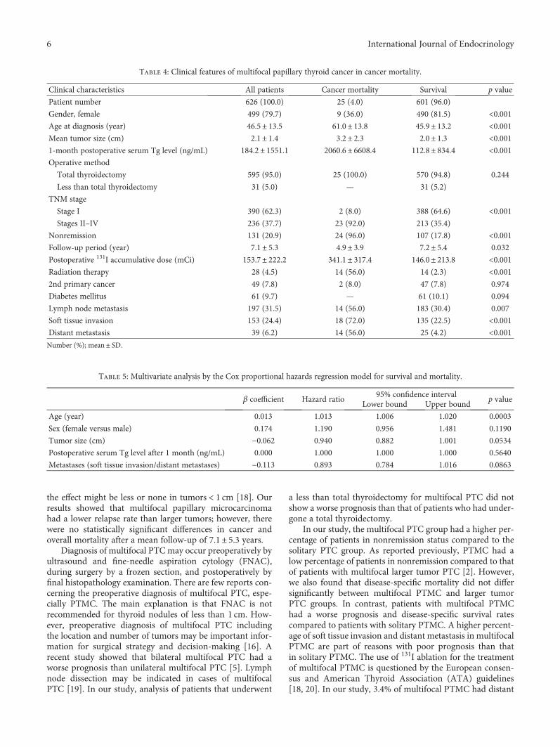
Of the 626 patients with multifocal PTC, 25 (4%) patientsdied during a mean follow-up period of 7.1± 5.3 years. Acomparison of risk factors between the cancer mortalityand survival groups revealed that male sex, older age, largertumor size, higher postoperative serum levels of Tg, andadvanced TNM stage differed significantly between thesegroups (Table 4). Multivariant analyses were performedusing the Cox proportional hazards regression model andrevealed that only age differed significantly between thesurvival and mortality groups (Table 5). The 5-, 10-, and20-year survival rates of the 2536 patients with PTC were97.3%, 95.7%, and 91.6%, respectively. Figure 2 shows theKaplan-Meier survival curves of the patients in the multifocalPTMC, multifocal larger PTC, solitary PTMC, and solitarylarger PTC groups. The 5-, 10-, and 20-year survival ratesof the four groups were 98.9%, 96.4%, 99.7%, and 96.8%;93.7%, 95.2%, 99.7%, and 95.0%; and 93.7%, 91.4%, 99.0%,and 90.3%, respectively. The survival rates of patients withsolitary PTMC were significantly different from the survivalrates of patients in the other three groups (Figure 2). Therewere no significant differences in survival rates between theother three groups.
4. Discussion
Multifocal PTC is the most frequently diagnosed multifocal,well-differentiated thyroid cancer, although follicular andmedullary thyroid cancer may also present with PTC in thesame patient [13]. Patients may be categorized as havingmultifocal PTMC, multifocal larger PTC, and mixed multifo-cal PTMC with larger tumors. In our study, one-quarter ofthe PTC cases were multifocal. Although the mean tumorsize of the largest multifocal tumor was smaller than that inthe solitary PTC group, the clinical and TNM stages weremore advanced. In contrast to that in a recent study, the mul-tifocal group in our study had a higher nonremission rate anda lower percentage of patients were disease-free when com-pared to the solitary PTC group [14]. These discordantresults may due to the larger number of patients enrolledand the longer follow-up period in our study.
In our study, 23.5% of multifocal PTCs were microcarci-nomas. This ratio was close to the reported proportion ofPTMC in an earlier study [15]. The prognosis of multifocalPTC was better than the prognosis for multifocal follicularor Hurthle cell histology [16]. However, more data arerequired regarding the long-term therapeutic outcomes ofmultifocal PTC in microcarcinoma or larger tumors, as wellas how unilateral or bilateral tumors and the number oftumors in multifocal carcinoma may affect treatment out-comes [1, 17]. Our study response to the consensus reportof the European Society of Endocrine Surgeons suggests thatprognosis might be impaired in clinical multifocal PTC, but
Table 3: Clinical features of multifocal papillary thyroid cancer in nonremission or remission.
Clinical characteristics All patients Nonremission Remission p value
Patient number 626 (100.0) 131 (20.9) 495 (79.1)
Gender, female 499 (79.7) 84 (64.1) 415 (83.8) <0.001Age at diagnosis (year) 46.5± 13.5 47.8± 17.1 46.1± 12.4 0.214
Mean tumor size (cm) 2.1± 1.4 2.6± 1.7 2.0± 1.3 <0.0011-month postoperative serum Tg level (ng/mL) 184.2± 1551.1 770.2± 3286.7 25.3± 102.8 <0.001Operative method
Total thyroidectomy 595 (95.0) 127 (96.9) 468 (94.5) 0.260
Less than total thyroidectomy 31 (5.0) 4 (3.1) 27 (5.5)
TNM stage
Stage I 390 (62.3) 53 (40.5) 337 (68.1) <0.001Stage II 58 (9.3) 15 (11.5) 43 (8.7) 0.332
Stage III 67 (10.7) 14 (10.7) 53 (10.7) 0.995
Stage IV 111 (17.7) 49 (37.4) 62 (12.5) <0.001Follow-up period (year) 7.1± 5.3 7.4± 5.5 7.0± 5.3 0.478
Postoperative 131I accumulative dose (mCi) 153.7± 222.2 384.2± 366.4 92.8± 95.5 <0.001Radiation therapy 28 (4.5) 27 (20.6) 1 (0.2) <0.0012nd primary cancer 49 (7.8) 15 (11.5) 34 (6.9) 0.083
Diabetes mellitus 61 (9.7) 13 (9.9) 48 (9.7) 0.938
Overall mortality 49 (7.8) 29 (22.1) 20 (4.0) <0.001Cancer mortality 25 (4.0) 24 (18.3) 1 (0.2) <0.001Disease-free 221 (35.3) 16 (12.2) 205 (41.4) <0.001Number (%); mean ± SD.
5International Journal of Endocrinology
the effect might be less or none in tumors < 1 cm [18]. Ourresults showed that multifocal papillary microcarcinomahad a lower relapse rate than larger tumors; however, therewere no statistically significant differences in cancer andoverall mortality after a mean follow-up of 7.1± 5.3 years.
Diagnosis of multifocal PTCmay occur preoperatively byultrasound and fine-needle aspiration cytology (FNAC),during surgery by a frozen section, and postoperatively byfinal histopathology examination. There are few reports con-cerning the preoperative diagnosis of multifocal PTC, espe-cially PTMC. The main explanation is that FNAC is notrecommended for thyroid nodules of less than 1 cm. How-ever, preoperative diagnosis of multifocal PTC includingthe location and number of tumors may be important infor-mation for surgical strategy and decision-making [16]. Arecent study showed that bilateral multifocal PTC had aworse prognosis than unilateral multifocal PTC [5]. Lymphnode dissection may be indicated in cases of multifocalPTC [19]. In our study, analysis of patients that underwent
a less than total thyroidectomy for multifocal PTC did notshow a worse prognosis than that of patients who had under-gone a total thyroidectomy.
In our study, the multifocal PTC group had a higher per-centage of patients in nonremission status compared to thesolitary PTC group. As reported previously, PTMC had alow percentage of patients in nonremission compared to thatof patients with multifocal larger tumor PTC [2]. However,we also found that disease-specific mortality did not differsignificantly between multifocal PTMC and larger tumorPTC groups. In contrast, patients with multifocal PTMChad a worse prognosis and disease-specific survival ratescompared to patients with solitary PTMC. A higher percent-age of soft tissue invasion and distant metastasis in multifocalPTMC are part of reasons with poor prognosis than thatin solitary PTMC. The use of 131I ablation for the treatmentof multifocal PTMC is questioned by the European consen-sus and American Thyroid Association (ATA) guidelines[18, 20]. In our study, 3.4% of multifocal PTMC had distant
Table 4: Clinical features of multifocal papillary thyroid cancer in cancer mortality.
Clinical characteristics All patients Cancer mortality Survival p value
Patient number 626 (100.0) 25 (4.0) 601 (96.0)
Gender, female 499 (79.7) 9 (36.0) 490 (81.5) <0.001Age at diagnosis (year) 46.5± 13.5 61.0± 13.8 45.9± 13.2 <0.001Mean tumor size (cm) 2.1± 1.4 3.2± 2.3 2.0± 1.3 <0.0011-month postoperative serum Tg level (ng/mL) 184.2± 1551.1 2060.6± 6608.4 112.8± 834.4 <0.001Operative method
Total thyroidectomy 595 (95.0) 25 (100.0) 570 (94.8) 0.244
Less than total thyroidectomy 31 (5.0) — 31 (5.2)
TNM stage
Stage I 390 (62.3) 2 (8.0) 388 (64.6) <0.001Stages II–IV 236 (37.7) 23 (92.0) 213 (35.4)
Nonremission 131 (20.9) 24 (96.0) 107 (17.8) <0.001Follow-up period (year) 7.1± 5.3 4.9± 3.9 7.2± 5.4 0.032
Postoperative 131I accumulative dose (mCi) 153.7± 222.2 341.1± 317.4 146.0± 213.8 <0.001Radiation therapy 28 (4.5) 14 (56.0) 14 (2.3) <0.0012nd primary cancer 49 (7.8) 2 (8.0) 47 (7.8) 0.974
Diabetes mellitus 61 (9.7) — 61 (10.1) 0.094
Lymph node metastasis 197 (31.5) 14 (56.0) 183 (30.4) 0.007
Soft tissue invasion 153 (24.4) 18 (72.0) 135 (22.5) <0.001Distant metastasis 39 (6.2) 14 (56.0) 25 (4.2) <0.001Number (%); mean ± SD.
Table 5: Multivariate analysis by the Cox proportional hazards regression model for survival and mortality.
β coefficient Hazard ratio95% confidence interval
p valueLower bound Upper bound
Age (year) 0.013 1.013 1.006 1.020 0.0003
Sex (female versus male) 0.174 1.190 0.956 1.481 0.1190
Tumor size (cm) −0.062 0.940 0.882 1.001 0.0534
Postoperative serum Tg level after 1 month (ng/mL) 0.000 1.000 1.000 1.000 0.5640
Metastases (soft tissue invasion/distant metastases) −0.113 0.893 0.784 1.016 0.0863
6 International Journal of Endocrinology

metastasis. A more aggressive postoperative remnant abla-tion and longer follow-up period than that of solitary PTMCare indicated.
Several studies have assessed the clonal origin of multifo-cal PTC [21–24]. However, the results were inconsistent.BRAF gene mutation combined with X chromosome inac-tivation analyses has been used to evaluate the clonal ori-gins of tumors and has showed that bilateral, recurrent,and metastatic PTCs often arise from a single clone andthat intrathyroid metastasis may play an important rolein the development of bilateral tumors [21]. In contrast,assessment of multifocal thyroid tumors using geneticalteration analyses and miRNA profiling found that multi-focal PTC did not necessarily evolve from single PTC pro-genitor foci [20]. In the clinic, different histological patternslike follicular variant and classical PTC in different thyroidlobes are not unusual. Both intrathyroid lymphatic spread-ing and different clonal origins may be present in multifo-cal PTC [24]. This study has several strengths, including anearly 10-year follow-up period of a large number ofpatients, which has strengthened the conclusions. Thepatients enrolled were diagnosed and treated at a singlemedical center, which may make the data more consistent.However, due to the enrollment period of over 30 years,examination and therapeutic modalities may likely havechanged over time.
5. Conclusion
Multifocal PTMC had a lower recurrence rate than multifo-cal larger tumor PTC; however, there was no difference incancer-specific mortality rates. These patients need afollow-up because there is a risk of recurrence.
Conflicts of Interest
The authors declare that they have no conflicts of interest.
References
[1] N. Qu, L. Zhang, Q. H. Ji et al., “Number of tumor foci predictsprognosis in papillary thyroid cancer,” BMC Cancer, vol. 14,no. 12, p. 914, 2014.
[2] K. J. Kim, S. M. Kin, Y. S. Lee, W. Y. Chung, H. S. Chang, andC. S. Park, “Prognostic significance of tumor multifocality inpapillary thyroid carcinoma and its relationship with primarytumor size: a retrospective study of 2,309 consecutivepatients,” Annals of Surgical Oncology, vol. 22, no. 1,pp. 125–131, 2015.
[3] J. D. Lin, S. T. Chen, T. C. Chao, C. Hsueh, and H. F. Weng,“Diagnosis and therapeutic strategy of papillary thyroidmicrocarcinoma,” Archives of Surgery, vol. 140, no. 10,pp. 940–945, 2005.
[4] B. R. Haugen, E. K. Alexander, K. C. Bible et al., “2015American Thyroid Association management guidelines foradult patients with thyroid nodules and differentiated thyroidcancer: the American Thyroid Association guidelines taskforce on thyroid nodules and differentiated thyroid cancer,”Thyroid, vol. 26, no. 1, pp. 1–133, 2016.
[5] W. Wang, X. Su, K. He et al., “Comparison of the clinico-pathologic features and prognosis of bilateral versus unilateralmultifocal papillary thyroid cancer: an updated study withmore than 2000 consecutive patients,” Cancer, vol. 122,no. 2, pp. 198–206, 2016.
[6] S. F. Kuo, S. F. Lin, T. C. Chao, C. Hsueh, K. J. Lin, andJ. D. Lin, “Prognosis of multifocal papillary thyroid carci-noma,” International Journal of Endocrinology, vol. 809382,no. 12, p. 2013, 2013.
[7] E. Dunki-Jacobs, K. Grannan, S. McDonough, and A. M.Engel, “Clinically unsuspected papillary microcarcinomas ofthe thyroid: a common finding with favorable biology?” TheAmerican Journal of Surgery, vol. 203, no. 2, pp. 140–144,2012.
[8] A. Kiriakopoulos, A. Petralias, and D. Linos, “Multifocal ver-sus solitary papillary thyroid carcinoma,” World Journal ofSurgery, vol. 40, no. 9, pp. 2139–2143, 2016.
[9] J. D. Lin, S. F. Kuo, T. C. Chao, and C. Hsueh, “Incidental andnon-incidental papillary thyroid microcarcinoma,” Annals ofSurgical Oncology, vol. 15, no. 8, pp. 2287–2292, 2008.
[10] L. H. Sobin and UICC, TNM Classification of MalignantTumors, W. Ch, Ed., pp. 52–56, Wiley-Liss, New York, 2002.
[11] R. A. Delellis, R. V. Lloyd, P. U. Heitx, and C. Eng, “Pathologyand genetics of tumors of endocrine organs,” in World HealthOrganization of Tumours, pp. 73–76, IARC, Lyon, 2004.
[12] D. D. Zhang, X. H. Zhou, D. H. Freeman, and J. L. Freema, “Anon-parametric method for the comparison of partial areasunder ROC curves and its application to large health care datasets,” Statistics in Medicine, vol. 21, no. 5, pp. 701–715, 2002.
[13] K. Kaliszewski, A. Zubkiewicz-Kucharska, B. Wojtczak, andM. Strutyńska-Karpińska, “Multi- and unifocal thyroid micro-carcinoma: are there any differences?” Advances in Clinicaland Experimental Medicine, vol. 25, no. 3, pp. 485–492, 2016.
[14] A. A. Tam, D. Özdemir, N. Çuhacı et al., “Association ofmultifocality, tumor number, and total tumor diameter withclinicopathological features in papillary thyroid cancer,”Endocrine, vol. 53, no. 3, pp. 774–783, 2016.
0 10 20 30 4075
80
85
90
95
100
Multifocal PTMCMultifocal larger PTCSolitary PTMC
Solitary larger PTCTotal
p = 0.5443p = 0.0065
p = 0.4498
p = 0.7115p = 0.0001
p = 0.0001
Surv
ival
rate
(%)
Follow‒up period (year)
Figure 2: Kaplan-Meier survival curves of the four subject groups:multifocal PTMC, multifocal larger PTC, solitary PTMC, andsolitary larger PTC. PTMC: papillary thyroid microcarcinoma.
7International Journal of Endocrinology
[15] J. D. Lin, “Increased incidence of papillary thyroid microcarci-noma with decreased tumor size of thyroid cancer,” MedicalOncology, vol. 27, no. 2, pp. 510–518, 2010.
[16] E. J. Kuo, S. A. Roman, and J. A. Sosa, “Patients with follicularand Hurthle cell microcarcinomas have compromised sur-vival: a population level study of 22,738 patients,” Surgery,vol. 154, no. 6, pp. 1246–1253, 2013.
[17] H. J. Kim, S. Y. Sohn, H. W. Jang, S. W. Kim, and J. H. Chung,“Multifocality, but not bilaterality, is a predictor of diseaserecurrence/persistence of papillary thyroid carcinoma,”WorldJournal of Surgery, vol. 37, no. 2, pp. 376–384, 2013.
[18] M. Iacobone, S. Jansson, M. Barczyński, and P. Goretzki,“Multifocal papillary thyroid carcinoma - a consensus reportof the European Society of Endocrine Surgeons (ESES),”Langenbeck’s Archives of Surgery, vol. 399, no. 2, pp. 141–154, 2014.
[19] A. Al Afif, B. A. Williams, M. H. Rigby et al., “Multifocalpapillary thyroid cancer increases the risk of central lymphnode metastasis,” Thyroid, vol. 25, no. 9, pp. 1008–1012, 2016.
[20] E. Krčálová, J. Horáček, L. Kudlej et al., “Is radioiodineadministration in patients with papillary thyroid multifocalmicrocarcinoma unnecessary?” Endocrinology Diabetes &Metabolism Case Report, vol. 2016, no. 5, p. 150138, 2016.
[21] W.Wang, H.Wang, X. Teng et al., “Clonal analysis of bilateral,recurrent, and metastatic papillary thyroid carcinomas,”Human Pathology, vol. 41, no. 9, pp. 1299–1309, 2010.
[22] S. T. Aherne, P. C. Smyth, R. J. Flavin Russell SM et al.,“Geographical mapping of a multifocal thyroid tumour usinggenetic alteration analysis & miRNA profiling,” MolecularCancer, vol. 7, no. 12, p. 89, 2008.
[23] E. Kuhn, L. Teller, S. Piana, J. Rosai, and M. J. Merino, “Differ-ent clonal origin of bilateral papillary thyroid carcinoma, witha review of the literature,” Endocrine Pathology, vol. 23, no. 2,pp. 101–107, 2012.
[24] Z. Lu, J. Sheng, Y. Zhang et al., “Clonality analysis of multifocalpapillary thyroid carcinoma by using genetic profiles,” TheJournal of Pathology, vol. 239, no. 1, pp. 72–83, 2016.
8 International Journal of Endocrinology

Submit your manuscripts athttps://www.hindawi.com
Stem CellsInternational
Hindawi Publishing Corporationhttp://www.hindawi.com Volume 2014
Hindawi Publishing Corporationhttp://www.hindawi.com Volume 2014
MEDIATORSINFLAMMATION
of
Hindawi Publishing Corporationhttp://www.hindawi.com Volume 2014
Behavioural Neurology
EndocrinologyInternational Journal of
Hindawi Publishing Corporationhttp://www.hindawi.com Volume 2014
Hindawi Publishing Corporationhttp://www.hindawi.com Volume 2014
Disease Markers
Hindawi Publishing Corporationhttp://www.hindawi.com Volume 2014
BioMed Research International
OncologyJournal of
Hindawi Publishing Corporationhttp://www.hindawi.com Volume 2014
Hindawi Publishing Corporationhttp://www.hindawi.com Volume 2014
Oxidative Medicine and Cellular Longevity
Hindawi Publishing Corporationhttp://www.hindawi.com Volume 2014
PPAR Research
The Scientific World JournalHindawi Publishing Corporation http://www.hindawi.com Volume 2014
Immunology ResearchHindawi Publishing Corporationhttp://www.hindawi.com Volume 2014
Journal of
ObesityJournal of
Hindawi Publishing Corporationhttp://www.hindawi.com Volume 2014
Hindawi Publishing Corporationhttp://www.hindawi.com Volume 2014
Computational and Mathematical Methods in Medicine
OphthalmologyJournal of
Hindawi Publishing Corporationhttp://www.hindawi.com Volume 2014
Diabetes ResearchJournal of
Hindawi Publishing Corporationhttp://www.hindawi.com Volume 2014
Hindawi Publishing Corporationhttp://www.hindawi.com Volume 2014
Research and TreatmentAIDS
Hindawi Publishing Corporationhttp://www.hindawi.com Volume 2014
Gastroenterology Research and Practice
Hindawi Publishing Corporationhttp://www.hindawi.com Volume 2014
Parkinson’s Disease
Evidence-Based Complementary and Alternative Medicine
Volume 2014Hindawi Publishing Corporationhttp://www.hindawi.com