therapeutic application and gait training - ottobock · condition after incomplete paraplegia)....
TRANSCRIPT
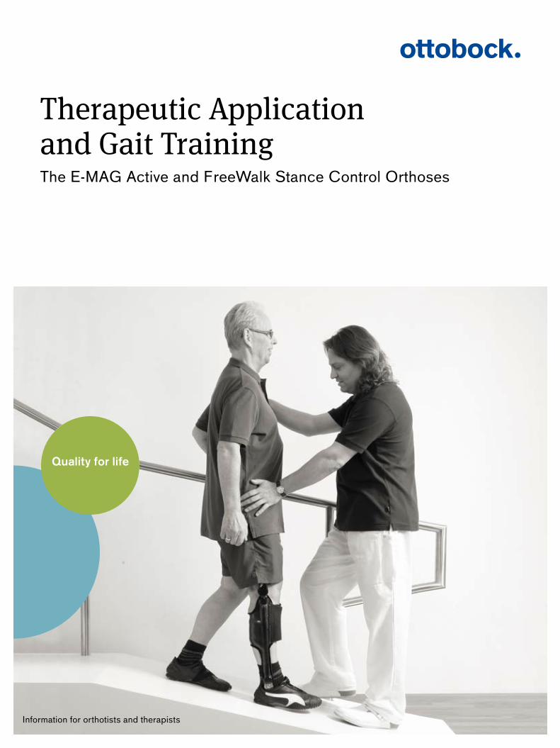
Therapeutic Application and Gait TrainingThe E-MAG Active and FreeWalk Stance Control Orthoses
Information for orthotists and therapists


Therapeutic Application and Gait Training | Ottobock 3
Contents
Introduction������������������������������������������������������������������������������������������������������������������������������������� �4Therapeutic�benefits����������������������������������������������������������������������������������������������������������������������� �6Relevant�underlying�diseases������������������������������������������������������������������������������������������������������� �7Biomechanics�of�the�human�gait�������������������������������������������������������������������������������������������������� �8Gait�cycle�with�stance�control�knee�joint�systems��������������������������������������������������������������������� �9Prerequisites�for�use�and�the�differences�between�knee-ankle-foot�orthoses�fitted�with�the�E-MAG�Active�joint�system�and�the�FreeWalk������������������������������������������������������������ �10• Muscle strength requirements������������������������������������������������������������������������������������������������������ �10• Testing muscle strength ��������������������������������������������������������������������������������������������������������������� �11• Mobility requirements ������������������������������������������������������������������������������������������������������������������� �13• Testing mobility����������������������������������������������������������������������������������������������������������������������������� �14Specific�therapy�to�improve�muscle�strength�and�mobility����������������������������������������������������� �16• Exercises to increase muscle strength ����������������������������������������������������������������������������������������� �16• Exercises to increase mobility ������������������������������������������������������������������������������������������������������ �18• Exercises to increase arthrogenous mobility in the foot, knee and hip joints ������������������������������ �19• Home exercises to increase muscle strength and mobility ���������������������������������������������������������� �20Handling�the�orthoses�and�starting�exercises�������������������������������������������������������������������������� �22• Donning the FreeWalk orthosis ���������������������������������������������������������������������������������������������������� �22• Donning a knee-ankle-foot orthosis with the E-MAG Active joint system������������������������������������� �23Standing�up�and�the�locking������������������������������������������������������������������������������������������������������� �24• Standing up with a bilaterally fitted E-MAG Active����������������������������������������������������������������������� �25Sitting�down�with�the�FreeWalk�orthosis����������������������������������������������������������������������������������� �26Sitting�down�with�the�E-MAG�Active������������������������������������������������������������������������������������������ �27• Sitting down with the E-MAG Active fitted bilaterally������������������������������������������������������������������� �28Gait�training����������������������������������������������������������������������������������������������������������������������������������� �29• Exercises for attaining even weight-distribution ����������������������������������������������������������������������������29• Stabilising exercises outside the parallel bars ������������������������������������������������������������������������������30• Practising initial heel contact up to stance phase �������������������������������������������������������������������������30• Exercise to practise the Lock Release function for the swing phase cycle with the FreeWalk ��������32• Exercise to practise the Lock Release function for the swing phase cycle
with a knee-ankle-foot orthosis using the E-MAG Active joint system ����������������������������������������� �33• Practising a double step with parallel bars and on a treadmill �����������������������������������������������������34Daily�activities������������������������������������������������������������������������������������������������������������������������������� �36• Walking backwards and sideways ����������������������������������������������������������������������������������������������� �36• Negotiating inclines/declines ������������������������������������������������������������������������������������������������������� �37• Climbing stairs ������������������������������������������������������������������������������������������������������������������������������38• Uneven ground ������������������������������������������������������������������������������������������������������������������������������39• Low-intensity sport ����������������������������������������������������������������������������������������������������������������������� �40Frequently�Asked�Questions������������������������������������������������������������������������������������������������������� �42• Functionality of the FreeWalk orthosis ������������������������������������������������������������������������������������������42• Functionality of a knee-ankle-foot orthosis fitted with the E-MAG Active joint system �����������������43
4 Ottobock | Therapeutic Application and Gait Training
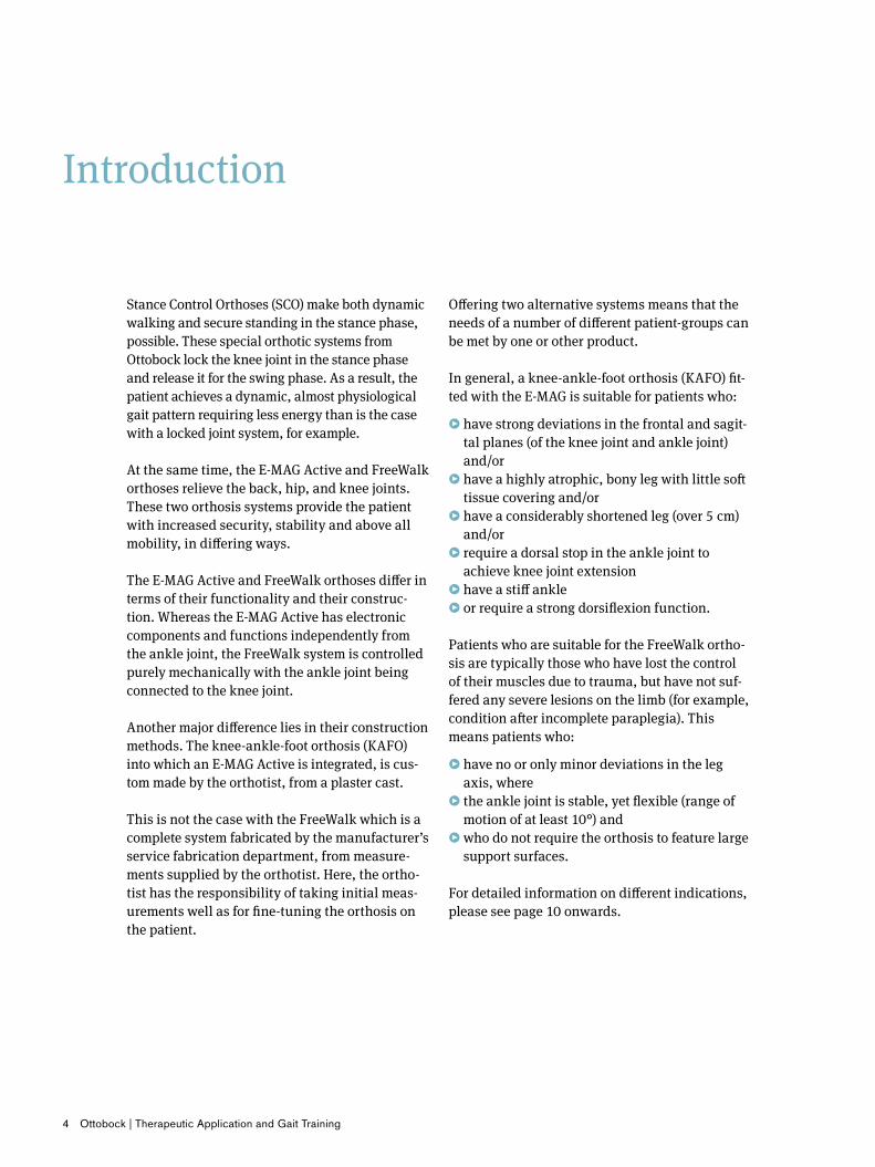
Stance Control Orthoses (SCO) make both dynamic walking and secure standing in the stance phase, possible. These special orthotic systems from Ottobock lock the knee joint in the stance phase and release it for the swing phase. As a result, the patient achieves a dynamic, almost physiological gait pattern requiring less energy than is the case with a locked joint system, for example.
At the same time, the E-MAG Active and FreeWalk orthoses relieve the back, hip, and knee joints. These two orthosis systems provide the patient with increased security, stability and above all mobility, in differing ways.
The E-MAG Active and FreeWalk orthoses differ in terms of their functionality and their construc-tion. Whereas the E-MAG Active has electronic components and functions independently from the ankle joint, the FreeWalk system is controlled purely mechanically with the ankle joint being connected to the knee joint.
Another major difference lies in their construction methods. The knee-ankle-foot orthosis (KAFO) into which an E-MAG Active is integrated, is cus-tom made by the orthotist, from a plaster cast.
This is not the case with the FreeWalk which is a complete system fabricated by the manufacturer’s service fabrication department, from measure-ments supplied by the orthotist. Here, the ortho-tist has the responsibility of taking initial meas-urements well as for fine-tuning the orthosis on the patient.
Offering two alternative systems means that the needs of a number of different patient-groups can be met by one or other product.
In general, a knee-ankle-foot orthosis (KAFO) fit-ted with the E-MAG is suitable for patients who:
• have strong deviations in the frontal and sagit-tal planes (of the knee joint and ankle joint) and/or
• have a highly atrophic, bony leg with little soft tissue covering and/or
• have a considerably shortened leg (over 5 cm) and/or
• require a dorsal stop in the ankle joint to achieve knee joint extension
• have a stiff ankle• or require a strong dorsiflexion function.
Patients who are suitable for the FreeWalk ortho-sis are typically those who have lost the control of their muscles due to trauma, but have not suf-fered any severe lesions on the limb (for example, condition after incomplete paraplegia). This means patients who:
• have no or only minor deviations in the leg axis, where
• the ankle joint is stable, yet flexible (range of motion of at least 10°) and
• who do not require the orthosis to feature large support surfaces.
For detailed information on different indications, please see page 10 onwards.
Introduction
Therapeutic Application and Gait Training | Ottobock 5
6 Ottobock | Therapeutic Application and Gait Training
The E-MAG Active knee joint system and the Free-Walk orthosis were developed for users who, due to partial paralysis or a complete failure of the knee extensors, are unable to stabilise their knees without compensatory measures.
The knee joint is often stabilised as a result of hyperextension achieved by compensatory acti-vation of the gluteal muscles (in other words, when the foot touches the ground, hip extension leads to knee extension).
As a result, severe ligament instabilities and arthritic symptoms in the knee joint will develop over time.
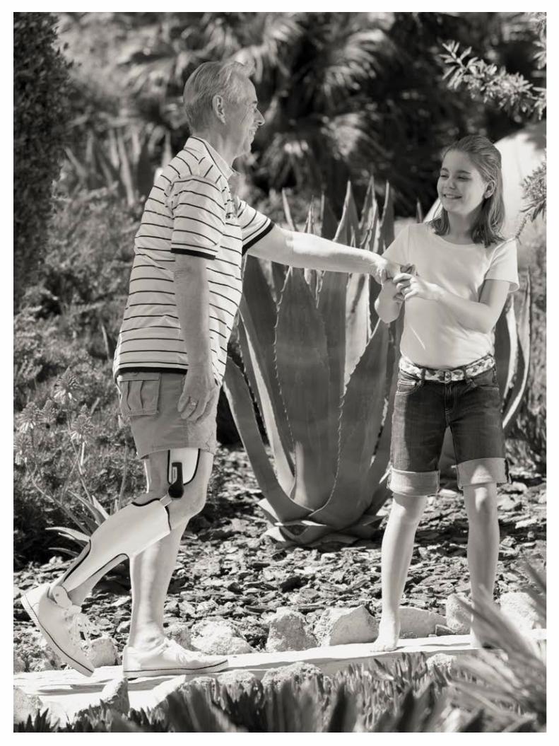
Therapeutic benefits
Stance control orthoses (SCO) can help correct these non-physiological movements. They provide the user with safe functionality and enable a largely normal gait.
There are numerous therapeutic benefits for paralysis patients: contractures and joint damage caused by immobilisation are prevented and muscle atrophy is reduced. Cardiovascular ability is retained for everyday activities. In the case of disorders affecting the central nervous system, the resumption of functions by other, unaffected brain regions (motor relearning, cortical reorgan-isation) can also be promoted. These are some of the areas in which our orthoses support the patient rehabilitation as well as their (re-)integration into their social and professional lives.
Therapeutic Application and Gait Training | Ottobock 7
Brain�diseases:• Cerebral palsy• Condition following a stroke• Condition following brain tumours• Condition following encephalitis/brain
abscesses• Condition following serious craniocerebral
trauma (CCT)• Multiple sclerosis• Ataxia in the context of diseases involving the
cerebellum (sporadic delayed atrophy of the cer-ebellar cortex, inherited cerebellar ataxia)
Diseases�of�the�spinal�cord:• Traumatic paraplegia • Incomplete hemispinal cord syndrome (Brown-
Séquard syndrome)• Condition following spinal cord tumours and
meningeal spinal tumours• Condition following paraplegie/paresis follow-
ing myelitis, abscesses• Spastic spinal paralysis• Amyotrophic lateral sclerosis (ALS)• Spinal muscular atrophies• Condition following acute poliomyelitis• Post-polio syndrome• Degenerative diseases (such as spinal stenosis,
stenosis of the intervertebral foramen, spondy-lolisthesis)
• Abnormalities of the spinal cord (such as verte-bral arch defects with spondylolisthesis, spina bifida cystica, meningocele, myelomeningocele)
• Funicular myelosis• Syringomyelia• Neural muscular atrophy• Anterior spinal artery syndrome• Condition following hernia of an intervertebral
disc
Relevant underlying diseases
Treatment with stance control orthoses is indicated for pareses or paralyses of muscles and muscle groups of the lower extremities. These may develop in the context of the following underlying diseases:
Muscular�diseases�(myopathies):• Muscular dystrophy (e. g. Duchenne MD)• Condition following polymyositis/dermatomy-
ositis• Other myopathies (also in the context of other
underlying diseases such as Cushing's myopa-thy)
• Myotonic muscular dystrophy (e. g. Morbus Curschmann-Steinert)
Diseases�affecting�the�peripheral�nervous�system:• Radicular syndromes (for example, condition
following hernia of the intervertebral disk, radiculitis and poly[neuro]radiculitis, Guillain-Barré syndrome)
• Condition following lesions of the lumbar and sacral plexuses
• Peripheral nerve lesions (e. g. femoral nerve, sciatic nerve, tibial nerve, obturator nerve, superior and inferior gluteal nerves)
• Polyneuropathy (e. g. asymmetrical diabetic polyneuropathy, alcoholic neuropathy, parain-fectious and paraneoplastic polyneuropathy)
• Hereditary motor and sensory neuropathy (HMSN)
It should be noted that the underlying dis-ease itself is not the deciding factor. The selection criteria for stance control orthosis are based purely on muscle status and joint mobility.
8 Ottobock | Therapeutic Application and Gait Training
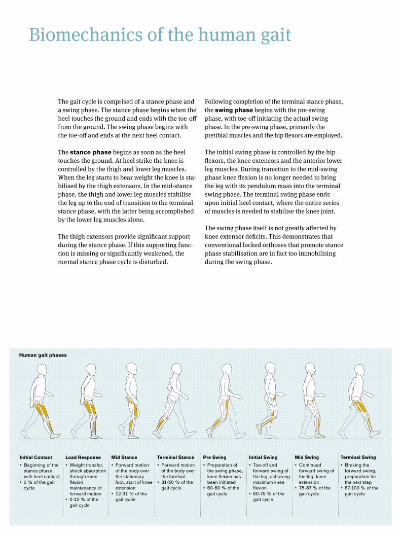
Biomechanics of the human gait
The gait cycle is comprised of a stance phase and a swing phase. The stance phase begins when the heel touches the ground and ends with the toe-off from the ground. The swing phase begins with the toe-off and ends at the next heel contact.
The stance�phase begins as soon as the heel touches the ground. At heel strike the knee is controlled by the thigh and lower leg muscles. When the leg starts to bear weight the knee is sta-bilised by the thigh extensors. In the mid-stance phase, the thigh and lower leg muscles stabilise the leg up to the end of transition to the terminal stance phase, with the latter being accomplished by the lower leg muscles alone.
The thigh extensors provide significant support during the stance phase. If this supporting func-tion is missing or significantly weakened, the normal stance phase cycle is disturbed.
Following completion of the terminal stance phase, the swing�phase begins with the pre-swing phase, with toe-off initiating the actual swing phase. In the pre-swing phase, primarily the pretibial muscles and the hip flexors are employed.
The initial swing phase is controlled by the hip flexors, the knee extensors and the anterior lower leg muscles. During transition to the mid-swing phase knee flexion is no longer needed to bring the leg with its pendulum mass into the terminal swing phase. The terminal swing phase ends upon initial heel contact, where the entire series of muscles is needed to stabilise the knee joint. The swing phase itself is not greatly affected by knee extensor deficits. This demonstrates that conventional locked orthoses that promote stance phase stabilisation are in fact too immobilising during the swing phase.
Human�gait�phases
Initial�Contact• Beginning of the
stance phase with heel contact
• 0 % of the gait cycle
Load�Response• Weight transfer,
shock absorption through knee flexion, maintenance of forward motion
• 0-12 % of the gait cycle
Mid�Stance• Forward motion
of the body over the stationary foot, start of knee extension
• 12-31 % of the gait cycle
Terminal�Stance• Forward motion
of the body over the forefoot
• 31-50 % of the gait cycle
Pre�Swing• Preparation of
the swing phase, knee flexion has been initiated
• 50-60 % of the gait cycle
Initial�Swing• Toe-off and
forward swing of the leg, achieving maximum knee flexion
• 60-75 % of the gait cycle
Mid�Swing�• Continued
forward swing of the leg, knee extension
• 75-87 % of the gait cycle
Terminal�Swing• Braking the
forward swing, preparation for the next step
• 87-100 % of the gait cycle
Therapeutic Application and Gait Training | Ottobock 9
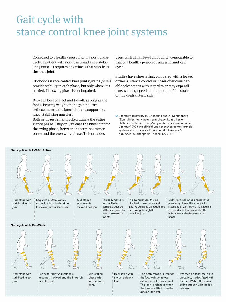
Gait cycle with stance control knee joint systems
Compared to a healthy person with a normal gait cycle, a patient with non-functional knee-stabil-ising muscles requires an orthosis that stabilises the knee joint.
Ottobock’s stance control knee joint systems (SCOs) provide stability in each phase, but only where it is needed. The swing phase is not impaired.
Between heel contact and toe-off, as long as the foot is bearing weight on the ground, the orthoses secure the knee joint and support the knee-stabilising muscles.Both orthoses remain locked during the entire stance phase. They only release the knee joint for the swing phase, between the terminal stance phase and the pre-swing phase. This provides
users with a high level of mobility, comparable to that of a healthy person during a normal gait cycle.
Studies have shown that, compared with a locked orthosis, stance control orthoses offer consider-able advantages with regard to energy expendi-ture, walking speed and reduction of the strain on the contralateral side.
• Literature review by B. Zacharias and A. Kannenberg “Zum klinischen Nutzen standphasenkontrollierter Orthesensysteme – Eine Analyse der wissenschaftlichen Literatur” (“On the clinical uses of stance control orthois systems – an analysis of the scientific literature”), published in Orthopädie Technik 6/2011.
Gait�cycle�with�E-MAG�Active
Gait�cycle�with�FreeWalk
Heel strike with stabilised knee joint.
Leg with E-MAG Active orthosis takes the load and the knee joint is stabilised.
Mid-stance phase with locked knee joint.
The body moves in front of the foot, complete extension of the knee joint; the lock is released at toe-off.
Pre-swing phase; the leg fitted with the orthosis and E-MAG Active is unloaded and can swing through the unlocked joint.
Mid to terminal swing phase: in the pre-swing phase, the knee joint is stabilised at 15° flexion, the knee joint is locked in full extension shortly before heel strike for the stance phase.
Heel strike with stabilised knee joint.
Leg with FreeWalk orthosis assumes the load and the knee joint is stabilised.
Mid-stance phase with locked knee joint.
Heel strike with the contralateral foot.
The body moves in front of the foot with complete extension of the knee joint. The lock is released when the toes are lifted from the ground (toe-off).
Pre-swing phase: the leg is unloaded, the leg fitted with the FreeWalk orthosis can swing through with the lock released.
10 Ottobock | Therapeutic Application and Gait Training
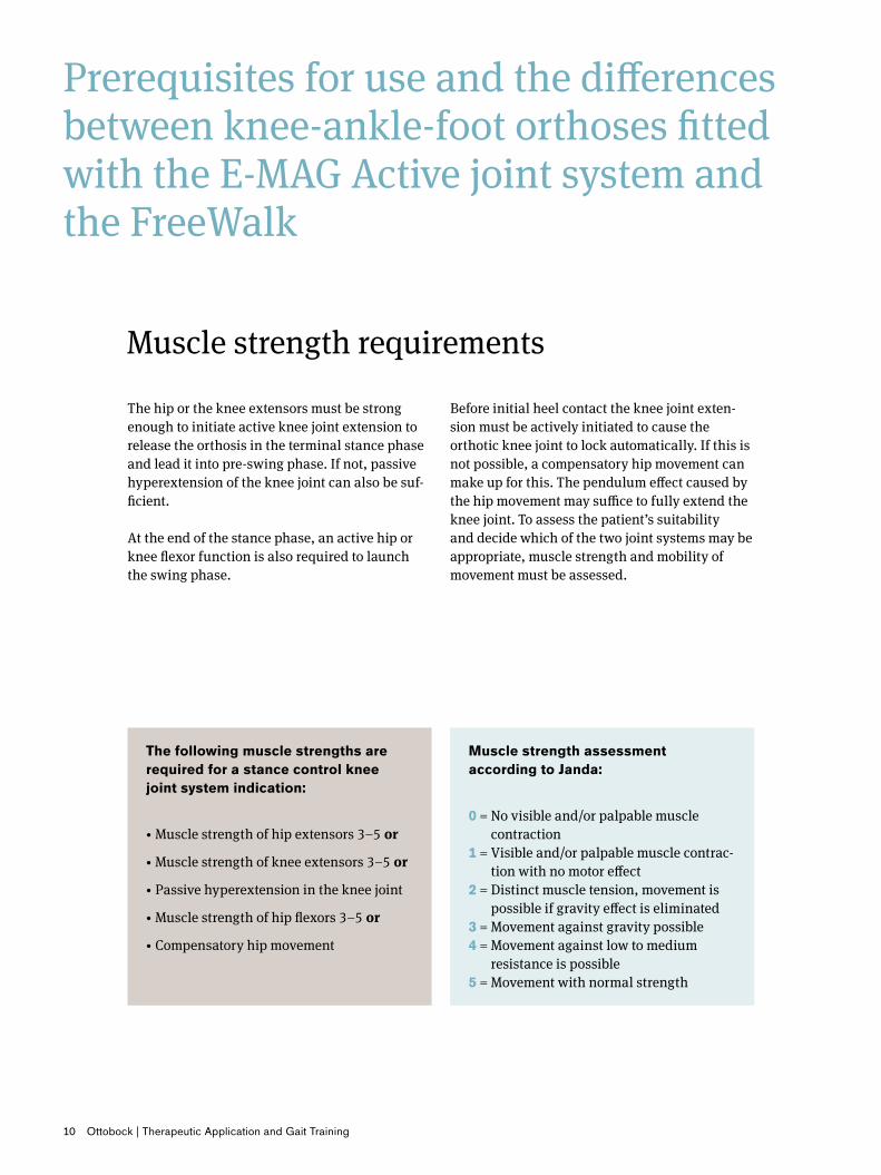
The hip or the knee extensors must be strong enough to initiate active knee joint extension to release the orthosis in the terminal stance phase and lead it into pre-swing phase. If not, passive hyperextension of the knee joint can also be suf-ficient.
At the end of the stance phase, an active hip or knee flexor function is also required to launch the swing phase.
Muscle�strength�assessment�according�to�Janda:
0 = No visible and/or palpable muscle contraction
1 = Visible and/or palpable muscle contrac-tion with no motor effect
2 = Distinct muscle tension, movement is possible if gravity effect is eliminated
3 = Movement against gravity possible4 = Movement against low to medium
resistance is possible5 = Movement with normal strength
Prerequisites for use and the differences between knee-ankle-foot orthoses fitted with the E-MAG Active joint system and the FreeWalk
Muscle strength requirements
Before initial heel contact the knee joint exten-sion must be actively initiated to cause the orthotic knee joint to lock automatically. If this is not possible, a compensatory hip movement can make up for this. The pendulum effect caused by the hip movement may suffice to fully extend the knee joint. To assess the patient’s suitability and decide which of the two joint systems may be appropriate, muscle strength and mobility of movement must be assessed.
The�following�muscle�strengths�are�required�for�a�stance�control�knee�joint�system�indication:
• Muscle strength of hip extensors 3–5 or
• Muscle strength of knee extensors 3–5 or
• Passive hyperextension in the knee joint
• Muscle strength of hip flexors 3–5 or
• Compensatory hip movement
Therapeutic Application and Gait Training | Ottobock 11
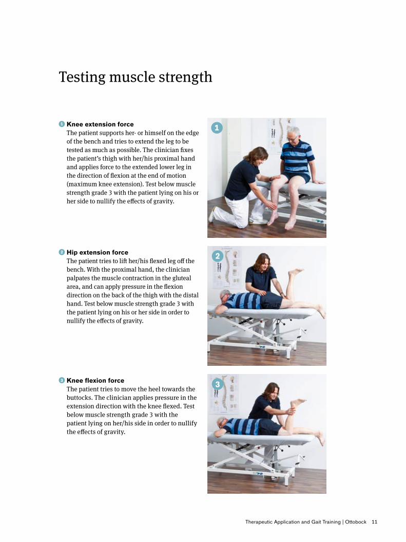
��Knee�extension�force�The patient supports her- or himself on the edge of the bench and tries to extend the leg to be tested as much as possible. The clinician fixes the patient’s thigh with her/his proximal hand and applies force to the extended lower leg in the direction of flexion at the end of motion (maximum knee extension). Test below muscle strength grade 3 with the patient lying on his or her side to nullify the effects of gravity.
��Hip�extension�force�The patient tries to lift her/his flexed leg off the bench. With the proximal hand, the clinician palpates the muscle contraction in the gluteal area, and can apply pressure in the flexion direction on the back of the thigh with the distal hand. Test below muscle strength grade 3 with the patient lying on his or her side in order to nullify the effects of gravity.
��Knee�flexion�force�The patient tries to move the heel towards the buttocks. The clinician applies pressure in the extension direction with the knee flexed. Test below muscle strength grade 3 with the patient lying on her/his side in order to nullify the effects of gravity.
Testing muscle strength

12 Ottobock | Therapeutic Application and Gait Training
Testing muscle strength
���Hip�flexion�force�The patient is asked to lift her/his thigh with the knee flexed towards her/his shoulder on the same side. At the end of the movement, the clinician exerts a resisting force in the extension direction, on the ventral side of the thigh. Test below muscle strength grade 3 with the patient lying on her/his side in order to nullify the effect of gravity.
����Assessment�of�muscle�strength�during�dorsiflexion�The patient is asked to pull the back of her/his foot up and inwards (dorsiflexion and supina-tion). At the end of the movement, the clinician exerts pressure in the plantar flexion direction. Test below muscle strength grade 3 with the patient lying on her/his side in order to nullify the effects of gravity.
��Checking�hyperextension�in�knee�joint�The clinician checks the maximum passive extensibility of the knee joint.
Therapeutic Application and Gait Training | Ottobock 13
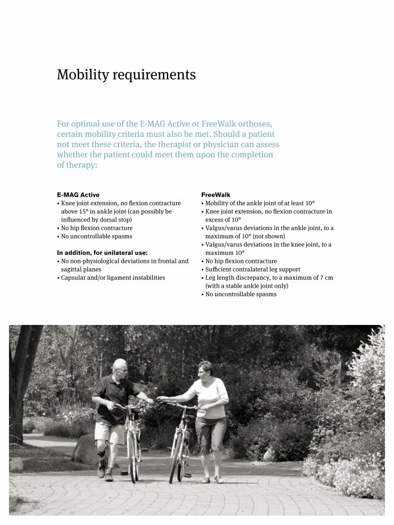
E-MAG�Active• Knee joint extension, no flexion contracture
above 15° in ankle joint (can possibly be influenced by dorsal stop)
• No hip flexion contracture• No uncontrollable spasms
In�addition,�for�unilateral�use:• No non-physiological deviations in frontal and
sagittal planes• Capsular and/or ligament instabilities
Mobility requirements
FreeWalk• Mobility of the ankle joint of at least 10°• Knee joint extension, no flexion contracture in
excess of 10°• Valgus/varus deviations in the ankle joint, to a
maximum of 10° (not shown)• Valgus/varus deviations in the knee joint, to a
maximum 10° • No hip flexion contracture• Sufficient contralateral leg support• Leg length discrepancy, to a maximum of 7 cm
(with a stable ankle joint only)• No uncontrollable spasms
For optimal use of the E-MAG Active or FreeWalk orthoses, certain mobility criteria must also be met. Should a patient not meet these criteria, the therapist or physician can assess whether the patient could meet them upon the completion of therapy:
14 Ottobock | Therapeutic Application and Gait Training
Testing mobility
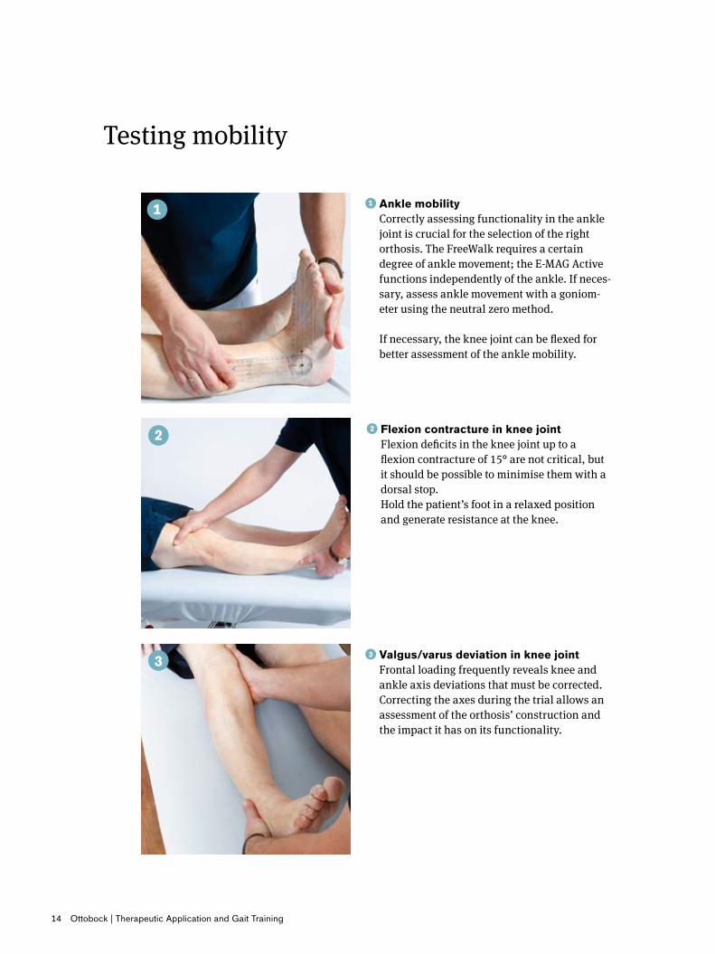
��Ankle�mobility�Correctly assessing functionality in the ankle joint is crucial for the selection of the right orthosis. The FreeWalk requires a certain degree of ankle movement; the E-MAG Active functions independently of the ankle. If neces-sary, assess ankle movement with a goniom-eter using the neutral zero method. If necessary, the knee joint can be flexed for better assessment of the ankle mobility.
��Valgus/varus�deviation�in�knee�joint� Frontal loading frequently reveals knee and ankle axis deviations that must be corrected. Correcting the axes during the trial allows an assessment of the orthosis’ construction and the impact it has on its functionality.
� ��Flexion�contracture�in�knee�joint�Flexion deficits in the knee joint up to a flexion contracture of 15° are not critical, but it should be possible to minimise them with a dorsal stop. Hold the patient’s foot in a relaxed position and generate resistance at the knee.

Therapeutic Application and Gait Training | Ottobock 15
����Leg�length�discrepancy�A critical difference between the FreeWalk and the E-MAG Active is their ankle or foot articula-tion. The FreeWalk orthosis requires a connection to the foot and hence can only be used to a limited extent if there is a leg length discrepancy. If the patient has a stable ankle joint, a maxi-mum leg length discrepancy of up to 7 cm is possible. Use of the E-MAG Active is independent of the foot and thus of a leg length discrepancy. This means that orthoprostheses and transtibial prostheses with knee control can also be used with the E-MAG Active. Differences in leg length should be eliminated as much as possible, but this equalisation should be carried out individ-ually for each patient and if necessary, in multiple steps.
���Test�for�hip�flexion�contracture�Hip flexion contractures are critical and are often overlooked. If possible, use the Thomas test (pictured).
16 Ottobock | Therapeutic Application and Gait Training
Specific therapy to improve muscle strength and mobility
If the prerequisites described above are not entirely met, certain weaknesses may be compen-sated for by strength and mobility therapy, mak-ing use of a stance control orthosis an option at some point in the future. For the purpose of improving therapy, a complimentary personal home exercise programme should be developed for the patient.
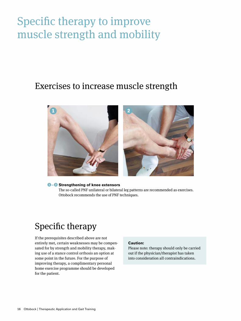
�–� ���Strengthening�of�knee�extensors�The so-called PNF unilateral or bilateral leg patterns are recommended as exercises. Ottobock recommends the use of PNF techniques.
Exercises to increase muscle strength
Caution:Please note: therapy should only be carried out if the physician/therapist has taken into consideration all contraindications.
Specific therapy

Therapeutic Application and Gait Training | Ottobock 17
�–� ���Strengthening�of�hip�extensors�and�flexors��to facilitate hip extension and abduction, the leg patterns can also be executed with the patient lying on her/his side with extended or flexed knee joint as well as in quad-ruped position.
Exercises to increase muscle strength

18 Ottobock | Therapeutic Application and Gait Training
Exercises to increase mobility
Physiotherapeutic techniques of manual therapy are suitable for improving mobility (in other words, passive joint mobilisation or muscle stretching techniques, depending on the diagnosis).
If muscle contractures are the cause of hypomo-bility, muscle stretching techniques are recom-mended to increase the range of motion (pictures 1–4).
�–� ���Increasing muscle mobility in the knee and ankle joints.
�–� ��Increasing muscle mobility in the knee and hip joints.
Therapeutic Application and Gait Training | Ottobock 19
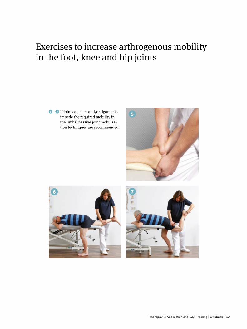
Exercises to increase arthrogenous mobility in the foot, knee and hip joints
�–� ���If joint capsules and/or ligaments impede the required mobility in the limbs, passive joint mobilisa-tion techniques are recommended.

20 Ottobock | Therapeutic Application and Gait Training
Home exercises to increase muscle strength and mobility
To further improve the body's constitution, the patient should do follow-up exercises at home to achieve sustainable benefits from the exercises carried out with a therapist.
Below are a few examples of mobility-enhancing exercises which patients can easily do at home. The more often the patient can do strengthening and mobilising exercises, the sooner she/he will be able to walk safely and physiologically with a stance control orthosis.
Caution: Please note: therapy should only be carried out if the physician/therapist has taken into consideration all contraindications.
Please note: the exercises shown here serve as a rough guideline only and must be adapted to the individual patient.
��Strengthening of knee joint extension using a resistance band. The unaffected leg should support the movement dur-ing execution.
��Strengthening hip extension using a resistance band.
Therapeutic Application and Gait Training | Ottobock 21
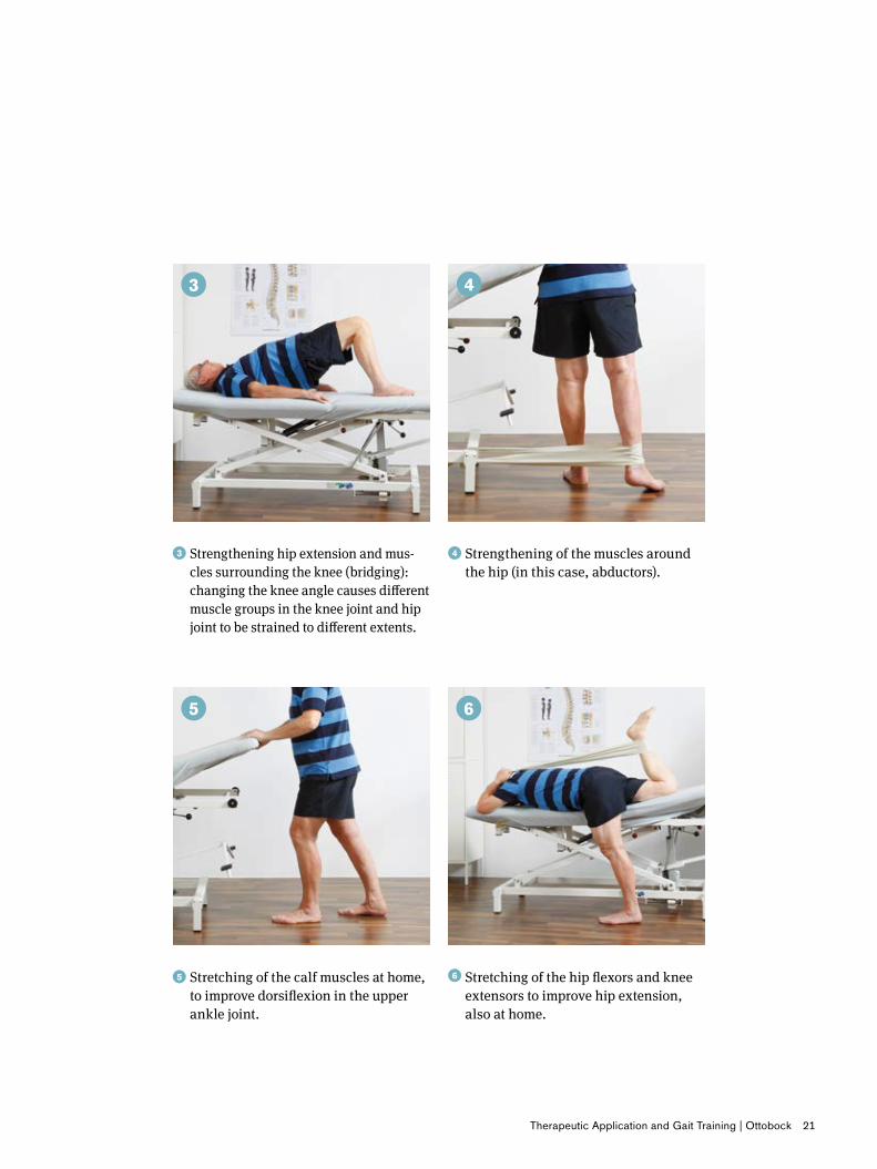
��Strengthening hip extension and mus-cles surrounding the knee (bridging): changing the knee angle causes different muscle groups in the knee joint and hip joint to be strained to different extents.
���Strengthening of the muscles around the hip (in this case, abductors).
��Stretching of the calf muscles at home, to improve dorsiflexion in the upper ankle joint.
��Stretching of the hip flexors and knee extensors to improve hip extension, also at home.
22 Ottobock | Therapeutic Application and Gait Training
Handling the orthoses and starting exercises
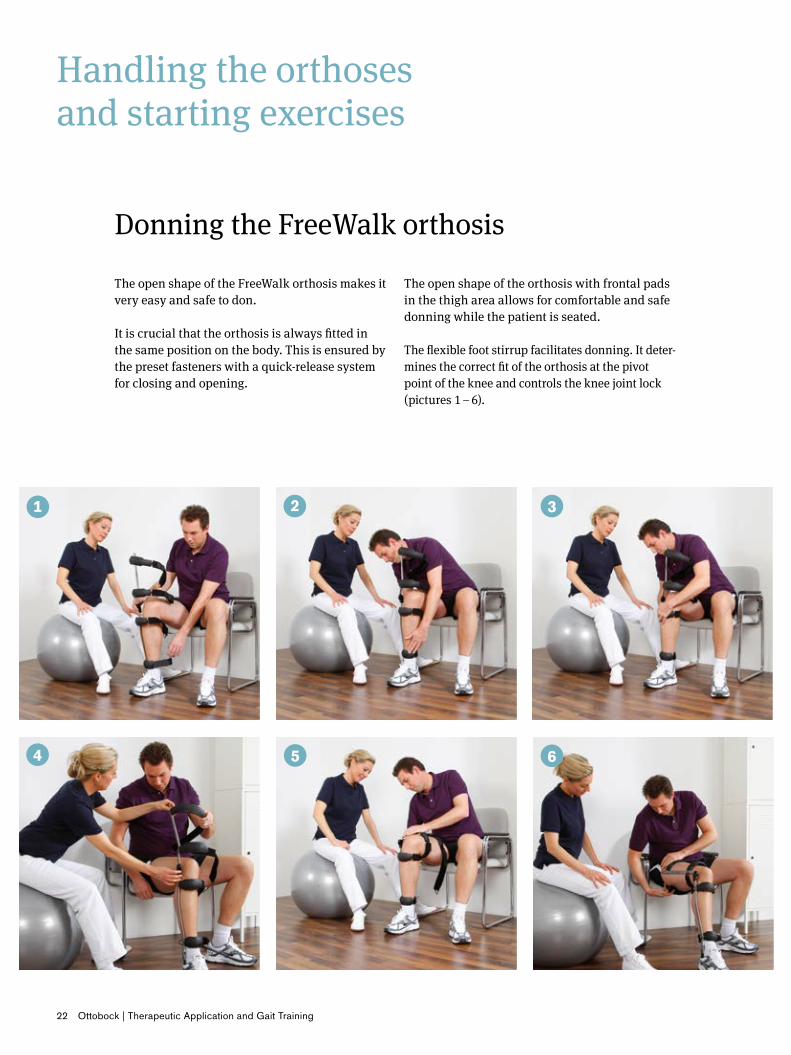
The open shape of the FreeWalk orthosis makes it very easy and safe to don.
It is crucial that the orthosis is always fitted in the same position on the body. This is ensured by the preset fasteners with a quick-release system for closing and opening.
Donning the FreeWalk orthosis
The open shape of the orthosis with frontal pads in the thigh area allows for comfortable and safe donning while the patient is seated.
The flexible foot stirrup facilitates donning. It deter-mines the correct fit of the orthosis at the pivot point of the knee and controls the knee joint lock (pictures 1 – 6).
Therapeutic Application and Gait Training | Ottobock 23
Donning a knee-ankle-foot orthosis with the E-MAG Active joint system
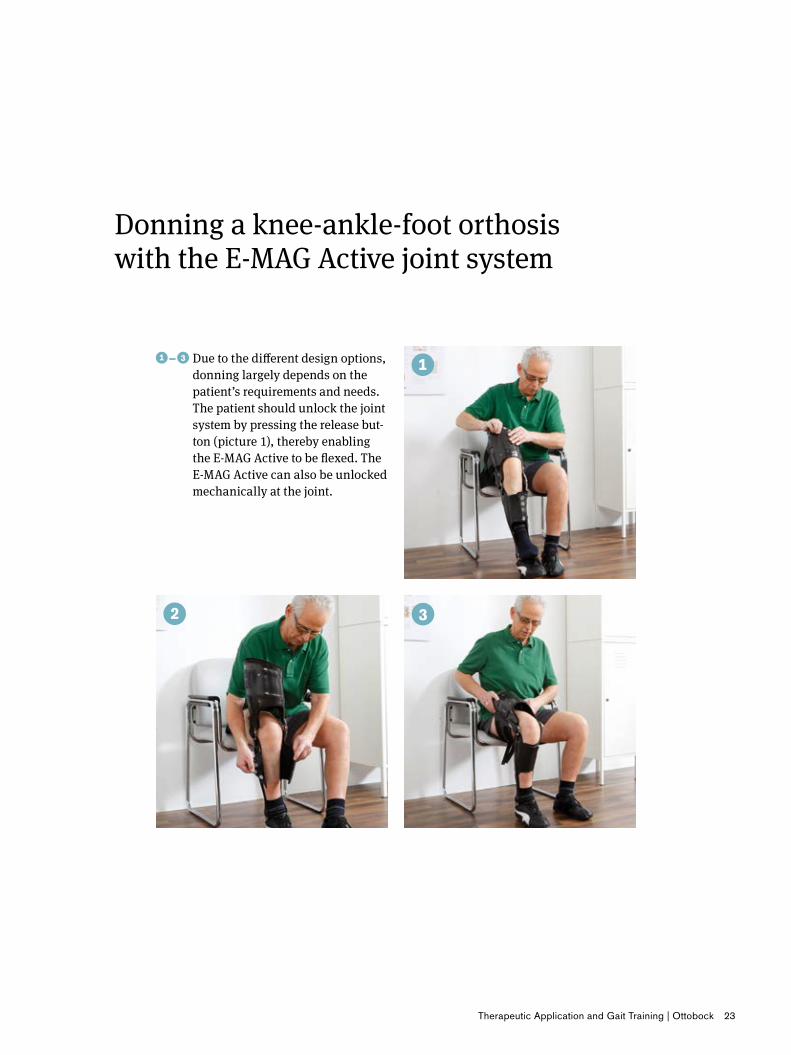
�–� ���Due to the different design options, donning largely depends on the patient’s requirements and needs. The patient should unlock the joint system by pressing the release but-ton (picture 1), thereby enabling the E-MAG Active to be flexed. The E-MAG Active can also be unlocked mechanically at the joint.

24 Ottobock | Therapeutic Application and Gait Training
Standing up and locking
�–� ��Standing�up�with�the�FreeWalk�orthosis�and�E-MAG�Active�The patient should support him- or herself with both hands when standing up. The contralateral leg should be moved further forward to achieve increased stability. The patient should then stand up, placing the heel of the fitted leg in front of the stance leg and moving the thigh backwards. This causes the knee joint to extend. After the orthosis has locked, and with the therapist’s assistance, the patient should try to stand evenly on both legs.
These exercises should be done first under the supervision of the therapist or orthotist and comprise standing up, locking, releasing and sitting down. These basic exercises enable the patient to get a feel for the locking func-tion of the orthosis. This make release easier when the first steps are taken.
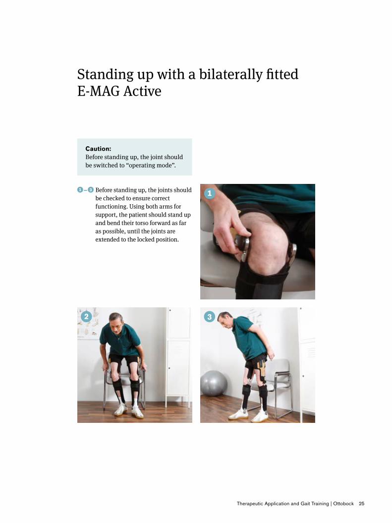
Caution: Before standing up, the joint should be switched to “operating mode”.
Therapeutic Application and Gait Training | Ottobock 25
�–� ���Before standing up, the joints should be checked to ensure correct functioning. Using both arms for support, the patient should stand up and bend their torso forward as far as possible, until the joints are extended to the locked position.
Standing up with a bilaterally fitted E-MAG Active
Caution: Before standing up, the joint should be switched to “operating mode”.
26 Ottobock | Therapeutic Application and Gait Training
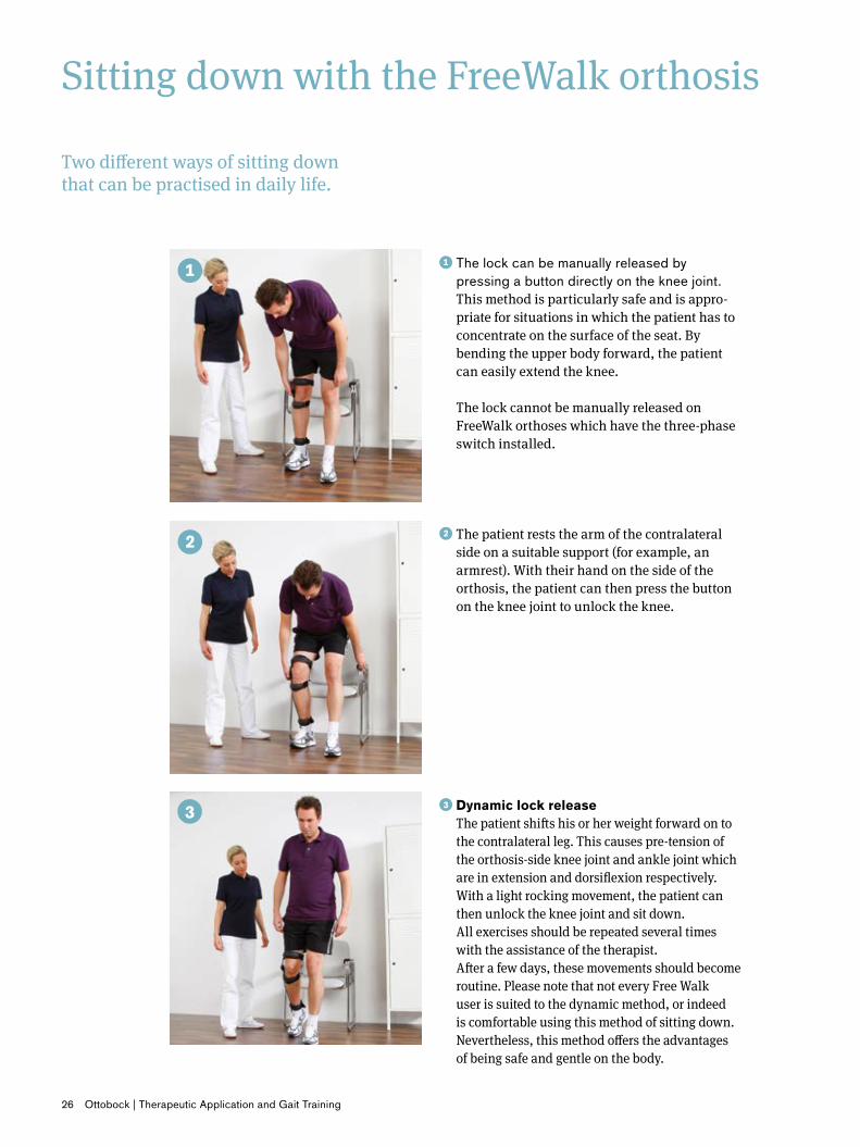
Sitting down with the FreeWalk orthosis
��The lock can be manually released by pressing a button directly on the knee joint. This method is particularly safe and is appro-priate for situations in which the patient has to concentrate on the surface of the seat. By bending the upper body forward, the patient can easily extend the knee. The lock cannot be manually released on FreeWalk orthoses which have the three-phase switch installed.
Two different ways of sitting down that can be practised in daily life.
���Dynamic�lock�release�The patient shifts his or her weight forward on to the contralateral leg. This causes pre-tension of the orthosis-side knee joint and ankle joint which are in extension and dorsiflexion respectively. With a light rocking movement, the patient can then unlock the knee joint and sit down. All exercises should be repeated several times with the assistance of the therapist. After a few days, these movements should become routine. Please note that not every Free Walk user is suited to the dynamic method, or indeed is comfortable using this method of sitting down. Nevertheless, this method offers the advantages of being safe and gentle on the body.
��The patient rests the arm of the contralateral side on a suitable support (for example, an armrest). With their hand on the side of the orthosis, the patient can then press the button on the knee joint to unlock the knee.

Therapeutic Application and Gait Training | Ottobock 27
Sitting down with the E-MAG Active
�–� ��Electromechanical�release�of�the�lock�is enabled by the lower button on the electronic unit of the E-MAG Active. As with the FreeWalk orthosis, this method is appropriate for situations in which the patient has to concentrate on sitting down. Before releasing the knee joint, the patient must be in a position with the knee extended. Some patients are able to extend their knees in a normal stance, but other patients have to consiously make a knee-extending movement.
�–� ��Dynamic�Lock�Release�As with the FreeWalk orthosis, here too the patient must move the contralateral leg further forward such that the orthosis is in a rear position, which activates the electronic unit and causes the E-MAG Active knee joint to release in a knee-extending position. As with the FreeWalk orthosis, the movement should be prac-tised with the help of the therapist to enable the patient to get accustomed to it.
The E-MAG Active also offers two different ways of sitting down that can be practised.
28 Ottobock | Therapeutic Application and Gait Training
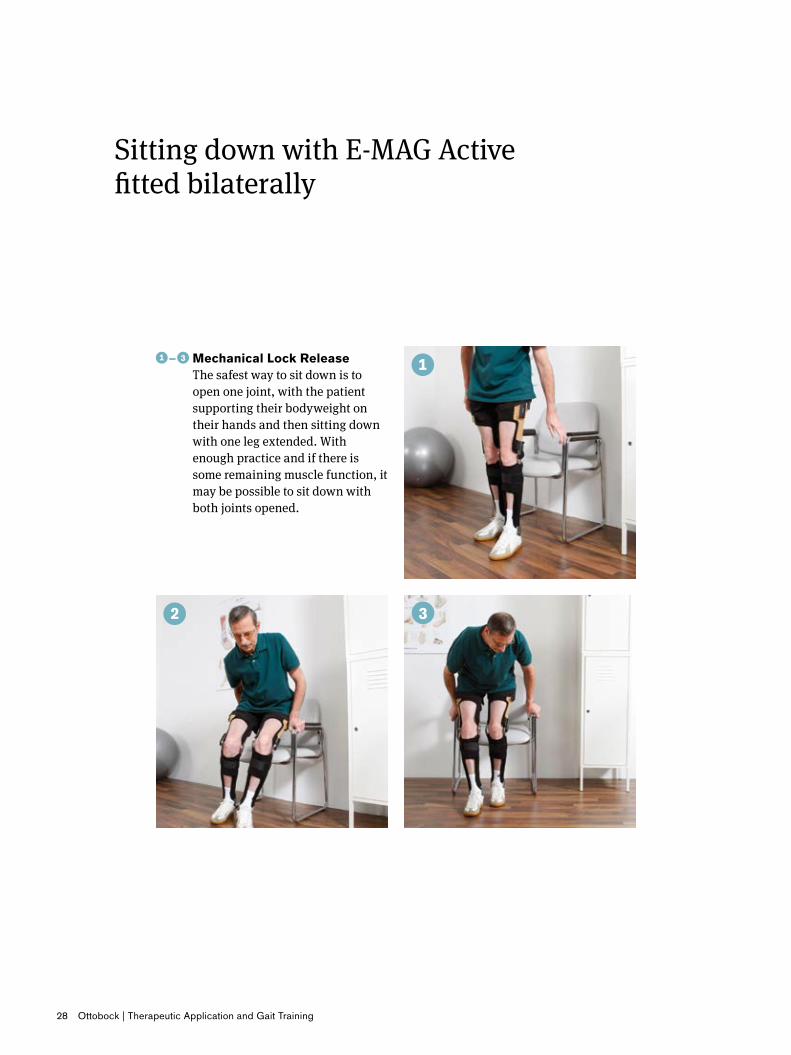
�–� ���Mechanical�Lock�Release�The safest way to sit down is to open one joint, with the patient supporting their bodyweight on their hands and then sitting down with one leg extended. With enough practice and if there is some remaining muscle function, it may be possible to sit down with both joints opened.
Sitting down with E-MAG Active fitted bilaterally

Therapeutic Application and Gait Training | Ottobock 29
Gait training
Before the user takes their first steps, priority should be given to correct weight-distribution on the orthosis. To prevent abnormal movements when the first steps are being taken, balancing exercises should be practised to determine the best possible weight-distribution on the sup-ported limb.
Exercises for attaining even weight-distribution
With the help of the visualised load-line and the hand-held control unit, the thera-pist or orthotist can give the patient pre-cise instructions for movements in order to determine the position of optimum load assumption.
Following this exercise the patient must practise standing on two legs stably and with uniform weight distribution. Various exercises can be carried out to achieve this.
The L.A.S.A.R. Posture is the ideal alternative to the inevitably inaccurate scales normally used for checking for correct weight distribution.
Stabilising�Exercise:The clinician applies a resisting force to the shoulder and/or pelvic girdle to help the patient practise achieving a stable stance.
30 Ottobock | Therapeutic Application and Gait Training
Stabilising exercises outside the parallel bars
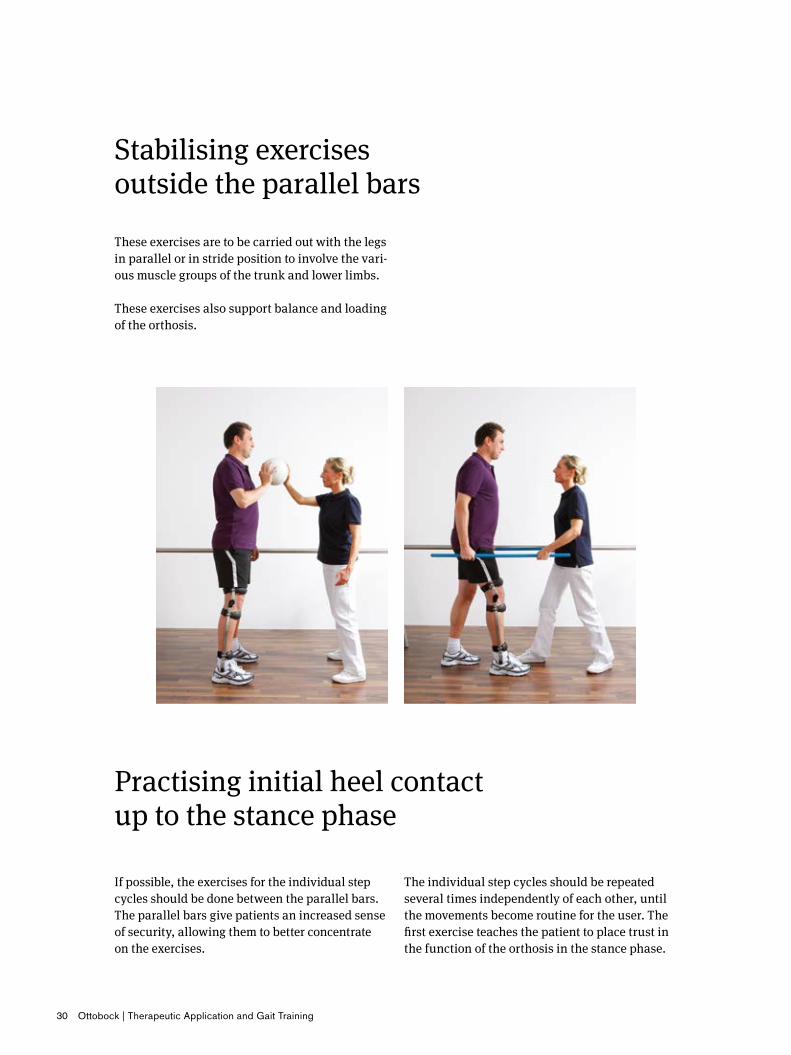
These exercises are to be carried out with the legs in parallel or in stride position to involve the vari-ous muscle groups of the trunk and lower limbs.
These exercises also support balance and loading of the orthosis.
Practising initial heel contact up to the stance phase
If possible, the exercises for the individual step cycles should be done between the parallel bars. The parallel bars give patients an increased sense of security, allowing them to better concentrate on the exercises.
The individual step cycles should be repeated several times independently of each other, until the movements become routine for the user. The first exercise teaches the patient to place trust in the function of the orthosis in the stance phase.
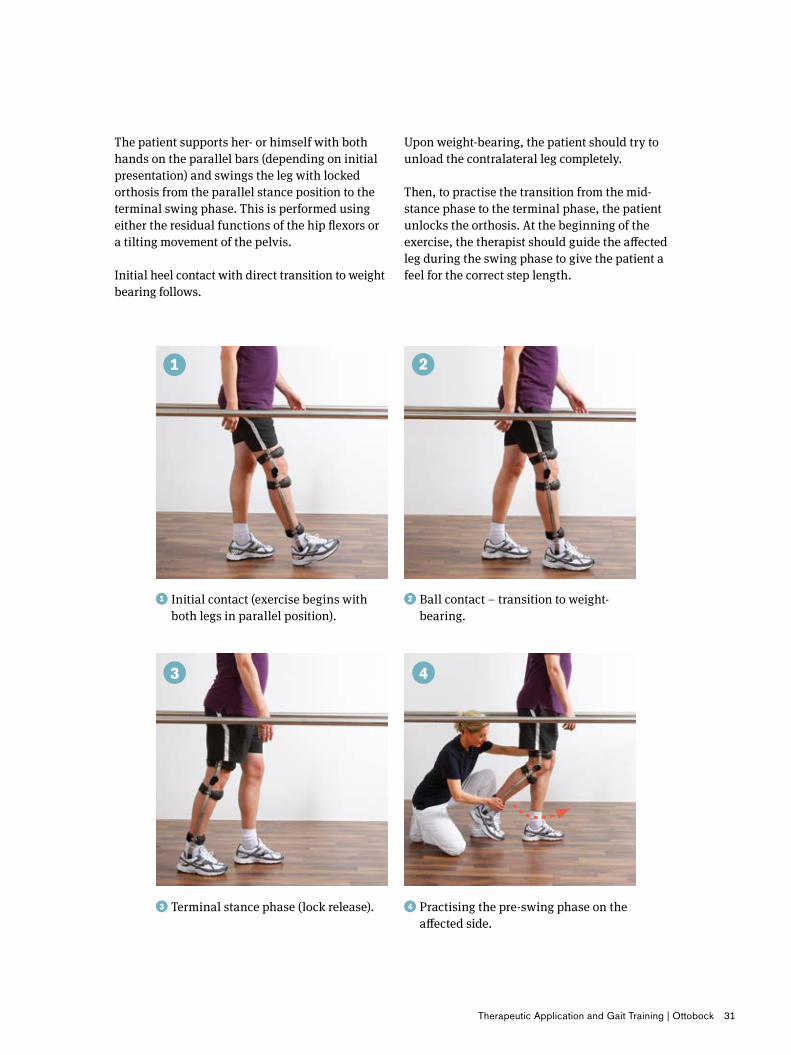
Therapeutic Application and Gait Training | Ottobock 31
Stabilising exercises outside the parallel bars
Practising initial heel contact up to the stance phase
Upon weight-bearing, the patient should try to unload the contralateral leg completely.
Then, to practise the transition from the mid-stance phase to the terminal phase, the patient unlocks the orthosis. At the beginning of the exercise, the therapist should guide the affected leg during the swing phase to give the patient a feel for the correct step length.
���Terminal stance phase (lock release). ��Practising the pre-swing phase on the affected side.
��Initial contact (exercise begins with both legs in parallel position).
��Ball contact – transition to weight-bearing.
The patient supports her- or himself with both hands on the parallel bars (depending on initial presentation) and swings the leg with locked orthosis from the parallel stance position to the terminal swing phase. This is performed using either the residual functions of the hip flexors or a tilting movement of the pelvis.
Initial heel contact with direct transition to weight bearing follows.
32 Ottobock | Therapeutic Application and Gait Training
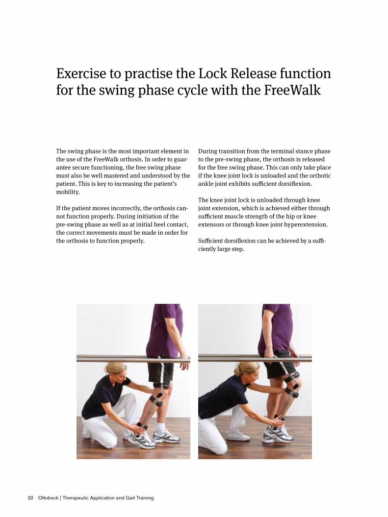
Exercise to practise the Lock Release function for the swing phase cycle with the FreeWalk
The swing phase is the most important element in the use of the FreeWalk orthosis. In order to guar-antee secure functioning, the free swing phase must also be well mastered and understood by the patient. This is key to increasing the patient’s mobility.
If the patient moves incorrectly, the orthosis can-not function properly. During initiation of the pre-swing phase as well as at initial heel contact, the correct movements must be made in order for the orthosis to function properly.
During transition from the terminal stance phase to the pre-swing phase, the orthosis is released for the free swing phase. This can only take place if the knee joint lock is unloaded and the orthotic ankle joint exhibits sufficient dorsiflexion.
The knee joint lock is unloaded through knee joint extension, which is achieved either through sufficient muscle strength of the hip or knee extensors or through knee joint hyperextension.
Sufficient dorsiflexion can be achieved by a suffi-ciently large step.
Therapeutic Application and Gait Training | Ottobock 33
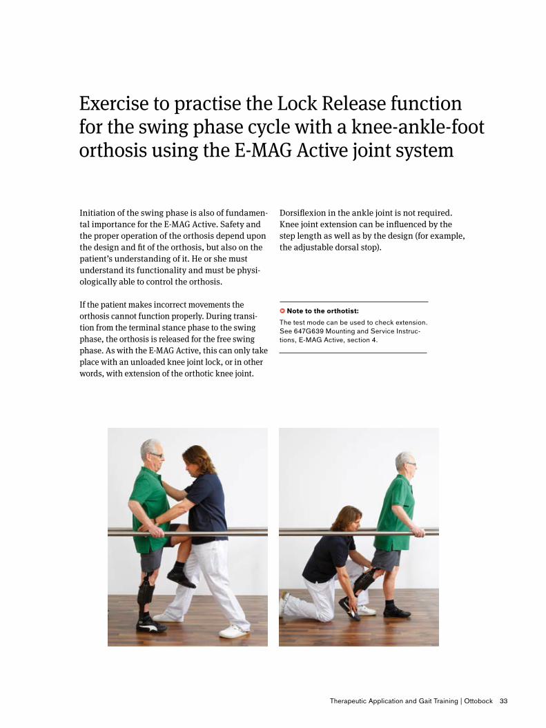
Exercise to practise the Lock Release function for the swing phase cycle with a knee-ankle-foot orthosis using the E-MAG Active joint system
Initiation of the swing phase is also of fundamen-tal importance for the E-MAG Active. Safety and the proper operation of the orthosis depend upon the design and fit of the orthosis, but also on the patient’s understanding of it. He or she must understand its functionality and must be physi-ologically able to control the orthosis.
If the patient makes incorrect movements the orthosis cannot function properly. During transi-tion from the terminal stance phase to the swing phase, the orthosis is released for the free swing phase. As with the E-MAG Active, this can only take place with an unloaded knee joint lock, or in other words, with extension of the orthotic knee joint.
Dorsiflexion in the ankle joint is not required. Knee joint extension can be influenced by the step length as well as by the design (for example, the adjustable dorsal stop).
• Note�to�the�orthotist:The test mode can be used to check extension. See 647G639 Mounting and Service Instruc-tions, E-MAG Active, section 4.
34 Ottobock | Therapeutic Application and Gait Training
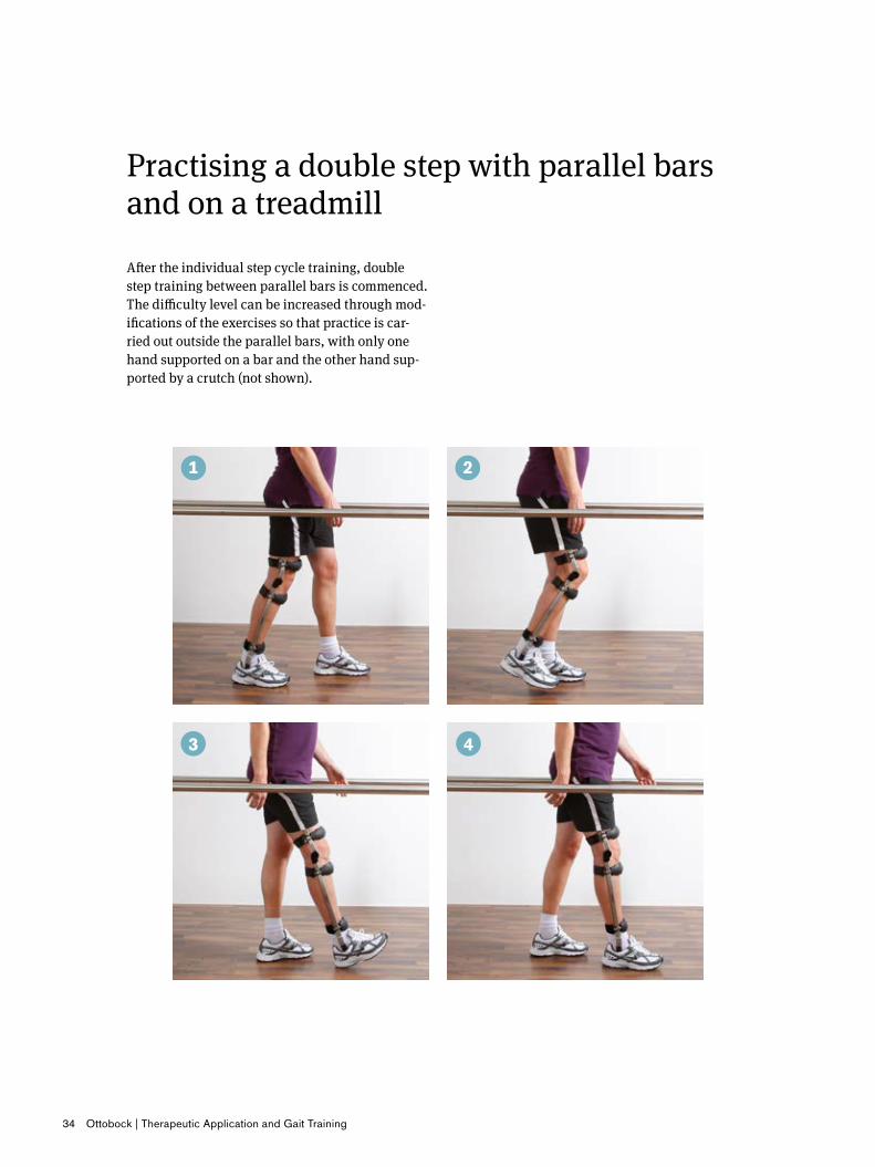
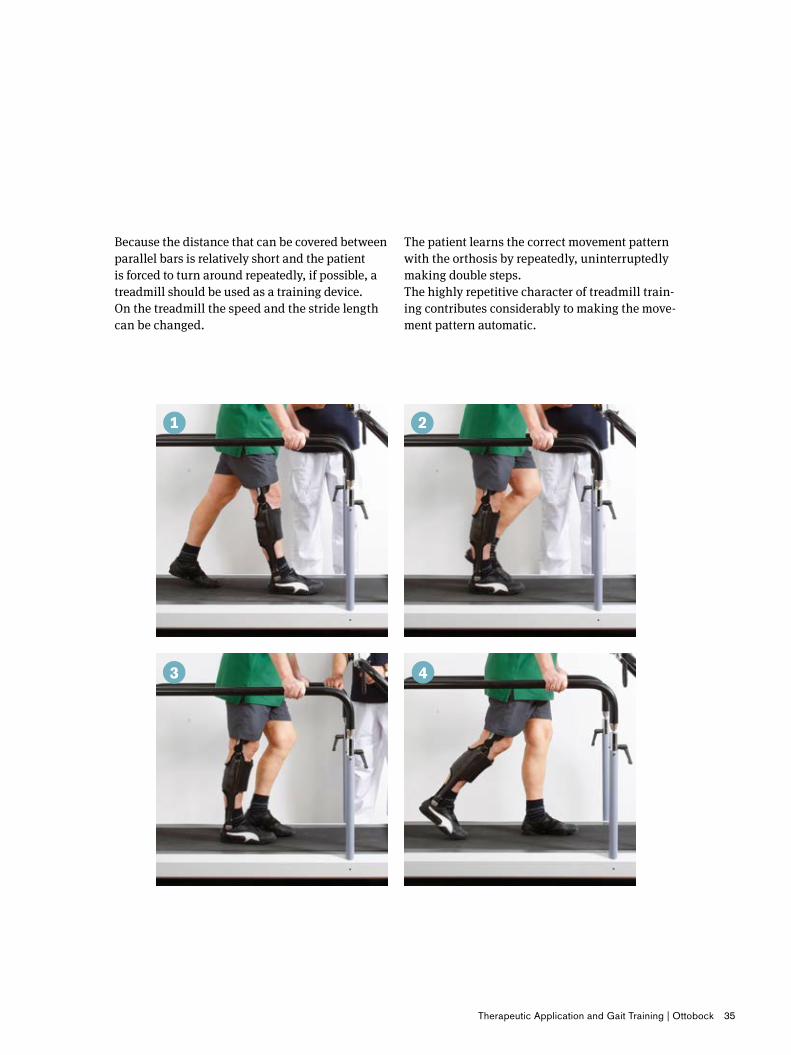
Practising a double step with parallel bars and on a treadmill
After the individual step cycle training, double step training between parallel bars is commenced. The difficulty level can be increased through mod-ifications of the exercises so that practice is car-ried out outside the parallel bars, with only one hand supported on a bar and the other hand sup-ported by a crutch (not shown).
Therapeutic Application and Gait Training | Ottobock 35
Because the distance that can be covered between parallel bars is relatively short and the patient is forced to turn around repeatedly, if possible, a treadmill should be used as a training device.On the treadmill the speed and the stride length can be changed.
The patient learns the correct movement pattern with the orthosis by repeatedly, uninterruptedly making double steps.The highly repetitive character of treadmill train-ing contributes considerably to making the move-ment pattern automatic.
36 Ottobock | Therapeutic Application and Gait Training
Daily Activities
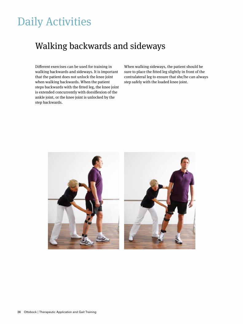
Different exercises can be used for training in walking backwards and sideways. It is important that the patient does not unlock the knee joint when walking backwards. When the patient steps backwards with the fitted leg, the knee joint is extended concurrently with dorsiflexion of the ankle joint, or the knee joint is unlocked by the step backwards.
Walking backwards and sideways
When walking sideways, the patient should be sure to place the fitted leg slightly in front of the contralateral leg to ensure that she/he can always step safely with the loaded knee joint.
Therapeutic Application and Gait Training | Ottobock 37
Negotiating inclines/declines
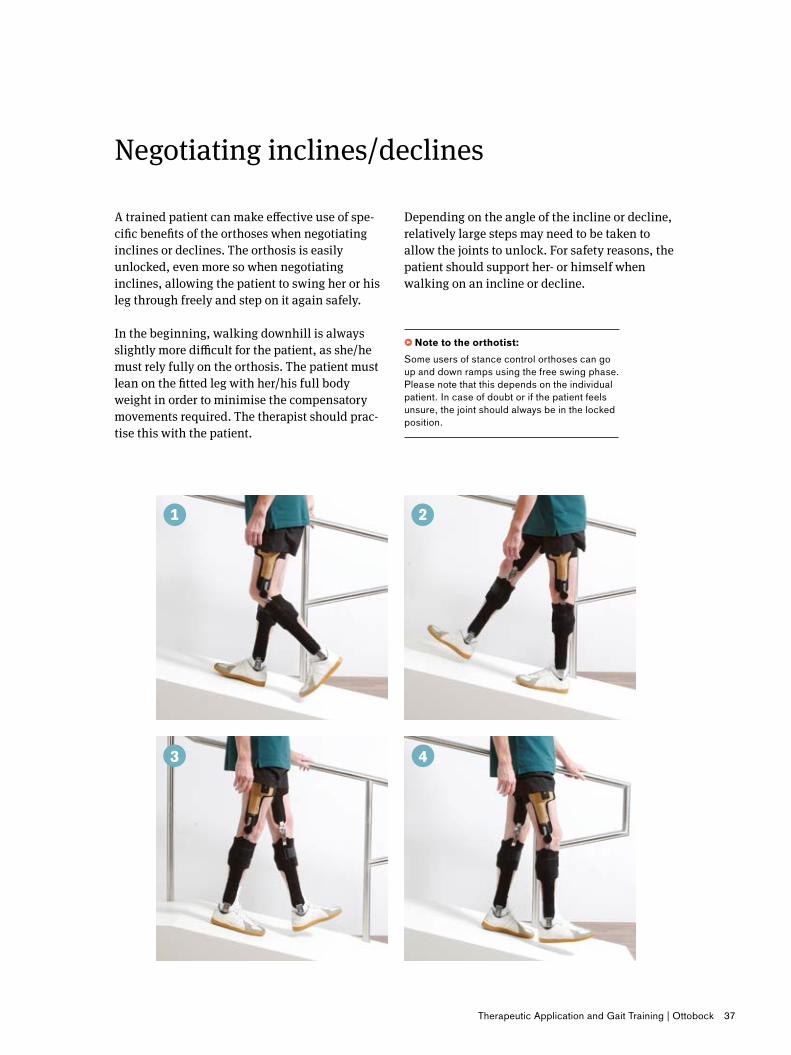
A trained patient can make effective use of spe-cific benefits of the orthoses when negotiating inclines or declines. The orthosis is easily unlocked, even more so when negotiating inclines, allowing the patient to swing her or his leg through freely and step on it again safely.
In the beginning, walking downhill is always slightly more difficult for the patient, as she/he must rely fully on the orthosis. The patient must lean on the fitted leg with her/his full body weight in order to minimise the compensatory movements required. The therapist should prac-tise this with the patient.
Depending on the angle of the incline or decline, relatively large steps may need to be taken to allow the joints to unlock. For safety reasons, the patient should support her- or himself when walking on an incline or decline.
• Note�to�the�orthotist:Some users of stance control orthoses can go up and down ramps using the free swing phase. Please note that this depends on the individual patient. In case of doubt or if the patient feels unsure, the joint should always be in the locked position.
38 Ottobock | Therapeutic Application and Gait Training
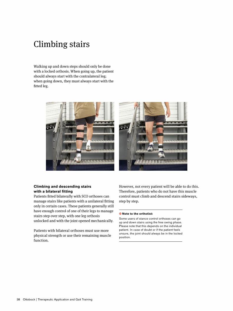
Walking up and down steps should only be done with a locked orthosis. When going up, the patient should always start with the contralateral leg; when going down, they must always start with the fitted leg.
Climbing stairs
However, not every patient will be able to do this. Therefore, patients who do not have this muscle control must climb and descend stairs sideways, step by step.
• Note�to�the�orthotist:Some users of stance control orthoses can go up and down stairs using the free swing phase. Please note that this depends on the individual patient. In case of doubt or if the patient feels unsure, the joint should always be in the locked position.
Climbing�and�descending�stairs��with�a�bilateral�fittingPatients fitted bilaterally with SCO orthoses can manage stairs like patients with a unilateral fitting only in certain cases. These patients generally still have enough control of one of their legs to manage stairs step over step, with one leg orthosis unlocked and with the joint opened mechanically.
Patients with bilateral orthoses must use more physical strength or use their remaining muscle function.
Therapeutic Application and Gait Training | Ottobock 39
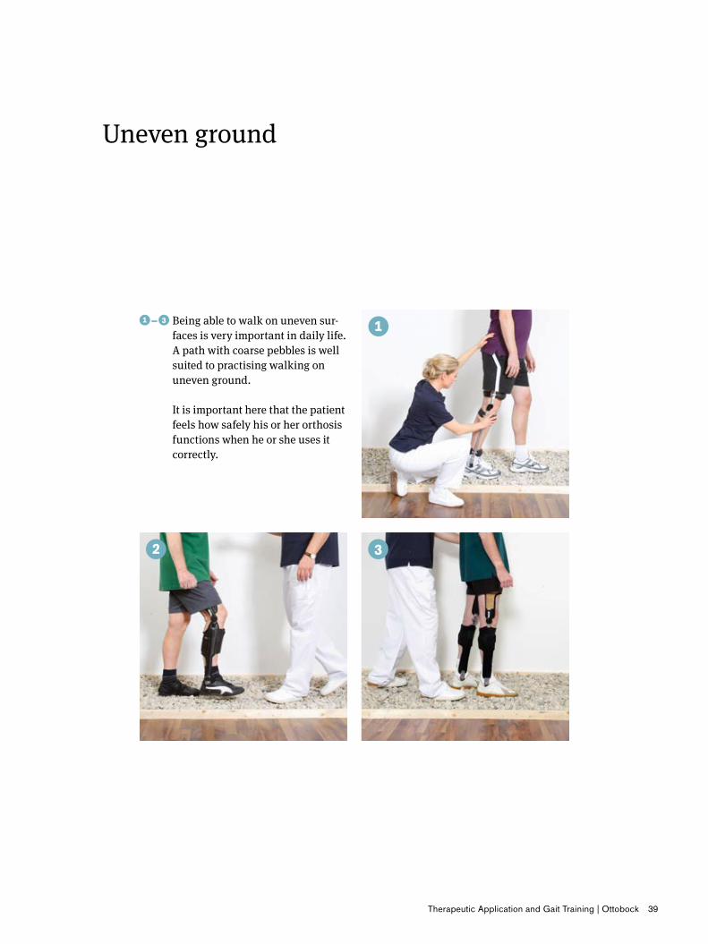
�–� ���Being able to walk on uneven sur-faces is very important in daily life. A path with coarse pebbles is well suited to practising walking on uneven ground. It is important here that the patient feels how safely his or her orthosis functions when he or she uses it correctly.
Uneven ground

40 Ottobock | Therapeutic Application and Gait Training
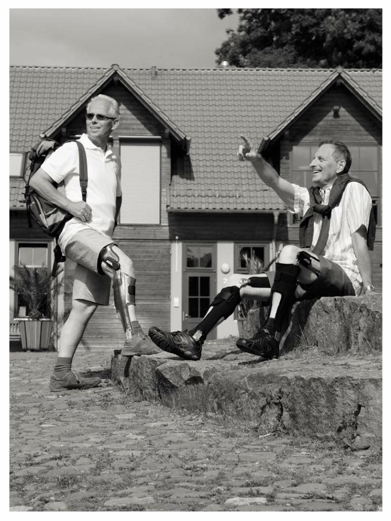
A trained user of such orthoses will have no prob-lem taking up low-activity sporting and leisure activities.
Uniform movements in particular, such as those involved in cycling, can easily be learned by the
Low-intensity sport
patient. Cycling on an exercise bike is a particu-larly good way to improve the patient’s fitness level. Other sports such as golf may also be suit-able for patients if they are approved by the phy-sician.
E-MAG�Active:�Lock�Release�functionThe E-MAG Active has a release function that allows the joint to be temporarily released, for example. for riding a bicycle.
Therapeutic Application and Gait Training | Ottobock 41
42 Ottobock | Therapeutic Application and Gait Training
Why�does�the�orthosis�sometimes�fail�to�unlock?Check that:• the knee is extended fully before toe-off occurs• the patient is able to reach the knee extension
stop on the orthosis• the orthotic knee joint is behind the load line• the ankle joint has sufficient stability• the pull-release cable has moved or is broken.
Why�does�the�orthosis�fail�to�lock?Check that:• the patient reaches the extension stop on the
orthotic knee joint before initial heel contact• the patient has sufficient muscle strength or a
sufficient hip movement to swing the leg into extension.
Are�some�parts�wearing�out�too�quickly?If the guide loops tear, check that: • the support tubes are correctly positioned,
for example, for patients with hyperextension of the knee the lower support tubes are mounted posteriorly.
How�should�the�three-phase�switch�be��operated�in�order�to�make�the�joint�freely�movable�(such�as�for�riding�bicycle)?�In contrast to locking the joint, where you press the switch down in the extended position, the joint must be in a flexed position to be released. Then press the switch down to make the joint freely movable.
Frequently Asked Questions
Functionality of the FreeWalk orthosis
When�should�the�orthosis�be�inspected?• The orthosis should be checked for wear by the
orthotist at least every 6 months.• All working parts such as plastic bearings, PU
dorsiflexion assist rings, cables, pads, and fas-teners can be replaced.
Why�has�the�foot�stirrup�on�the�FreeWalk�orthosis�broken?At the initial fitting with the FreeWalk orthosis, it has often been observed that patients need a rela-tively hard dorsal stop to be able to feel the switch threshold of the FreeWalk orthosis. However, this changes very quickly: a high dorsal force at the ankle is no longer necessary as patients develop an automatic feel for when unlocking occurs. It is therefore crucial that if the insole extends beyond the ball of the foot or as far as the toes, it is perhaps too stiff to cause a dorsal stop. If the force is per-sistently too high, then the foot stirrup, or in the worst-case scenario the ankle joint, will break. For this reason, the orthosis’ design and functionality should be assessed, and necessary changes made, (for example, switching to a softer forefoot) no later than two weeks after patient’s initial fitting.
In the event of any further concerns, please do not hesitate to contact your Ottobock representative.
Therapeutic Application and Gait Training | Ottobock 43
•��Note�to�the�orthotist:�Before it is completed, the orthosis should be tested again.
Why�does�the�orthosis�sometimes�fail�to�unlock?Check that:• the patient achieves full knee extension of the
orthotic knee joint prior to toe-off• the patient is able to reach the knee extension
stop on the orthosis• the alignment of the orthosis is correct, i.e. the
orthotic knee joint is behind the load line• the dorsal stop in the ankle joint is sufficient• if no ankle joint is present, the alignment of the
foot in relation to the lower leg or • the sole of the foot is too flexible and cannot be
used to affect knee extension• the electronic unit, the knee joint or the cables
are intact• the battery is in good order or charged.
The latest generation of the E-MAG Active offers twice the safety with the so-called PreLock func-tion: Even if your patient does not achieve exten-sion straightaway, the system is already secured at 15° of flexion during the swing phase in the flexion direction. In extension, the joint swings freely to the extension stop and then finally locks.
Functionality of the knee-ankle-foot orthosis with the E-MAG Active joint system
At�the�same�time,�you�should�try�to�prevent�the�patient�from�continually�stepping�into�15°�of�flexion��If�this�is�occurring,�the�following�items�should�be�checked:• Is the patient reaching the extension stop of the
orthosis at all, particularly prior to direct heel strike?
• Is the patient physically able to reach this extension stop?
• Does the patient possess sufficient function concerning muscle strength, compensation, contractures, etc.?
• Is the orthosis correctly fitted and aligned?• Is there a mechanical problem in the joint?• Is the bicycle mode switched off?
Are�some�parts�wearing�out�too�quickly?If the plastic bearings are wearing out too quickly, check that:• the joints are parallel• the orthosis is properly constructed• there is moisture or signs of oxidation in the
electronic components• the components have been installed correctly.
How�often�should�the�orthosis�be�inspected?• According to the Instructions for Use, the ortho-
sis shall be subject to inspection following a 6 monthly maintenance schedule. (Where the medial support 17B206 is used)
• For unilateral use, the inspection interval is shortened to every three months.
Otto Bock HealthCare GmbH Max-Näder-Straße 15 · 37115 Duderstadt/Germany T +49 5527 848-0 · F +49 5527-848 1524 [email protected] · www.ottobock.com
© O
ttobo
ck ·
646A
214=
GB
-04-
1605
· Te
chni
cal c
hang
es re
serv
ed.
Please contact us if you have any further questions or would like more information.