theperiplasmictransaminaseptaaof pseudomonas … such as pathogenic pseudomonas aeruginosa or plant...
TRANSCRIPT

The periplasmic transaminase PtaA of Pseudomonasfluorescens converts the glutamic acid residue at thepyoverdine fluorophore to �-ketoglutaric acidReceived for publication, August 15, 2017, and in revised form, September 13, 2017 Published, Papers in Press, September 14, 2017, DOI 10.1074/jbc.M117.812545
Michael T. Ringel‡, Gerald Dräger§, and Thomas Brüser‡1
From the ‡Institute of Microbiology, Leibniz Universität Hannover, Herrenhäuser Strasse 2, 30419 Hannover, Germany and the§Institute of Organic Chemistry, Leibniz Universität Hannover, Schneiderberg 1 B, 30167 Hannover, Germany
Edited by F. Peter Guengerich
The periplasmic conversion of ferribactin to pyoverdine isessential for siderophore biogenesis in fluorescent pseudo-monads, such as pathogenic Pseudomonas aeruginosa or plantgrowth-promoting Pseudomonas fluorescens. The non-ribo-somal peptide ferribactin undergoes cyclizations and oxidationsthat result in the fluorophore, and a strictly conserved fluoro-phore-bound glutamic acid residue is converted to a range ofvariants, including succinamide, succinic acid, and �-ketoglu-taric acid residues. We recently discovered that the pyridoxalphosphate-containing enzyme PvdN is responsible for the gen-eration of the succinamide, which can be hydrolyzed to succinicacid. Based on this, a distinct unknown enzyme was postulatedto be responsible for the conversion of the glutamic acid to �-ke-toglutaric acid. Here we report the identification and character-ization of this enzyme in P. fluorescens strain A506. In silicoanalyses indicated a periplasmic transaminase in fluorescentpseudomonads and other proteobacteria that we termed PtaAfor “periplasmic transaminase A.” An in-frame-deleted ptaAmutant selectively lacked the �-ketoglutaric acid form ofpyoverdine, and recombinant PtaA complemented this pheno-type. The ptaA/pvdN double mutant produced exclusively theglutamic acid form of pyoverdine. PtaA is homodimeric andcontains a pyridoxal phosphate cofactor. Mutation of the active-site lysine abolished PtaA activity and affected folding as well asTat-dependent transport of the enzyme. In pseudomonads,the occurrence of ptaA correlates with the occurrence of �-ke-toglutaric acid forms of pyoverdines. As this enzyme is notrestricted to pyoverdine-producing bacteria, its catalysis ofperiplasmic transaminations is most likely a general tool for spe-cific biosynthetic pathways.
Iron ions play important roles in all living organisms, but inmany habitats their availability is limited due to low solubility ofFe(III) oxide hydrates (1). Pyoverdines are siderophores thatpermit growth of many pseudomonads under iron limitation.Examples include pathogenic Pseudomonas aeruginosa as well
as non-pathogenic or even plant-growth promoting Pseudomo-nas fluorescens strains (2). All pyoverdines originate from non-ribosomally synthesized peptides that are translocated intothe periplasm where a quinoline fluorophore is formed (3, 4).The many known “fluorescent pseudomonads” produce a mul-titude of distinct pyoverdine variants that differ generally in thesequence of the peptide chain and the nature of a fluorophore-attached residue that is synthesized as a glutamic acid andusually converted to succinamide, succinic acid, or �-keto-glutaric acid and in rare cases to malamide and malic acid (4).While the formation of the fluorophore is known to dependon the activity of the periplasmic enzyme PvdP (5), thechemical modification of the glutamic acid that is amide-bonded to the fluorophore via its �-carboxylic acid has longbeen puzzling.
We recently revealed that the periplasmic enzyme PvdN isresponsible for the direct conversion of the glutamic acid tosuccinamide by a novel PLP2-catalyzed reaction (6). From thesefindings, it was concluded that there had to exist a yet unknowntransaminase in the periplasm that should, in competition withthe PvdN-catalyzed succinamide formation, convert the glu-tamic acid residue to �-ketoglutaric acid (6). Here we demon-strate that this enzyme indeed exists. Initially, we identifiedin silico a unique candidate that was predicted to possess allrequired characteristics. The cofactor content, transport,and quaternary structure of this enzyme were experimen-tally confirmed. We inactivated the corresponding gene byscarless in-frame deletion and showed that the �-ketoglu-taric acid variant was indeed absent in the mutant strain, aphenotype that was fully complemented in trans. Furtheranalyses indicate that in pseudomonads the occurrence ofthis transaminase correlates with �-ketoglutaric acid-con-taining pyoverdines. However, there is strong evidence foradditional functions of this transaminase in the periplasm,which is why we term this enzyme “periplasmic transami-nase A” (PtaA).
This work was supported by the German Science Foundation (DeutscheForschungsgemeinschaft) GRK1798 “Signaling at the Plant-Soil Interface”and Project BR 2285/7-1. The authors declare that they have no conflicts ofinterest with the contents of this article.
This article contains supplemental Figs. S1 and S2.1 To whom correspondence should be addressed. Tel.: 49-511-762-5945; Fax:
49-511-762-5287; E-mail: [email protected].
2 The abbreviations used are: PLP, pyridoxal phosphate; CAS, chrome azurol S;EDDHA, ethylenediamine di(o-hydroxy)phenylacetic acid; IEF, isoelectricfocusing; ONC, overnight culture; Tat, twin-arginine translocation; UPLC,ultraperformance liquid chromatography; PVD, pyoverdine; DSC, differen-tial scanning calorimetry; SEC, size-exclusion chromatography; MALS-RI,multiangle light scattering/refractive index; CAA, casamino acid; a.m.u.,atomic mass units.
croARTICLE
18660 J. Biol. Chem. (2017) 292(45) 18660 –18671
© 2017 by The American Society for Biochemistry and Molecular Biology, Inc. Published in the U.S.A.
by guest on May 5, 2019
http://ww
w.jbc.org/
Dow
nloaded from

Results
In silico prediction of a pyoverdine-modifying periplasmictransaminase in P. fluorescens A506
Recent genetic and biochemical analyses of our model strainP. fluorescens A506 indicated the existence of a yet unknownperiplasmic enzyme that converts the fluorophore-attachedglutamic acid residue of its pyoverdine (PVDA506) into �-keto-glutaric acid (6). We approached the identification of thisenzyme initially by combining in silico tools, anticipating thatthe conversion should be carried out by a PLP-containingtransaminase, and like the PLP-containing PvdN (6), thisenzyme should be Tat-dependently transported into theperiplasm.
A TATFIND v.1.4 analysis (7) predicted the presence of 29putative Tat substrates in the proteome of P. fluorescens A506of which three are most likely false positives. The functionalannotation based on the InterPro web service (8, 9) indicatedthat two of these predicted Tat substrates were sequence-re-lated to transaminases, namely PvdN and PflA506_4424 (Table1). PvdN has been structurally and functionally characterized(6, 10). It contains a PLP cofactor and catalyzes the unusualamine-retaining oxidative decarboxylation of the fluorophore-attached glutamic acid residue of pyoverdine to succinamide.PvdN thus has already been demonstrated not to be involved inthe searched-for transamination reaction. Therefore, the onlyremaining candidate was PflA506_4424, which we will hereaf-ter term PtaA to avoid confusion and to facilitate reading.
To gain a better understanding of PtaA’s properties, wesearched all genomes contained in the Pseudomonas GenomeDatabase (11) for homologs of PtaA. To reduce the bias by over-represented sequences within the identified homologs, we per-formed a redundancy reduction with an identity threshold of
0.95 utilizing the CD-HIT web service (12). Thereafter, thesequences were aligned using the T-Coffee web service (13),and the multiple sequence alignment was visualized assequence logo with WebLogo 3 (14). From the results of theseanalyses, it can be inferred that Tat signal peptides are commonin PtaA homologs (Fig. 1A). Additionally, the initial classifica-tion of PtaA by the InterPro web service (8, 9) allowed us toidentify the active-site lysine residue Lys-224 in a highly con-served sequence pattern (TFSK(I/L)YG(M/L)AGAR), which ishighlighted in the sequence logo (Fig. 1B). Furthermore, theInterPro predictions suggested that PtaA likely forms ahomodimer.
PtaA is a periplasmic homodimeric enzyme that requirescytoplasmic cofactor assembly for folding and transport
To identify the predicted PLP cofactor, PtaA was overpro-duced in its mature form in Escherichia coli and purified asdescribed under “Experimental procedures.” By means of a PLPbinding assay, we removed the PLP cofactor from PtaA anddetected the PLP oxime via its fluorescence (Fig. 2). The pre-dicted periplasmic localization was addressed in comple-mented P. fluorescens mutant strains that were constructed forsubsequent functional analyses (described below). We carriedout an in-frame deletion of the gene encoding PtaA in the wild-type P. fluorescens A506 strain as well as in a �pvdN deletionstrain that was generated previously (6). In P. fluorescens A506,the ptaA gene is flanked by genes encoding a predicted DNA-binding protein upstream in the opposite direction and aputative autoinducer-binding transcriptional regulator down-stream in the same direction. The in-frame deletion removedthe sequence from codons 5 to 357 of the coding region to avoidany potential polar effects.
Table 1Potential Tat substrates in P. fluorescens A506Potential Tat substrates were identified with TATFIND v.1.4 (7) and classified using the InterPro web service (8, 9). ABC, ATP-binding cassette.
Locus tag Classification
PflA506_0002a DNA polymerase III, �-chainPflA506_0050 Gluconate 2-dehydrogenase subunit 3PflA506_0159 Pectin lyase/hemagglutininPflA506_0418 Flavin-dependent halogenasePflA506_0574 Leu/Ile/Val-binding protein/urea ABC transporter, substrate-binding protein UrtA-likePflA506_0673 Acid phosphatase, AcpAPflA506_0796 Alkaline phosphatase D-related/metallo-dependent phosphatase-likePflA506_0832 Rieske iron--sulphur proteinPflA506_0959 Mannose-1-phosphate guanylyltransferase/mannose-6-phosphate isomerasePflA506_1047 Multicopper oxidase (CumA)PflA506_1182 Glucan biosynthesis, MdoDPflA506_1897 Alkaline phosphatase D-related/metallo-dependent phosphatase-likePflA506_2032a Hypothetical protein/no classification possiblePflA506_2213 Oxidoreductase molybdopterin-binding subunit, LorB-relatedPflA506_2582 Deferrochelatase/peroxidase EfeBPflA506_2836 Periplasmic binding protein-like II/Aliphatic sulfonate-binding protein-relatedPflA506_2902 Dienelactone hydrolasePflA506_3071 Copper chaperone SCO1/SenCPflA506_3083 PvdP (uncharacterized domain, dicopper center, tyrosinase family protein)PflA506_3085 PvdN (aminotransferase class V domain)PflA506_3359 Leucine-binding protein domainPflA506_3490 Aldehyde oxidase/xanthine dehydrogenase, molybdopterin-bindingPflA506_3831 Copper-resistance protein CopAPflA506_4010a 3-Oxoacyl-[acyl-carrier-protein] synthase 2PflA506_4424 Aminotransferase, class I/class IIPflA506_4509 Oxidoreductase, molybdopterin-binding domain (YedY)PflA506_4922 Six-bladed �-propeller, TolB-like/protein of unknown function, DUF839PflA506_5086 Hypothetical protein/no classification possiblePflA506_5390 N-Acetylmuramoyl-L-alanine amidase
a This hit might represent a false positive in the opinion of the authors.
Periplasmic transamination of pyoverdines
J. Biol. Chem. (2017) 292(45) 18660 –18671 18661
by guest on May 5, 2019
http://ww
w.jbc.org/
Dow
nloaded from

With the �ptaA, �pvdN, and �ptaA/�pvdN strains in ourhands, we constructed complementation vectors for recombi-nant production of functional PtaA or a PtaA(K224A) variantthat is expected to abolish the PLP-dependent activity. The con-structs were C-terminally tagged with a Strep-tag II to facilitatedetection and purification.
Western blot detection in subcellular fractions proved thatPtaA is indeed localized in the periplasmic fraction. Interest-ingly, the PtaA(K224A) variant accumulated in the membranefraction, suggesting folding defects that are known to influencethe efficiency and accuracy of Tat-dependent translocation(Ref. 15 and Fig. 3A). The precursor form still carrying theN-terminal signal peptide could be distinguished from themature form in which the signal peptide was cleaved off. Theseobservations were made with both the single- and the double-mutant strains. To examine whether folding defects werecaused by the K224A exchange that could influence transport,
the protein was analyzed by differential scanning calorimetry(DSC) (Fig. 3C). The proteins used for this study were purifiedvia affinity chromatography and subsequent size-exclusionchromatography (SEC). A Coomassie-stained SDS-PAGE gel ofthe purified proteins is depicted in Fig. 3B. The DSC data indi-cated that covalent PLP binding stabilized the overall proteinstructure to some extent since the unfolding transition temper-ature of the PtaA(K224A) variant was lowered from 70.8 to69.0 °C in comparison with the wild-type PtaA. More impor-tantly, a folded subdomain that gives rise to a minor peak atapproximately 60 °C was almost absent in the PtaA(K224A)variant, which can be explained by a stabilization of the bindingpocket in wild-type PtaA by the covalently attached PLP cofac-tor. To assess whether PtaA forms a homodimer, we analyzedthe molecular size via SEC coupled with static multiangle lightscattering/refractive index (MALS-RI) detectors as detailedunder “Experimental procedures.” The experiment allowed usto calculate the approximate molecular mass from the dataobtained (Fig. 3D). Since the size of mature monomeric PtaAand PtaA(K224A) (with the Strep-tag II) amounts to 37.3 and37.2 kDa, respectively, the measured molecular masses of73.2 � 1.0 kDa for PtaA and 71.4 � 1.1 kDa for PtaA(K224A)correlate well with the calculated molecular masses of thedimers when the influence of protein shape on MALS-derivedvalues is considered. From these data, it can be concluded thatboth the wild-type PtaA and the PtaA(K224A) variant formhomodimeric structures.
PtaA is not essential for the overall pyoverdine production andphysiological function
The influence of the �ptaA deletion on formation/export ofPVDA506 was assessed by means of a semiquantitative pyover-dine production assay (Fig. 4A). Furthermore, in the sameassay, we included the analysis of �pvdN single- and �ptaA/�pvdN double-deletion strains and the above mentioned intrans complementation systems in the analyses. The resultsobtained (Fig. 4A) demonstrate that the deletions do not affect
Figure 1. Analysis of the N-terminal Tat signal peptide of PtaA and its conserved PLP-binding site, the lysine residue Lys-224. A, depiction of theN-terminal Tat signal peptide of PtaA containing the eponymous twin-arginine motif (ZRRX��; Z denotes a polar amino acid, R is an L-arginine residue, X is anyamino acid, and � is a hydrophobic amino acid; Ref. 30). Furthermore, a subsection of the sequence logo of the signal peptide region demonstrates theconserved twin-arginine motif. B, sequence logo of the active-site lysine, Lys-224, and the sequence in its vicinity. The process to generate the sequence logois detailed under “Experimental procedures.”
Figure 2. PLP binding assay. Shown are the fluorescence spectrum of thePLP oxime as generated by transimination with hydroxylamine with maturePtaA purified from E. coli (continuous line), the reference spectrum of the PLPoxime (dotted line), and buffer as well as PLP negative control spectra (dashedlines), indicating that PtaA binds PLP. Due to different sensitivity modes, spec-tra are to be qualitatively compared. AU, arbitrary units.
Periplasmic transamination of pyoverdines
18662 J. Biol. Chem. (2017) 292(45) 18660 –18671
by guest on May 5, 2019
http://ww
w.jbc.org/
Dow
nloaded from
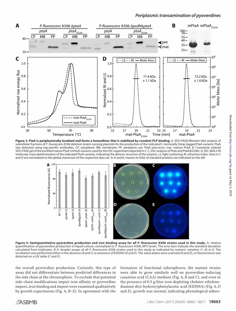
the overall pyoverdine production. Certainly, this type ofassay did not differentiate between predicted differences inthe side chain at the chromophore. To exclude that potentialside-chain modifications impair iron affinity or pyoverdineimport, iron binding and import were examined qualitativelyby growth experiments (Fig. 4, B–E). In agreement with the
formation of functional siderophores, the mutant strainswere able to grow similarly well on pyoverdine-inducingcasamino acid (CAA) medium (Fig. 4, B and C), and even inthe presence of 0.5 g/liter iron-depleting chelator ethylene-diamine di(o-hydroxy)phenylacetic acid (EDDHA) (Fig. 4, Dand E), growth was normal, indicating physiological sidero-
Figure 3. PtaA is periplasmically localized and forms a homodimer that is stabilized by covalent PLP binding. A, SDS-PAGE/Western blot analysis ofsubcellular fractions of P. fluorescens A506 deletion strains carrying plasmids for the production of the indicated C-terminally Strep-tagged PtaA variants. PtaAwas detected using tag-specific antibodies. CP, cytoplasm; MB, membrane; PP, periplasm; pre, PtaA precursor; mat, mature PtaA. B, Coomassie stainedSDS-PAGE gel of the purified mature PtaA (mPtaA) variants used for the DSC experiment depicted in C. C, DSC analysis of PtaA and PtaA(K224A). D, SEC-MALS-RImolecular mass determination of the indicated PtaA variants, indicating the dimeric structure of the enzyme. LS, light scattering; RI, refractive index. Data in Cand D are normalized to the global maximum of the respective data set. In A and B, masses (in kDa) of standard proteins are indicated on the left.
Figure 4. Semiquantitative pyoverdine production and iron binding assay for all P. fluorescens A506 strains used in this study. A, relativequantification of pyoverdine production in liquid culture, normalized to P. fluorescens A506 (WT) levels. The error bars indicate the standard deviationcalculated from triplicates. B–E, droplet assays of all P. fluorescens A506 strains used in this study as indicated by numeric notation (1– 8) in A. Theincubation was performed either in the absence (B and C) or presence of EDDHA (D and E). The same plates were scanned (B and D), or fluorescence wasdetected on a UV table (C and E).
Periplasmic transamination of pyoverdines
J. Biol. Chem. (2017) 292(45) 18660 –18671 18663
by guest on May 5, 2019
http://ww
w.jbc.org/
Dow
nloaded from

phore functionality of the pyoverdines produced in themutant strains.
PtaA is the searched-for transaminase
To examine whether PtaA is responsible for the predictedside-chain modification, we isolated the pyoverdines from allstrains and analyzed the molecular composition via ultraperfor-
mance liquid chromatography (UPLC)-coupled high-resolu-tion mass spectrometry (HR-MS) and isoelectric focusing (IEF)gels in combination with the chrome azurol S (CAS) overlayassay. The results verified our hypothesis about the nature ofthe modification (Fig. 5, A–D). For convenience, the structuresof all pyoverdine variants identified in this study are denoted inFig. 5, B and D, and the assigned numbers 1-6 will be used to
Figure 5. The enzyme PtaA is the periplasmic transaminase that converts the glutamic acid side chain of pyoverdine into the �-ketoglutaric acidvariant. A, mass spectrometry results showing the UPLC chromatograms filtered for the masses of the succinamide, �-ketoglutaric acid, and glutamic acidvariants on the left. On the right, the corresponding mass spectra of the peak at 0.6 min are depicted. The mass peaks were assigned to the structures shownin B and D. B, structure of the pyoverdine backbone, the intramolecular cyclized succinic acid variant, and the non-cyclized pyoverdine precursor ferribactin.The residues R1– 4 correspond to the substructures shown in D. C, IEF gels visualized by pyoverdine fluorescence (left) and a CAS overlay assay (right). Numbersdenote the identified pyoverdine variants, whose structures are represented in B in conjunction with D.
Periplasmic transamination of pyoverdines
18664 J. Biol. Chem. (2017) 292(45) 18660 –18671
by guest on May 5, 2019
http://ww
w.jbc.org/
Dow
nloaded from

unambiguously attribute the compounds discussed. Themasses of compounds 1-6 were determined by HR-MS andcompared with the calculated molecular masses (denoted inparentheses) of the respective molecules: 1, 1190.544 atomicmass units (a.m.u.) (1190.544 a.m.u.); 2, 1189.510 a.m.u.(1189.513 a.m.u.); 3, 1160.534 a.m.u. (1160.534 a.m.u.); 4,1161.520 a.m.u. (1161.518 a.m.u.); 5, 1143.507 a.m.u. (1143.507a.m.u.); 6, 1178.581 a.m.u. (1178.581 a.m.u.).
To analyze whether the identified structures were truly dis-tinct molecules and not artifacts of MS (e.g. dehydrations), weused a shallower gradient for elution and could thereby provethat all compounds have a distinct elution profile (supplemen-tal Fig. S1). From the MS results, it was obvious that, in theabsence of PtaA, no �-ketoglutaric acid variant (2) of PVDA506was produced (Fig. 5A). The equilibrium between the side-chain modifications was clearly shifted in favor of the succina-mide variant (3) of PVDA506. Additionally, the unmodified glu-tamic acid precursor (1) of PVDA506 was detectable, indicatingan incomplete turnover by PvdN. It should be noted that fur-ther pyoverdine species could by detected by MS, including thehydrolysis product of the succinamide variant, namely the suc-cinic acid variant (4); traces of the intramolecular cyclizationproduct thereof (5); and the non-cyclized pyoverdine precursorferribactin (6). As expected from our previously published anal-ysis of PvdN (6), the �ptaA/�pvdN double-deletion strain pro-duced neither the succinamide variant (3) or further derivativesthereof nor the �-ketoglutaric acid variant (2) of PVDA506.Consequently, the only detectable variants were the fluoro-phore-containing precursor with the original glutamic acid res-idue (1) and its non-cyclized precursor ferribactin (6). Toexamine the validity of our MS data interpretation by an inde-pendent method, we performed IEF experiments in combina-tion with the CAS overlay assay (Fig. 5C). The assignments ofthe bands are denoted on the sides of the images using thedefinitions given in Fig. 5D. The results obtained by IEF corre-late well with the data obtained by MS. Noticeably, the wild-type strain produced an excess of the �-ketoglutaric acid vari-ant (2), whereas the succinamide variant (3) was a minor sideproduct. The �pvdN deletion strain did not produce the suc-cinamide variant (3) anymore, and all pyoverdine was con-verted into the �-ketoglutaric acid variant (2) by PtaA. The�ptaA deletion strain in turn produced at least three variants ofpyoverdine, namely the unmodified glutamic acid precursor(1), the succinamide variant (3), and the hydrolysis productthereof, the succinic acid variant (4). It should be noted that thelower band in the �ptaA strain could easily be mistaken for the�-ketoglutaric acid variant (2) due to its nearly identical pI, butthe succinic acid variant (4) migrated slightly higher. Further-more, in the �ptaA/�pvdN double-deletion strain, no succinicacid variant (4) was present due to the lack of its precursor, thesuccinamide variant (3). This lane unambiguously clarifies theassignment of the closely migrating �-ketoglutaric acid (2) andsuccinic acid variants (4) of PVDA506.
The �ptaA phenotype was entirely complemented in transby the ptaA complementation strain, whereas the PtaA(K224A)variant did not complement the phenotype and was thereforeinactive. This conclusion can be deduced from the comparisonof the IEF pyoverdine band patterns of these strains with the
patterns obtained for the wild-type and the �ptaA deletionstrains. The same holds true for the complementation of the�ptaA/�pvdN double-deletion strains as the in trans com-plementation with ptaA resulted in the quantitative turn-over of the glutamic acid precursor (1) to the �-ketoglutaricacid variant (2), whereas the strain with PtaA(K224A) pro-duced only the unmodified glutamic acid precursor (1).Moreover, the CAS overlay assay indicated that the side-chain modifications did not alter the iron affinity of thepyoverdine variants as can be inferred from the identicalband patterns in comparison with fluorescence imaging ofthe IEF gels (Fig. 4C).
Discussion
More than 30 years ago, an �-ketoglutaric acid was identifiedfor the first time as an amide-bonded residue at the fluorophoreof a pyoverdine in strains of P. aeruginosa and P. fluorescens (16,17). Also the at that time already known succinamide (18) andsuccinic acid residues were detected at this position in thosestudies. Interestingly, pyoverdines of P. aeruginosa strainsgreatly differ in their peptide moiety but generally have thethree above mentioned residues in common that are amidicallyattached to their fluorophore (19). Also a glutamic acid res-idue bound to the fluorophore via its �-carboxylic acid wasdiscovered at this position (20) as well as malamide (21),malic acid (22), and an intramolecular cyclized succinate(23). These variations of the pyoverdine are made in a spe-cies- and strain-specific manner (24). Based on the large var-iability of amino acid sequences and fluorophore-attachedside chains, IEF-based siderotyping methods have beendeveloped that use the strain-specific characteristics for sys-tematic purposes (25).
The glutamic acid residue is always the first residue in ferri-bactins, initially discovered by Hohlneicher et al. (26). This glu-tamic acid residue is myristoylated for transport into theperiplasm where it is deacylated by PvdQ (27–29). After forma-tion of the fluorophore, it thus must be the glutamic acidresidue that is initially present at this position, and the foundsuccinamide, succinic acid, �-ketoglutaric acid, malamide,and malic acid residues must result from glutamic acidmodifications.
Biochemistry of PtaA
It was clear from our recent study on PvdN (6) that anunknown enzyme had to be responsible for the formation of the�-ketoglutaric acid. Using P. fluorescens A506 as model orga-nism, we have now identified PtaA as the periplasmic transam-inase responsible for the formation of the �-ketoglutaric acidresidue in PVDA506 (2). PtaA is a PLP-containing enzyme (Fig.2) that is transported together with its covalently bound cofac-tor via the Tat pathway in a folded conformation (Fig. 3). Thefolded state is influenced by cofactor binding to position Lys-224 (Fig. 3C), which is most likely the reason for a partial mis-localization of the mutated protein in the membrane (Fig. 3A).A similar effect had been already observed with PvdN, whichalso requires PLP binding for Tat-dependent transport (6). Inthe case of PvdN, the exchange of the active-site Lys resultedeven in a complete mislocalization of the enzyme in the mem-
Periplasmic transamination of pyoverdines
J. Biol. Chem. (2017) 292(45) 18660 –18671 18665
by guest on May 5, 2019
http://ww
w.jbc.org/
Dow
nloaded from

brane fraction (6). PvdN and PtaA are both homodimericenzymes (Ref. 6 and Fig. 3D) with their PLP most likely boundclose to the dimer interface (Ref. 6 and Fig. 6C). This class ofenzymes includes therefore good examples for typical Tat sub-strates that need to be folded prior to translocation (30). Theypossess twin-arginine signal peptides that can be used for iden-
tification purposes in addition to transaminase sequence spec-ifications (Fig. 1).
Fig. 6A summarizes the now established modifications of thefluorophore-attached glutamic acid residue of pyoverdines.The transamination probably proceeds via a standard transam-inase mechanism, which is interesting to exist in the periplasm
Figure 6. Role and mechanism of PtaA in periplasmic pyoverdine tailoring. A, beginning from the glutamic acid variant (1) of PVDA506, two competingtailoring pathways are present. Transamination by PtaA results in the �-ketoglutaric acid variant (2) PVDA506, whereas the PvdN modification results in thesuccinamide variant (3), which can be partially hydrolyzed to the succinic acid (4) and then intramolecularly cyclized (6). B, proposed mechanism of transam-ination as catalyzed by PtaA in the periplasm, postulating a carbonyl compound for regeneration of PLP. C, structure of PtaA as calculated by homologymodeling, highlighting the Lys-224 residue and the PLP cofactor. The model was visualized with UCSF Chimera.
Periplasmic transamination of pyoverdines
18666 J. Biol. Chem. (2017) 292(45) 18660 –18671
by guest on May 5, 2019
http://ww
w.jbc.org/
Dow
nloaded from

as it requires a yet unknown periplasmic carbonyl compoundthat we postulate to exist (Fig. 6B). As the Tat transport and itsPLP requirement indicate that PtaA assembles PLP inside thecytoplasm to fold to an active enzyme, PLP must be regeneratedfrom PtaA-bound pyridoxamine inside the periplasm. Theenzymes PvdN and PtaA are responsible for the initial conver-sions of the glutamic acid pyoverdine variant. The existence ofthese competing enzyme reactions explains the reported regu-latory differences for the formation of succinamide and �-ke-toglutaric acid forms of pyoverdine (31). As a side aspect, theglutamic acid variant of pyoverdine that is produced by the�ptaA/�pvdN double mutant can be very helpful for drugdelivery by “Trojan horse” antibiotics (32) because bulky sidechains may be attached to this residue without affecting uptake(33). Malamide is likely formed by a hydroxylating enzyme act-ing on succinamide. The acids succinic acid and malic acid arehydrolysis products of their respective amides. A non-enzy-matic hydrolysis has been observed and postulated to beresponsible for the formation of these acids in cultures (34). Itcertainly might be that amidases contribute to the rates of thesereactions.
PtaA, a transaminase for pyoverdines and other periplasmiccompounds
In P. aeruginosa PAO1, the homolog of PtaA (53% sequenceidentity) is encoded by the locus PA2531, which is PvdS-depen-dently up-regulated in response to iron (35); hence this genebelongs to the regulon of PvdS, the � factor that mediatespyoverdine production (36). An “iron starvation box” forPvdS binding (37) has been identified in the promoter region ofPA2531 (35). We found the consensus box as describedfor P. aeruginosa PAO1 by Ochsner et al. (35) also inthe ptaA promoter region of P. fluorescens A506, namely“TAAATN16CGT.” However, the genetic context of ptaA is notconserved between P. aeruginosa PAO1 and P. fluorescens
A506. While the iron-responsive regulation of ptaA correlateswith its function in pyoverdine modification, its usually pvd-unrelated genomic environment is suggestive for additionalfunctions. This interpretation is further strengthened by a pre-vious study on chorismate mutases that included the detectionof transaminase activity in the periplasm of P. aeruginosa PAO1(38). In that study, the periplasmic transaminase activity couldbe assigned to PA2531, the above mentioned PtaA homolog.PA2531 is dimeric just like PtaA from P. fluorescens in our study(Fig. 3D). At that time, the authors could not know about its rolein pyoverdine modification, which is now clarified by our study.They used �-ketoglutaric acid and phenylalanine as substratesfor the transaminase assay. Therefore, PtaA homologs cannotbe highly specific for pyoverdine substrates and are likely to beinvolved in other conversions as well. In support of this conclu-sion, ptaA homologs are also present in some acidobacteria and�-proteobacteria, such as Zymomonas mobilis (supplementalFig. S2), that do not produce any pyoverdines. Therefore, thisperiplasmic transaminase must be able to exert its function fordistinct physiological purposes, possibly in conjunction withthe generation of distinct secondary metabolites.
Distribution of PtaA among pseudomonads
As summarized in Fig. 7, we searched for ptaA homologsin all Pseudomonas genomes available in the PseudomonasGenome Database (11) (see “Experimental procedures” fordetails). In agreement with the known occurrence of �-ketoglu-taric acid forms of pyoverdines, we found this gene in almostall sequenced P. aeruginosa strains (2193 out of 2225 genomes;�99%). The absence of hits in the �1% of strains without this genemight be due to the inclusion of draft genomes in the analysis.
In the more diverse groups of P. fluorescens and Pseudomo-nas putida strains, only 45 out of 106 genomes and 13 out of 66genomes, respectively, encode PtaA, suggesting that the pres-ence of the �-ketoglutaric acid variant of pyoverdine is not
Figure 7. Distribution of PtaA homologs within the genus Pseudomonas. Shown is an evaluation of the distribution of PtaA within the genus Pseudomonas.Numbers to the right of the bar graph indicate the ratio between genomes positive for the presence of PtaA and the total number of analyzed genomes of therespective species.
Periplasmic transamination of pyoverdines
J. Biol. Chem. (2017) 292(45) 18660 –18671 18667
by guest on May 5, 2019
http://ww
w.jbc.org/
Dow
nloaded from

strictly conserved in these species. In agreement with this pre-diction, P. fluorescens Pf0-1 has no PtaA and, as expected, hasbeen shown not to contain the �-ketoglutaric acid (24). On thecontrary, there are strains that form �-ketoglutaric acid pyover-dine variants but not any succinamide/succinate variants (24),such as the genome-sequenced P. putida strain H8234. We foundthat this strain has a pvdMO-ptaA operon instead of the com-monly found pvdMNO operon. Therefore, in the rarely foundstrains that do not possess the succinamide/succinic acid modifi-cation pathway, ptaA can be associated with the pvd gene clusterthat encodes the periplasmic pyoverdine maturation enzymes. Inconclusion, some Pseudomonas species with at least the exceptionof P. aeruginosa show the interesting characteristic of containingeither only the PtaA pathway or only the PvdN pathway (39).Strains with both pathways acting in parallel also exist. We did notfind a strain that produces pyoverdine without modifying its glu-tamic acid. The reason for the apparent requirement for suchmodifications is unknown and must be unrelated to iron affinityand uptake (Ref. 40 and Fig. 4). Future studies will have to clarifythe exact functions of these modifications as well as the additionalroles that PtaA can have in the periplasm of proteobacteria.
Experimental procedures
Strains and growth conditions
For physiological studies, P. fluorescens A506 was used. Forcloning E. coli DH5� � pir� and for expression E. coli Rosetta 2(DE3) pLysSRARE2 were utilized. P. fluorescens A506 was cul-tivated at 30 °C, whereas E. coli strains were cultivated at 37 °Cunless noted otherwise. The standard cultivation medium wasLB (1% (w/v) tryptone, 1% (w/v) NaCl, 0.5% (w/v) yeast extract).If necessary, the appropriate antibiotics were added to the cul-tivation media at the following final concentrations: 100 �g/mlampicillin, 25 �g/ml chloramphenicol, 50 �g/ml kanamycin,and 20 �g/ml tetracycline.
The production of pyoverdine and the pyoverdine plateassay, pertaining to P. fluorescens A506, were carried out asdescribed previously (6). For relative pyoverdine quantification,5-ml LB overnight cultures (ONCs) of P. fluorescens A506 wereinoculated from cryocultures and incubated at 30 °C at 180 rpm
overnight. Thereafter, 50 of ml CAA medium (41) (5 g/litercasamino acids, 5 mM K2HPO4, 1 mM MgSO4) precultures in100-ml Erlenmeyer flasks with one baffle were inoculated with50 �l of the respective ONC and incubated for �16 h at 30 °Cand 180 rpm. 2 ml of the respective preculture were sedimentedat 16,000 � g for 2 min at room temperature and subsequentlywashed twice with 1.5 ml of CAA medium. Afterward, theOD600 of the cell suspensions was adjusted to 1.0 with CAAmedium, and for each sample three 4.5-ml CAA cultures (with-out antibiotics) were inoculated with 0.5 ml of the respectiveadjusted cell suspension. The cultures were then incubatedovernight at 30 °C and 180 rpm. These cultures were sedi-mented by centrifugation at 3,260 � g and 4 °C for 10 min in15-ml screw-top plastic tubes. 2.7 ml of the supernatant weremixed with 300 �l of a 1 M HEPES buffer, pH 8.0, and trans-ferred into 1-cm acrylic cuvettes. The samples were measuredwith a Jasco FP-6500 spectrofluorometer using the followingsettings for acquisition: excitation at 405 nm, emission at 460nm, bandwidth of 3 nm for both excitation and emission, and0.5 s response time in “low-sensitivity” mode.
Genetic methods and plasmids
The construction of all scarless and markerless deletions inP. fluorescens A506 were performed as described previously (6).Furthermore, complementation and production vectors as wellas constructs with single-point mutations (pME6010 andpEXH5 derivatives) were generated according to our previouslypublished procedure (6). All primers used for cloning are listedin Table 2. Constructs were verified by restriction analysis andsequencing. E. coli DH5� � pir� cells were rendered competentand transformed as described previously (42).
Biochemical methods
For analysis of subcellular fractionations and protein overex-pression by SDS-PAGE, successive Western blotting, or in-gelcolloidal Coomassie staining, standard protocols were used(43– 46). Western blots were developed according to the man-ufacturer’s instructions using StrepMAB-Classic (IBA, Göttin-gen, Germany) as primary antibody and anti-mouse-HRP con-
Table 2Primers used in this studyoePCR, overlap extension PCR.
Name SequenceRestriction
site Purpose
PfA506-4424-F1-MR ATAGCCGGATCCTAGACAGGTAGCGCCAAATCACG BamHI Forward primer for ptaA left flanking regionPfA506-4424-R1-MR AGGAGTAGTCACCATGGTGCGTGTCACGCAGGTGGTCTGA
TCAGGCGGCGCTATAGCTGReverse primer for ptaA left flanking region
PfA506-4424-F2-MR GACACGCACCATGGTGACTACTCCTTG Forward primer for ptaA right flanking regionPfA506-4424-R2-MR GGCCGCGAATTCTTTGTACAGATTCTTGATTTCCATCTTG EcoRI Reverse primer for ptaA right flanking regionPfA506-4424-DF-MR TTTGAGTGCTGCGCCTATTG ptaA genomic deletion control primerPfA506-4424-DR-MR CGCGGCAATGCTGGTGAAGATATAG ptaA genomic deletion control primerPfA506-4424-F-MR TCATCGCATATGGTGCGTGTCAGTCGTCGATCC NdeI Forward primer for cloning PtaA-coding region
into pEXH5PfA506-4424-strep-R-MR GGCCGCAAGCTTTTACTTTTCGAACTGCGGGTGGCTCCAG
ACCACCTGCGTCGCAAAGGCCTCGCHindIII Reverse primer for cloning PtaA-coding region
into pEXH5PfA506-4424_K224A-F-MR CTGGTGCTGCGCACCTTCTCCGCCATCTAC Primer for K224A exchange by oePCRPfA506-4424_K224A-R-MR CCGGCCATGCCGTAGATGGCGGAGAAGGTG Primer for K224A exchange by oePCRPfA506-mat4424-F-MR GATATACATATGAGCCCAGCGCCGACAAAATCTGACC NdeI Forward primer for cloning mature PtaA-coding
region into pEXH5pEXH5-RBS-F-MR GGCGCGGGATCCGTTTAACTTTAAGAAGGAGATATAC BamHI Forward primer for subcloning from pEXH5
into pME6010pEXH5-strep-term-HindIII-R-MR CCCCTTAAGCTTAAAAAAAACCCCGCCCTGTCAGGGGCGG
GGTTTTTTTTTTTACTTTTCGAACTGCGGGTGGCTCCHindIII Reverse primer for subcloning from pEXH5
into pME6010
Periplasmic transamination of pyoverdines
18668 J. Biol. Chem. (2017) 292(45) 18660 –18671
by guest on May 5, 2019
http://ww
w.jbc.org/
Dow
nloaded from

jugate (Carl Roth, Karlsruhe, Germany) as secondary antibody.Images were acquired with the MF-ChemiBIS 4.2 imaging sys-tem (DNR Bio-Imaging Systems, Jerusalem, Israel).
The overproduction of mature PtaA and mature PtaA(K224A)was performed using E. coli Rosetta 2 (DE3) pLysSRARE2 asdescribed previously (47) with minor modifications. Briefly, therespective construct was transformed into E. coli Rosetta 2(DE3) pLysSRARE2 by the transformation and storage so-lution method (48). After recovery, the cells were spread onMDAG-11 plates (47) and incubated at 37 °C overnight. A 5-mlMDAG-135 (47) ONC was then inoculated with a single colonyand incubated at 180 rpm and 37 °C. For overproduction, two0.5-liter ZYM-5052 preheated (37 °C) autoinducer mediumcultures (47) in 3-liter Erlenmeyer flasks with four baffles wereeach inoculated with 0.5 ml of the ONC. The cultures wereincubated for 3 h at 200 rpm and 37 °C. Subsequently, the tem-perature was decreased to 30 °C, and the cultures were incu-bated for approximately 19 h at 200 rpm. The cells were har-vested, and the respective protein was purified by means of four1.5-ml gravity-flow Strep-Tactin�-Sepharose columns (IBA) asdescribed previously (6). The purified proteins were concen-trated using Vivaspin� 6 concentrators with a cutoff of 10 kDa(Sartorius, Göttingen, Germany). For molecular weight deter-mination, a Pharmacia FPLC (LKB Pump P-500, V-7 valve,50-�l sample loop) was connected to a size-exclusion chroma-tography column (Superdex 200 Increase 10/300 GL, GEHealthcare) coupled to MALS (miniDAWN TREOS, WyattTechnology Europe GmbH, Dernbach, Germany) and refrac-tive index detectors (Shodex RI-101, Showa Denko EuropeGmbH, Munich, Germany). The flow rate of the mobile phase(PBS; 10 mM phosphate buffer, pH 7.4, 140 mM NaCl) was set to0.75 ml/min. The chromatograms were recorded and analyzedwith ASTRA 6.1 software (Wyatt Technology). For DSC, aNANO DSC in conjunction with a degassing station from TAInstruments (Lindon, UT) was used. For preparation of DSCsamples, the protein elution peak (19-min retention time) fromthe SEC-MALS-RI system was collected by hand and subse-quently concentrated with Vivaspin 6 concentrators (cutoff, 10kDa). The protein concentration was determined using Roti�-Nanoquant according to the instruction manual (Carl Roth)using a SpectraMax M3 spectrophotometer (Molecular De-vices, Biberach an der Riss, Germany). The protein of interestwas diluted with degassed PBS to a concentration of �1.5mg/ml prior to analysis. The DSC reference cell was filled withdegassed PBS. Analyses covered the temperature range from 20to 110 °C at a heating rate of 1 °C/min with a 900-s pre-equili-bration time at a constant pressure of 3 atm.
The subcellular fractionation of P. fluorescens A506 cultureswas performed as described previously (49) with slight modifi-cations. 50-ml cultures were grown to an OD600 of 1 and frac-tionated into 1-ml fractions that were further analyzed withoutprecipitation.
For PLP cofactor detection, a method modified from that ofOjha et al. (50) was used. The purified mature PtaA enzyme in100 mM potassium phosphate buffer, pH 7.2, was treated with 5mM hydroxylamine and incubated for �72 h at 4 °C. The sam-ple was loaded onto a Vivaspin 6 concentrator with 5-kDa cut-off, and the PLP oxime in the flow-through was detected at
446-nm emission (scan from 400 to 700 nm) with an excitationof 353 nm, 3-nm bandwidth for both excitation and emission,0.2-s response time, and three accumulations in “high-sensitiv-ity” mode using a Jasco FP-6500 spectrofluorometer. Referencesolutions with the buffer only or with 1 mM PLP in the samebuffer were treated identically. PLP- or PLP oxime-containingreference spectra were recorded with the same settings but in“medium-sensitivity” mode.
The extraction of pyoverdine, IEF analysis in conjunctionwith the CAS overlay assay, and UPLC-MS analysis were per-formed as described previously (6). To prove that the identifiedcompounds are not artifacts of the MS, an adjusted linear gra-dient profile was utilized as follows: solvent A (double-distilledwater with 0.1% (v/v) formic acid) and solvent B (acetonitrilewith 0.1% (v/v) formic acid) at a flow rate of 0.6 ml/min; 2% B(0.0 min), 10% B (10.0 min), 90% B (10.5 min), 90% B (13.0 min),2% B (13.5 min), and 2% B (20.0 min). The electrospray ioniza-tion voltage was set to 100 V, and the injection volume was 5 �l.All other instrument parameters and all instrument hardwarewere identical to the method described previously (6) exceptthat an ACQUITY UPLC Column Manager (Waters) was addi-tionally installed on the instrument.
Bioinformatics methods
To perform an initial distribution analysis of PtaA within thegenus Pseudomonas, we downloaded all amino acid sequencesof all complete and draft genomic sequences from the Pseu-domonas Genome Database (11) and used TATFIND v.1.4 (7)to search for potential Tat substrates. Subsequently, phmmerv3.1b (51) was used to search for PtaA homologs (cutoff,1e�45) within all potential Tat substrates. Then a taxonomytable reduced to species level was constructed by means of in-house developed software. Thereafter, the ratio between thetotal number of genomes of one species and the number ofgenomes of the same species carrying a potential PtaA homologwas calculated.
To construct the sequence logos, the sequence redundancyof all PtaA homologs was reduced by means of the CD-HITalgorithm contained in the CD-HIT Suite (12) using a similaritythreshold of 0.95. Then the resulting sequences were alignedwith the T-Coffee web server (13). The sequence alignment wassubsequently visualized with WebLogo 3 (14) by plotting theprobability of each amino acid at each alignment position. Forfurther investigations into the distribution of PtaA homologsacross other bacterial species, a profile hidden Markov model(pHMM) was generated from the multiple sequence alignmentwith hmmbuild v.3.1b (51). Then all available referencesequences were searched with the generated model (E-valuecutoff ,1e�100) using the HMMER web server (52, 53). Theresulting sequences were then analyzed regarding their phylo-genetic relationship using the Phylogeny.fr3 web service (54) inconjunction with the ETE toolkit (55) for visualization.
To initially predict the enzyme class and active-site lysineresidue, Lys-224, the InterPro web service (8, 9) was used. TheSWISS-MODEL web service (56) was used to model the struc-
3 Please note that the JBC is not responsible for the long-term archiving andmaintenance of this site or any other third party-hosted site.
Periplasmic transamination of pyoverdines
J. Biol. Chem. (2017) 292(45) 18660 –18671 18669
by guest on May 5, 2019
http://ww
w.jbc.org/
Dow
nloaded from

ture of PtaA (template, Protein Data Bank code 3LY1), andUCSF Chimera (57) in conjunction with APBS (58) was used tovisualize the structure of PtaA.
Author contributions—M. T. R. performed the experiments, pre-pared the figures, and analyzed the data together with T. B. G. D.performed the MS analyses. T. B. conceived and coordinated thestudy. T. B. and M. T. R. wrote the paper. All authors reviewed theresults and approved the final version of the manuscript.
Acknowledgment—We thank Sybille Traupe for technical support.
References1. Andrews, S. C., Robinson, A. K., and Rodríguez-Quiñones, F. (2003) Bac-
terial iron homeostasis. FEMS Microbiol. Rev. 27, 215–2372. Cézard, C., Farvacques, N., and Sonnet, P. (2015) Chemistry and biology of
pyoverdines, Pseudomonas primary siderophores. Curr. Med. Chem. 22,165–186
3. Gulick, A. M. (2017) Nonribosomal peptide synthetase biosynthetic clus-ters of ESKAPE pathogens. Nat. Prod. Rep. 34, 981–1009
4. Schalk, I. J., and Guillon, L. (2013) Pyoverdine biosynthesis and secretionin Pseudomonas aeruginosa: implications for metal homeostasis. Environ.Microbiol. 15, 1661–1673
5. Nadal-Jimenez, P., Koch, G., Reis, C. R., Muntendam, R., Raj, H., Jeroni-mus-Stratingh, C. M., Cool, R. H., and Quax, W. J. (2014) PvdP is a tyro-sinase that drives maturation of the pyoverdine chromophore in Pseu-domonas aeruginosa. J. Bacteriol. 196, 2681–2690
6. Ringel, M. T., Dräger, G., and Brüser, T. (2016) PvdN enzyme catalyzes aperiplasmic pyoverdine modification. J. Biol. Chem. 291, 23929 –23938
7. Rose, R. W., Brüser, T., Kissinger, J. C., and Pohlschröder, M. (2002) Ad-aptation of protein secretion to extremely high-salt conditions by exten-sive use of the twin-arginine translocation pathway. Mol. Microbiol. 45,943–950
8. Jones, P., Binns, D., Chang, H.-Y., Fraser, M., Li, W., McAnulla, C., Mc-William, H., Maslen, J., Mitchell, A., Nuka, G., Pesseat, S., Quinn, A. F.,Sangrador-Vegas, A., Scheremetjew, M., Yong, S.-Y., et al. (2014) Inter-ProScan 5: genome-scale protein function classification. Bioinformatics30, 1236 –1240
9. Finn, R. D., Attwood, T. K., Babbitt, P. C., Bateman, A., Bork, P., Bridge,A. J., Chang, H.-Y., Dosztányi, Z., El-Gebali, S., Fraser, M., Gough, J., Haft,D., Holliday, G. L., Huang, H., Huang, X., et al. (2017) InterPro in 2017—beyond protein family and domain annotations. Nucleic Acids Res. 45,D190 –D199
10. Drake, E. J., and Gulick, A. M. (2016) 1.2 Å resolution crystal structure ofthe periplasmic aminotransferase PvdN from Pseudomonas aeruginosa.Acta Crystallogr. F Struct. Biol. Commun. 72, 403– 408
11. Winsor, G. L., Griffiths, E. J., Lo, R., Dhillon, B. K., Shay, J. A., and Brink-man, F. S. (2016) Enhanced annotations and features for comparing thou-sands of Pseudomonas genomes in the Pseudomonas genome database.Nucleic Acids Res. 44, D646 –D653
12. Huang, Y., Niu, B., Gao, Y., Fu, L., and Li, W. (2010) CD-HIT Suite: a webserver for clustering and comparing biological sequences. Bioinformatics26, 680 – 682
13. Di Tommaso, P., Moretti, S., Xenarios, I., Orobitg, M., Montanyola, A.,Chang, J.-M., Taly, J.-F., and Notredame, C. (2011) T-Coffee: a web serverfor the multiple sequence alignment of protein and RNA sequences usingstructural information and homology extension. Nucleic Acids Res. 39,W13–W17
14. Crooks, G. E., Hon, G., Chandonia, J.-M., and Brenner, S. E. (2004) We-bLogo: a sequence logo generator. Genome Res. 14, 1188 –1190
15. Halbig, D., Wiegert, T., Blaudeck, N., Freudl, R., and Sprenger, G. A. (1999)The efficient export of NADP-containing glucose-fructose oxidoreduc-tase to the periplasm of Zymomonas mobilis depends both on an intacttwin-arginine motif in the signal peptide and on the generation of a struc-tural export signal induced by cofactor binding. Eur. J. Biochem. 263,543–551
16. Briskot, G., Taraz, K., and Budzikiewicz, H. (1986) Pyoverdine type sidero-phores from Pseudomonas aeruginosa. Z. Naturforsch. C 41, 497–506
17. Poppe, K., Taraz, K., and Budzikiewicz, H. (1987) Pyoverdine type sidero-phores from Pseudomonas fluorescens. Tetrahedron 43, 2261–2272
18. Teintze, M., Hossain, M. B., Barnes, C. L., Leong, J., and van der Helm, D.(1981) Structure of ferric pseudobactin: a siderophore from a plant growthpromoting Pseudomonas. Biochemistry 20, 6446 – 6457
19. Meyer, J. M., Stintzi, A., De Vos D., Cornelis, P., Tappe, R., Taraz, K., andBudzikiewicz, H. (1997) Use of siderophores to type pseudomonads: thethree Pseudomonas aeruginosa pyoverdine systems. Microbiology 143,35– 43
20. Geisen, K., Taraz, K., and Budzikiewicz, H. (1992) New pyoverdin typesiderophores from Pseudomonas fluorescens. Monatsh. Chem. 123,151–178
21. Yang, C. C., and Leong, J. (1984) Structure of pseudobactin 7SR1, a sid-erophore from a plant-deleterious Pseudomonas. Biochemistry 23,3534 –3540
22. Demange, P., Bateman, A., Mertz, C., Dell, A., Piémont, Y., and Abdallah,M. A. (1990) Bacterial siderophores: structures of pyoverdins Pt, sidero-phores of Pseudomonas tolaasii NCPPB 2192, and pyoverdins Pf, sidero-phores of Pseudomonas fluorescens CCM 2798. Identification of an un-usual natural amino acid. Biochemistry 29, 11041–11051
23. Lenz, C., Amann, C., Briskot, G., Taraz, K., and Budzikiewicz, H. (2000)Succinopyoverdins—a new variety of the pyoverdin chromophore. Z.Naturforsch. C 55, 146 –152
24. Meyer, J.-M., Gruffaz, C., Raharinosy, V., Bezverbnaya, I., Schäfer, M., andBudzikiewicz, H. (2008) Siderotyping of fluorescent Pseudomonas: molec-ular mass determination by mass spectrometry as a powerful pyoverdinesiderotyping method. Biometals 21, 259 –271
25. Koedam, N., Wittouck, E., Gaballa, A., Gillis, A., Höfte, M., and Cor-nelis, P. (1994) Detection and differentiation of microbial siderophoresby isoelectric focusing and chrome azurol S overlay. Biometals 7,287–291
26. Hohlneicher, U., Hartmann, R., Taraz, K., and Budzikiewicz, H. (1992)The structure of ferribactin from Pseudomonas fluorescens ATCC 13525.Z. Naturforsch. B 47, 1633–1638
27. Yeterian, E., Martin, L. W., Guillon, L., Journet, L., Lamont, I. L., andSchalk, I. J. (2010) Synthesis of the siderophore pyoverdine in Pseu-domonas aeruginosa involves a periplasmic maturation. Amino Acids38, 1447–1459
28. Drake, E. J., and Gulick, A. M. (2011) Structural characterization andhigh-throughput screening of inhibitors of PvdQ, an NTN hydrolase in-volved in pyoverdine synthesis. ACS Chem. Biol. 6, 1277–1286
29. Hannauer, M., Schäfer, M., Hoegy, F., Gizzi, P., Wehrung, P., Mislin, G. L.,Budzikiewicz, H., and Schalk, I. J. (2012) Biosynthesis of the pyoverdinesiderophore of Pseudomonas aeruginosa involves precursors with a myr-istic or a myristoleic acid chain. FEBS Lett. 586, 96 –101
30. Hou, B., and Brüser, T. (2011) The Tat-dependent protein translocationpathway. Biomol. Concepts 2, 507–523
31. Schäfer, H., Taraz, K., and Budzikiewicz, H. (1991) On the genesis of thedicarboxylic acids bound amidically to the chromophore of the pyover-dins. Z. Naturforsch. C 46, 398 – 406
32. Schalk, I. J., and Mislin, G. L. A. (2017) Bacterial iron uptake pathways:gates for the import of bactericide compounds. J. Med. Chem. 60,4573– 4576
33. Schons, V., Atkinson, R. A., Dugave, C., Graff, R., Mislin, G. L., Rochet, L.,Hennard, C., Kieffer, B., Abdallah, M. A., and Schalk, I. J. (2005) Thestructure-activity relationship of ferric pyoverdine bound to its outermembrane transporter: implications for the mechanism of iron uptake.Biochemistry 44, 14069 –14079
34. Budzikiewicz, H. (2004) Siderophores of the Pseudomonadaceae sensustricto (fluorescent and non-fluorescent Pseudomonas spp.), in Progress inthe Chemistry of Organic Natural Products (Falk, H., and Kirby, G. W.,eds), pp. 81–237, Springer, Vienna, Austria
35. Ochsner, U. A., Wilderman, P. J., Vasil, A. I., and Vasil, M. L. (2002)GeneChip� expression analysis of the iron starvation response in Pseu-domonas aeruginosa: identification of novel pyoverdine biosynthesisgenes. Mol. Microbiol. 45, 1277–1287
Periplasmic transamination of pyoverdines
18670 J. Biol. Chem. (2017) 292(45) 18660 –18671
by guest on May 5, 2019
http://ww
w.jbc.org/
Dow
nloaded from
36. Cunliffe, H. E., Merriman, T. R., and Lamont, I. L. (1995) Cloning andcharacterization of pvdS, a gene required for pyoverdine synthesis in Pseu-domonas aeruginosa: PvdS is probably an alternative � factor. J. Bacteriol.177, 2744 –2750
37. Wilson, M. J., McMorran, B. J., and Lamont, I. L. (2001) Analysis of pro-moters recognized by PvdS, an extracytoplasmic-function � factor proteinfrom Pseudomonas aeruginosa. J. Bacteriol. 183, 2151–2155
38. Calhoun, D. H., Bonner, C. A., Gu, W., Xie, G., and Jensen, R. A. (2001)The emerging periplasm-localized subclass of AroQ chorismate mutases,exemplified by those from Salmonella typhimurium and Pseudomonasaeruginosa. Genome Biol. 2, RESEARCH0030
39. Gwose, I., and Taraz, K. (1992) Pyoverdine from Pseudomonas putida. Z.Naturforsch. B 47, 487–502
40. Meyer, J. M. (2000) Pyoverdines: pigments, siderophores and potentialtaxonomic markers of fluorescent Pseudomonas species. Arch. Microbiol.174, 135–142
41. Ochsner, U. A., Snyder, A., Vasil, A. I., and Vasil, M. L. (2002) Effectsof the twin-arginine translocase on secretion of virulence factors,stress response, and pathogenesis. Proc. Natl. Acad. Sci. U.S.A. 99,8312– 8317
42. Inoue, H., Nojima, H., and Okayama, H. (1990) High efficiency transfor-mation of Escherichia coli with plasmids. Gene. 96, 23–28
43. Laemmli, U. K. (1970) Cleavage of structural proteins during the assemblyof the head of bacteriophage T4. Nature 227, 680 – 685
44. Burnette, W. N. (1981) “Western blotting”: electrophoretic transfer ofproteins from sodium dodecyl sulfate-polyacrylamide gels to unmodifiednitrocellulose and radiographic detection with antibody and radioiodi-nated protein A. Anal. Biochem. 112, 195–203
45. Towbin, H., Staehelin, T., and Gordon, J. (1979) Electrophoretic transferof proteins from polyacrylamide gels to nitrocellulose sheets: procedureand some applications. Proc. Natl. Acad. Sci. U.S.A. 76, 4350 – 4354
46. Neuhoff, V., Arold, N., Taube, D., and Ehrhardt, W. (1988) Improvedstaining of proteins in polyacrylamide gels including isoelectric focusinggels with clear background at nanogram sensitivity using Coomassie Bril-liant Blue G-250 and R-250. Electrophoresis 9, 255–262
47. Studier, F. W. (2014) Stable expression clones and auto-induction forprotein production in E. coli. Methods Mol. Biol. 1091, 17–32
48. Chung, C. T., Niemela, S. L., and Miller, R. H. (1989) One-step preparationof competent Escherichia coli: transformation and storage of bacterial cellsin the same solution. Proc. Natl. Acad. Sci. U.S.A. 86, 2172–2175
49. Ize, B., Viarre, V., and Voulhoux, R. (2014) Cell fractionation. MethodsMol. Biol. 1149, 185–191
50. Ojha, S., Wu, J., LoBrutto, R., and Banerjee, R. (2002) Effects of hemeligand mutations including a pathogenic variant, H65R, on the propertiesof human cystathionine �-synthase. Biochemistry 41, 4649 – 4654
51. Eddy, S. R. (2011) Accelerated profile HMM searches. PLoS Comput. Biol.7, e1002195
52. Finn, R. D., Clements, J., and Eddy, S. R. (2011) HMMER web server:interactive sequence similarity searching. Nucleic Acids Res. 39,W29 –W37
53. Finn, R. D., Clements, J., Arndt, W., Miller, B. L., Wheeler, T. J., Schreiber,F., Bateman, A., and Eddy, S. R. (2015) HMMER web server: 2015 update.Nucleic Acids Res. 43, W30 –W38
54. Dereeper, A., Guignon, V., Blanc, G., Audic, S., Buffet, S., Chevenet, F.,Dufayard, J.-F., Guindon, S., Lefort, V., Lescot, M., Claverie, J.-M., andGascuel, O. (2008) Phylogeny.fr: robust phylogenetic analysis for the non-specialist. Nucleic Acids Res. 36, W465–W469
55. Huerta-Cepas, J., Serra, F., and Bork, P. (2016) ETE 3: reconstruction,analysis, and visualization of phylogenomic data. Mol. Biol. Evol. 33,1635–1638
56. Biasini, M., Bienert, S., Waterhouse, A., Arnold, K., Studer, G., Schmidt,T., Kiefer, F., Gallo Cassarino, T., Bertoni, M., Bordoli, L., and Schwede, T.(2014) SWISS-MODEL: modelling protein tertiary and quaternary struc-ture using evolutionary information. Nucleic Acids Res. 42, W252–W258
57. Pettersen, E. F., Goddard, T. D., Huang, C. C., Couch, G. S., Greenblatt,D. M., Meng, E. C., and Ferrin, T. E. (2004) UCSF Chimera—a visualiza-tion system for exploratory research and analysis. J. Comput. Chem. 25,1605–1612
58. Baker, N. A., Sept, D., Joseph, S., Holst, M. J., and McCammon, J. A. (2001)Electrostatics of nanosystems: application to microtubules and the ribo-some. Proc. Natl. Acad. Sci. U.S.A. 98, 10037–10041
Periplasmic transamination of pyoverdines
J. Biol. Chem. (2017) 292(45) 18660 –18671 18671
by guest on May 5, 2019
http://ww
w.jbc.org/
Dow
nloaded from

Michael T. Ringel, Gerald Dräger and Thomas Brüser-ketoglutaric acidαglutamic acid residue at the pyoverdine fluorophore to
converts thePseudomonas fluorescensThe periplasmic transaminase PtaA of
doi: 10.1074/jbc.M117.812545 originally published online September 14, 20172017, 292:18660-18671.J. Biol. Chem.
10.1074/jbc.M117.812545Access the most updated version of this article at doi:
Alerts:
When a correction for this article is posted•
When this article is cited•
to choose from all of JBC's e-mail alertsClick here
Supplemental material:
http://www.jbc.org/content/suppl/2017/09/14/M117.812545.DC1
http://www.jbc.org/content/292/45/18660.full.html#ref-list-1
This article cites 57 references, 9 of which can be accessed free at
by guest on May 5, 2019
http://ww
w.jbc.org/
Dow
nloaded from