their phytochemistry, pharmacological activity and … · 2017-05-22 · molecules review...
TRANSCRIPT
molecules
Review
Phenylethanoid Glycosides: Research Advances inTheir Phytochemistry, Pharmacological Activityand PharmacokineticsZhenzhen Xue and Bin Yang *
Institute of Chinese Materia Medica, China Academy of Chinese Medical Sciences, Beijing 100700, China;[email protected]* Correspondence: [email protected]; Tel.: +86-10-6401-4411 (ext. 2848)
Academic Editor: Derek J. McPheeReceived: 28 June 2016; Accepted: 26 July 2016; Published: 29 July 2016
Abstract: Phenylethanoid glycosides (PhGs) are widely distributed in traditional Chinese medicinesas well as in other medicinal plants, and they were characterized by a phenethyl alcohol (C6-C2)moiety attached to a β-glucopyranose/β-allopyranose via a glycosidic bond. The outstanding activityof PhGs in diverse diseases proves their importance in medicinal chemistry research. This reviewsummarizes new findings on PhGs over the past 10 years, concerning the new structures, theirbioactivities, including neuroprotective, anti-inflammatory, antioxidant, antibacterial and antivirus,cytotoxic, immunomodulatory, and enzyme inhibitory effects, and pharmacokinetic properties.
Keywords: phenylethanoid glycosides; novel structures; bioactivity; pharmacokinetics
1. Introduction
Phenylethanoid glycosides (PhGs) are a class of water-soluble compounds widely distributed intraditional Chinese medicines (TCMs), as well as other medicinal plants. They have been detectedin roots, stems, leaves, flowers, fruits and seeds without organ selectivity, while their concentrationsin each organ may vary a lot [1,2]. As their names suggest, PhGs are characterized by a phenethylalcohol (C6-C2) moiety attached to a β-glucopyranose/β-allopyranose via a glycosidic bond. The corestructures are often abundantly decorated with substituents such as aromatic acids (e.g., caffeic acid,coumaric acid, cinnamic acid, ferulic acid, and isoferulic acid) and various saccharides (e.g., rhamnose,xylose, apiose, glucose, lyxose, allose and arabinose) through ester or glycosidic linkages, respectively.The outstanding activity of PhGs in diverse diseases proves their importance in medicinal chemistryresearch. Several reviews on PhGs regarding their isolation and purification, structure elucidation,chemotaxonomy and biotransformations, and pharmacological activities have been reported [3,4].Recently, interest in PhGs has been growing, with a significantly increasing volume of literaturedescribing PhGs1 novel structures, diverse bioactivities, and evident roles in the prevention andtreatment of various human diseases as well as their pharmacokinetics having been reported. Such richinformation prompted us to review papers on novel PhG structures, their pharmacological activitiesand pharmacokinetics published in the last decade.
2. Phytochemistry
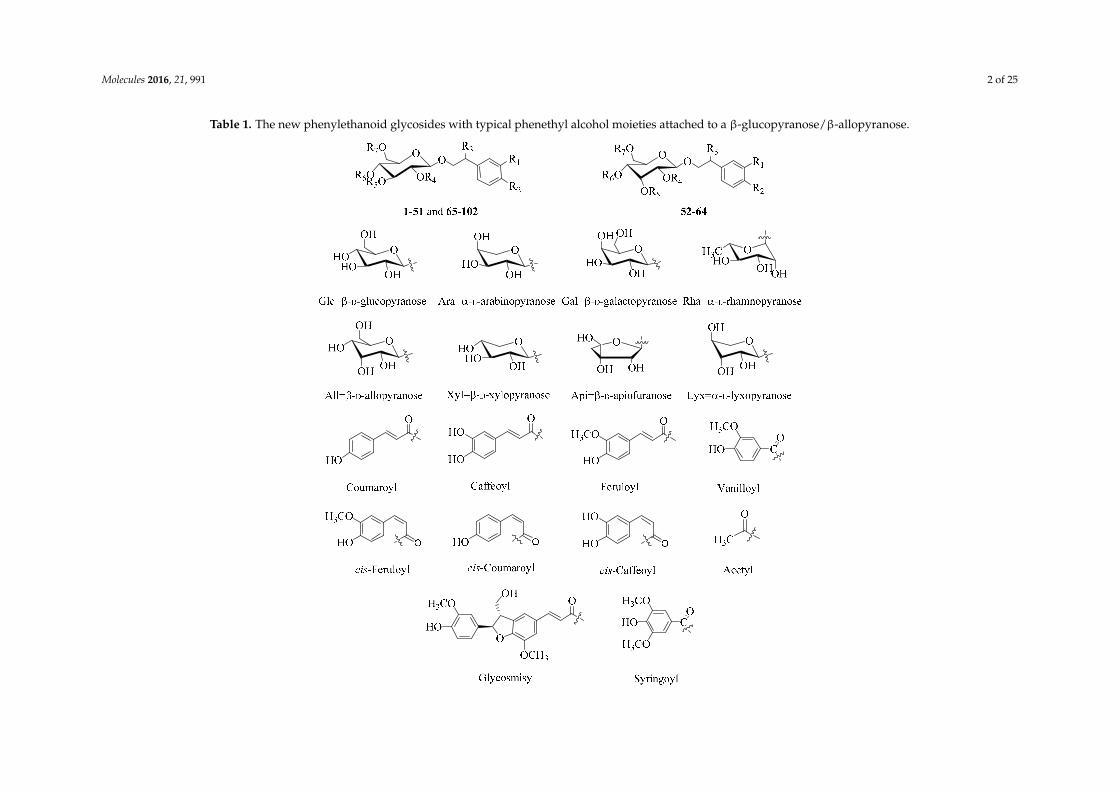
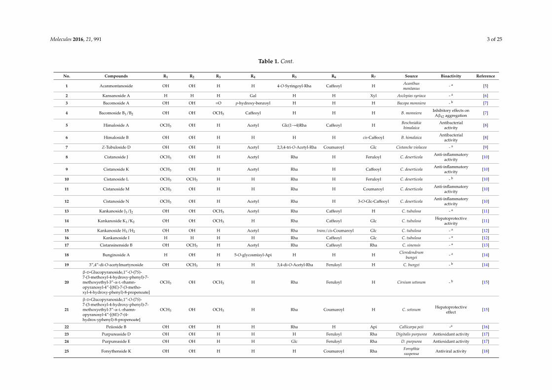
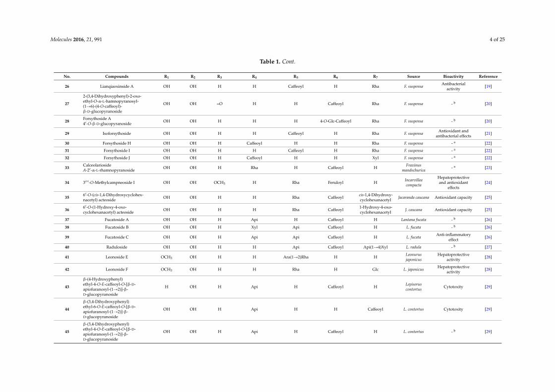
Since a 2008 review [4], more than 100 new PhGs have been isolated and identified.Compared with the known PhGs reported in [4], some of the new ones differed in their core structures,while others differed in the number and/or position of the substituents. The new PhGs with a typicalphenethyl alcohol (C6-C2) moiety attached to a β-glucopyranose/β-allopyranose are listed in Table 1.
Molecules 2016, 21, 991; doi:10.3390/molecules21080991 www.mdpi.com/journal/molecules
Molecules 2016, 21, 991 2 of 25
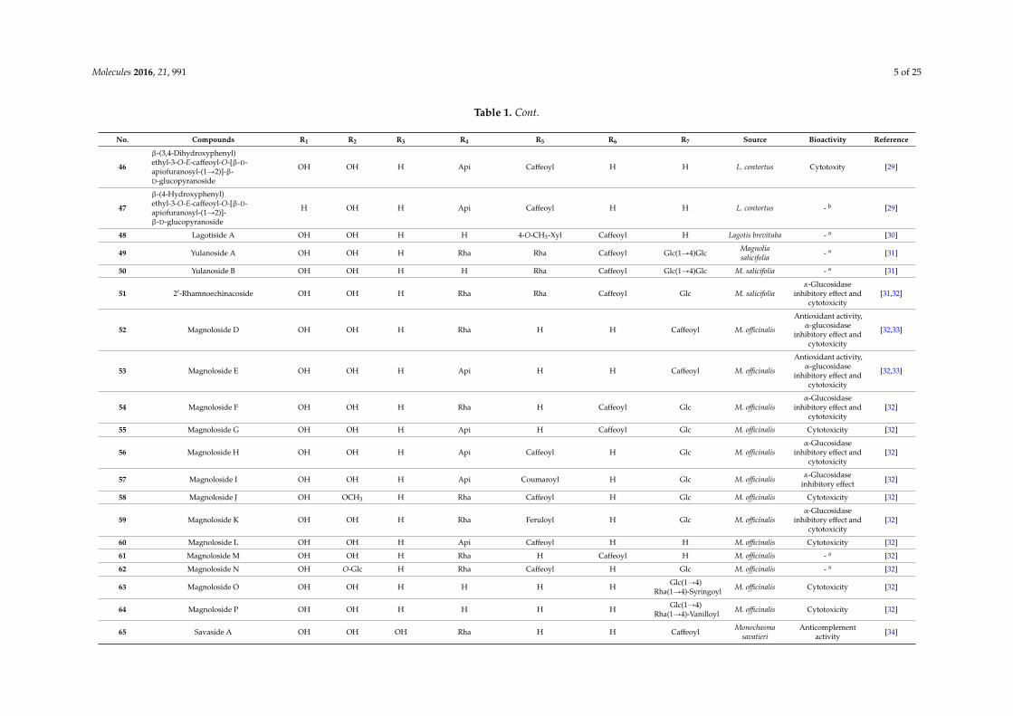
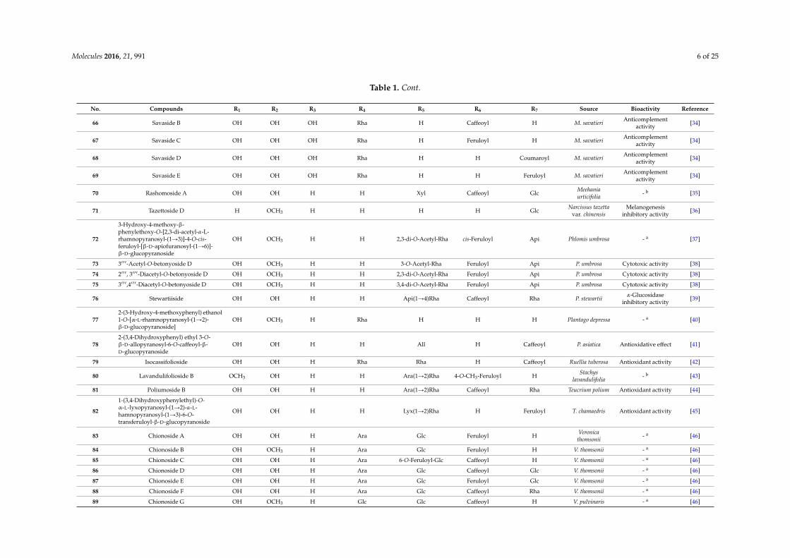
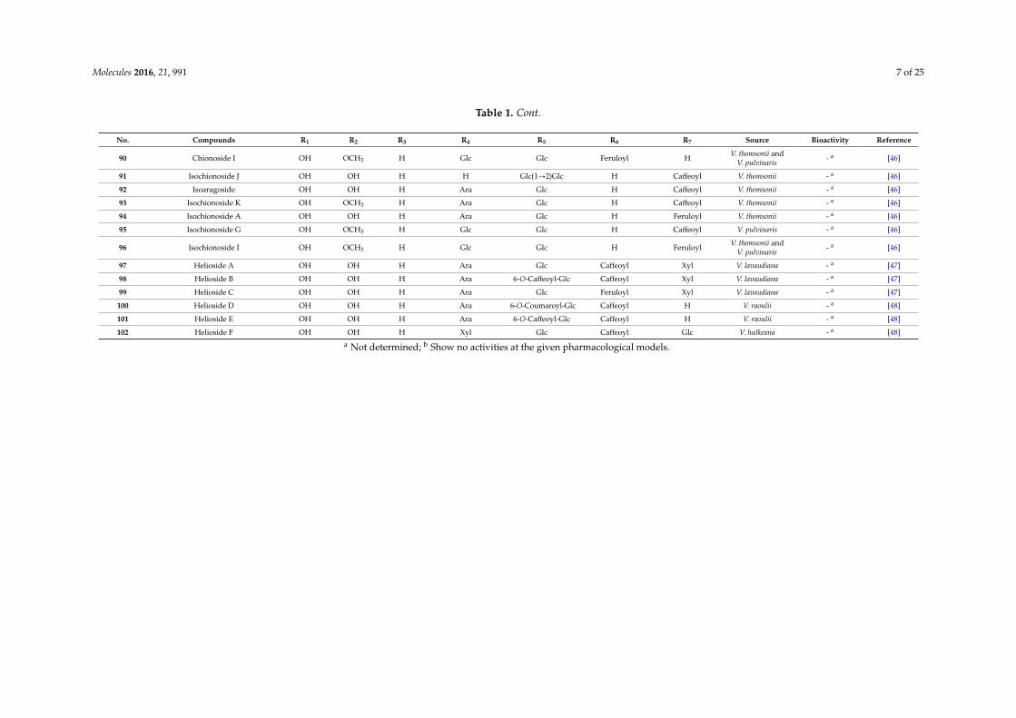
Table 1. The new phenylethanoid glycosides with typical phenethyl alcohol moieties attached to a β-glucopyranose/β-allopyranose.
Molecules 2016, 21, 991 2 of 23
Table 1. The new phenylethanoid glycosides with typical phenethyl alcohol moieties attached to a β-glucopyranose/β-allopyranose.
Molecules 2016, 21, 991 3 of 25
Table 1. Cont.
No. Compounds R1 R2 R3 R4 R5 R6 R7 Source Bioactivity Reference
1 Acanmontanoside OH OH H H 4-O-Syringoyl-Rha Caffeoyl H Acanthusmontanus - a [5]
2 Kansanoside A H H H Gal H H Xyl Asclepias syriaca - a [6]
3 Bacomoside A OH OH =O p-hydroxy-benzoyl H H H Bacopa monniera - b [7]
4 Bacomoside B1/B2 OH OH OCH3 Caffeoyl H H H B. monniera Inhibitory effects onAβ42 aggregation [7]
5 Himaloside A OCH3 OH H Acetyl Glc(1Ñ4)Rha Caffeoyl H Boschniakiahimalaica
Antibacterialactivity [8]
6 Himaloside B OH OH H H H H cis-Caffeoyl B. himalaica Antibacterialactivity [8]
7 Z-Tubuloside D OH OH H Acetyl 2,3,4-tri-O-Acetyl-Rha Coumaroyl Glc Cistanche violacea - a [9]
8 Cistanoside J OCH3 OH H Acetyl Rha H Feruloyl C. deserticola Anti-inflammatoryactivity [10]
9 Cistanoside K OCH3 OH H Acetyl Rha H Caffeoyl C. deserticola Anti-inflammatoryactivity [10]
10 Cistanoside L OCH3 OCH3 H H Rha H Feruloyl C. deserticola - b [10]
11 Cistanoside M OCH3 OH H H Rha H Coumaroyl C. deserticola Anti-inflammatoryactivity [10]
12 Cistanoside N OCH3 OH H Acetyl Rha H 3-O-Glc-Caffeoyl C. deserticola Anti-inflammatoryactivity [10]
13 Kankanoside J1/J2 OH OH OCH3 Acetyl Rha Caffeoyl H C. tubulosa - a [11]
14 Kankanoside K1/K2 OH OH OCH3 H Rha Caffeoyl Glc C. tubulosa Hepatoprotectiveactivity [11]
15 Kankanoside H1/H2 OH OH H Acetyl Rha trans/cis-Coumaroyl Glc C. tubulosa - a [12]
16 Kankanoside I H H H H Rha Caffeoyl Glc C. tubulosa - a [12]
17 Cistansinenside B OH OCH3 H Acetyl Rha Caffeoyl Rha C. sinensis - a [13]
18 Bunginoside A H OH H 5-O-glycosmisyl-Api H H H Clerodendrumbungei - a [14]
19 3”,4”-di-O-acetylmartynoside OH OCH3 H H 3,4-di-O-Acetyl-Rha Feruloyl H C. bungei - b [14]
20
β-D-Glucopyranoside,1”-O-(7S)-7-(3-methoxyl-4-hydroxy-phenyl)-7-methoxyethyl-3”-α-L-rhamn-opyranosyl-4”-[(8E)-7-(3-metho-xyl-4-hydroxy-phenyl)-8-propenoate]
OCH3 OH OCH3 H Rha Feruloyl H Cirsium setosum - b [15]
21
β-D-Glucopyranoside,1”-O-(7S)-7-(3-methoxyl-4-hydroxy-phenyl)-7-methoxyethyl-3”-α-L-rhamn-opyranosyl-4”-[(8E)-7-(4-hydrox-yphenyl)-8-propenoate]
OCH3 OH OCH3 H Rha Coumaroyl H C. setosum Hepatoprotectiveeffect [15]
22 Peiioside B OH OH H H Rha H Api Callicarpa peii -a [16]
23 Purpureaside D OH OH H H H Feruloyl Rha Digitalis purpurea Antioxidant activity [17]
24 Purpureaside E OH OH H H Glc Feruloyl Rha D. purpurea Antioxidant activity [17]
25 Forsythenside K OH OH H H H Coumaroyl Rha Forsythiasuspensa Antiviral activity [18]
Molecules 2016, 21, 991 4 of 25
Table 1. Cont.
No. Compounds R1 R2 R3 R4 R5 R6 R7 Source Bioactivity Reference
26 Lianqiaoxinside A OH OH H H Caffeoyl H Rha F. suspensa Antibacterialactivity [19]
27
2-(3,4-Dihydroxyphenyl)-2-oxo-ethyl-O-α-L-hamnopyranosyl-(1Ñ6)-(4-O-caffeoyl)-β-D-glucopyranoside
OH OH =O H H Caffeoyl Rha F. suspensa - b [20]
28 Forsythoside A41-O-β-D-glucopyranoside OH OH H H H 4-O-Glc-Caffeoyl Rha F. suspensa - b [20]
29 Isoforsythoside OH OH H H Caffeoyl H Rha F. suspensa Antioxidant andantibacterial effects [21]
30 Forsythoside H OH OH H Caffeoyl H H Rha F. suspensa - a [22]
31 Forsythoside I OH OH H H Caffeoyl H Rha F. suspensa - a [22]
32 Forsythoside J OH OH H Caffeoyl H H Xyl F. suspensa - a [22]
33 CalceolariosideA-21-α-L-rhamnopyranoside OH OH H Rha H Caffeoyl H Fraxinus
mandschurica - a [23]
34 3111-O-Methylcampneoside I OH OH OCH3 H Rha Feruloyl H Incarvilleacompacta
Hepatoprotectiveand antioxidant
effects[24]
35 61-O-(cis-1,4-Dihydroxycyclohex-nacetyl) acteoside OH OH H H Rha Caffeoyl cis-1,4-Dihydroxy-
cyclohexanacetyl Jacaranda caucana Antioxidant capacity [25]
36 61-O-(1-Hydroxy-4-oxo-cyclohexanacetyl) acteoside OH OH H H Rha Caffeoyl 1-Hydroxy-4-oxo-
cyclohexanacetyl J. caucana Antioxidant capacity [25]
37 Fucatoside A OH OH H Api H Caffeoyl H Lantana fucata - b [26]
38 Fucatoside B OH OH H Xyl Api Caffeoyl H L. fucata - b [26]
39 Fucatoside C OH OH H Api Api Caffeoyl H L. fucata Anti-inflammatoryeffect [26]
40 Raduloside OH OH H H Api Caffeoyl Api(1Ñ4)Xyl L. radula - b [27]
41 Leonoside E OCH3 OH H H Ara(1Ñ2)Rha H H Leonurusjaponicus
Hepatoprotectiveactivity [28]
42 Leonoside F OCH3 OH H H Rha H Glc L. japonicus Hepatoprotectiveactivity [28]
43
β-(4-Hydroxyphenyl)ethyl-4-O-E-caffeoyl-O-[β-D-apiofuranosyl-(1Ñ2)]-β-D-glucopyranoside
H OH H Api H Caffeoyl H Lepisoruscontortus Cytotoxity [29]
44
β-(3,4-Dihydroxyphenyl)ethyl-6-O-E-caffeoyl-O-[β-D-apiofuranosyl-(1Ñ2)]-β-D-glucopyranoside
OH OH H Api H H Caffeoyl L. contortus Cytotoxity [29]
45
β-(3,4-Dihydroxyphenyl)ethyl-4-O-E-caffeoyl-O-[β-D-apiofuranosyl-(1Ñ2)]-β-D-glucopyranoside
OH OH H Api H Caffeoyl H L. contortus - b [29]
Molecules 2016, 21, 991 5 of 25
Table 1. Cont.
No. Compounds R1 R2 R3 R4 R5 R6 R7 Source Bioactivity Reference
46
β-(3,4-Dihydroxyphenyl)ethyl-3-O-E-caffeoyl-O-[β-D-apiofuranosyl-(1Ñ2)]-β-D-glucopyranoside
OH OH H Api Caffeoyl H H L. contortus Cytotoxity [29]
47
β-(4-Hydroxyphenyl)ethyl-3-O-E-caffeoyl-O-[β-D-apiofuranosyl-(1Ñ2)]-β-D-glucopyranoside
H OH H Api Caffeoyl H H L. contortus - b [29]
48 Lagotiside A OH OH H H 4-O-CH3-Xyl Caffeoyl H Lagotis brevituba - a [30]
49 Yulanoside A OH OH H Rha Rha Caffeoyl Glc(1Ñ4)Glc Magnoliasalicifolia - a [31]
50 Yulanoside B OH OH H H Rha Caffeoyl Glc(1Ñ4)Glc M. salicifolia - a [31]
51 21-Rhamnoechinacoside OH OH H Rha Rha Caffeoyl Glc M. salicifoliaα-Glucosidase
inhibitory effect andcytotoxicity
[31,32]
52 Magnoloside D OH OH H Rha H H Caffeoyl M. officinalis
Antioxidant activity,α-glucosidase
inhibitory effect andcytotoxicity
[32,33]
53 Magnoloside E OH OH H Api H H Caffeoyl M. officinalis
Antioxidant activity,α-glucosidase
inhibitory effect andcytotoxicity
[32,33]
54 Magnoloside F OH OH H Rha H Caffeoyl Glc M. officinalisα-Glucosidase
inhibitory effect andcytotoxicity
[32]
55 Magnoloside G OH OH H Api H Caffeoyl Glc M. officinalis Cytotoxicity [32]
56 Magnoloside H OH OH H Api Caffeoyl H Glc M. officinalisα-Glucosidase
inhibitory effect andcytotoxicity
[32]
57 Magnoloside I OH OH H Api Coumaroyl H Glc M. officinalis α-Glucosidaseinhibitory effect [32]
58 Magnoloside J OH OCH3 H Rha Caffeoyl H Glc M. officinalis Cytotoxicity [32]
59 Magnoloside K OH OH H Rha Feruloyl H Glc M. officinalisα-Glucosidase
inhibitory effect andcytotoxicity
[32]
60 Magnoloside L OH OH H Api Caffeoyl H H M. officinalis Cytotoxicity [32]
61 Magnoloside M OH OH H Rha H Caffeoyl H M. officinalis - a [32]
62 Magnoloside N OH O-Glc H Rha Caffeoyl H Glc M. officinalis - a [32]
63 Magnoloside O OH OH H H H H Glc(1Ñ4)Rha(1Ñ4)-Syringoyl M. officinalis Cytotoxicity [32]
64 Magnoloside P OH OH H H H H Glc(1Ñ4)Rha(1Ñ4)-Vanilloyl M. officinalis Cytotoxicity [32]
65 Savaside A OH OH OH Rha H H Caffeoyl Monochasmasavatieri
Anticomplementactivity [34]
Molecules 2016, 21, 991 6 of 25
Table 1. Cont.
No. Compounds R1 R2 R3 R4 R5 R6 R7 Source Bioactivity Reference
66 Savaside B OH OH OH Rha H Caffeoyl H M. savatieri Anticomplementactivity [34]
67 Savaside C OH OH OH Rha H Feruloyl H M. savatieri Anticomplementactivity [34]
68 Savaside D OH OH OH Rha H H Coumaroyl M. savatieri Anticomplementactivity [34]
69 Savaside E OH OH OH Rha H H Feruloyl M. savatieri Anticomplementactivity [34]
70 Rashomoside A OH OH H H Xyl Caffeoyl Glc Meehaniaurticifolia - b [35]
71 Tazettoside D H OCH3 H H H H Glc Narcissus tazettavar. chinensis
Melanogenesisinhibitory activity [36]
72
3-Hydroxy-4-methoxy-β-phenylethoxy-O-[2,3-di-acetyl-α-L-rhamnopyranosyl-(1Ñ3)]-4-O-cis-feruloyl-[β-D-apiofuranosyl-(1Ñ6)]-β-D-glucopyranoside
OH OCH3 H H 2,3-di-O-Acetyl-Rha cis-Feruloyl Api Phlomis umbrosa - a [37]
73 3111-Acetyl-O-betonyoside D OH OCH3 H H 3-O-Acetyl-Rha Feruloyl Api P. umbrosa Cytotoxic activity [38]
74 2111, 3111-Diacetyl-O-betonyoside D OH OCH3 H H 2,3-di-O-Acetyl-Rha Feruloyl Api P. umbrosa Cytotoxic activity [38]
75 3111,4111-Diacetyl-O-betonyoside D OH OCH3 H H 3,4-di-O-Acetyl-Rha Feruloyl Api P. umbrosa Cytotoxic activity [38]
76 Stewartiiside OH OH H H Api(1Ñ4)Rha Caffeoyl Rha P. stewartii α-Glucosidaseinhibitory activity [39]
772-(3-Hydroxy-4-methoxyphenyl) ethanol1-O-[α-L-rhamnopyranosyl-(1Ñ2)-β-D-glucopyranoside]
OH OCH3 H Rha H H H Plantago depressa - a [40]
782-(3,4-Dihydroxyphenyl) ethyl 3-O-β-D-allopyranosyl-6-O-caffeoyl-β-D-glucopyranoside
OH OH H H All H Caffeoyl P. asiatica Antioxidative effect [41]
79 Isocassifolioside OH OH H Rha Rha H Caffeoyl Ruellia tuberosa Antioxidant activity [42]
80 Lavandulifolioside B OCH3 OH H H Ara(1Ñ2)Rha 4-O-CH3-Feruloyl H Stachyslavandulifolia - b [43]
81 Poliumoside B OH OH H H Ara(1Ñ2)Rha Caffeoyl Rha Teucrium polium Antioxidant activity [44]
82
1-(3,4-Dihydroxyphenylethyl)-O-α-L-lyxopyranosyl-(1Ñ2)-α-L-hamnopyranosyl-(1Ñ3)-6-O-transferuloyl-β-D-glucopyranoside
OH OH H H Lyx(1Ñ2)Rha H Feruloyl T. chamaedris Antioxidant activity [45]
83 Chionoside A OH OH H Ara Glc Feruloyl H Veronicathomsonii - a [46]
84 Chionoside B OH OCH3 H Ara Glc Feruloyl H V. thomsonii - a [46]
85 Chionoside C OH OH H Ara 6-O-Feruloyl-Glc Caffeoyl H V. thomsonii - a [46]
86 Chionoside D OH OH H Ara Glc Caffeoyl Glc V. thomsonii - a [46]
87 Chionoside E OH OH H Ara Glc Feruloyl Glc V. thomsonii - a [46]
88 Chionoside F OH OH H Ara Glc Caffeoyl Rha V. thomsonii - a [46]
89 Chionoside G OH OCH3 H Glc Glc Caffeoyl H V. pulvinaris - a [46]
Molecules 2016, 21, 991 7 of 25
Table 1. Cont.
No. Compounds R1 R2 R3 R4 R5 R6 R7 Source Bioactivity Reference
90 Chionoside I OH OCH3 H Glc Glc Feruloyl H V. thomsonii andV. pulvinaris - a [46]
91 Isochionoside J OH OH H H Glc(1Ñ2)Glc H Caffeoyl V. thomsonii - a [46]
92 Isoaragoside OH OH H Ara Glc H Caffeoyl V. thomsonii - a [46]
93 Isochionoside K OH OCH3 H Ara Glc H Caffeoyl V. thomsonii - a [46]
94 Isochionoside A OH OH H Ara Glc H Feruloyl V. thomsonii - a [46]
95 Isochionoside G OH OCH3 H Glc Glc H Caffeoyl V. pulvinaris - a [46]
96 Isochionoside I OH OCH3 H Glc Glc H Feruloyl V. thomsonii andV. pulvinaris - a [46]
97 Helioside A OH OH H Ara Glc Caffeoyl Xyl V. lavaudiana - a [47]
98 Helioside B OH OH H Ara 6-O-Caffeoyl-Glc Caffeoyl Xyl V. lavaudiana - a [47]
99 Helioside C OH OH H Ara Glc Feruloyl Xyl V. lavaudiana - a [47]
100 Helioside D OH OH H Ara 6-O-Coumaroyl-Glc Caffeoyl H V. raoulii - a [48]
101 Helioside E OH OH H Ara 6-O-Caffeoyl-Glc Caffeoyl H V. raoulii - a [48]
102 Helioside F OH OH H Xyl Glc Caffeoyl Glc V. hulkeana - a [48]
a Not determined; b Show no activities at the given pharmacological models.
Molecules 2016, 21, 991 8 of 25
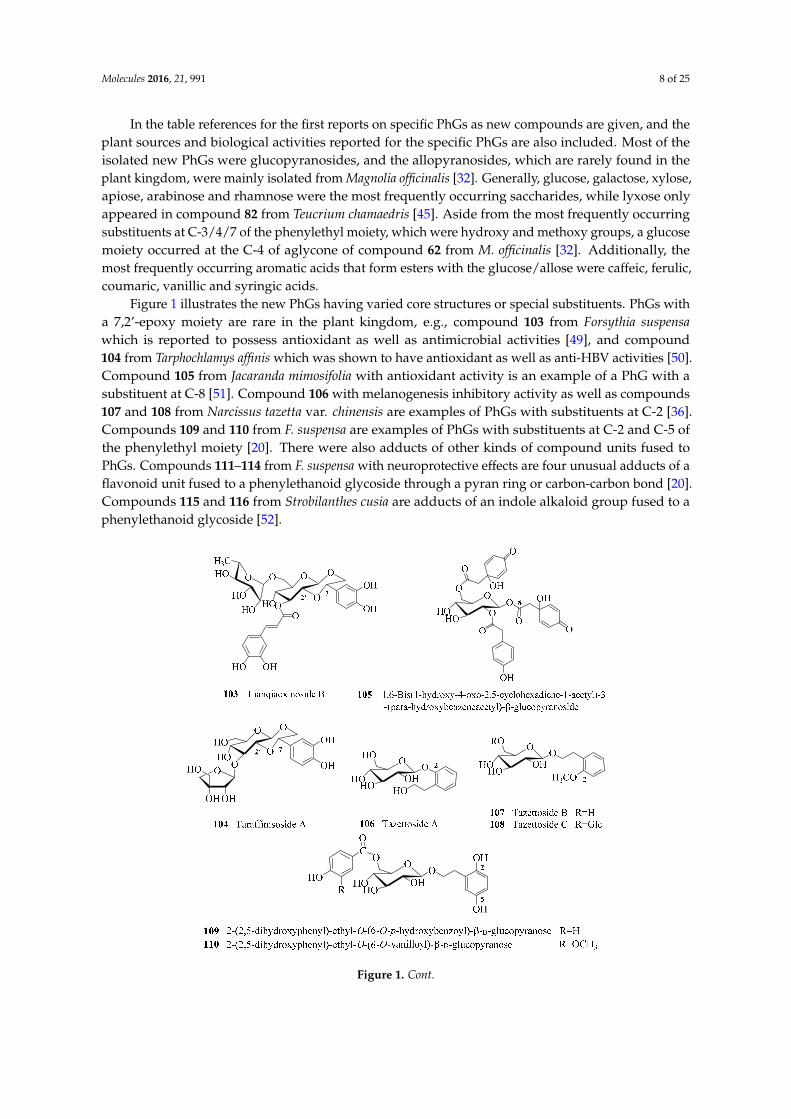
In the table references for the first reports on specific PhGs as new compounds are given, and theplant sources and biological activities reported for the specific PhGs are also included. Most of theisolated new PhGs were glucopyranosides, and the allopyranosides, which are rarely found in theplant kingdom, were mainly isolated from Magnolia officinalis [32]. Generally, glucose, galactose, xylose,apiose, arabinose and rhamnose were the most frequently occurring saccharides, while lyxose onlyappeared in compound 82 from Teucrium chamaedris [45]. Aside from the most frequently occurringsubstituents at C-3/4/7 of the phenylethyl moiety, which were hydroxy and methoxy groups, a glucosemoiety occurred at the C-4 of aglycone of compound 62 from M. officinalis [32]. Additionally, themost frequently occurring aromatic acids that form esters with the glucose/allose were caffeic, ferulic,coumaric, vanillic and syringic acids.
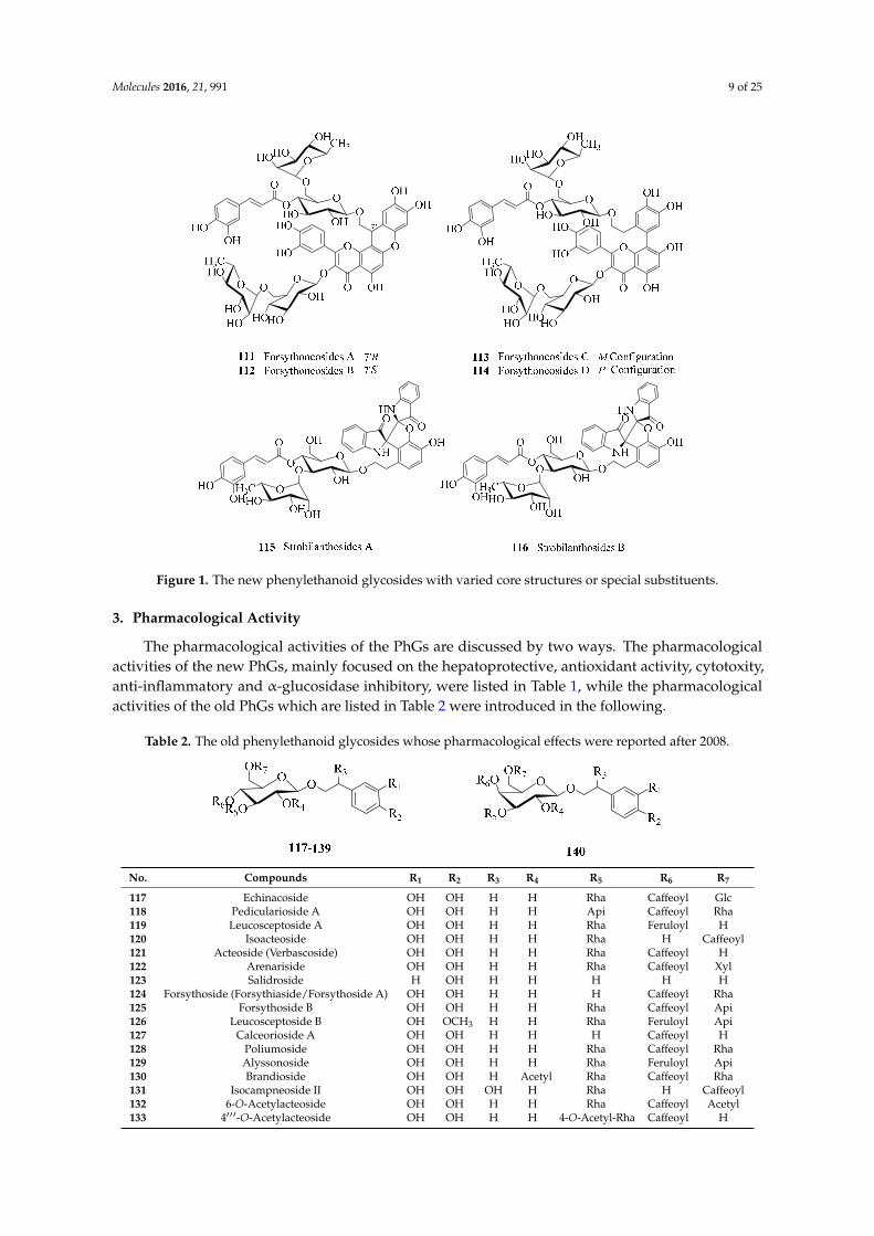
Figure 1 illustrates the new PhGs having varied core structures or special substituents. PhGs witha 7,2’-epoxy moiety are rare in the plant kingdom, e.g., compound 103 from Forsythia suspensawhich is reported to possess antioxidant as well as antimicrobial activities [49], and compound104 from Tarphochlamys affinis which was shown to have antioxidant as well as anti-HBV activities [50].Compound 105 from Jacaranda mimosifolia with antioxidant activity is an example of a PhG with asubstituent at C-8 [51]. Compound 106 with melanogenesis inhibitory activity as well as compounds107 and 108 from Narcissus tazetta var. chinensis are examples of PhGs with substituents at C-2 [36].Compounds 109 and 110 from F. suspensa are examples of PhGs with substituents at C-2 and C-5 ofthe phenylethyl moiety [20]. There were also adducts of other kinds of compound units fused toPhGs. Compounds 111–114 from F. suspensa with neuroprotective effects are four unusual adducts of aflavonoid unit fused to a phenylethanoid glycoside through a pyran ring or carbon-carbon bond [20].Compounds 115 and 116 from Strobilanthes cusia are adducts of an indole alkaloid group fused to aphenylethanoid glycoside [52].
Molecules 2016, 21, 991 7 of 23
In the table references for the first reports on specific PhGs as new compounds are given, and the plant sources and biological activities reported for the specific PhGs are also included. Most of the isolated new PhGs were glucopyranosides, and the allopyranosides, which are rarely found in the plant kingdom, were mainly isolated from Magnolia officinalis [32]. Generally, glucose, galactose, xylose, apiose, arabinose and rhamnose were the most frequently occurring saccharides, while lyxose only appeared in compound 82 from Teucrium chamaedris [45]. Aside from the most frequently occurring substituents at C-3/4/7 of the phenylethyl moiety, which were hydroxy and methoxy groups, a glucose moiety occurred at the C-4 of aglycone of compound 62 from M. officinalis [32]. Additionally, the most frequently occurring aromatic acids that form esters with the glucose/allose were caffeic, ferulic, coumaric, vanillic and syringic acids.
Figure 1 illustrates the new PhGs having varied core structures or special substituents. PhGs with a 7,2′-epoxy moiety are rare in the plant kingdom, e.g., compound 103 from Forsythia suspensa which is reported to possess antioxidant as well as antimicrobial activities [49], and compound 104 from Tarphochlamys affinis which was shown to have antioxidant as well as anti-HBV activities [50]. Compound 105 from Jacaranda mimosifolia with antioxidant activity is an example of a PhG with a substituent at C-8 [51]. Compound 106 with melanogenesis inhibitory activity as well as compounds 107 and 108 from Narcissus tazetta var. chinensis are examples of PhGs with substituents at C-2 [36]. Compounds 109 and 110 from F. suspensa are examples of PhGs with substituents at C-2 and C-5 of the phenylethyl moiety [20]. There were also adducts of other kinds of compound units fused to PhGs. Compounds 111–114 from F. suspensa with neuroprotective effects are four unusual adducts of a flavonoid unit fused to a phenylethanoid glycoside through a pyran ring or carbon-carbon bond [20]. Compounds 115 and 116 from Strobilanthes cusia are adducts of an indole alkaloid group fused to a phenylethanoid glycoside [52].
Figure 1. Cont.
Molecules 2016, 21, 991 9 of 25Molecules 2016, 21, 991 8 of 23
Figure 1. The new phenylethanoid glycosides with varied core structures or special substituents.
3. Pharmacological Activity
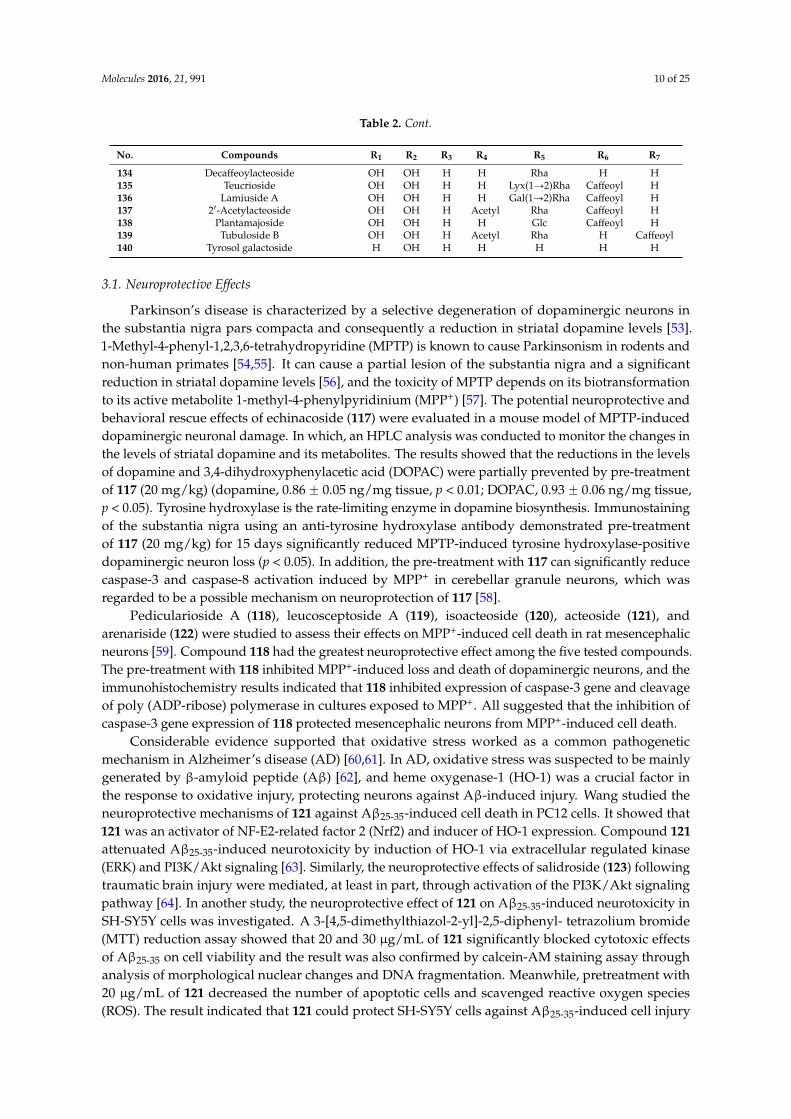
The pharmacological activities of the PhGs are discussed by two ways. The pharmacological activities of the new PhGs, mainly focused on the hepatoprotective, antioxidant activity, cytotoxity, anti-inflammatory and α-glucosidase inhibitory, were listed in Table 1, while the pharmacological activities of the old PhGs which are listed in Table 2 were introduced in the following.
Table 2. The old phenylethanoid glycosides whose pharmacological effects were reported after 2008.
No. Compounds R1 R2 R3 R4 R5 R6 R7
117 Echinacoside OH OH H H Rha Caffeoyl Glc 118 Pedicularioside A OH OH H H Api Caffeoyl Rha 119 Leucosceptoside A OH OH H H Rha Feruloyl H 120 Isoacteoside OH OH H H Rha H Caffeoyl 121 Acteoside (Verbascoside) OH OH H H Rha Caffeoyl H 122 Arenariside OH OH H H Rha Caffeoyl Xyl 123 Salidroside H OH H H H H H 124 Forsythoside (Forsythiaside/Forsythoside A) OH OH H H H Caffeoyl Rha 125 Forsythoside B OH OH H H Rha Caffeoyl Api 126 Leucosceptoside B OH OCH3 H H Rha Feruloyl Api 127 Calceorioside A OH OH H H H Caffeoyl H 128 Poliumoside OH OH H H Rha Caffeoyl Rha 129 Alyssonoside OH OH H H Rha Feruloyl Api 130 Brandioside OH OH H Acetyl Rha Caffeoyl Rha 131 Isocampneoside II OH OH OH H Rha H Caffeoyl 132 6-O-Acetylacteoside OH OH H H Rha Caffeoyl Acetyl 133 4′′′-O-Acetylacteoside OH OH H H 4-O-Acetyl-Rha Caffeoyl H 134 Decaffeoylacteoside OH OH H H Rha H H
Molecules 2016, 21, x FOR PEER REVIEW 11 of 27
Figure 1. The new phenylethanoid glycosides with varied core structures or special substituents.
Figure 1 illustrates the new PhGs having varied core structures or special substituents. PhGs with a 7,2′-epoxy moiety are rare in the plant kingdom, e.g., compound 103 from Forsythia suspensa which is reported to possess antioxidant as well as antimicrobial activities [49], and compound 104 from Tarphochlamys affinis which was shown to have antioxidant as well as anti-HBV activities [50]. Compound 105 from Jacaranda mimosifolia with antioxidant activity is an example of a PhG with a substituent at C-8 [51]. Compound 106 with melanogenesis inhibitory activity as well as compounds 107 and 108 from Narcissus tazetta var. chinensis are examples of PhGs with substituents at C-2 [36]. Compounds 109 and 110 from F. suspensa are examples of PhGs with substituents at C-2 and C-5 of the phenylethyl moiety [20]. There were also adducts of other kinds of compound units fused to PhGs. Compounds 111–114 from F. suspensa with neuroprotective effects are four unusual adducts of a flavonoid unit fused to a phenylethanoid glycoside through a pyran ring or carbon-carbon bond [20]. Compounds 115 and 116 from Strobilanthes cusia are adducts of an indole alkaloid group fused to a phenylethanoid glycoside [52].
3. Pharmacological Activity
The pharmacological activities of the PhGs are discussed by two ways. The pharmacological activities of the new PhGs, mainly focused on the hepatoprotective, antioxidant activity, cytotoxity, anti-inflammatory and α-glucosidase inhibitory, were listed in Table 1, while the pharmacological activities of the old PhGs which are listed in Table 2 were introduced in the following.
3.1. Neuroprotective Effects
Parkinson’s disease is characterized by a selective degeneration of dopaminergic neurons in the substantia nigra pars compacta and consequently a reduction in striatal dopamine levels [53]. 1-Methyl-4-phenyl-1,2,3,6-tetrahydropyridine (MPTP) is known to cause Parkinsonism in rodents and non-human primates [54,55]. It can cause a partial lesion of the substantia nigra and a significant reduction in striatal dopamine levels [56], and the toxicity of MPTP depends on its biotransformation to its active metabolite 1-methyl-4-phenylpyridinium (MPP+) [57]. The potential neuroprotective and behavioral rescue effects of echinacoside (117) were evaluated in a mouse model of MPTP-induced dopaminergic neuronal damage. In which, an HPLC analysis was conducted to monitor the changes in the levels of striatal dopamine and its metabolites. The results showed that the reductions in the levels of dopamine and 3,4-dihydroxyphenylacetic acid (DOPAC) were partially prevented by pre-treatment of 117 (20 mg/kg) (dopamine, 0.86 ± 0.05 ng/mg tissue, p < 0.01; DOPAC, 0.93 ± 0.06 ng/mg tissue, p < 0.05). Tyrosine hydroxylase is the rate-limiting enzyme in dopamine biosynthesis. Immunostaining of the substantia nigra using an anti-tyrosine hydroxylase antibody demonstrated pre-treatment of 117 (20 mg/kg) for 15 days significantly reduced MPTP-induced tyrosine hydroxylase-positive dopaminergic neuron loss (p < 0.05). In addition, the pre-treatment with 117 can significantly reduce caspase-3 and caspase-8 activation induced by MPP+ in cerebellar granule neurons, which was regarded to be a possible mechanism on neuroprotection of 117 [58].
Figure 1. The new phenylethanoid glycosides with varied core structures or special substituents.
3. Pharmacological Activity
The pharmacological activities of the PhGs are discussed by two ways. The pharmacologicalactivities of the new PhGs, mainly focused on the hepatoprotective, antioxidant activity, cytotoxity,anti-inflammatory and α-glucosidase inhibitory, were listed in Table 1, while the pharmacologicalactivities of the old PhGs which are listed in Table 2 were introduced in the following.
Table 2. The old phenylethanoid glycosides whose pharmacological effects were reported after 2008.
Molecules 2016, 21, 991 8 of 23
Figure 1. The new phenylethanoid glycosides with varied core structures or special substituents.
3. Pharmacological Activity
The pharmacological activities of the PhGs are discussed by two ways. The pharmacological activities of the new PhGs, mainly focused on the hepatoprotective, antioxidant activity, cytotoxity, anti-inflammatory and α-glucosidase inhibitory, were listed in Table 1, while the pharmacological activities of the old PhGs which are listed in Table 2 were introduced in the following.
Table 2. The old phenylethanoid glycosides whose pharmacological effects were reported after 2008.
No. Compounds R1 R2 R3 R4 R5 R6 R7
117 Echinacoside OH OH H H Rha Caffeoyl Glc 118 Pedicularioside A OH OH H H Api Caffeoyl Rha 119 Leucosceptoside A OH OH H H Rha Feruloyl H 120 Isoacteoside OH OH H H Rha H Caffeoyl 121 Acteoside (Verbascoside) OH OH H H Rha Caffeoyl H 122 Arenariside OH OH H H Rha Caffeoyl Xyl 123 Salidroside H OH H H H H H 124 Forsythoside (Forsythiaside/Forsythoside A) OH OH H H H Caffeoyl Rha 125 Forsythoside B OH OH H H Rha Caffeoyl Api 126 Leucosceptoside B OH OCH3 H H Rha Feruloyl Api 127 Calceorioside A OH OH H H H Caffeoyl H 128 Poliumoside OH OH H H Rha Caffeoyl Rha 129 Alyssonoside OH OH H H Rha Feruloyl Api 130 Brandioside OH OH H Acetyl Rha Caffeoyl Rha 131 Isocampneoside II OH OH OH H Rha H Caffeoyl 132 6-O-Acetylacteoside OH OH H H Rha Caffeoyl Acetyl 133 4′′′-O-Acetylacteoside OH OH H H 4-O-Acetyl-Rha Caffeoyl H 134 Decaffeoylacteoside OH OH H H Rha H H
No. Compounds R1 R2 R3 R4 R5 R6 R7
117 Echinacoside OH OH H H Rha Caffeoyl Glc118 Pedicularioside A OH OH H H Api Caffeoyl Rha119 Leucosceptoside A OH OH H H Rha Feruloyl H120 Isoacteoside OH OH H H Rha H Caffeoyl121 Acteoside (Verbascoside) OH OH H H Rha Caffeoyl H122 Arenariside OH OH H H Rha Caffeoyl Xyl123 Salidroside H OH H H H H H124 Forsythoside (Forsythiaside/Forsythoside A) OH OH H H H Caffeoyl Rha125 Forsythoside B OH OH H H Rha Caffeoyl Api126 Leucosceptoside B OH OCH3 H H Rha Feruloyl Api127 Calceorioside A OH OH H H H Caffeoyl H128 Poliumoside OH OH H H Rha Caffeoyl Rha129 Alyssonoside OH OH H H Rha Feruloyl Api130 Brandioside OH OH H Acetyl Rha Caffeoyl Rha131 Isocampneoside II OH OH OH H Rha H Caffeoyl132 6-O-Acetylacteoside OH OH H H Rha Caffeoyl Acetyl133 4111-O-Acetylacteoside OH OH H H 4-O-Acetyl-Rha Caffeoyl H
Molecules 2016, 21, 991 10 of 25
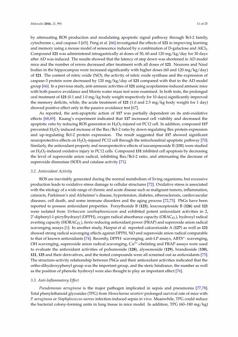
Table 2. Cont.
No. Compounds R1 R2 R3 R4 R5 R6 R7
134 Decaffeoylacteoside OH OH H H Rha H H135 Teucrioside OH OH H H Lyx(1Ñ2)Rha Caffeoyl H136 Lamiuside A OH OH H H Gal(1Ñ2)Rha Caffeoyl H137 21-Acetylacteoside OH OH H Acetyl Rha Caffeoyl H138 Plantamajoside OH OH H H Glc Caffeoyl H139 Tubuloside B OH OH H Acetyl Rha H Caffeoyl140 Tyrosol galactoside H OH H H H H H
3.1. Neuroprotective Effects
Parkinson’s disease is characterized by a selective degeneration of dopaminergic neurons inthe substantia nigra pars compacta and consequently a reduction in striatal dopamine levels [53].1-Methyl-4-phenyl-1,2,3,6-tetrahydropyridine (MPTP) is known to cause Parkinsonism in rodents andnon-human primates [54,55]. It can cause a partial lesion of the substantia nigra and a significantreduction in striatal dopamine levels [56], and the toxicity of MPTP depends on its biotransformationto its active metabolite 1-methyl-4-phenylpyridinium (MPP+) [57]. The potential neuroprotective andbehavioral rescue effects of echinacoside (117) were evaluated in a mouse model of MPTP-induceddopaminergic neuronal damage. In which, an HPLC analysis was conducted to monitor the changes inthe levels of striatal dopamine and its metabolites. The results showed that the reductions in the levelsof dopamine and 3,4-dihydroxyphenylacetic acid (DOPAC) were partially prevented by pre-treatmentof 117 (20 mg/kg) (dopamine, 0.86 ˘ 0.05 ng/mg tissue, p < 0.01; DOPAC, 0.93 ˘ 0.06 ng/mg tissue,p < 0.05). Tyrosine hydroxylase is the rate-limiting enzyme in dopamine biosynthesis. Immunostainingof the substantia nigra using an anti-tyrosine hydroxylase antibody demonstrated pre-treatmentof 117 (20 mg/kg) for 15 days significantly reduced MPTP-induced tyrosine hydroxylase-positivedopaminergic neuron loss (p < 0.05). In addition, the pre-treatment with 117 can significantly reducecaspase-3 and caspase-8 activation induced by MPP+ in cerebellar granule neurons, which wasregarded to be a possible mechanism on neuroprotection of 117 [58].
Pedicularioside A (118), leucosceptoside A (119), isoacteoside (120), acteoside (121), andarenariside (122) were studied to assess their effects on MPP+-induced cell death in rat mesencephalicneurons [59]. Compound 118 had the greatest neuroprotective effect among the five tested compounds.The pre-treatment with 118 inhibited MPP+-induced loss and death of dopaminergic neurons, and theimmunohistochemistry results indicated that 118 inhibited expression of caspase-3 gene and cleavageof poly (ADP-ribose) polymerase in cultures exposed to MPP+. All suggested that the inhibition ofcaspase-3 gene expression of 118 protected mesencephalic neurons from MPP+-induced cell death.
Considerable evidence supported that oxidative stress worked as a common pathogeneticmechanism in Alzheimer’s disease (AD) [60,61]. In AD, oxidative stress was suspected to be mainlygenerated by β-amyloid peptide (Aβ) [62], and heme oxygenase-1 (HO-1) was a crucial factor inthe response to oxidative injury, protecting neurons against Aβ-induced injury. Wang studied theneuroprotective mechanisms of 121 against Aβ25-35-induced cell death in PC12 cells. It showed that121 was an activator of NF-E2-related factor 2 (Nrf2) and inducer of HO-1 expression. Compound 121attenuated Aβ25-35-induced neurotoxicity by induction of HO-1 via extracellular regulated kinase(ERK) and PI3K/Akt signaling [63]. Similarly, the neuroprotective effects of salidroside (123) followingtraumatic brain injury were mediated, at least in part, through activation of the PI3K/Akt signalingpathway [64]. In another study, the neuroprotective effect of 121 on Aβ25-35-induced neurotoxicity inSH-SY5Y cells was investigated. A 3-[4,5-dimethylthiazol-2-yl]-2,5-diphenyl- tetrazolium bromide(MTT) reduction assay showed that 20 and 30 µg/mL of 121 significantly blocked cytotoxic effectsof Aβ25-35 on cell viability and the result was also confirmed by calcein-AM staining assay throughanalysis of morphological nuclear changes and DNA fragmentation. Meanwhile, pretreatment with20 µg/mL of 121 decreased the number of apoptotic cells and scavenged reactive oxygen species(ROS). The result indicated that 121 could protect SH-SY5Y cells against Aβ25-35-induced cell injury
Molecules 2016, 21, 991 11 of 25
by attenuating ROS production and modulating apoptotic signal pathway through Bcl-2 family,cytochrome c, and caspase-3 [65]. Peng et al. [66] investigated the effects of 121 in improving learningand memory using a mouse model of senescence induced by a combination of D-galactose and AlCl3.Compound 121 was administered intragastrically at doses of 30, 60 and 120 mg/kg/day for 30 daysafter AD was induced. The results showed that the latency of step down was shortened in AD modelmice and the number of errors decreased after treatment with all doses of 121. Neurons and Nisslbodies in the hippocampus were increased significantly with higher doses (60 and 120 mg/kg/day)of 121. The content of nitric oxide (NO), the activity of nitric oxide synthase and the expression ofcaspase-3 protein were decreased by 120 mg/kg/day of 121 compared with that in the AD modelgroup [66]. In a previous study, anti-amnesic activities of 121 using scopolamine-induced amnesic micewith both passive avoidance and Morris water maze test were examined. In both tests, the prolongedoral treatment of 121 (0.1 and 1.0 mg/kg body weight respectively for 10 days) significantly improvedthe memory deficits, while, the acute treatment of 121 (1.0 and 2.5 mg/kg body weight for 1 day)showed positive effect only in the passive avoidance test [67].
As reported, the anti-apoptotic action of 117 was partially dependent on its anti-oxidativeeffects [68,69]. Kuang’s experiment indicated that 117 increased cell viability and decreased theapoptotic ratio by reducing ROS generation in H2O2-injured rat PC12 cell. In addition, compound 117prevented H2O2-induced increase of the Bax/Bcl-2 ratio by down-regulating Bax protein expressionand up-regulating Bcl-2 protein expression. The result suggested that 117 showed significantneuroprotective effects on H2O2-injured PC12 cell through the mitochondrial apoptotic pathway [70].Similarly, the antioxidant property and neuroprotective effects of isocampneoside II (131) were studiedon H2O2-induced oxidative injury in PC12 cells. Compound 131 inhibited cell apoptosis by decreasingthe level of superoxide anion radical, inhibiting Bax/Bcl-2 ratio, and attenuating the decrease ofsuperoxide dismutase (SOD) and catalase activity [71].
3.2. Antioxidant Activity
ROS are inevitably generated during the normal metabolism of living organisms, but excessiveproduction leads to oxidative stress damage to cellular structures [72]. Oxidative stress is associatedwith the etiology of a wide range of chronic and acute disease such as malignant tumors, inflammation,cataracts, Parkinson’s and Alzheimer’s disease, hypertension, diabetes, atherosclerosis, cardiovasculardiseases, cell death, and some immune disorders and the aging process [72,73]. PhGs have beenreported to possess antioxidant properties. Forsythoside B (125), leucosceptoside B (126) and 121were isolated from Verbascum xanthophoeniceum and exhibited potent antioxidant activities in 2,21-diphenyl-1-picrylhydrazyl (DPPH), oxygen radical absorbance capacity (ORACFL), hydroxyl radicalaverting capacity (HORACFL), ferric-reducing antioxidant power (FRAP) and superoxide anion radicalscavenging assays [1]. In another study, Harput et al. reported calceorioside A (127) as well as 121showed strong radical scavenging effects against DPPH, NO and superoxide anion radical comparableto that of known antioxidants [74]. Recently, DPPH¨ scavenging, anti-LP assays, ABTS+¨ scavenging,OH scavenging, superoxide anion radical scavenging, Cu2+-chelating and FRAP assays were usedto evaluate the antioxidant activities of poliumoside (128), alyssonoside (129), brandioside (130),121, 125 and their derivatives, and the tested compounds were all screened out as antioxidants [75].The structure-activity relationship between PhGs and their antioxidant activities indicated that theortho-dihydroxyphenyl group was the important group, and the steric hindrance, the number as wellas the position of phenolic hydroxyl were also thought to play an important effect [76].
3.3. Anti-Inflammatory Effect
Pseudomonas aeruginosa is the major pathogen implicated in sepsis and pneumonia [77,78].Total phenylethanoid glycosides (TPG) from Monochasma savatieri prolonged survival rate of mice withP. aeruginosa or Staphylococcus aureus infection-induced sepsis in vivo. Meanwhile, TPG could reducethe bacterial colony-forming units in lung tissue in mice model. In addition, TPG (60–180 mg/kg)
Molecules 2016, 21, 991 12 of 25
had significantly reduced xylene-induced ear edema and cotton pellet-induced granulomat formationat a dose-dependent manner. Furthermore, the treatment of TPG (1.5 g/kg) for 15 days did notcause any death of rat and no organic toxicity at the dose equal to approximately 284 times ofclinical dose used [79]. It was reported that compound induced HO-1 in macrophages throughp38 mitogen-activated protein kinase (MAPK)/Nrf2 signaling and decreased the release of highmobility group box 1 (HMGB1) in lipopolysaccharide (LPS)-stimulated Raw264.7 cells and in cecalligation and puncture (CLP)-induced septic mice. In vitro, compound 121 not only inhibited therelease of HMGB1, the production of inducible nitric oxide synthase and NO, but also induced HO-1expression in a concentration-dependent manner; in vivo, it increased survival and decreased theHMGB1 levels of serum and lung in CLP-induced sepsis [80]. In another study, the anti-inflammatoryactivity, the anti-nociceptive activity, and the wound healing activity of 121 were studied usinga carrageenan-induced hind paw edema model in vivo, a p-benzoquinone-induced abdominalconstriction test, and incision and excision models in vivo, respectively [81]. It was previously reportedthat 121 was more active than ibuprofen in the writhing test (67.6% and 50.0% at equimolar doses)and showed similar effects in the tail flick (topic and oral) at equivalent dose to ibuprofen [82].Moreover, compound 121 was found to be active in a carrageenan-induced hind paw edema modeland in p-benzoquinone-induced writhing in mice [83]. Penido et al., revealed that 121 exhibited apotent inhibitory effect on LPS-induced total leucocyte, neutrophil and eosinophil accumulation inthe pelural cavity along with a potent antiulcerogenic activity against diclofenac-induced gastriculcers at 100 mg/kg [84]. Meanwhile, the histological scores indicated that treatment with 121ameliorated intestinal inflammation in both acute and chronic dextran sulphate sodium-inducedcolitis in vivo through inhibition of oxidative burst activity [85]. Cell adhesion molecules (CAMs) playa role in the pathogenesis of atherosclerosis and inflammation. Compounds and 6-O-acetyl-acteoside(132) inhibited IL-1β-activated expression of intercellular CAM-1 and vascular CAM-1 (VCAM-1) inhuman umbilical vein endothelial cells (HUVECs). Compounds 121 and dose-dependently inhibitedVCAM-1 gene promoter activity in IL-1β-activated HUVECs and their inhibition on IL-1β-activatedexpression of CAMs was manifested by decreased phosphorylation of ERK and c-Jun N-terminalkinase (JNK) [86]. Georgiev et al., studied anti-inflammatory properties of and forsythoside (124)towards human keratinocytes. Compounds 121 and 124 were both equally effective inhibitors ofIL-8 release at 50 mM, with more than 90% reduction of IL-8 at spontaneous levels. Meanwhile, theysignificantly and dose-dependently impaired the release of IFN-γ-induced MCP-1 and IP-10 as wellas significantly reduced background and IFN-γ-induced levels of IL-8 mRNA [87]. In addition, theprotective effect of 123 on ethanol-induced acute gastric ulcer and H2O2-induced gastric epithelialcell damage were investigated. Intragastrical treatment with 123 inhibited the overproduction ofpro-inflammatory cytokines (interleukin-6, interleukin-1β and tumor necrosis factor-α), enhancedantioxidant activity and alleviated acute gastric ulcer as well as gastric epithelial cell damage throughthe MAPK/NF-κB pathway [88].
3.4. Antibacterial and Antivirus Activity
The antimicrobial activity of TPG from M. savatieri was studied in vivo and in vitro. In vitro,TPG showed significant bacteriostatic properties against S. aureus, P. aeruginosa, Escherichia coli,Enterococcus faecalis, and Streptococcus pneumoniae at a concentration between 0.0625 and 16 mg/mL [79].The anti-influenza virus effect of TPG from Ligustrum purpurascens was reported in vivo and in vitro.In vivo, C57BL/6J mice were given oral administration of TPG once daily for five successive days.TPG significantly decreased the mouse lung index (p < 0.05), alleviated influenza-induced lethalityand clinical symptoms, and subsequently enhanced mouse survival (p < 0.05). In vitro, TPG inhibitedinfluenza A virus H1N1 infection of MDCK cells in a hemagglutination assay [89]. Besides, manypure PhGs also possessed potent antibacterial activity. Compounds 121 and 125 showed considerableantibacterial activities against all strains of S. aureus with the minimum inhibitory concentration (MIC)values ranging from 64 µg/L to 256 µg/L. Particularly, the activities of 121 (MIC = 2.1 ˆ 10´4 and
Molecules 2016, 21, 991 13 of 25
4.1 ˆ 10´4 M) and 125 (MIC = 3.4 ˆ 10´4 M) against SA 1199B (NorA) and XU 212 (TetK/MecA),respectively, were comparable to those of the positive control, norfloxacin (MIC = 1.0 ˆ 10´4 and2.5 ˆ 10´5 M) [90]. In addition, 4111-O-acetylacteoside (133) and 121 possessed significant inhibition ofthe formation of bacterial biofilms by E. coli UTI89 [91]. The antifungal/antimicrobial effect of PhGsmay be largely due to the presence of phenolic hydroxyls which have high affinity with proteins [92].
3.5. Anti-Tumor Activity
The effects of 123 on the growth of human breast cancer in vitro and in vivo were evaluated, andit was found that 123 inhibited the proliferation of breast adenocarcinoma (MCF-7) cells with halfmaximal inhibitory concentration (IC50) value of 19.48 µM, and promoted the apoptosis of MCF-7 cellsin a dose-dependent manner by increasing the activity of caspase, up-regulating the Bax expression,and down-regulating the Bcl-2 expression. In addition, compound 123 significantly diminished notonly the weight but also the volume of tumor (p < 0.05) in a nude mouse mode. Compound 123inhibited the intracellular ROS formation and MAPK pathway activation, which may contribute tothe inhibition of tumor growth [93]. Compound 121 was reported to be a potent anti-cancer drugin the treatment of fibrosarcoma metastasis. It inhibited phorbol-12-myristate-13-acetate-inducedmatrix metalloproteinase-9 expression via Ca2+-dependent calmodulin-dependent protein kinase(CaMK)/ERK and JNK/nuclear factor-κB (NF-κB)-signaling pathways [94]. Cytotoxic activities of121 and 127 against human larynx epidermoid carcinoma, human rhabdomyosarcoma and humanMCF-7 cell lines were determined with the IC50 from 36.24 µg/mL to 64.6 µg/mL, and apoptotic celldeath was observed in histological analysis [74]. In another study, compounds 119, 120, 121, 125, 129,and decaffeoyl-acteoside (134) from Marrubium thessalum were assayed by MTT and 3H-thymidineincorporation assays, and 120 and 121 showed tumor toxicity, while, they also showed low toxicityagainst peripheral blood mononuclear cells [95].
3.6. Immunomodulatory Effect
Autoimmune hepatitis (AIH) is a severe form of hepatitis. Studies have indicated thatinflammatory cytokines and T lymphocytes play important roles in the pathogenesis of AIH [96,97].Concanavalin A-induced hepatitis in a mouse model was regarded as the immune-mediated liverinjury that resembles AIH occurring in human [98]. Hu et al., reported the intravenous (i.v.) injection of123 dramatically reduced the levels of alanine aminotransferase and aspartic transaminase in the abovementioned mouse model, and partly suppressed the secretion of proinflammatory cytokines throughdownregulating the activity of NF-κB. Meanwhile, compound 123 altered the distribution of CD4+ andCD8+ T lymphocyte in the liver and spleen through regulating CXCL-10 and decreased the severityof liver injuries [99]. Song extracted TPG from L. purpurascens and tested the immune enhancementeffect of the TPG using serum hemolysin antibody, phagocytosis, splenocyte antibody production, andNK cells activity assays. Mice treated with TPG showed an increase in the haemagglutination titre,the antibody production of spleen cells, MΦ phagocytosis of chicken RBCs and NK cell activity [100].Huang et al., established a screening model of immunological activity by using dendritic cells as targetcells to investigate the effects of 120 and 121 on the phenotypic and functional maturation of dendriticcells. Expressions of major histocompatibility complex (MHC) class II and costimulatory moleculeswere used as indicators of successful maturation, and dendritic cells treated with 120 and 121 expressedhigh level of class II MHC and costimulatory molecule CD86 (B7-2). In addition, increased naïveT cell stimulatory activity and decreased endocytosis further confirmed the functional maturation ofdendritic cells [101].
3.7. Enzyme Inhibitory Activity
Prescott et al. found that 121, teucrioside (135) and lamiuside A (136) (caffeoyl phenylethanoidglycosides) were direct calcineurin inhibitors when assayed both in the presence and absence ofcalmodulin using p-nitrophenyl phosphate as substrate [102]. In Georgiev’s study, compound 125
Molecules 2016, 21, 991 14 of 25
and the phenylethanoid fractions from the Devil’s claw cultures showed higher butyrylcholinesteraseinhibitory activity than that of galanthamine [103]. Compound 131 was found to significantly inhibitrecombinant human aldose reductase with an IC50 value of 9.72 µM. Furthermore, it inhibited sorbitolformation in a rat lens incubated with a high concentration of glucose [104]. Meanwhile, the effect ofpure PhG on improving glucose tolerance was also performed in vivo and in vitro. Compounds 117and 121 inhibited the increase in postprandial blood glucose levels in starchloaded mice at dosesof 250–500 mg/kg p.o. and also significantly improved glucose tolerance in starchloaded mice after2 weeks of continuous administration at doses of 125 and/or 250 mg/kg/day p.o. without producingsignificant changes in body weight or food intake. In vitro, nine of pure PhGs demonstrated potentrat lens aldose reductase inhibitory activity. In particular, 21-acetyl-acteoside (137) (0.071 µM) wassimilar to that of epalrestat (0.072 µM), a clinical aldose reductase inhibitor [105]. In an alloxan-induceddiabetic mice model, compound 123 significantly reduced fasting blood glucose, total cholesterol,triglyceride and methane dicarboxylic aldehyde levels, and at same time increased serum insulinlevels, SOD, glutathione peroxidase and catalase activities [106].
3.8. Other Pharmacological Effects
The effect of 121 on a 42-mer amyloid β protein aggregation was examined by usingthe thioflavin-T assay, transmission electron microscopy, and circular dichroism spectroscopy.Compound 121 strongly inhibited the aggregation of 42-mer amyloid β protein in a dose-dependentmanner [107]. In another study, compound 121 appeared an inhibitory effect on DHT-induced secretionof both free and total prostate-specific antigen at all tested concentration in an in vitro model of humanprostate epithelium [108]. He et al. studied the vasorelaxant activity of 117 and the results highlightedthat 117 could evoke a significant endothelium-dependent vasorelaxation action mediated through theNO-cGMP pathway in an isolated rat thoracic aorta ring [109].
4. Pharmacokinetics
4.1. Pharmacokinetics of Echinacoside (117) and Acteoside (121)
Compounds 117 and 121 are the major PhGs in Herba Cistanchis, and 117 is widely present inplants. 117 contained additional glucose linking to C-6 of core saccharide compared with 121, and bothof them exhibited good bioactivities [58,59,64,65]. In Caco-2 cell monolayer model, compounds 117,120 and 121 were primarily transported via poorly absorbed passive diffusion down a concentrationgradient without efflux [110], which was consistent with the result that the caffeic acid conjugatespermeated poorly through the Caco-2 monolayers [111]. Though the absorption of 117 was poor,it was significantly increased when 117 was combined with verapamil and clove oil both in situ andin vitro [112].
PhGs were characterized by low intestinal absorption due to their physicochemical characteristicssuch as molecular sizes, degrees of polymerization and solubilities [113], but it is a growingrecognition that not only the absorbed PhGs but also their metabolites may contribute to theirpharmacological activities [114,115]. For example, the hydrolyzing metabolites of 117 and 121, suchas hydroxytyrosol (HT) and 3-hydroxyphenylpropionic acid (3-HPP), possessed antioxidant [116,117],neuroprotective [118–120], and anti-inflammatory activities [121,122]. Identification of 117’smetabolites produced by human intestinal bacteria, biliary metabolites as well as urinary and fecal oneswas reported. Eight phase II metabolites of parent compound (methyl ethers, glucuronides, and minorsulfates) were isolated and identified unambiguously from rat bile sample after i.v. administrationof 117 [123]. Unlike the metabolites in rat bile, besides the phase II metabolites of parent compound,the degradation products and their glucuronic acid, sulfate, and methyl conjugations were identifiedin rat urine and feces [124]. PhGs were reported to be transformed by the intestinal bacteria beforebeing absorbed into blood [125]. Compound 117 was found to be stable in simulated gastric juiceand intestinal juice, whereas it could be metabolized by intestinal bacteria. Thirteen metabolites
Molecules 2016, 21, 991 15 of 25
of compound 117 and five possible metabolic pathways, including hydroxylation, dehydroxylation,reduction, deglycosylation, and acetylation were identified using UPLC-quadrupole time-of-flightmass spectrometry (UPLC-Q-TOF-MS) with MSE technology and MetaboLynx software. In addition,HT and 3-HPP were found to be bioactive metabolites of 117. The fact that HT and 3-HPP possessedbiological functions similar to those of 117, could potentially explain that 117 has prominent bioactivitybut poor bioavailability [126].
Up to thirty-five metabolites were observed in the urine samples of rats orally administeredwith compound 121, through processes of oxidization, glucuronidation, sulfation, and methylation.Interestingly, the metabolism of 121 occurred much quickly than those of the degradation products,while the concentrations of metabolites from the degradation products were much higher than thatof 121 [127]. The metabolic profiles of 121 produced by human or rat intestinal bacteria or intestinalenzyme in vitro were also reported. 3-HPP (56.13%), HT (24.77%) and reduction 121 or its isomers(18.07%) were the main products of 121 produced by the action of human bacteria, while 3-HPP(55.75%) and 134 (36.31%) were the main products of 121 produced by rat bacteria. The content ofmetabolite produced by intestinal enzyme was lower than that produced by intestinal bacteria, whichindicated that intestinal bacteria had more impact on the absorption and metabolism of 121 than thatof intestinal enzyme [128,129].
Further pharmacokinetic study was also reported to offer suitable references in PhGs’ clinicalapplications. Compound 121 was absorbed fast with low peak area, and the integral area under drugconcentration-time curve (AUC) was small, which indicated few 121 were absorbed into the circulatorysystem. Its moderate elimination made less possibility of organ injury [130,131]. Interestingly, doublepeaks were seen from concentration-time curve of 121 in rat plasma [131,132]. And its absolutebioavailability was 0.12% [133]. The absorption of 117 was also fast with lower peak area, andelimination was faster than that of 121, but the absolute bioavailability of 117 with a value of 0.83%was a bit higher than that of 121 [134]. The different results of 117 and 121 may be ascribed to theirstructural difference, i.e., more than one glucose existed in the C-6 of 117, which meant that 117 waseasier to be hydrolyzed and resulted in lower peak area as well as faster elimination. Another issuewas that the value of Tmax of 117 obtained from the study performed by Yang [135] was prolonged to90 min compared to Jia’s study [134]. Jia’s study was conducted in three groups of rats collected todevelop a full pharmacokinetic profile whereas in Yang’s study the full pharmacokinetic profile wasobtained from a group of rats.
With the development of analysis and extraction technology, more and more sensitive and specificmethods were reposted. What’s more, simultaneous determination of more than one chemical markerand their pharmacokinetic studies were also reported. The microemulsion liquid chromatography(MELC) method [136] and the two-phase hollow fiber liquid phase microextraction coupled with amagnetofluid technique [137] for simultaneous determination of 117, 120, 121 and tubuloside B (139)in rat plasma after oral administration of Cistanche salsa extract by HPLC were developed. In theMELC method, the calibration curve for the four PhGs was linear in the range of 10–1000 ng/mLwith the correlation coefficients greater than 0.9994. The RSDs of intra-day and inter-day precisionwere below 8.64% and the limits of detection (LOD) for the four PhGs were 0.4–1.3 ng/mL (S/N = 3).Under the MELC method, the calibration curve for PhGs was linear in the range of 0.1–100 ng/mLwith correlation coefficients greater than 0.9996. The RSDs of intra-day and inter-day precision werebelow 8.74% and the LOD for the four PhGs were 8–15 pg/mL (S/N = 3).
4.2. Pharmacokinetics of Salidroside (123) and p-Tyrosol
Guo et al. [138] established an HPLC-tandem mass spectrometry method to determine 123and its aglycone metabolite p-tyrosol in rat plasma after i.v. (50 mg/kg) and intragastric gavage (i.g.)(100 mg/kg) administration of 123 to rats. Both 123 and p-tyrosol were detected after i.v. administration,the T1/2 of elimination phase was prolonged 1.34 fold to 1.64 ˘ 0.30 h for p-tyrosol, comparing withthat of 0.70 ˘ 0.21 h for 123. According to AUC0-8 data, about 2% of 123 was present as the aglycone
Molecules 2016, 21, 991 16 of 25
metabolite, p-tyrosol, in plasma. On the other hand, only 123 was detected after i.g. administration,with T1/2 value at 1.32 ˘ 0.22 h. It indicated that 123 was eliminated quickly after both i.v. and i.g.administrations in vivo. In addition, 123 may metabolize to p-tyrosol after i.g. administration, whereasit may be further metabolized to other metabolites, and resulted in undetectable p-tyrosol in the plasmasample [138]. The speculation was verified by Hu’s experiment, in which 123 and its deglycosylationphase I metabolite p-tyrosol were further metabolized to glucuronidation and sulfation products andmainly excreted through the urine excretion pathway [139]. Later, Guo’s research team studied themetabolism of 123 and p-tyrosol in liver tissues after i.v. administration of 123 (50 mg/kg) to rats, inwhich T1/2 values were 0.54 ˘ 0.06 h and 0.92 ˘ 0.03 h for 123 and p-tyrosol, respectively. In addition,the higher mean residence time and clearance (CL) values of p-tyrosol suggested that p-tyrosol waseliminated more slowly than 123 in liver tissues [140]. These differences in the pharmacokineticsparameters of 123 and p-tyrosol might be attributed to their chemical properties. Compound 123 ismade up of aglycone p-tyrosol and a glucopyranose through glycosidic linkage, which makes it morewater-soluble and consequently leads to a more rapid elimination than its aglycone [141]. The samegoes for the deconjugation of flavonoid glucuronides, which could also lead to prolonged circulationand enhanced bioactivity in in vitro studies [141,142]. The elimination of 123 in rats was fast but slow(T1/2, 120.0 min) in beagle dogs after a single i.v. at a dose of 75 mg/kg [143], which indicated speciesdifference existed in metabolism of 123. In addition, different dosages and administrative patternsmight affect the bioavailability of 123. The bioavailability of 123 was calcaluted as 51.97% at dosagesof 100 mg/kg i.g. and 50 mg/kg i.v. administration [138], 32.1% at dosages of 12 mg/kg oral and i.v.administration [144] and 98.0% at dosages of 25 mg/kg oral and 5 mg/kg i.v. administration [145].
4.3. Pharmacokinetics of Forsythoside (124)
It was found that 124 was rapidly absorbed into the circulation system and reached its peakconcentration (Cmax, 122.2 ˘ 45.4 ng/mL) at around 20 min following oral administration (100 mg/kg)in rats. Similarly, its absolute bioavailability was also quite low with a value of 0.5% [146]. The potentialhydrolysis in the gastrointestinal tract, poor permeability through the intestinal epithelial membraneand first-pass effect in the liver might be responsible for the low bioavailability of 124. Though thelow permeability of 124 leads to low oral bioavailability of 124 [147,148], water-soluble chitosan atdosage of 50 mg/kg improved the bioavailability of 124 and the antioxidant activity in vivo [149].Meanwhile, the metabolism and bioactivity studies of 124 also showed that its metabolites HT anddihydrocaffeic acid exhibited more potent anti-complement, antimicrobial and antiendotoxin effectsthan itself [150].
The pharmacokinetic characteristics of 124 in dogs after i.v. administration of 5, 10 or 20 mg/kg of124, respectively, were also reported. The AUC and Cmax increased proportionally with the increasingdoses, but CL and T1/2 were not dose-dependence. The result that 124 was eliminated quickly andits T1/2 was short, clued to that 124 should be given by continuous i.v. infusion to maintain clinicaleffect. Meanwhile, the relative large values of distribution volume (Vd, 1.10–1.90 L/kg) suggested that124 was easily to distribute into tissues, which was beneficial to the treatment of infectious diseases intissues [151]. It’s worth noting that T1/2 and Vd of 124 in dogs were different from those in rats [152],the species difference existed and deep reason needed further investigation.
The pharmacokinetics and hepatobiliary excretion of 124 in rats were also reported. The resultsindicated that hepatobiliary excretion was an important excretion path for 124. Furthermore, thedisposition of 124 in blood and bile suggested that there was rapid exchange and equilibrationbetween the blood and hepatobiliary systems [153].
A comparative pharmacokinetic study of 124 in rats after administration of Shuang-huang-lian(SHL) solutions via i.v., peroral or intratracheal routes was reported [154]. The plasma concentration of124 reached the peak at 45 min with Cmax of 35.0 ˘ 7.1 ng/mL after oral administration of 1000 mg/kgSHL solutions. The absolute bioavailability was determined to be 0.72% for 124. Whereas, theintratracheal delivery produced the peak plasma concentration within 5 min, and the absolute

Molecules 2016, 21, 991 17 of 25
bioavailability of 124 via pulmonary route was determined to be 25.8%. The absorption characteristicof 124 from the respiratory tract was distinct from that via the peroral route. Compared to peroraladministration, pulmonary delivered chemical markers more rapidly and thoroughly absorbed.
4.4. Pharmacokinetics of Other PhGs
Plantamajoside (138) was a unique compound that characterizes Plantago asiatica. The meanplasma concentration-time profile of 138 in rats after oral administration of 10 g/kg (dry herbweight equivalent) was reported. The pharmacokinetic results showed 138 was quickly absorbedin rats with the time of 16.7 min to maximum plasma concentration (Cmax, 172.3 ˘ 35.1 ng/mL).The elimination rate constants was 0.28 ˘ 0.01 L/h and T1/2 was 2.46 ˘ 1.0 h [132]. Pharmacokineticsof tyrosol galactoside (140) following oral and i.v. administration both at a dose of 60 mg/kg wereperformed [155]. The oral bioavailability of 140 was about 27.9%, which was similar to that ofcompound 123 calculated at dosages of 12 mg/kg oral and i.v. administration [144].
5. Conclusions
The structural diversity of PhGs and the resulting biological properties, including neuroprotective,anti-inflammatory, antioxidant, anti-aging, memory enhancement, antibacterial, antivirus, cytotoxic,immunomodulatory, and enzyme inhibitory effects are attractive to those engaged in drug discovery.Pure PhGs and herbs rich in PhGs have been shown to possess multiple medical functions in vitro andin vivo. The poor permeability through the intestinal epithelial membrane, hydrolysis by enzymes inthe gastrointestinal tract, and interaction with the enriched intestinal bacteria are the three possiblereasons for the poor bioavailability of PhGs. Metabolic studies revealed that PhGs could be presumedto act as prodrugs, which were easily hydrolyzed in vivo and mainly metabolized into degradationproducts. There is a growing recognition that not only the absorbed parent PhGs, but also theirmetabolites may have the potential to be the effective ingredients, while most pharmacokinetic studieshave focused on prototype compounds rather than their metabolites, so intensive studies of metabolitepharmacokinetics are required to shed light on the mechanisms underlying their systemic health effectsof these compounds and confirm their clinical potential.
Acknowledgments: The research was financially supported by an 863 Project (2014AA022201) and a BasicResearch Program of the Ministry of S&T of China (2015FY111500).
Author Contributions: Z.Z. Xue and B. Yang conceived and designed the paper; Z.Z. Xue wrote the paper andB. Yang reviewed the paper.
Conflicts of Interest: The authors declare no conflict of interest.
References
1. Georgiev, M.I.; Alipieva, K.; Orhan, I.; Abrashev, R.; Denev, P.; Angelova, M. Antioxidant and cholinesterasesinhibitory activities of Verbascum xanthophoeniceum Griseb and its phenylethanoid glycosides. Food Chem.2011, 128, 100–105. [CrossRef] [PubMed]
2. Kirmizibekmez, H.; Ariburnu, E.; Masullo, M.; Festa, M.; Capasso, A.; Yesilada, E.; Piacente, S.Iridoid, phenylethanoid and flavonoid glycosides from Sideritis trojana. Fitoterapia 2012, 83, 130–136.[CrossRef] [PubMed]
3. Jimenez, C.; Riguera, R. Phenylethanoid glycosides in plants: Structure and biological activity. Nat. Prod. Rep.1994, 11, 591–606. [CrossRef] [PubMed]
4. Fu, G.M.; Pang, H.H.; Wong, Y.H. Naturally occurring phenylethanoid glycosides: Potential leads for newtherapeutics. Curr. Med. Chem. 2008, 15, 2592–2613. [CrossRef] [PubMed]
5. Noiarsa, P.; Ruchirawat, S.; Kanchanapoom, T. Acanmontanoside, a new phenylethanoid diglycoside fromAcanthus montanus. Molecules 2010, 15, 8967–8972. [CrossRef] [PubMed]
6. Araya, J.J.; Kindscher, K.; Timmermann, B.N. Cytotoxic cardiac glycosides and other compounds fromAsclepias syriaca. J. Nat. Prod. 2012, 75, 400–407. [CrossRef] [PubMed]
Molecules 2016, 21, 991 18 of 25
7. Ohta, T.; Nakamura, S.; Nakashima, S.; Oda, Y.; Matsumoto, T.; Fukaya, M.; Yano, M.; Yoshikawa, M.;Matsuda, H. Chemical structures of constituents from the whole plant of Bacopa monniera. J. Nat. Med. 2016,70, 404–411. [CrossRef] [PubMed]
8. Wan, J.F.; Yuan, J.Q.; Mei, Z.N.; Yang, X.Z. Phenolic glycosides from Boschniakia himalaica. Chin. Chem. Lett.2012, 23, 579–582. [CrossRef]
9. Bougandoura, A.; D’Abrosca, B.; Ameddah, S.; Scognamiglio, M.; Mekkiou, R.; Fiorentino, A.; Benayache, S.;Benayache, F. Chemical constituents and in vitro anti-infammatory activity of Cistanche violacea Desf(Orobanchaceae) extract. Fitoterapia 2016, 109, 248–253. [CrossRef] [PubMed]
10. Nan, Z.D.; Zeng, K.W.; Shi, S.P.; Zhao, M.B.; Jiang, Y.; Tu, P.F. Phenylethanoid glycosides withanti-inflammatory activities from the stems of Cistanche deserticola cultured in Tarim desert. Fitoterapia2013, 89, 167–174. [CrossRef] [PubMed]
11. Pan, Y.N.; Morikawa, T.; Ninomiya, K.; Imura, K.; Yuan, D.; Yoshikawa, M.; Muraoka, O.Bioactive constituents from Chinese nature medicines: Four new acylated phenylethanoid oligoglycosides,kankanosides J1, J2, K1 and K2 from stems of Cistanche tubulosa. Chem. Pharm. Bull. 2010, 58, 575–578.[CrossRef] [PubMed]
12. Morikawa, T.; Pan, Y.; Ninomiya, K.; Imura, K.; Matsuda, H.; Yoshikawa, M.; Yuan, D.;Muraoka, O. Acylated phenylethanoid oligoglycosides with hepatoprotective activity from the desertplant Cistanche tubulosa. Bioorg. Med. Chem. 2010, 18, 1882–1890. [CrossRef] [PubMed]
13. Liu, X.M.; Li, J.; Jiang, Y.; Zhao, M.B.; Tu, P.F. Chemical constituents from Cistanche sinensis (Orobanchaceae).Biochem. Syst. Ecol. 2013, 47, 21–24. [CrossRef]
14. Liu, Q.; Hu, H.J.; Li, P.F.; Yang, Y.B.; Wu, L.H.; Chou, G.X.; Wang, Z.T. Diterpenoids and phenylethanoidglycosides from the roots of Clerodendrum bungei and their inhibitory effects against angiotensin convertingenzyme and α-glucosidase. Phytochemistry 2014, 103, 196–202. [CrossRef] [PubMed]
15. Ma, Q.G.; Guo, Y.M.; Luo, B.M.; Liu, W.M.; Wei, R.R.; Yang, C.X.; Ding, C.H.; Xu, X.F.; He, M.H.Hepatoprotective phenylethanoid glycosides from Cirsium setosum. Nat. Prod. Res. 2015, 11, 1–6. [CrossRef][PubMed]
16. Wu, A.Z.; Zhai, Y.J.; Zhao, Z.X.; Zhang, C.X.; Lin, C.Z.; Zhu, C.C. Phenylethanoid glycosides from the stemsof Callicarpa peii (hemostatic drug). Fitoterapia 2013, 84, 237–241. [CrossRef] [PubMed]
17. Jin, Q.L.; Jin, H.G.; Shin, J.E.; Hong, J.; Woo, E.R. Phenylethanoid glycosides from Digitals purpurea L.Bull. Korean Chem. Soc. 2011, 32, 1721–1724. [CrossRef]
18. Li, C.; Dai, Y.; Zhang, S.X.; Duan, Y.H.; Liu, M.L.; Chen, L.Y.; Yao, X.S. Quinoid glycosides fromForsythia suspensa. Phytochemistry 2014, 104, 105–113. [CrossRef] [PubMed]
19. Kuang, H.X.; Xia, Y.G.; Yang, B.Y.; Liang, J.; Zhang, Q.B.; Li, G.Y. A new caffeoyl phenylethanoid glycosidesfrom the unripe fruits of Forsythia suspense. Chin. J. Nat. Med. 2009, 7, 278–282. [CrossRef]
20. Zhang, F.; Yang, Y.N.; Song, X.Y.; Shao, S.Y.; Feng, Z.M.; Jiang, J.S.; Li, L.; Chen, N.H.; Zhang, P.C.Forsythoneosides A-D, neuroprotective phenethanoid and flavone glycoside heterodimers from the fruits ofForsythia suspensa. J. Nat. Prod. 2015, 78, 2390–2397. [CrossRef] [PubMed]
21. Qu, H.H.; Zhang, Y.M.; Chai, X.Y.; Sun, W.J. Isoforsythiaside, an antioxidant and antibacterial phenylethanoidglycoside isolated from Forsythia suspensa. Bioorg. Chem. 2012, 40, 87–91. [CrossRef] [PubMed]
22. Wang, F.N.; Ma, Z.Q.; Liu, Y.; Guo, Y.Z.; Gu, Z.W. New phenylethanoid glycosides from the fruits ofForsythia suspense (Thunb.) Vahl. Molecules 2009, 14, 1324–1331. [CrossRef] [PubMed]
23. Chen, Y.J.; Zhang, H.G.; Li, X. Phenylethanoid glycosides from the bark of Fraxinus mandschurica.Chem. Nat. Compd. 2009, 3, 330–332. [CrossRef]
24. Wu, H.F.; Zhu, Y.D.; Zhang, L.J.; Zou, Q.Y.; Chen, L.; Shen, T.; Wang, X.F.; Ma, G.X.; Hu, B.R.; Hu, W.C.; et al.A new phenylethanoid glycoside from Incarvillea compacta. J. Asian Nat. Prod. Res. 2015, 2, 1–7.
25. Martin, F.; Hay, A.E.; Condoretty, V.R.Q.; Cressend, D.; Reist, M.; Gupta, M.P.; Carrupt, P.A.; Hostettmann, K.Antioxidant phenylethanoid glycosides and a neolignan from Jacaranda caucana. J. Nat. Prod. 2009, 72,852–856. [CrossRef] [PubMed]
26. Julia, L.S.; Piccinelli, A.L.; Marzocco, S.; Leitão, S.G.; Lotti, C.; Autore, G.; Rastrelli, L. Phenylethanoidglycosides from Lantana fucata with in vitro anti-inflammatory activity. J. Nat. Prod. 2009, 72, 1424–1428.
27. Sena Filho, J.G.; Nimmo, S.L.; Xavier, H.S.; Barbosa-Filho, J.M.; Cichewicz, R.H. Phenylethanoid and lignanglycosides from polar extracts of Lantana, a genus of verbenaceous plants widely used in traditional herbaltherapies. J. Nat. Prod. 2009, 72, 1344–1347. [CrossRef] [PubMed]
Molecules 2016, 21, 991 19 of 25
28. Li, Y.X.; Chen, Z.; Feng, Z.M.; Yang, Y.N.; Jiang, J.S.; Zhang, P.C. Hepatoprotective glycosides fromLeonurus japonicus Houtt. Carbohydr. Res. 2012, 348, 42–46. [CrossRef] [PubMed]
29. Yang, J.H.; Kondratyuk, T.P.; Jermihov, K.C.; Marler, L.E.; Qiu, X.; Choi, Y.; Cao, H.M.; Yu, R.; Sturdy, M.;Huang, R.; et al. Bioactive compounds from the fern Lepisorus contortus. J. Nat. Prod. 2011, 74, 129–136.[CrossRef] [PubMed]
30. Hao, F.; Deng, J.; Wang, Y.H. A new phenylethanoid glucoside from Lagotis brevituba. Chin. J. Chin. Mater. Med.2009, 16, 2054–2056.
31. Porter, E.A.; Kite, G.C.; Veitch, N.C.; Geoghegan, I.A.; Larsson, S.; Simmonds, M.S.J. Phenylethanoidglycosides in tepals of Magnolia salicifolia and their occurrence in flowers of Magnoliaceae. Phytochemistry2015, 117, 185–193. [CrossRef] [PubMed]
32. Xue, Z.Z.; Yan, R.Y.; Yang, B. Phenylethanoid glycosides and phenolic glycosides from stem bark ofMagnolia officinalis. Phytochemistry 2016, 127, 50–62. [CrossRef] [PubMed]
33. Yu, S.X.; Yan, R.Y.; Liang, R.X.; Wang, W.H.; Yang, B. Bioactive polar compounds from stem bark ofMagnolia officinalis. Fitoterapia 2012, 83, 356–361. [CrossRef] [PubMed]
34. Li, M.; Shi, M.F.; Liu, Y.L.; Xu, Q.M.; Yang, S.L. Phenylethanoid glycosides from Monochasma savatieri andtheir anticomplement activity through the classical pathway. Planta Med. 2012, 78, 1381–1386. [CrossRef][PubMed]
35. Murata, T.; Miyase, T.; Yoshizaki, F. New phenolic compounds from Meehania urticifolia. J. Nat. Med. 2011, 65,385–390. [CrossRef] [PubMed]
36. Morikawa, T.; Ninomiya, K.; Kuramoto, H.; Kamei, I.; Yoshikawa, M.; Muraoka, O. Phenylethanoid andphenylpropanoid glycosides with melanogenesis inhibitory activity from the flowers of Narcissus tazetta varchinensis. J. Nat. Med. 2015, 70, 89–101. [CrossRef] [PubMed]
37. Deng, R.X.; Duan, W.L.; Liu, P.; Yang, Y.L.; Yin, W.P. Secondary metabolites from the roots of Phlomis umbrosa.J. Asian Nat. Prod. Res. 2011, 13, 230–237. [CrossRef] [PubMed]
38. Liu, P.; Deng, R.X.; Duan, H.Q.; Yin, W.P.; Zhao, T.Z. Phenylethanoid glycosides from the roots ofPhlomis umbrosa. J. Asian Nat. Prod. Res. 2009, 11, 69–74. [CrossRef] [PubMed]
39. Jabeen, B.; Riaz, N.; Saleem, M.; Naveed, M.A.; Ashraf, M.; Alam, U.; Rafiq, H.M.; Tareen, R.B.;Jabbar, A. Isolation of natural compounds from Phlomis stewartii showing α-glucosidase inhibitory activity.Phytochemistry 2013, 96, 443–448. [CrossRef] [PubMed]
40. Yu, C.Y.; Sun, Y.C.; Chen, G. A new phenylethanoid glucoside from Plantago depressa Willd. Nat. Prod. Res.2013, 27, 609–612. [CrossRef] [PubMed]
41. Amakura, Y.; Yoshimura, A.; Yoshimura, M.; Yoshida, T. Isolation and characterization of phenolicantioxidants from Plantago Herb. Molecules 2012, 17, 5459–5466. [CrossRef] [PubMed]
42. Phakeovilay, C.; Disadee, W.; Sahakitpichan, P.; Sitthimonchai, S.; Kittakoop, P.; Ruchirawat, S.;Kanchanapoom, T. Phenylethanoid and flavone glycosides from Ruellia tuberosa L. J. Nat. Med. 2013,67, 228–233. [CrossRef] [PubMed]
43. Delazara, A.; Delnavaziab, M.R.; Naharc, L.; Moghadama, S.B.; Mojarabab, M.; Guptad, A.; Williamsd, A.S.;Rahmane, M.M.; Sarkerf, S.D. Lavandulifolioside B: A new phenylethanoid glycoside from the aerial partsof Stachys lavandulifolia Vahl. Nat. Prod. Res. 2011, 25, 8–16. [CrossRef] [PubMed]
44. Marino, S.D.; Festa, C.; Zollo, F.; Incollingo, F.; Raimo, G.; Evangelista, G.; Iorizzi, M. Antioxidant activity ofphenolic and phenylethanoid glycosides from Teucrium polium L. Food Chem. 2012, 133, 21–28. [CrossRef]
45. Pacifico, S.; D’Abrosca, B.; Pascarella, M.T.; Letizia, M.; Uzzo, P.; Piscopo, V.; Fiorentino, A.Antioxidant efficacy of iridoid and phenylethanoid glycosides from the medicinal plant Teucrium chamaedrisin cell-free systems. Bioorgan. Med. Chem. 2009, 17, 6173–6179. [CrossRef] [PubMed]
46. Taskova, R.M.; Kokubun, T.; Ryan, Y.K.G.; Garnock-jones, P.J.; Jensen, S.R. Phenylethanoid and iridoidglycosides in the New Zealand snow hebes (Veronica, Plantaginaceae). Chem. Pharm. Bull. 2010, 58, 703–711.[CrossRef] [PubMed]
47. Taskova, R.M.; Kokubun, T.; Ryan, K.G.; Garnock-Jones, P.J.; Jensen, S.R. Iridoid and phenylethanoidglucosides from Veronica lavaudiana. J. Nat. Prod. 2011, 74, 1477–1483. [CrossRef] [PubMed]
48. Taskova, R.M.; Kokubun, T.; Garnock-Jones, P.J.; Jensen, S.R. Iridoid and phenylethanoid glycosides in theNew Zealand sun hebes (Veronica; Plantaginaceae). Phytochemistry 2012, 77, 209–217. [CrossRef] [PubMed]
49. Kuang, H.X.; Xia, Y.G.; Liang, J.; Yang, B.Y.; Wang, Q.H. Lianqiaoxinoside B, a novel caffeoyl phenylethanoidglycoside from Forsythia suspensa. Molecules 2011, 16, 5674–5681. [CrossRef] [PubMed]
Molecules 2016, 21, 991 20 of 25
50. Zhou, X.L.; Wen, Q.W.; Lin, X.; Zhang, S.J.; Li, Y.X.; Guo, Y.J.; Huang, B. A new phenylethanoid glycosidewith antioxidant and anti-HBV activity from Tarphochlamys affinis. Arch. Pharm. Res. 2014, 37, 600–605.[CrossRef] [PubMed]
51. Rana, A.; Bhangalia, S.; Singh, H.P. A new phenylethanoid glucoside from Jacaranda mimosifolia. Nat. Prod. Res.2013, 27, 1167–1173. [CrossRef] [PubMed]
52. Gu, W.; Zhang, Y.; Hao, X.J.; Yang, F.M.; Sun, Q.Y.; Morris-Natschke, S.L.; Lee, K.H.; Wang, Y.H.; Long, C.L.Indole alkaloid glycosides from the aerial parts of Strobilanthes cusia. J. Nat. Prod. 2014, 77, 2590–2594.[CrossRef] [PubMed]
53. Oertel, W.H.; Ellgring, H. Parkinson1s disease-medical education and psychosocial aspects.Patient Educ. Couns. 1995, 26, 71–79. [CrossRef]
54. Seniuk, N.A.; Tatton, W.G.; Greenwood, C.E. Dose-dependent destruction of the coeruleus-cortical andnigral-striatal projections by MPTP. Brain Res. 1990, 527, 7–20. [CrossRef]
55. Hantraye, P.; Varastet, M.; Peschanski, M.; Riche, D.; Cesaro, P.; Willer, J.C.; Maziere, M. Stable Parkinsoniansyndrome and uneven loss of striatal dopamine fibres following chronic MPTP administration in baboons.Neuroscience 1993, 53, 169–178. [CrossRef]
56. Heikkila, R.E.; Hess, A.; Duvoisin, R.C. Dopaminergic neurotoxicity of 1-methyl-4-phenyl-1,2,5,6-tetrahydropyridine in mice. Science 1984, 224, 1451–1453. [CrossRef] [PubMed]
57. Chiba, K.; Trevor, A.; Castagnoli, N., Jr. Metabolism of the neurotoxic tertiary amine, MPTP, by brainmonoamine oxidase. Biochem. Biophys. Res. Commun. 1984, 120, 574–578. [CrossRef]
58. Geng, X.C.; Tian, X.F.; Tu, P.F.; Pu, X.P. Neuroprotective effects of echinacoside in the mouse MPTP model ofParkinson’s disease. Eur. J. Pharmacol. 2007, 564, 66–74. [CrossRef] [PubMed]
59. Li, Y.Y.; Lu, J.H.; Li, Q.; Zhao, Y.Y.; Pu, X.P. Pedicularioside A from Buddleia lindleyana inhibits cell deathinduced by 1-methyl-4-phenylpyridinium ions (MPP+) in primary cultures of rat mesencephalic neurons.Eur. J. Pharmacol. 2008, 579, 134–140. [CrossRef] [PubMed]
60. Markesbery, W. Oxidative stress hypothesis in Alzheimer’s disease. Free Radic. Biol. Med. 1997, 23, 134–147.[CrossRef]
61. Reddy, P.H. Amyloid precursor protein-mediated free radicals and oxidative damage: Implications for thedevelopment and progression of Alzheimer’s disease. J. Neurochem. 2006, 96, 1–13. [CrossRef] [PubMed]
62. Behl, C.; Davis, J.; Lesley, R.; Schubert, D. Hydrogen peroxide mediates amyloid beta protein toxicity. Cell1994, 77, 817–827. [CrossRef]
63. Wang, H.Q.; Xu, Y.X.; Zhu, C.Q. Upregulation of heme oxygenase-1 by acteoside through ERK and PI3K/Akt pathway confer neuroprotection against beta-amyloid-induced neurotoxicity. Neurotox. Res. 2012, 21,368–378. [CrossRef] [PubMed]
64. Chen, S.F.; Tsai, H.J.; Hung, T.H.; Chen, C.C.; Lee, C.Y.; Wu, C.H.; Wang, P.Y.; Liao, N.C. Salidroside improvesbehavioral and histological outcomes and reduces apoptosis via PI3K/Akt signaling after experimentaltraumatic brain injury. PLoS ONE 2012, 7, e45763. [CrossRef] [PubMed]
65. Wang, H.Q.; Xu, Y.X.; Yan, J.; Zhao, X.Y.; Sun, X.B.; Zhang, Y.P.; Guo, J.C.; Zhu, C.Q. Acteoside protectshuman neuroblastoma SH-SY5Y cells against β-amyloid-induced cell injury. Brain Res. 2009, 1283, 139–147.[CrossRef] [PubMed]
66. Peng, X.M.; Gao, L.; Huo, S.X.; Liu, X.M.; Yan, M. The mechanism of memory enhancement of acteoside(verbascoside) in the senescent mouse model induced by a combination of gal and AlCl3. Phytother. Res.2015, 29, 1137–1144. [CrossRef] [PubMed]
67. Lee, K.Y.; Jeong, E.J.; Lee, H.S.; Kim, Y.C. Acteoside of Callicarpa dichotoma attenuates scopolamine-inducedmemory impairments. Biol. Pharm. Bull. 2006, 29, 71–74. [CrossRef] [PubMed]
68. Butterfield, D.A.; Howard, B.; Yatin, S.; Koppal, T.; Drake, J.; Hensley, K.; Aksenov, M.; Aksenova, M.;Subramaniam, R.; Varadarajan, S.; et al. Elevated oxidative stress in models of normal brain aging andAlzheimer’s disease. Life Sci. 1999, 65, 1883–1892. [CrossRef]
69. Deng, M.; Zhao, J.Y.; Tu, P.F.; Jiang, Y.; Li, Z.B.; Wang, Y.H. Echinacoside rescues the SHSY5Y neuronal cellsfrom TNF alpha-induced apoptosis. Eur. J. Pharmacol. 2004, 505, 11–18. [CrossRef] [PubMed]
70. Kuang, R.; Sun, Y.G.; Yuan, W.; Lei, L.; Zheng, X.X. Protective effects of echinacoside, one of thephenylethanoid glycosides, on H2O2-induced cytotoxicity in PC12 cells. Planta Med. 2009, 75, 1499–1504.[CrossRef] [PubMed]
Molecules 2016, 21, 991 21 of 25
71. Si, C.L.; Shen, T.; Jiang, Y.Y.; Wu, L.; Yu, G.J.; Ren, X.D.; Xu, G.H.; Hu, W.C. Antioxidant properties andneuroprotective effects of isocampneoside II on hydrogen peroxide-induced oxidative injury in PC12 cells.Food Chem. Toxicol. 2013, 59, 145–152. [CrossRef] [PubMed]
72. Halliwell, B.; Gutteridge, J.M. Oxygen toxicity, oxygen radicals, transition metals and disease. Biochem. J.1984, 219, 1–14. [CrossRef] [PubMed]
73. Nie, W.; Zhu, P.; Huang, L.; Ye, J. The research progress of Callicarpa kwangtungensis Chun. Mod. Chin. Med.2011, 13, 37–39.
74. Harput, U.S.; Genc, Y.; Saracoglu, I. Cytotoxic and antioxidative activities of Plantago lagopus L. andcharacterization of its bioactive compounds. Food Chem. Toxicol. 2012, 50, 1554–1559. [CrossRef] [PubMed]
75. Cai, H.; Xie, Z.Y.; Liu, G.H.; Sun, X.M.; Peng, G.T.; Lin, B.Q.; Liao, Q.F. Isolation, identification and activitiesof natural antioxidants from Callicarpa kwangtungensis Chun. PLoS ONE 2014, 9, e93000. [CrossRef] [PubMed]
76. Yang, J.H.; Hu, J.P.; Rena, K.; Du, N.S. Structure-activity relationships of phenylethanoid glycosides in plantsof Cistanche salsa on antioxidative activity. Zhong Yao Cai 2009, 32, 1067–1069. [PubMed]
77. Lodise, T.P., Jr.; Patel, N.; Kwa, A.; Graves, J.; Furuno, J.P.; Graffunder, E.; Lomaestro, B.; McGregor, J.C.Predictors of 30-day mortality among patients with Pseudomonas aeruginosa blood stream infection: Impact ofdelayed appropriate antibiotic selection. Antimicrob. Agents Chem. 2007, 51, 3510–3515. [CrossRef] [PubMed]
78. Kerr, K.G.; Snelling, A.M. Pseudomonas aeruginosa: A formidable and ever-present adversary. J. Hosp. Infect.2009, 73, 338–344. [CrossRef] [PubMed]
79. Liu, Y.L.; He, W.J.; Mo, L.; Shi, M.F.; Zhu, Y.Y.; Pan, S.; Li, X.R.; Xu, Q.M.; Yang, S.L.Antimicrobial, anti-inflammatory activities and toxicology of phenylethanoid glycosides fromMonochasma savatieri Franch Ex Maxim. J. Ethnopharmacol. 2013, 149, 431–437. [CrossRef] [PubMed]
80. Seo, E.S.; Oh, B.K.; Pak, J.H.; Yim, S.H.; Gurunathan, S.; Kim, Y.P.; Lee, K.J. Acteoside improves survival incecal ligation and puncture-induced septic mice via blocking of high mobility group box 1 release. Mol. Cells2013, 35, 348–354. [CrossRef] [PubMed]
81. Akdemir, Z.; Kahraman, C.; Tatlı, II.; Akkol, E.K.; Süntar, I.; Keles, H. Bioassay-guided isolation ofanti-inflammatory, antinociceptive and wound healer glycosides from the flowers of Verbascum mucronatumLam. J. Ethnopharmacol. 2011, 136, 436–443. [CrossRef] [PubMed]
82. Backhouse, N.; Delporte, C.; Apablaza, C.; Farías, M.; Goïty, L.; Arrau, S.; Negrete, R.; Castro, C.; Miranda, H.Antinociceptive activity of Buddleja globose (matico) in several models of pain. J. Ethnopharmacol. 2008, 119,160–165. [CrossRef] [PubMed]
83. Güvenç, A.; Okada, Y.; Kupeli Akkol, E.; Duman, H.; Okuyama, T.; Calis, I. Investigations ofanti-inflammatory, antinociceptive, antioxidant and aldose reductase inhibitory activities of phenoliccompounds from Sideritis brevibracteata. Food Chem. 2010, 118, 686–692. [CrossRef]
84. Penido, C.; Costa, K.A.; Futuro, D.O.; Paiva, S.R.; Kaplan, M.A.C.; Figueiredo, M.R.; Henriques, M.G.M.O.Anti-inflammatory and anti-ulcerogenic properties of Stachytarpheta cayennensis. J. Ethnopharmacol. 2006, 104,225–233. [CrossRef] [PubMed]
85. Hausmann, M.; Obermeier, F.; Paper, D.H.; Balan, K.; Dunger, N.; Menzel, K.; Falk, W.; Schoelmerich, J.;Herfarth, H.; Rogler, G. In vivo treatment with the herbal phenylethanoid acteoside ameliorates intestinalinflammation in dextran sulphate sodium-induced colitis. Clin. Exp. Immunol. 2007, 148, 373–381. [CrossRef][PubMed]
86. Chen, C.H.; Song, T.Y.; Liang, Y.C.; Hu, M.L. Acteoside and 6-O-acetylacteoside downregulate cell adhesionmolecules induced by IL-1β through inhibition of ERK and JNK in human vascular endothelial cells. J. Agric.Food Chem. 2009, 57, 8852–8859. [CrossRef] [PubMed]
87. Georgiev, M.I.; Pastore, S.; Lulli, D.; Alipieva, K.; Kostyuk, V.; Potapovich, A.; Panetta, M.; Korkina, L.Verbascum xanthophoeniceum-derived phenylethanoid glycosides are potent inhibitors of inflammatorychemokines in dormant and interferon-gamma-stimulated human keratinocytes. J. Ethnopharmacol. 2012,144, 754–760. [CrossRef] [PubMed]
88. Chang, X.Y.; Luo, F.; Jiang, W.J.; Zhu, L.P.; Gao, J.; He, H.; Wei, T.T.; Gong, S.L.; Yan, T.H. Protective activityof salidroside against ethanol-induced gastric ulcer via the MAPK/NF-κB pathway in vivo and in vitro.Int. Immunopharmac. 2015, 28, 604–615. [CrossRef] [PubMed]
89. Hu, X.P.; Shao, M.M.; Song, X.; Wu, X.L.; Qi, L.; Zheng, K.; Fan, L.; Liao, C.H.; Li, C.Y.; He, J.; et al.Anti-influenza virus effects of crude phenylethanoid glycosides isolated from Ligustrum purpurascens viainducing endogenous interferon-γ. J. Ethnopharmacol. 2015, 179, 128–136. [CrossRef] [PubMed]
Molecules 2016, 21, 991 22 of 25
90. Nazemiyeh, H.; Rahman, M.M.; Gibbons, S.; Nahar, L.; Delazar, A.; Ghahramani, M.A.; Talebpour, A.H.;Sarker, S.D. Assessment of the antibacterial activity of phenylethanoid glycosides from Phlomis lanceolateagainst multiple-drug-resistant strains of Staphylococcus aureus. J. Nat. Med. 2008, 62, 91–95. [CrossRef][PubMed]
91. Ye, M.; Zhao, Y.; Norman, V.L.; Starks, C.M.; Rice, S.M.; Goering, M.G.; O1Neil-Johnson, M.; Eldridge, G.R.;Hu, J.F. Antibiofilm phenylethanoid glycosides from Penstemon centranthifolius. Phytother. Res. 2010, 24,778–781. [PubMed]
92. Prusky, D.; Keen, N.T. Involvement of preformed antifungal compounds in the resistance of subtropicalfruits to fungal decay. Plant. Dis. 1993, 77, 114–119. [CrossRef]
93. Zhao, G.; Shi, A.P.; Fan, Z.M.; Du, Y. Salidroside inhibits the growth of human breast cancer in vitro andin vivo. Oncol. Rep. 2015, 33, 2553–2560. [CrossRef] [PubMed]
94. Hwang, Y.P.; Kim, H.G.; Choi, J.H.; Park, B.H.; Jeong, M.H.; Jeong, T.C.; Jeong, H.G. ACT inhibitsPMA-induced matrix metalloproteinase-9 expression via CaMK/ERK- and JNK/NF-κB-dependent signaling.Mol. Nutr. Food Res. 2011, 55, 103–116. [CrossRef] [PubMed]
95. Argyropoulou, A.; Samara, P.; Tsitsilonis, O.; Skaltsa, H. Polar constituents of Marrubium thessalum Boiss. &Heldr. (Lamiaceae) and their cytotoxic/cytostatic activity. Phytother. Res. 2012, 26, 1800–1806. [PubMed]
96. Wolf, D.; Hallmann, R.; Sass, G.; Sixt, M.; Küsters, S.; Fregien, B.; Trautwein, C.; Tiegs, G. TNF-κ-inducedexpression of adhesion molecules in the liver is under the control of TNFR1-relevance for concanavalinA-induced hepatitis. J. Immunol. 2001, 166, 1300–1307. [CrossRef] [PubMed]
97. Zuo, D.M.; Yu, X.F.; Guo, C.Q.; Wang, H.X.; Qian, J.; Yi, H.F.; Lu, X.L.; Lv, Z.P.; Subjeck, J.R.; Zhou, H.P.; et al.Scavenger receptor A restrains T-cell activation and protects against concanavalin A-induced hepatic injury.Hepatology 2013, 57, 228–238. [CrossRef] [PubMed]
98. Tiegs, G.; Hentschel, J.; Wendel, A. A T cell-dependent experimental liver injury in mice inducible byconcanavalin A. J. Clin. Investig. 1992, 90, 196–203. [CrossRef] [PubMed]
99. Hu, B.J.; Zou, Y.; Liu, S.S.; Wang, J.; Zhu, J.L.; Li, J.B.; Bo, L.L.; Deng, X.M. Salidroside attenuates concanavalinA-induced hepatitis via modulating cytokines secretion and lymphocyte migration in mice. Mediat. Inflamm.2014, 2014, 314081. [CrossRef] [PubMed]
100. Song, X.; Li, C.Y.; Zeng, Y.; Wu, H.Q.; Huang, Z.; Zhang, J.; Hong, R.S.; Chen, X.X.; Wang, L.Y.;Hu, X.P.; et al. Immunomodulatory effects of crude phenylethanoid glycosides from Ligustrum purpurascens.J. Ethnopharmacol. 2012, 144, 584–591. [CrossRef] [PubMed]
101. Huang, D.F.; Tang, Y.F.; Nie, S.P.; Wan, Y.; Xie, M.Y.; Xie, X.M. Effect of phenylethanoid glycosides andpolysaccharides from the seed of Plantago asiatica L. on the maturation of murine bone marrow-deriveddendritic cells. Eur. J. Pharmacol. 2009, 620, 105–111. [CrossRef] [PubMed]
102. Prescott, T.A.K.; Veitch, N.C.; Simmonds, M.S.J. Direct inhibition of calcineurin by caffeoyl phenylethanoidglycosides from Teucrium chamaedrys and Nepeta cataria. J. Ethnopharmacol. 2011, 137, 1306–1310. [CrossRef][PubMed]
103. Georgiev, M.I.; Alipieva, K.; Orhan, I.E. Cholinesterases inhibitory and antioxidant activities ofHarpagophytum procumbens from in vitro systems. Phytother. Res. 2012, 26, 313–316. [CrossRef] [PubMed]
104. Kim, J.K.; Lee, Y.S.; Kim, S.H.; Bae, Y.S.; Lim, S.S. Inhibition of aldose reductase by phenylethanoid glycosideisolated from the seeds of Paulownia coreana. Biol. Pharm. Bull. 2011, 34, 160–163. [CrossRef] [PubMed]
105. Morikawa, T.; Ninomiya, K.; Imamura, M.; Akaki, J.; Fujikura, S.; Pan, Y.; Yuan, D.; Yoshikawa, M.; Jia, X.G.;Li, Z.; et al. Acylated phenylethanoid glycosides, echinacoside and acteoside from Cistanche tubulosa, improveglucose tolerance in mice. J. Nat. Med. 2014, 68, 561–566. [CrossRef] [PubMed]
106. Li, F.L.; Tang, H.; Xiao, F.R.; Gong, J.L.; Peng, Y.; Meng, X.L. Protective effect of salidroside fromRhodiolae. Radix on diabetes-induced oxidative stress in mice. Molecules 2011, 16, 9912–9924. [CrossRef][PubMed]
107. Kurisu, M.; Miyamae, Y.; Murakami, K.; Han, J.; Isoda, H.; Irie, K.; Shigemori, H. Inhibition of amyloidβ aggregation by acteoside, a phenylethanoid glycoside. Biosci. Biotechnol. Biochem. 2013, 77, 1329–1332.[CrossRef] [PubMed]
108. Marcoccia, D.; Georgiev, M.I.; Alipieva, K.I.; Lorenzetti, S. Inhibition of the DHT-induced PSA secretionby Verbascum xanthophoeniceum and Serenoa repens extracts in human LNCaP prostate epithelial cells.J. Ethnopharmacol. 2014, 155, 616–625. [CrossRef] [PubMed]
Molecules 2016, 21, 991 23 of 25
109. He, W.J.; Fang, T.H.; Ma, X.; Zhang, K.; Ma, Z.Z.; Tu, P.F. Echinacoside elicits endothelium-dependentrelaxation in rat aortic rings via an NO-cGMP pathway. Planta Med. 2009, 75, 1400–1404. [CrossRef][PubMed]
110. Gao, Y.; Zong, C.J.; Liu, F.; Fang, L.; Cai, R.L.; Shi, Y.; Chen, X.; Qi, Y. Evaluation of the intestinal transport ofa phenylethanoid glycoside-rich extract from Cistanche deserticola across the Caco-2 cell monolayer model.PLoS ONE 2015, 10, e0116490. [CrossRef] [PubMed]
111. Matthias, A.; Blanchfield, J.T.; Penman, K.G.; Toch, I.; Lang, C.S.; de Voss, J.J.; Lehmann, R.P.; Clin, J.Permeability studies of alkylamides and caffeic acid conjugates from echinacea using a Caco-2 cell monolayermodel. J. Clin. Pharm. Ther. 2004, 29, 7–13. [CrossRef] [PubMed]
112. Shen, J.Y.; Yang, X.L.; Yang, Z.L.; Kou, J.P.; Li, F. Enhancement of absorption and bioavailability ofechinacoside by verapamil or clove oil. Drug Des. Dev. Ther. 2015, 9, 4685–4693.
113. Martin, K.R.; Appel, C.L. Polyphenols as dietary supplements: A double-edged sword. Nutr. Diet. Suppl.2010, 2, 1–12. [CrossRef]
114. Guo, J.; Xue, C.; Duan, J.A.; Qian, D.; Tang, Y.; You, Y. Anticonvulsant, antidepressant-like activity ofAbelmoschus manihot ethanol extract and its potential active components in vivo. Phytomedicine 2011, 18,1250–1254. [CrossRef] [PubMed]
115. Uhlenhut, K.; Hogger, P. Facilitated cellular uptake and suppression of inducible nitric oxide synthase by ametabolite of maritime pine bark extract (Pycnogenol). Free Radic. Biol. Med. 2012, 53, 305–313. [CrossRef][PubMed]
116. Gordon, M.H.; Paiva-Martins, F.; Almeida, M. Antioxidant activity of hydroxytyrosol acetate compared withthat of other olive oil polyphenols. J. Agric. Food Chem. 2001, 49, 2480–2485. [CrossRef] [PubMed]
117. Gómez-Ruiz, J.Á.; Leake, D.S.; Ames, J.M. In vitro antioxidant activity of coffee compounds and theirmetabolites. J. Agric. Food Chem. 2007, 55, 6962–6969.
118. Koo, K.A.; Kim, S.H.; Oh, T.H.; Kim, Y.C. Acteoside and its aglycones protect primary cultures of rat corticalcells from glutamate-induced excitotoxicity. Life Sci. 2006, 79, 709–716. [CrossRef] [PubMed]
119. Schaffer, S.; Podstawa, M.; Visioli, F.; Bogani, P.; Muller, W.E.; Eckert, G.P. Hydroxytyrosol-rich olive millwastewater extract protects brain cells in vitro and ex vivo. J. Agric. Food Chem. 2007, 55, 5043–5049.[CrossRef] [PubMed]
120. Wang, D.; Ho, L.; Faith, J.; Ono, K.; Janle, E.M.; Lachcik, P.J.; Cooper, B.R.; Jannasch, A.H.; D1Arcy, B.R.;Williams, B.A. Role of intestinal microbiota in the generation of polyphenol-derived phenolic acid mediatedattenuation of Alzheimer’s disease β-amyloid oligomerization. Mol. Nutr. Food Res. 2015, 59, 1025–1040.[CrossRef] [PubMed]
121. Larrosa, M.; Luceri, C.; Vivoli, E.; Pagliuca, C.; Lodovici, M.; Moneti, G.; Dolara, P. Polyphenol metabolitesfrom colonic microbiota exert anti-inflammatory activity on different inflammation models. Mol. Nutr.Food Res. 2009, 53, 1044–1054. [CrossRef] [PubMed]
122. Richard, N.; Arnold, S.; Hoeller, U.; Kilpert, C.; Wertz, K.; Schwager, J. Hydroxytyrosol is the majoranti-inflammatory compound in aqueous olive extracts and impairs cytokine and chemokine production inmacrophages. Planta Med. 2011, 77, 1890–1897. [CrossRef] [PubMed]
123. Jia, C.Q.; Shi, H.M.; Jin, W.; Zhang, K.; Jiang, Y.; Zhao, M.B.; Tu, P.F. Metabolism of echinacoside, a goodantioxidant, in rats: Isolation and identification of its biliary metabolites. Drug Metab. Dispos. 2009, 37,431–438. [CrossRef] [PubMed]
124. Wang, Y.X.; Hao, H.P.; Wang, G.J.; Tu, P.F.; Jiang, Y.; Liang, Y.; Dai, L.; Yang, H.; Lai, L.; Zheng, C.N.; et al.An approach to identifying sequential metabolites of a typical phenylethanoid glycoside, echinacoside,based on liquid chromatography-ion trap-time of flight mass spectrometry analysis. Talanta 2009, 80, 572–580.[CrossRef] [PubMed]
125. Lei, L.; Song, Z.H.; Tu, P.F.; Li, Y.Z.; Wu, L.J.; Chen, F.K. Separation of echinacoside by reversed-phasepreparative high performance liquid chromatography. Chin. J. Chromatogr. 2001, 19, 200–202.
126. Li, Y.; Zhou, G.S.; Xing, S.H.; Tu, P.F.; Li, X.B. Identification of echinacoside metabolites produced byhuman intestinal bacteria using ultraperformance liquid chromatography-quadrupole time-of-flight massspectrometry. J. Agric. Food Chem. 2015, 63, 6764–6771. [CrossRef] [PubMed]
127. Qi, M.; Xiong, A.Z.; Li, P.F.; Yang, Q.M.; Yang, Li.; Wang, Z.T. Identification of acteoside and its majormetabolites in rat urine by ultra-performance liquid chromatography combined with electrospray ionizationquadrupole time-of-flight tandem mass spectrometry. J. Chromatogr. B 2013, 940, 77–85. [CrossRef] [PubMed]
Molecules 2016, 21, 991 24 of 25
128. Cui, Q.L.; Pan, Y.N.; Xu, X.T.; Zhang, W.J.; Wu, X.; Qu, S.H.; Liu, X.Q. The metabolic profile of acteosideproduced by human or rat intestinal bacteria or intestinal enzyme in vitro employed UPLC-Q-TOF-MS.Fitoterapia 2016, 109, 67–74. [CrossRef] [PubMed]
129. Li, Y.; Zhou, G.S.; Peng, Y.; Tu, P.F.; Li, X.B. Screening and identification of three typical phenylethanoidglycosides metabolites from Cistanches Herba by human intestinal bacteria using UPLC/Q-TOF-MS.J. Pharm. Biomed. 2016, 118, 167–176. [CrossRef] [PubMed]
130. Wu, P.P.; Huo, S.X.; Gao, L.; Li, J.M.; Lin, J.; Cai, L.M.; Yan, M.; Huang, Y.; KAISAIER, A. Pharmacokineticstudy on acetoside in rats. Chin. J. Chin. Mater. Med. 2012, 37, 417–420.
131. Gan, P.; Huo, S.X.; Bai, P.; Gao, L.; Peng, X.M.; He, Y.; Yan, M. Pharmacokinetics and tissue distribution ofacteoside in rats. Chin. Pharmacol. Bull. 2014, 30, 417–420.
132. Li, Y.J.; Gan, L.; Li, G.Q.; Deng, L.; Zhang, X.S.; Deng, Y.L. Pharmacokinetics of plantamajoside and acteosidefrom Plantago asiatica in rats by liquid chromatography-mass spectrometry. J. Pharm. Biomed. 2014, 89,251–256. [CrossRef] [PubMed]
133. Wu, Y.T.; Lin, L.C.; Sung, J.S.; Tsai, T.H. Determination of acetoside in Cistanche deserticola and Boschniakiarossica and its pharmacokinetics in freely-moving rats using LC-MS/MS. J. Chromatogr. B 2006, 844, 89–95.[CrossRef] [PubMed]
134. Jia, C.Q.; Shi, H.M.; Wu, X.M.; Li, Y.Z.; Chen, J.J.; Tu, P.F. Determination of echinacoside in rat serum byreversed-phase high-performance liquid chromatography with ultraviolet detection and its application topharmacokinetics and bioavailability. J. Chromatogr. B 2006, 844, 308–313. [CrossRef] [PubMed]
135. Yang, H.; Wang, G.J.; Hao, H.P.; Tu, P.F.; Jiang, Y.; Wang, Q.; Zhang, Y.; Zheng, C.N.; Wang, Y.X.; Dai, L.A sensitive and specific liquid chromatography/tandem mass spectrometry method for determination ofechinacoside and its pharmacokinetic application in rats. Biomed. Chromatogr. 2009, 23, 630–637. [CrossRef][PubMed]
136. Zhou, J.; Zhang, Q.; Sun, J.B.; Wang, F.Q.; Zeng, P. Simultaneous separation and determination of fourphenylethanoid glycosides in rat plasma sample after oral administration of Cistanche salsa extract bymicroemulsion liquid chromatography. J. Chromatogr. B 2014, 951–952, 24–31. [CrossRef] [PubMed]
137. Zhou, J.; Zhang, Q.; Sun, J.B.; Sun, X.L.; Ping, Z. Two-phase hollow fiber liquid phase microextraction basedon magnetofluid for simultaneous determination of echinacoside, tubuloside B, acteoside and isoacteosidein rat plasma after oral administration of Cistanche salsa extract by high performance liquid chromatography.J. Pharm. Biomed. 2014, 94, 30–35.
138. Guo, N.; Hu, Z.W.; Fan, X.X.; Zheng, J.; Zhang, D.H.; Xu, T.; Yu, T.; Wang, Y.; Li, H.Y.Simultaneous determination of salidroside and its aglycone metabolite p-tyrosol in rat plasma by liquidchromatography-tandem mass spectrometry. Molecules 2012, 17, 4733–4754. [CrossRef] [PubMed]
139. Hu, Z.W.; Wang, Z.M.; Liu, Y.; Wu, Y.; Han, X.J.; Zheng, J.; Yan, X.F.; Wang, Y. Metabolite profile ofsalidroside in rats by ultraperformance liquid chromatography coupled with quadrupole time-of-flight massspectrometry and high-performance liquid chromatography coupled with quadrupole-linear ion trap massspectrometry. J. Agric. Food Chem. 2015, 63, 8999–9005. [CrossRef] [PubMed]
140. Guo, N.; Ding, W.M.; Wang, Y.; Hu, Z.W.; Wang, Z.M.; Wang, Y. An LC–MS/MS method for the determinationof salidroside and its metabolite p-tyrosol in rat liver tissues. Pharm. Biol. 2014, 52, 637–645. [CrossRef][PubMed]
141. Bartholome, R.; Haenen, G.; Hollman, P.C.H.; Bast, A.; Dagnelie, P.C.; Roos, D.; Keijer, J.; Kroon, P.A.;Needs, P.W.; Arts, I.C.W. Deconjugation kinetics of glucuronidated phase II flavonoid metabolites byβ-glucuronidase from neutrophils. Drug Metab. Pharm. 2010, 25, 379–387. [CrossRef]
142. Day, A.J.; Bao, Y.; Morgan, M.R.; Williamson, G. Conjugation position of quercetin glucuronides and effecton biological activity. Free Radic. Biol. Med. 2000, 29, 1234–1243. [CrossRef]
143. Mao, Y.; Zhang, X.R.; Zhang, X.D.; Lu, G.C. Development of an HPLC method for the determinationof salidroside in beagle dog plasma after administration of salidroside injection: Application to apharmacokinetics study. J. Sep. Sci. 2007, 30, 3218–3222. [CrossRef] [PubMed]
144. Yu, S.; Liu, L.; Wen, T.; Liu, Y.C.; Wang, D.L.; He, Y.X.; Liang, Y.; Liu, X.D.; Xie, L.; Wang, G.J.; Wei, W.Z.Development and validation of a liquid chromatographic/electrospray ionization mass spectrometricmethod for the determination of salidroside in rat plasma: Application to the pharmacokinetics study.J. Chromatogr. B 2008, 861, 10–15. [CrossRef] [PubMed]
Molecules 2016, 21, 991 25 of 25
145. Chang, Y.W.; Yao, H.T.; Hsieh, S.H.; Lu, T.J.; Yeh, T.K. Quantitative determination of salidroside in rat plasmaby on-line solid-phase extraction integrated with high-performance liquid chromatography/electrosprayionization tandem mass spectrometry. J. Chromatogr. B 2007, 857, 164–169. [CrossRef] [PubMed]
146. Wang, G.N.; Pan, R.L.; Liao, Y.H.; Chen, Y.; Tang, J.T.; Chang, Q. An LC-MS/MS method for determination offorsythiaside in rat plasma and application to a pharmacokinetic study. J. Chromatogr. B 2010, 878, 102–106.[CrossRef] [PubMed]
147. Zhou, W.; Di, L.Q.; Wang, J.; Shan, J.J.; Liu, S.J.; Ju, W.Z.; Cai, B.C. Intestinal absorption of forsythoside A inin situ single-pass intestinal perfusion and in vitro Caco-2 cell models. Acta Pharm. Sin. 2012, 33, 1069–1079.[CrossRef] [PubMed]
148. Li, Y.X.; Zhang, R.Q.; Peng, C. Assessment and modulation of forsythiaside absorption with MDCKII cellsand validation with in situ intestinal experiment. Eur. J. Drug Metab. Pharm. 2012, 37, 179–186. [CrossRef][PubMed]
149. Zhou, W.; Qin, K.M.; Shan, J.J.; Ju, W.Z.; Liu, S.J.; Cai, B.C.; Di, L.Q. Improvement of intestinal absorptionof forsythoside A in weeping forsythia extract by various absorption enhancers based on tight junctions.Phytomedicine 2012, 20, 47–58. [CrossRef] [PubMed]
150. Xing, S.H.; Peng, Y.; Wang, M.Y.; Chen, D.F.; Li, X.B. In vitro human fecal microbial metabolism offorsythoside A and biological activities of its metabolites. Fitoterapia 2014, 99, 159–165. [CrossRef] [PubMed]
151. Shi, R.; Xuan, Z.; Ma, Y.; Liu, Y.; Lu, H.; Sun, T. Pharmacokinetics of forsythoside after intravenousadministration in beagle dogs. Eur. J. Drug Metab. Pharm. 2009, 34, 101–105. [CrossRef]
152. Li, Y.X.; Jiang, X.H.; Liang, H.Y.; Li, X. Determination of forsythiaside in rat plasma by high-performanceliquid chromatography and its application to pharmacokinetic studies. Biomed. Chromatogr. 2008, 22, 361–366.[CrossRef] [PubMed]
153. Chu, Y.; Wang, X.Y.; Guo, J.H.; Li, W.; Ma, X.H.; Zhu, Y.H. Pharmacokinetic study of unbound forsythiasidein rat blood and bile by microdialysis coupled with HPLC method. Eur. J. Drug Metab. Pharm. 2012, 37,173–177. [CrossRef] [PubMed]
154. Ye, J.X.; Wei, W.; Quan, L.H.; Liu, C.Y.; Chang, Q.; Liao, Y.H. An LC–MS/MS method for the simultaneousdetermination of chlorogenic acid, forsythiaside A and baicalin in rat plasma and its application topharmacokinetic study of Shuang-huang-lian in rats. J. Pharm. Biomed. 2010, 52, 625–630. [CrossRef][PubMed]
155. Wang, Q.W.; Wang, Y.; Deng, Y.T.; Shi, T.R.; Liu, X.Y.; Sun, X.L.; Li, X.Y.; Zhou, D. A rapid determination ofdrug candidate tyrosol galactoside in rat plasma by HPLC and its application to the pharmacokinetics study.Eur. J. Drug Metab. Pharmacokinet. 2011, 35, 131–136. [CrossRef] [PubMed]
© 2016 by the authors; licensee MDPI, Basel, Switzerland. This article is an open accessarticle distributed under the terms and conditions of the Creative Commons Attribution(CC-BY) license (http://creativecommons.org/licenses/by/4.0/).