the yeast rapid trna decay pathway primarily monitors the
TRANSCRIPT

The yeast rapid tRNA decay pathwayprimarily monitors the structuralintegrity of the acceptor and T-stemsof mature tRNA
Joseph M. Whipple, Elizabeth A. Lane, Irina Chernyakov,1 Sonia D’Silva, and Eric M. Phizicky2
Department of Biochemistry and Biophysics, Center for RNA Biology, University of Rochester School of Medicine, Rochester,New York 14642, USA
tRNAs, like other RNAs, are subject to quality control steps during and after biosynthesis. We previouslydescribed a rapid tRNA degradation (RTD) pathway in which the 59–39 exonucleases Rat1 and Xrn1 degrademature tRNAVal(AAC) in yeast mutants lacking m7G and m5C, and mature tRNASer(CGA) in mutants lacking Umand ac4C. To understand how the RTD pathway selects substrate tRNAs among different tRNAs lacking the samemodifications, we used a genetic screen to examine tRNASer(CGA) variants. Our results suggest that RTD substraterecognition in vivo depends primarily on the stability of the acceptor and T-stems, and not the anti-codon stem,and does not necessarily depend on modifications, since fully modified tRNAs are subject to RTD if appropriatelydestabilized. We found that weaker predicted stability of the acceptor and T-stems of tRNAs is strongly correlatedwith RTD sensitivity, increased RNase T2 sensitivity of this region of the tRNA in vitro, and increased exposure ofthe 59 end to phosphatase. We also found that purified Xrn1 selectively degrades RTD substrate tRNAs in vitrounder conditions in which nonsubstrates are immune. These results suggest that tRNAs have evolved not only foraccurate translation, but for resistance to attack by RTD.
[Keywords: Saccharomyces cerevisiae; Xrn1; Met22; Rat1; tRNA; quality control]
Supplemental material is available for this article.
Received March 13, 2011; revised version accepted April 26, 2011.
Surveillance of RNA quality and clearance of aberrantRNAs is crucial in all studied organisms. AberrantmRNAs are subject to nuclear surveillance and degrada-tion, or are exported to the cytoplasm and subsequentlydegraded by a number of mechanisms (Doma and Parker2007; Isken and Maquat 2007; Houseley and Tollervey2009). Quality control pathways also recognize aberrantstable noncoding RNAs during biogenesis. A nuclearRNA surveillance pathway in yeast recognizes both pre-tRNAs and pre-rRNAs that are misprocessed (Kadabaet al. 2004; Kuai et al. 2004), appending a poly(A) tail totheir 39 ends via the TRAMP complex to trigger 39–59
degradation by the nuclear exosome and Rrp6 (Fang et al.2004; Kadaba et al. 2004; LaCava et al. 2005; Vanacovaet al. 2005; Schneider et al. 2007). The best-characterizedtRNA substrate for this nuclear RNA surveillance pathwayis pre-tRNAi
Met lacking 1-methyladenosine (m1A) at posi-
tion 58 (Kadaba et al. 2006), although other pre-tRNA sub-strates have been identified that are misprocessed prior tosplicing (Copela et al. 2008).
Stable RNAs also undergo surveillance after matura-tion. Mature rRNA that is defective for translation issubject to surveillance by a nonfunctional rRNA degrada-tion pathway (NRD) (LaRiviere et al. 2006; Cole et al. 2009;Fujii et al. 2009), and mature tRNA species lacking certainmodifications are subject to rapid tRNA decay (RTD)(Alexandrov et al. 2006; Chernyakov et al. 2008). Thus,mature tRNAVal(AAC) is rapidly deacylated and degradedat 37°C in a trm8-D trm4-D strain, in which tRNAs lackm5C (5-methylcytidine) and m7G46 (7-methylguanosine),resulting in a temperature-sensitive growth phenotype(Alexandrov et al. 2006). Similarly, mature tRNASer(CGA)
and tRNASer(UGA) are degraded at 37°C in strains lackingUm44 (29-O-methyluridine) and ac4C12 (4-acetylcytidine)due to lack of TRM44 and TAN1, resulting in a tempera-ture-sensitive phenotype (Kotelawala et al. 2008).
Genetic evidence demonstrates that RTD is mediatedby Met22 and the 59–39 exonucleases Rat1 and Xrn1, sincedeletion of MET22 alone—or a combination of a RAT1mutation and XRN1 deletion—prevents the degradation of
1Present address: Department of Molecular Biology and Genetics, WeillInstitute for Cell and Molecular Biology, Cornell University, Ithaca, NY14853, USA.2Corresponding author.E-MAIL [email protected]; FAX (585) 271-2683.Article is online at http://www.genesdev.org/cgi/doi/10.1101/gad.2050711.
GENES & DEVELOPMENT 25:1173–1184 � 2011 by Cold Spring Harbor Laboratory Press ISSN 0890-9369/11; www.genesdev.org 1173
Cold Spring Harbor Laboratory Press on February 6, 2022 - Published by genesdev.cshlp.orgDownloaded from

tRNAVal(AAC) in a trm8-D trm4-D strain, restores itsaminoacylation, and rescues the growth defect at 37°C,and since deletion of MET22 prevents the degradation oftRNASer(CGA) and tRNASer(UGA) in a trm44-D tan1-D strainand rescues its growth defect at 37°C (Chernyakov et al.2008). Rat1 and Xrn1 are the predominant 59–39 exonucle-ases in the nucleus and cytoplasm, respectively, and are pre-sumed to be responsible for degradation, whereas Met22 islikely only indirectly involved in RTD, since its sub-strate, adenosine-59,39-bisphosphate, is a known inhibitorof Xrn1 and Rat1 activity in vitro (Dichtl et al. 1997).
One major question about the RTD pathway is the basisfor its stringent substrate specificity. It is clear, based onprior analysis, that the missing modifications are notthemselves sufficient to trigger RTD. Thus, in a trm8-Dtrm4-D strain, only tRNAVal(AAC) is degraded, whereas eachof the other three tRNA species known to have m7G46 andm5C49 [tRNAi
Met, tRNAPhe(GAA), and tRNAVal(CAC)] arestable, as are tRNALys(UUU), tRNAMet, and tRNACys, theother tRNAs known to have m7G46 and m5C at otherpositions (Alexandrov et al. 2006; Chernyakov et al. 2008).Likewise, only tRNASer(CGA) and tRNASer(UGA) are de-graded in a trm44-D tan1-D strain, whereas tRNASer(IGA)
and tRNASer(GCU) are stable (Kotelawala et al. 2008),although all four tRNASer species lack Um44 and ac4C12
in this strain (Dunin-Horkawicz et al. 2006). Thus, tounderstand how tRNA integrity is monitored in the cell,and how this pathway is regulated to prevent degradationof all cellular tRNAs, it is crucial to understand how theRTD pathway discriminates substrate tRNAs from non-substrate tRNAs.
To identify the determinants that are important forRTD in vivo, we used a simple gene replacement assay toexamine individual tRNASer(CGA) variants for their abilityto support life. We show here that the stability of thecombined acceptor and T-stems is the major element oftRNA substrate recognition by the RTD machinery, andsuggest that modifications exert their effects on RTD bymodulating the stability of the tertiary fold, thereby affect-ing the stability of the acceptor and T-stems of the tRNA.Furthermore, we provide biochemical evidence that RTDsubstrates have a more exposed acceptor stem, T-stem, and59 end, and are preferentially degraded by purified Xrn1.These results suggest that the acceptor stem and T-stem oftRNAs have evolved in part to be stable enough to avoidattack by the major cellular 59–39 exonucleases, in additionto the numerous other evolved structural and functionalconstraints of tRNA.
Results
To understand the mechanisms by which the RTD pathwaydiscriminates substrate tRNAs from nonsubstrate tRNAs,we focused on the differential sensitivity of tRNASer speciesto degradation by this pathway. Since all four tRNASer
species have both ac4C12 and Um44, but only tRNASer(CGA)
and tRNASer(UGA) are degraded in trm44-D tan1-D mu-tants, their susceptibility to RTD must be due to somefactor other than these modifications, such as sequenceor structure. To evaluate these differences, we compared
the RTD substrate tRNASer(CGA) [which is nearly identi-cal in sequence to tRNASer(UGA)] and the nonsubstratetRNASer(IGA) (Fig. 1A).
There are 13 nucleotide differences between tRNASer(CGA)
and tRNASer(IGA), eight of which occur in the acceptor stemand T-stem–loop, which normally stack on one another toform a continuous helix. Six of these differences impactfour base pairs, two of which are predicted to substan-tially weaken the structure of tRNASer(CGA) relative to
Figure 1. Substitution of G:U pairs with G:C pairs in theacceptor stem of tRNASer(CGA) restores wild-type growth toa trm44-D tan1-D strain and prevents tRNASer(CGA) degradation.(A) Schematic of tRNASer(CGA) and tRNASer(IGA) secondarystructures, with ac4C12 and Um44 highlighted. (B) Replacementof G2:U71 or U6:G67 in tRNASer(CGA) with G:C restores growth ofa trm44-D tan1-D strain at 37°C. Strains were grown overnightin YPD medium at 28°C, adjusted to an OD600 of ;0.5, andserially diluted 10-fold, and 2 mL was spotted onto YPD andgrown for 2 d at the indicated temperatures. Each image isderived from cells on the same plate. (C) G:C-substitutedtRNASer(CGA) variants are resistant to degradation in a trm44-Dtan1-D strain. Strains were grown in YPD, treated with thi-olutin, and shifted to 37°C; cells from the indicated time pointswere harvested; and 3 mg of bulk RNA was analyzed byNorthern blot analysis.
Whipple et al.
1174 GENES & DEVELOPMENT
Cold Spring Harbor Laboratory Press on February 6, 2022 - Published by genesdev.cshlp.orgDownloaded from

tRNASer(IGA): The U6:G67 pair in tRNASer(CGA) is C6:G67 intRNASer(IGA), and the U51:U63 mismatch in tRNASer(CGA) isA51:U63 in tRNASer(IGA). Since the other two base pairdifferences are predicted to be only mildly stabilizing orneutral [C5:G68 in tRNASer(CGA) vs. A5:U68 in tRNASer(IGA),and A7:U66 in tRNASer(CGA) vs. U7:A66 in tRNASer(IGA)], theoverall effect is to destabilize the structure of the combinedacceptor and T-stems of tRNASer(CGA) by 4.5 kcal/mol, ascalculated using the RNAstructure program (Reuter andMathews 2010). Because of the presumed importance ofthe 59 end of tRNA for Rat1 and Xrn1 exonucleaseactivity, we reasoned that the weaker predicted stabilityof the acceptor and T-stems of tRNASer(CGA) might be thereason that tRNASer(CGA) is a substrate for RTD, whereastRNASer(IGA) is resistant. In contrast, the only remainingbase pair difference between the two tRNAs is N28:N42 inthe anti-codon stem, and this pair would increase, ratherthan decrease, the stability of the anti-codon stem oftRNASer(CGA) relative to tRNASer(IGA).
To determine the contribution of structural features oftRNA to RTD substrate specificity, we constructed a sen-sitive genetic system to screen tRNASer(CGA) variants invivo. tRNASer(CGA) is ideal as an RTD reporter because it isthe one known RTD substrate that is encoded by a singlecopy-essential gene, SUP61 (Etcheverry et al. 1982), andbecause expression of tRNASer(CGA) alone substantiallyrescues the temperature-sensitive growth defect of atrm44-D tan1-D strain (Kotelawala et al. 2008). We there-fore developed a shuffle assay in which a tRNASer(CGA)
variant is integrated at the ADE2 locus of a sup61-D[URA3 SUP61] strain and transformants are tested forgrowth after 5-fluoroorotic acid selection against theURA3 SUP61 plasmid (Supplemental Fig. S1).
A major determinant of RTD specificity for tRNASer
species is the predicted stability of the acceptorand T-stems
We found that a number of tRNASer(CGA) variants that in-crease the predicted stability of the acceptor and T-stemsrescue the temperature sensitivity of a trm44-D tan1-Dstrain and prevent degradation of mature tRNASer(CGA) bythe RTD pathway. Thus, substitution of G2:U71 by G2:C71 intRNASer(CGA), or of U6:G67 by C6:G67, completely restoresgrowth of a trm44-D tan1-D strain at 37°C, whereas the samestrain with wild-type tRNASer(CGA) is temperature-sensitiveat 33°C (Fig. 1B). Furthermore, these tRNASer(CGA) variantsare fully stable in trm44-D tan1-D strains 8 h after the shift to37°C [although both wild-type and variant tRNASer(CGA)
constructs at the ADE2 locus maintain lower steady-statelevels of tRNASer(CGA) relative to wild-type tRNASer(CGA) atits normal locus], whereas the wild-type tRNASer(CGA) in thecontrol trm44-D tan1-D strain is almost completely de-graded after 3 h (Fig. 1C). As expected, tRNASer(UGA) isdegraded with the same time course in all of the trm44-Dtan1-D strains, and tRNASer(IGA) and tRNASer(GCU) are stable(Fig. 1C). Similarly, each of three tRNASer(CGA) variants thatpair the U51:U63 mismatch in the T-stem (Fig. 2A) com-pletely restores growth to a trm44-D tan1-D strain and isstable after a temperature shift (Fig. 2B,C).
We also found that the degree of growth rescue of atrm44-D tan1-D strain is strongly correlated with thepredicted increase in stability of the acceptor and T-stemsof each tRNASer(CGA) variant. The healthy growth ob-served at 37°C with G2:C71, C6:G67, or N51:N963 variantsis associated with predicted stabilization of the acceptorand T-stems by 2.3–6.1 kcal/mol (Fig. 3, rows 2,4,5,7,9).Variants with more modest increases in predicted stabil-ity of these stems only restore growth at a more limitedrange of temperatures. Thus, replacement of G2:U71 byA2:U71 stabilizes the tRNASer(CGA) by 0.6 kcal/mol, andthe resulting strain is healthy only up to 35°C (Fig. 3, cf.
Figure 2. Substitution of the U51–U63 T-stem mismatch intRNASer(CGA) with a G:C or A:U pair restores growth to a trm44-Dtan1-D strain and prevents tRNASer(CGA) degradation. (A) Re-placement of U51–U63 of tRNASer(CGA) with G:C or A:U restoresgrowth of a trm44-D tan1-D strain at 37°C. Strains were grownand plated as in Figure 1B. (B) tRNASer(CGA) variants are resistantto degradation in a trm44-D tan1-D strain upon replacement ofU51–U63 of tRNASer(CGA) with G:C or A:U. Strains were grownand treated as described in Figure 1C, and 3 mg of bulk RNA wasanalyzed by Northern blot. (C) Quantification of tRNASer(CGA)
and tRNASer(UGA) levels 8 h after temperature shift. Levels oftRNASer species were normalized to tRNAPhe levels in the samesample, and then divided by the normalized levels of the sametRNA species in the wild-type parent strain (BY4741) prior totemperature shift.
Specificity of yeast rapid tRNA decay
GENES & DEVELOPMENT 1175
Cold Spring Harbor Laboratory Press on February 6, 2022 - Published by genesdev.cshlp.orgDownloaded from

rows 12 and 15); replacement of U6:G67 by U6:A67 stabi-lizes the tRNA by 0.3 kcal/mol, and supports growth onlyweakly at 35°C (Fig. 3, rows 13,15).
Since increased predicted stability of the acceptor andT-stems of tRNASer(CGA) restores growth and preventstRNA decay in a trm44-D tan1-D strain, destabilization ofthese same regions should result in exaggerated temper-ature sensitivity or lethality due to RTD. Consistent withthis prediction, substitution of A7:U66 with G7:U66 reducesthe predicted stability of tRNASer(CGA) by 1.3 kcal/mol andcauses lethality in a trm44-D tan1-D strain, and deletion ofMET22 rescues growth up to 35°C (Figs. 3 [rows 15,22],4A). Since a met22-D mutation phenocopies the effectof an xrn1-D rat1 double mutant in inhibiting severalprocessing and degradation phenotypes, including RTD(Dichtl et al. 1997; Petfalski et al. 1998; Chernyakov et al.2008), the met22-D-mediated rescue of the exaggeratedgrowth defect caused by the G7:U66 variant is almostcertainly due to RTD.
The importance of the acceptor and T-stems to RTDsubstrate specificity extends to other tRNASer familymembers and fully modified tRNASer
Based on our analysis of the acceptor and T-stems oftRNASer(CGA), it seemed likely that tRNASer(IGA) was
resistant to RTD because of the high predicted stabil-ity of these stems. To test this prediction, we constructedtRNASer(IGA/CGA) hybrids containing the body oftRNASer(IGA) and the anti-codon loop (residues 32–38) of tRNASer(CGA). As anticipated, the wild-typetRNASer(IGA/CGA) hybrid restores growth of a trm44-Dtan1-D strain at 37°C (Fig. 4B, row 2). In contrast, de-stabilization of the hybrid by substitution of C6:G67 withU6:G67 results in temperature-sensitive growth in thetrm44-D tan1-D strain at 35°C that is almost fully rescuedby a met22-D mutation at 37°C (Fig. 4B, rows 4,6), in-dicating that the weakened tRNASer(IGA/CGA) hybridis a substrate for RTD. As observed with tRNASer(CGA)
variants, we found that further destabilization of thetRNASer(IGA/CGA) hybrid results in a lethal phenotypethat is not rescued by a met22-D mutation. Since similarexperiments with tRNASer(GCU/CGA) hybrids yield similarresults (Fig. 3), and since tRNASer(UGA) is virtually iden-tical to tRNASer(CGA) and is similarly subject to RTD, weconclude that all four tRNASer species require similardeterminants to define them as substrates of the RTDpathway in a trm44-D tan1-D strain, albeit with slightidiosyncrasies.
We also found substantial evidence that the RTD path-way targets tRNA variants that are fully modified. Thus,a met22-D mutation suppresses or partially suppresses
Figure 3. Analysis of tRNASer variants with substitutions in the acceptor stem and T-stem. Strains with different tRNASer variantswere grown overnight, serially diluted, plated on YPD, and incubated for 2–3 d at 25°C, 30°C, 33°C, 35°C, and 37°C, and growth wasexamined and compared with the predicted stability of the acceptor and T-stems (Reuter and Mathews 2010). The highest temperatureat which growth was observed is listed, and, in each case, healthy growth is observed at the next lower temperature. For thetRNASer(CGA) C51:U63 variant, growth was evaluated only by replica plating. (+++) Healthy growth; (+) poor but distinct growth; (green)healthy growth at 37°C; (yellow) intermediate growth; (red) lethal; (N/A) met22-D derivative strain not made, since healthy growth wasobserved without suppression; (N/D) strain not made.
Whipple et al.
1176 GENES & DEVELOPMENT
Cold Spring Harbor Laboratory Press on February 6, 2022 - Published by genesdev.cshlp.orgDownloaded from

the growth defect of eight different tRNASer variants in anotherwise wild-type background in which tRNAs are pre-sumably fully modified, including four tRNASer(CGA) var-iants with mutations at different positions of the acceptorstem (Fig. 3, rows 14,22,24,26), two tRNASer(CGA) T-stemvariants (Fig. 3, rows 16,23), one tRNASer(IGA/CGA) acceptorstem variant (Fig. 3, row 10), and one tRNASer(GCU/CGA)
acceptor stem variant (Fig. 3, row 21). These results pro-vide strong evidence that RTD surveillance primarilymonitors the stability of the acceptor and T stems oftRNASer variants, and not necessarily modification status.
RTD sensitivity of tRNASer(CGA) variants is predictableregardless of the position of the acceptor stemand T-stem mutations, and is modestly butpredictably influenced by ac4C and Um
A consideration of the results from all 28 tRNASer variants[as well as the wild-type tRNASer(CGA) and the two wild-type hybrid tRNAs] underscores the importance of theacceptor and T-stems for RTD substrate recognition (Fig.3). All eight variants that restore healthy growth toa trm44-D tan1-D strain have among the highest predictedstabilities of their acceptor and T-stems, and each of the sixvariants in this group that were analyzed in vivo has stablelevels of tRNASer(CGA). Furthermore, 12 of the 19 variantsthat cause lethality or poor growth in either the wild-typeor trm44-D tan1-D strain are at least partially rescued bya met22-D mutation (one was not tested), and all but one ofthese has intermediate levels of predicted stability of theacceptor and T-stems. Moreover, of the seven variantsthat cause lethality or poor growth and are not rescued bya met22-D mutation in either strain, five are predictedto be among the least stable variants in the acceptor andT-stems (Fig. 3, rows 25,27,29–31), are less stable in thesestems than any other cytoplasmic yeast tRNA (Supple-mental Table S1), and are presumably below the level of
permissible stability for the cell. We conclude that thestrong correlation between growth phenotype conferredby a tRNASer variant and the predicted stability of theacceptor and T-stems is primarily due to RTD.
This conclusion is reinforced by the variety of muta-tions we examined, which include alterations of six of theseven base pairs in the acceptor stem and three of the fivebase pairs in the T-stem, and various combinations of thesechanges with a range of predicted stabilities. Furthermore,almost all of the base pair changes within the acceptorstem and T-stem do not themselves perturb tRNA func-tion, since tRNASer(CGA) variants with seven of the ninealtered base pairs in the acceptor or T-stem are functionalin at least one variant combination (Fig. 3), and since oneof the remaining two altered base pairs (A4:U69 to C4:U69)is found in another tRNASer species. Moreover, allsix tRNASer(GCU/CGA) and tRNASer(IGA/CGA) acceptor andT-stem variants that were tested contain substitutionsthat are found in tRNASer(CGA).
Further examination of Figure 3 also indicates that theUm44 and ac4C12 modifications stabilize tRNASer, since,in the 16 cases where there is a growth difference betweenthe wild-type and the corresponding trm44-D tan1-Dstrains, the wild-type strain invariably grows better. Thisis true for all three species of tRNASer examined, and istrue whether or not the strains also have a met22-Dmutation. From a comparison of the predicted stabilitiesof variants with similar growth properties in a trm44-Dtan1-D and a wild-type background, we infer that lack ofUm44 and ac4C12 reduces the apparent stability of theacceptor and T-stems of tRNASer by ;1.0–1.5 kcal/mol,although we argue below that this occurs as an indirectresult of perturbation of tertiary structure.
We note that a trm44-D tan1-D strain containing a wild-type tRNASer(CGA) variant is unhealthy (temperature-sensitive at 33°C) and near the limit for viability in ourgenetic screen (see Fig. 3, rows 15–31 in this background),yet is completely rescued by a met22-D mutation,whereas the corresponding wild-type strain with a wild-type tRNASer(CGA) variant is healthy at 37°C. We there-fore infer that, if variants have only very minor defects (ofany type), they will cause lethality in a trm44-D tan1-Dstrain that is fully suppressed by a met22-D mutation, butwill have no growth defect in a wild-type strain. Variantswith this set of phenotypes can have only a minor role inRTD, at best. Of the strains listed in Figure 3, only thetRNASer(CGA) variants U3:G70 G2:C71 and U5:G68 C6:G67
(Fig. 3, rows 11,19) have such a minor role in RTD, basedon this consideration.
The anti-codon stem has no measurable effect on RTDspecificity, and the variable stem has a limited effect
Contrary to our observations with variants in the acceptorand T-stems, stabilization of the tRNASer(CGA) anti-codonstem by substitution of either A27:U43 or A29:U41 with thecorresponding G:C pair does not measurably rescue thegrowth defect of a trm44-D tan1-D strain (Table 1, cf. row1 and rows 2,3). Indeed, an anti-codon stem containingboth of these G:C substitutions also does not measurably
Figure 4. Destabilization of the acceptor stem of tRNASer(CGA)
and tRNASer(IGA) results in a temperature-sensitive growth de-fect that is rescued by a met22-D mutation. Strains were grownand sampled as in Figure 1B. (A) Substitution of A7:U66 byG7:U66 in tRNASer(CGA). (B) Substitution of C6:G67 by U6:G67 intRNASer(IGA/CGA).
Specificity of yeast rapid tRNA decay
GENES & DEVELOPMENT 1177
Cold Spring Harbor Laboratory Press on February 6, 2022 - Published by genesdev.cshlp.orgDownloaded from

restore growth in a trm44-D tan1-D strain (Table 1, row 4),despite substantial predicted stabilization (3.8 kcal/mol)of this stem. The lack of involvement of the anti-codonstem in RTD is consistent with the initiation of RTD bya 59–39 exonuclease and the lack of known interactions ofthis stem with other portions of the tRNA molecule.
The variable arm also appears to have only a modesteffect on RTD substrate recognition. Because the variablestem is comprised entirely of G:C base pairs, we couldanalyze only destabilizing mutations and mutations inthe loop. Since substitution of Ce3:Ge7 by Ue3:Ae7 (and/orreplacement of Ce5 by Ue5) causes lethality in a trm44-Dtan1-D strain that is rescued by a met22-D mutation, butcauses no phenotype in wild-type cells (Table 1, rows 5–7),its RTD effect is modest, as we argued above. However,replacement of G45:C46 by G45:U46 impairs growth ina wild-type strain and is very modestly suppressed by amet22-D mutation (Table 1, row 8). This result suggeststhat the G45:U46 substitution inhibits tRNA function by amechanism that is primarily independent of RTD, al-though a small portion can be ascribed to RTD. Since thislimited RTD effect of the G45:U46 substitution in wild-type cells is modestly increased if the tRNA also has boththe Ue3:Ae7 and the Ue5 mutations (Table 1, row 10 butnot row 9), we conclude that the variable arm has adistinct but limited role in RTD, unless wholesale changesare made to the arm.
Comparable experiments in the D-stem–loop were in-conclusive, since lethal phenotypes were observed by thesubstitutions we tested, which were not suppressed bya met22-D mutation (Table 1).
A destabilizing mutation in the T-stem of a tRNATyr
species elicits RTD
We also provide evidence that SUP4-3amts strains (Rasse-Messenguy and Fink 1973) are temperature-sensitive for
suppression because the tRNATyr suppressor encoded bySUP4 in this strain is degraded by RTD. Thus, SUP4-3amts
strains suppress the auxotrophy of his4-am and trp1-amnonsense mutations at 30°C and 25°C but not at 35°C,whereas deletion of MET22 restores suppression of bothmutations up to 37°C (Supplemental Fig. S2). We quan-titatively evaluated suppression using a dual luciferasereporter, with a nonsense amber (UAG) or ochre (UAA)codon between the firefly and Renilla luciferase genes(Salas-Marco and Bedwell 2005). At 25°C, suppression in aSUP4-3amts strain is 7.6% efficient, similar to that ob-served for a control SUP4-1oc suppressor and substan-tially more than background (0.35%), whereas, at 35°C,suppression is at 0.46%, near background levels (Table 2).Consistent with our plating results, a met22-D mutationimproves suppression by SUP4-3amts from 0.46% to2.9% at 35°C, and from 7.6% to 22.0% at 25°C (Table2). In addition to the expected CUA anti-codon, we foundthat the SUP4-3amts allele has a C52:C62 mismatch in theT-stem (instead of C52:G62) that is predicted to destabilizethe acceptor and T-stems from �26.2 to �18.9 kcal/mol(Supplemental Table S1), and separate experiments showthat temperature-sensitive suppression requires this mis-match (data not shown).
Table 1. Analysis of tRNASer(CGA) variants with substitutions outside the acceptor stem and T-stem
Base pairing
Viability of strain expressing variant
RowSerinespecies Wild type Variant
tRNAstem
trm44-Da
tan1-D
trm44-Da
tan1-Dmet22-D Wild typea met22-Da
1 CGA wild type wild type +++ 30°C +++ 37°C +++ 37°C N/A2 CGA A27:U43 G27:C43 Anti-codon +++ 30°C +++ 37°C +++ 37°C N/A3 CGA A29:U41 G29:C41 Anti-codon +++ 30°C +++ 37°C +++ 37°C N/A4 CGA A27:U43 A29:U41 G27:C43 G29:C41 Anti-codon +++ 30°C +++ 37°C +++ 37°C N/A5 CGA Ce3:Ge7 Ue3:Ae7 Variable Lethal +++ 37°C +++ 37°C N/A6 CGA Ce5 Ue5 Variable Lethal +++ 37°C +++ 37°C N/A7 CGA Ce3:Ge7 Ce5 Ue3:Ae7 Ue5 Variable Lethal +++ 37°C +++ 37°C N/A8 CGA G45:C46 G45:U46 Variable Lethal ++ 33°C,b + 35°C ++ 33°C,b + 35°C +++ 33°C,b ++ 35°C9 CGA G45:C46 Ce3:Ge7 G45:U46 Ue3:Ae7 Variable Lethal ++ 25°C,b + 30°C ++ 33°C,b + 35°C ++ 33°C,b + 35°C10 CGA G45:C46
Ce3:Ge7 Ce5
G45:U46
Ue3:Ae7 Ue5
Variable Lethal Lethal ++ 25°C,b + 30°C ++ 33°C,b + 35°C
11 CGA C11:G24 U11:G24 D Lethal Lethal Lethal Lethal12 CGA G13 U13:A22 D Lethal Lethal Lethal Lethal
aValues indicate the highest temperature at which growth was observed.bValues indicate growth at the next lowest temperature tested.(N/A) Strain not made, since no suppression could be scored.
Table 2. Deletion of MET22 restores suppression by SUP4-3ts
at the nonpermissive temperature
Readthrough
StrainRluc-Flucconstruct 25°C 35°C
Wild type UAA 0.22% 6 0.03% N/DSUP4-1 UAA 7.4% 6 1.9% N/DWild type UAG 0.35% 6 0.09% 0.35% 6 0.05%SUP4-3ts UAG 7.6% 6 0.9% 0.46% 6 0.06%SUP4-3ts met22-D UAG 22.0% 6 1.9% 2.9% 6 0.42%
(N/D) Not determined.
Whipple et al.
1178 GENES & DEVELOPMENT
Cold Spring Harbor Laboratory Press on February 6, 2022 - Published by genesdev.cshlp.orgDownloaded from
RTD sensitivity of tRNASer(CGA) variants is correlatedwith a more accessible acceptor stem and T-stemand a more exposed 59 end
To explicitly demonstrate that the acceptor and T-stems ofsubstrate tRNAs have biochemical properties that wouldpromote degradation by the RTD pathway, we examinedthree representative purified tRNASer(CGA) species fromboth wild-type and trm44-D tan1-D strains with widelyvarying ranges of predicted acceptor and T-stem stabilityand corresponding susceptibility to RTD: tRNASer(CGA)
G2:C71 C6:G67 (�22.1 kcal/mol), which is resistant toRTD in both trm44-D tan1-D and wild-type strains; wild-type tRNASer(CGA) (�17.1 kcal/mol), which is only sensi-tive to RTD in a trm44-D tan1-D strain; and tRNASer(CGA)
G7:U66 (�15.8 kcal/mol), which is sensitive to RTD in bothstrains. All of these purified tRNA species have the fullcomplement of expected modifications (data not shown).
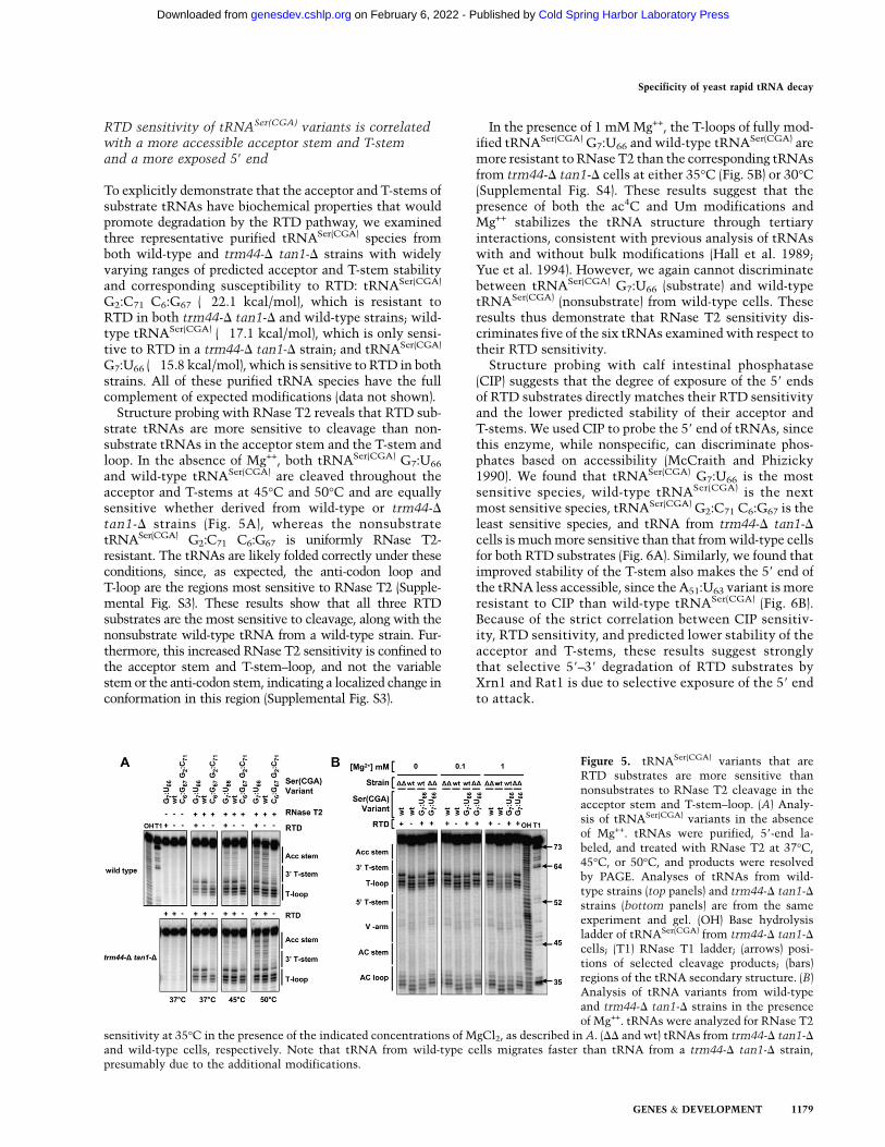
Structure probing with RNase T2 reveals that RTD sub-strate tRNAs are more sensitive to cleavage than non-substrate tRNAs in the acceptor stem and the T-stem andloop. In the absence of Mg++, both tRNASer(CGA) G7:U66
and wild-type tRNASer(CGA) are cleaved throughout theacceptor and T-stems at 45°C and 50°C and are equallysensitive whether derived from wild-type or trm44-Dtan1-D strains (Fig. 5A), whereas the nonsubstratetRNASer(CGA) G2:C71 C6:G67 is uniformly RNase T2-resistant. The tRNAs are likely folded correctly under theseconditions, since, as expected, the anti-codon loop andT-loop are the regions most sensitive to RNase T2 (Supple-mental Fig. S3). These results show that all three RTDsubstrates are the most sensitive to cleavage, along with thenonsubstrate wild-type tRNA from a wild-type strain. Fur-thermore, this increased RNase T2 sensitivity is confined tothe acceptor stem and T-stem–loop, and not the variablestem or the anti-codon stem, indicating a localized change inconformation in this region (Supplemental Fig. S3).
In the presence of 1 mM Mg++, the T-loops of fully mod-ified tRNASer(CGA) G7:U66 and wild-type tRNASer(CGA) aremore resistant to RNase T2 than the corresponding tRNAsfrom trm44-D tan1-D cells at either 35°C (Fig. 5B) or 30°C(Supplemental Fig. S4). These results suggest that thepresence of both the ac4C and Um modifications andMg++ stabilizes the tRNA structure through tertiaryinteractions, consistent with previous analysis of tRNAswith and without bulk modifications (Hall et al. 1989;Yue et al. 1994). However, we again cannot discriminatebetween tRNASer(CGA) G7:U66 (substrate) and wild-typetRNASer(CGA) (nonsubstrate) from wild-type cells. Theseresults thus demonstrate that RNase T2 sensitivity dis-criminates five of the six tRNAs examined with respect totheir RTD sensitivity.
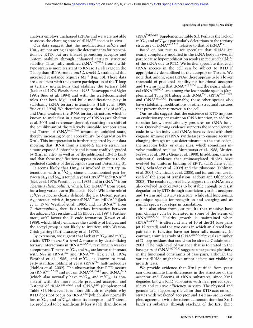
Structure probing with calf intestinal phosphatase(CIP) suggests that the degree of exposure of the 59 endsof RTD substrates directly matches their RTD sensitivityand the lower predicted stability of their acceptor andT-stems. We used CIP to probe the 59 end of tRNAs, sincethis enzyme, while nonspecific, can discriminate phos-phates based on accessibility (McCraith and Phizicky1990). We found that tRNASer(CGA) G7:U66 is the mostsensitive species, wild-type tRNASer(CGA) is the nextmost sensitive species, tRNASer(CGA) G2:C71 C6:G67 is theleast sensitive species, and tRNA from trm44-D tan1-Dcells is much more sensitive than that from wild-type cellsfor both RTD substrates (Fig. 6A). Similarly, we found thatimproved stability of the T-stem also makes the 59 end ofthe tRNA less accessible, since the A51:U63 variant is moreresistant to CIP than wild-type tRNASer(CGA) (Fig. 6B).Because of the strict correlation between CIP sensitiv-ity, RTD sensitivity, and predicted lower stability of theacceptor and T-stems, these results suggest stronglythat selective 59–39 degradation of RTD substrates byXrn1 and Rat1 is due to selective exposure of the 59 endto attack.
Figure 5. tRNASer(CGA) variants that areRTD substrates are more sensitive thannonsubstrates to RNase T2 cleavage in theacceptor stem and T-stem–loop. (A) Analy-sis of tRNASer(CGA) variants in the absenceof Mg++. tRNAs were purified, 59-end la-beled, and treated with RNase T2 at 37°C,45°C, or 50°C, and products were resolvedby PAGE. Analyses of tRNAs from wild-type strains (top panels) and trm44-D tan1-Dstrains (bottom panels) are from the sameexperiment and gel. (OH) Base hydrolysisladder of tRNASer(CGA) from trm44-D tan1-Dcells; (T1) RNase T1 ladder; (arrows) posi-tions of selected cleavage products; (bars)regions of the tRNA secondary structure. (B)Analysis of tRNA variants from wild-typeand trm44-D tan1-D strains in the presenceof Mg++. tRNAs were analyzed for RNase T2
sensitivity at 35°C in the presence of the indicated concentrations of MgCl2, as described in A. (DD and wt) tRNAs from trm44-D tan1-Dand wild-type cells, respectively. Note that tRNA from wild-type cells migrates faster than tRNA from a trm44-D tan1-D strain,presumably due to the additional modifications.
Specificity of yeast rapid tRNA decay
GENES & DEVELOPMENT 1179
Cold Spring Harbor Laboratory Press on February 6, 2022 - Published by genesdev.cshlp.orgDownloaded from

Xrn1 acts on tRNASer(CGA) RTD substrates in vivoand in vitro
To determine the role of 59–39 exonucleases in the degra-dation of tRNASer(CGA) in vivo, we examined the effect ofan xrn1-D mutation in a trm44-D tan1-D strain. We foundthat the temperature sensitivity of the trm44-D tan1-Dstrain is almost completely rescued by an xrn1-D mutationat 37°C (Supplemental Fig. S5A), and that tRNASer(CGA)
and tRNASer(UGA) are almost completely stable in thesestrains (Supplemental Fig. S5B,C), suggesting that most ofthe RTD in a trm44-D tan1-D strain is due to Xrn1.
To examine the role of Xrn1 directly, we purified Xrn1and analyzed its activity with the same set of RTDsubstrate and nonsubstrate tRNAs analyzed with RNaseT2 and CIP. We found that the sensitivity of 39 end-labeled tRNAs to Xrn1 digestion exactly parallels sensi-tivity to CIP treatment and growth phenotype in all sixtRNA species tested (Fig. 7). Thus, tRNASer(CGA) G7:U66
from trm44-D tan1-D cells is the most sensitive to Xrn1cleavage, with 86% of the full-length tRNA partially orcompletely degraded, followed by tRNASer(CGA) froma trm44-D tan1-D strain, tRNASer(CGA) G7:U66 from wild-type cells, tRNASer(CGA) from wild-type cells, and thentRNASer(CGA) G2:C71 C6:G67 from either strain (Fig. 7). Ineach case, most of the observed product appears to be pCp,based on size, while treatment with a mock purification
fraction yields no degradation (Fig. 7). These results exactlymatch the order of sensitivity observed with CIP (cf. Figs. 7and 6A).
We also found that Xrn1 degrades tRNAVal(AAC)
from trm8-D trm4-D cells much more efficiently thantRNAVal(AAC) from wild-type cells (Supplemental Fig. S6).This result demonstrates that the discrimination of RTDsubstrates by Xrn1 in vitro extends to other tRNAs knownto be subject to this pathway (Chernyakov et al. 2008).
Discussion
Here, we provided evidence that the RTD pathway acts todegrade substrate tRNAs that have slipped below a criti-cal threshold of stability through mutation in their accep-tor stem or T-stem or lack of certain modifications. Thepredicted stability of the acceptor and T-stems in thetRNASer family is strongly correlated with RTD resis-tance, as measured by growth and in vivo stability ofstabilizing tRNA variants, for a large number of variantscontaining different acceptor and T-stem mutations.Furthermore, sensitivity to RTD directly correlates withsensitivity of tRNA variants to RNase T2 in the accep-tor stem and T-stem–loop region, exposure of the 59 endto phosphatase, and sensitivity to degradation by purifiedXrn1. Thus, we conclude that a primary reason that RTDoccurs in vivo is because of a less stable acceptor andT-stem, resulting in exposure of the 59 end of tRNA toattack by Xrn1 or Rat1. We note, however, that theseconclusions do not take into account the role of chargingin substrate recognition by RTD, since our in vitro
Figure 6. tRNASer(CGA) variants that are substrates for RTD aremore sensitive than nonsubstrates to removal of a 59 phosphate.(A) Analysis of tRNASer(CGA) acceptor stem variants. tRNASer(CGA)
variants purified from wild-type and trm44-D tan1-D strains were59 end-labeled and treated with CIP as indicated, and releasedphosphate was separated from tRNA by TLC and quantified.(Dashed lines) tRNA from trm44-D tan1-D cells; (solid lines)tRNA from wild-type cells. (B) Analysis of tRNASer(CGA) T-stemvariant A51:U63.
Figure 7. Xrn1 degrades RTD substrate tRNAs in vitro withthe same specificity as RTD in vivo. Purified tRNASer(CGA)
variants were 39 end-labeled and treated with the indicatedamounts of purified Xrn1, and products were resolved on a 20%polyacrylamide/7M urea gel. (-) buffer only; (v) equivalentvolume of mock purification; (a) RNase T1; (b) base hydrolysisladder.
Whipple et al.
1180 GENES & DEVELOPMENT
Cold Spring Harbor Laboratory Press on February 6, 2022 - Published by genesdev.cshlp.orgDownloaded from
analysis employs uncharged tRNAs and we were not ableto assess the charging state of tRNASer species in vivo.
Our data suggest that the modifications ac4C12 andUm44 are not acting as specific determinants for recogni-tion by RTD, but are instead promoting acceptor andT-stem stability through enhanced tertiary structurestability. Thus, fully modified tRNASer(CGA) from a wild-type strain is more resistant to RNase T2 cleavage in theT-loop than tRNA from a tan1-D trm44-D strain, and thisincreased resistance requires Mg++ (Fig. 5B). These dataare consistent with the known participation of the T-loopin tertiary interactions that stabilize the tertiary fold(Jack et al. 1976; Westhof et al. 1985; Basavappa and Sigler1991; Biou et al. 1994) and with the well-documentedroles that both Mg++ and bulk modifications play instabilizing tRNA tertiary interactions (Hall et al. 1989;Yue et al. 1994). We therefore suggest that lack of ac4C12
and Um44 weakens the tRNA tertiary structure, which isknown to melt first in a variety of tRNAs (see Sheltonet al. 2001 and references therein), resulting in a shift ofthe equilibrium of the relatively unstable acceptor stemand T-stem of tRNASer(CGA) toward an unfolded state,thereby increasing 59 end accessibility for degradation byXrn1. This interpretation is further supported by our datashowing that tRNA from a trm44-D tan1-D strain hasa more exposed 59 phosphate and is more readily degradedby Xrn1 in vitro, as well as by the additional 1.0–1.5 kcal/mol that these modifications appear to contribute to thepredicted stability of the acceptor stem and T-stem (Fig. 3).
It seems likely that Um44 is involved in tertiary in-teractions with m2,2G26, since a noncanonical pair be-tween N44 and N26 is found in yeast tRNAPhe and tRNAAsp
(Jack et al. 1976; Westhof et al. 1985) and in tRNASer fromThermus thermophilus, which, like tRNASer from yeast,has a long variable arm (Biou et al. 1994). While the role ofac4C12 is not as clearly defined, the U12 D-stem partnerA23 interacts with A9 in yeast tRNAPhe and tRNAAsp (Jacket al. 1976; Westhof et al. 1985), and, in tRNASer fromT. thermophilus, there is a tertiary interaction betweenthe adjacent G13 residue and G9 (Biou et al. 1994). Further-more, ac4C favors the 39 endo formation (Kawai et al.1989), which likely enhances the stability of helices, andthe acetyl group is not likely to interfere with Watson-Crick pairing (Parthasarathy et al. 1978).
By extension, we suggest that lack of m7G46 and m5C49
elicits RTD in trm8-D trm4-D mutants by destabilizingtertiary interactions in tRNAVal(AAC), resulting in weakeracceptor and T-stems. m7G46 and A46 are known to interactwith N22 in tRNAPhe and tRNAAsp (Jack et al. 1976;Westhof et al. 1985), and m5C49 is known to mod-estly stabilize folding of yeast tRNAPhe half-molecules(Nobles et al. 2002). The observation that RTD occurson tRNAVal(AAC) and not on tRNAVal(CAC) and tRNAi
Met
(which also normally have m7G46 and m5C49) is con-sistent with the more stable predicted acceptor andT-stems of tRNAVal(CAC) and tRNAi
Met (SupplementalTable S1). However, it is more difficult to explain whyRTD does not occur on tRNAPhe (which also normallyhas m7G46 and m5C49), since its acceptor and T-stemsare predicted to be significantly less stable than those of
tRNAVal(AAC) (Supplemental Table S1). Perhaps the lack ofm7G46 and m5C49 is particularly deleterious to the tertiarystructure of tRNAVal(AAC) relative to that of tRNAPhe.
Based on our results, we speculate that tRNAs arenearly completely modified in the tRNA body in vivo, inpart because hypomodification results in reduced half-lifeof the tRNA due to RTD. We further speculate that eachtRNA species in the cell can be subject to RTD ifappropriately destabilized in the acceptor or T-stem. Wenote that, among yeast tRNAs, there appears to be a lowerthreshold of predicted stability for functional acceptorand T-stems, and that tRNASer(CGA) and the nearly identi-cal tRNASer(UGA) are among the least stable species (Sup-plemental Table S1), along with tRNAMet, tRNAAla(UGC),and tRNASer(GCU). Presumably, these other species alsohave stabilizing modifications or other structural featuresthat prevent their turnover in the cell.
Our results suggest that the existence of RTD imposesan evolutionary constraint on tRNA function, in additionto other known evolutionary pressures on tRNA func-tion. Overwhelming evidence supports the second geneticcode, in which individual tRNAs have evolved with theircognate aminoacyl tRNA synthetases to ensure accuratecharging through unique determinants in the anti-codon,the acceptor helix, or other sites, which sometimes in-volve modified residues (Muramatsu et al. 1988; Musier-Forsyth et al. 1991; Giege et al. 1998). In addition, there issubstantial evidence that aminoacylated tRNAs haveevolved for uniform binding of EF-Tu (LaRiviere et al.2001; Schrader et al. 2009) and the ribosome (Fahlmanet al. 2004; Olejniczak et al. 2005), and for uniform use ineach of the steps of translation (Ledoux and Uhlenbeck2008). The results reported here suggest that tRNAs havealso evolved in eukaryotes to be stable enough to resistdegradation by RTD through a sufficiently stable acceptorand T-stem and tertiary structure, while still functioningas unique species for recognition and charging and assimilar species for steps in translation.
It is also clear from our results that massive basepair changes can be tolerated in some of the stems oftRNASer(CGA). Healthy growth is maintained whentRNASer(CGA) is altered at any of 10 of the 24 base pairs(of 12 tested), and the two cases in which an altered basepair fails to function have not been fully examined. Incontrast, a similar study of tRNAArg(CCG) reveals a numberof D-loop residues that could not be altered (Geslain et al.2003). The high level of variance that is tolerated in thebase pairs of tRNASer(CGA) suggests unexpected plasticityin the functional constraints of base pairs, although thevariant tRNAs might have minor defects not visible bygrowth tests.
We provide evidence that Xrn1 purified from yeastcan discriminate fine differences in the structure of theacceptor and T-stems of tRNA substrates, since Xrn1degrades known RTD substrates with near-perfect spec-ificity and relative efficiency in vitro. The physical andgenetic data supporting the claim that RTD acts on sub-strates with weakened acceptor and T-stems are in com-plete agreement with the recent demonstration that Xrn1binds its substrate through stacking of the first three
Specificity of yeast rapid tRNA decay
GENES & DEVELOPMENT 1181
Cold Spring Harbor Laboratory Press on February 6, 2022 - Published by genesdev.cshlp.orgDownloaded from
nucleotides of a single-stranded substrate within theXrn1 active site (Jinek et al. 2011). The observed completedegradation of tRNA substrates by purified Xrn1 is alsoconsistent with the known processivity of Xrn1 (Stevens1980; Chang et al. 2011; Jinek et al. 2011), although wenote that Xrn1 appears to stall at Gm18 and Um44, theonly sites of 29-O-methylation in our substrate tRNAs(Fig. 7).
It is interesting to consider that Xrn1 activity is balancedso finely as to catalyze degradation of hypomodified ordestabilized tRNAs (Chernyakov et al. 2008) and 18SrRNA during nonfunctional rRNA decay (NRD) (Coleet al. 2009), as well as to catalyze 59 end processing of 5.8SRNA, 25S rRNA, and several snoRNAs (Henry et al. 1994;Petfalski et al. 1998; Geerlings et al. 2000; Lee et al. 2003),while avoiding the degradation of mature functioningrRNAs, snoRNAs, and tRNAs. Perhaps Xrn1 activity inRTD is localized to P-bodies, as for 18S rRNA NRD (Coleet al. 2009) and no-go decay (Doma and Parker 2006), orperhaps activity is biochemically limited by accessibilityof the 59 end (Jinek et al. 2011) or by regulatory factors,much as Rai1 modulates the activity of Rat1 (Xiang et al.2009). Our results thus may suggest not only that tRNAshave evolved to be stable enough in their acceptor andT-stems to avoid RTD, but, conversely, that Xrn1 andRat1/Rai1 have coevolved to avoid activity on tRNAsand other stable cellular RNAs.
Materials and methods
Yeast strains
The sup61-D [URA3 CEN SUP61] strains were generated bystandard methods. The MATa trm44-DTnatMX tan1-DTkanMX
(JMW119) strain (Supplemental Table S2) and its parent, BY4741(JMW007), were transformed with a URA3 CEN SUP61 plasmid(JW031), followed by transformation with a sup61-DTbleR frag-ment made by PCR amplification of the pUG66 bleR cassette(primers in Supplemental Table S3) to generate JMW221 (JMW119sup61-DTbleR [URA3 CEN SUP61]) and JMW223 (BY4741sup61-DTbleR [URA3 CEN SUP61]). Derivatives of JMW221and JMW223, and plasmids used to make them, are listed inSupplemental Table S4.
tRNASer variants were integrated into JMW221 and JMW223by transformation of a StuI fragment from JW132-derived plas-mids containing a tRNASer variant gene and a MET15 markerflanked by ADE2 sequences, followed by selection on SD-Metmedium and screening for Ade� transformants. Then, the URA3CEN SUP61 plasmid was selected against on SD mediumcontaining 750 mg/mL 5-flouroorotic acid. When necessary, amet22-DThphMX cassette was introduced by standard methodsafter PCR amplification of DNA from the knockout strain.
Construction of plasmids containing tRNASer variantsfor integration into yeast
To express tRNASer variants, we constructed a tRNA expressioncassette that allows for replacement of the tRNAHis sequencewith another tRNA sequence, starting with a plasmid harbor-ing a BamH1 fragment containing the tRNAHis(GUG) gene[tH(GUG)G2] and flanking sequences. Using QuikChange(Stratagene), a XhoI site was introduced 22 base pairs 59 of the +1site of the mature tRNA sequence, and a BglII site was introduced
21 base pairs 39 of residue 73 to generate MAB812A. Then, weconstructed an integrating vector derived from yIPlac111 byligation of an ADE2 59 fragment, the BamHI tRNA expressioncassette 39 of the ADE2 fragment, an ADE2 39 fragment 39 of thetRNA cassette, and a MET15 marker ligated between the tRNAcassette and the 39 ADE2 fragment to generate plasmid JW132.Variant tRNASer sequences were then constructed in JW132using overlapping DNA oligonucleotides (Supplemental TableS5) by treatment with T4 PNK, mixing at 150 mM NaCl, meltingfor 5 min at 95°C, slow cooling to 50°C, and ligation into JW132digested with XhoI and BglII.
Overproduction and purification of tRNAs
tRNASer variants were overexpressed using a URA3 leu2-d mul-ticopy vector (pYEX4T-1) after ligation of the correspondingBamH1 fragment from a JW132-derived plasmid. Strains weregrown at 28°C in SD �Ura medium, followed by growth inSD �Ura �Leu medium, and 300 OD cell pellets were used topurify tRNASer(CGA) variants from bulk RNA using 59-biotinylatedoligonucleotides (Supplemental Table S3) as described (Jackmanet al. 2003).
Northern blot analysis
Strains expressing tRNASer variants were grown in YPD mediumat 28°C, thiolutin (5 mg/mL) was added at OD600 = 1.5, theculture was shifted 10 min later to 37°C, and cells (2 mL) wereharvested as indicated, washed, and quick-frozen. Bulk RNA wasprepared using hot phenol (Alexandrov et al. 2006), and RNA wasseparated by 10% PAGE in 8 M urea and TBE buffer, followed bytransfer to Hybond N+ membrane, UV cross-linking, and hybrid-ization with 59 32P-labeled DNA probes (Supplemental Table S3),as described (Alexandrov et al. 2006).
XRN1 cloning, expression, and purification
XRN1 was cloned into BG2663, a derivative of BG2483, to expressXRN1 under PGAL1 control, and fused at its C terminus to acomplex tag containing a protease 3C site, an HA epitope, His6,and the ZZ domain of protein A (Quartley et al. 2009). Plasmidswere transformed into strain BCY123 (Macbeth et al. 2005),transformants were grown and induced for expression, and Xrn1was purified by IgG Sepharose chromatography and 3C proteasecleavage, as described (Quartley et al. 2009), followed by dialysisin 20 mM Tris-Cl (pH 7.5), 150 mM NaCl, 1 mM DTT, 1 mMMgCl2, and 50% glycerol, and storage at �20°C.
RNase T2 structure probing
tRNA was 59 end-labeled (Jackman et al. 2003); melted in buffercontaining 150 mM NaCl and 10 mM TrisCl (pH 7.5) at 95°C;refolded for 10 min at the reaction temperature with or withoutMgCl2; incubated for 1 h in 10-mL mixtures containing 150 mMNaCl, 10 mM TrisCl (pH 7.5), 1 mg/mL carrier RNA, MgCl2 asindicated, and 1 mL of RNase T2 (diluted in 50 mM sodiumacetate at pH 5.2, 40% glycerol); and resolved on 8% poly-acrylamide gels containing 7 M urea.
Phosphatase assays
tRNA was 59 end-labeled (Jackman et al. 2003), refolded, andincubated for 30 min at 37°C in 40-mL mixtures containing150 mM NaCl, 25 mM Tris-Cl (pH 8), 1 mM MgCl2, 10 mg/mLbovine serum albumin (BSA), and 4 mL of CIP (diluted in buffercontaining 150 mM NaCl, 25 mM Tris-Cl at pH 8, 100 mg/mL
Whipple et al.
1182 GENES & DEVELOPMENT
Cold Spring Harbor Laboratory Press on February 6, 2022 - Published by genesdev.cshlp.orgDownloaded from
BSA). Reactions were stopped by freezing on dry ice and extractedwith cold phenol, and 5 mL of aqueous layer was applied to a PEIcellulose TLC plate (EMD) and resolved in buffer containing 1.3 Msodium formate, followed by visualization on a Typhoon Phos-phorImager (GE Healthcare) and quantification using Imagequant.
Xrn1 and luciferase assays
RNA was 39 pCp-labeled, refolded, and incubated for 20 min at37°C in 40-mL mixtures containing 150 mM NaCl, 25 mM Tris-Cl(pH 8), 2 mM MgCl2, 0.5 mM DTT, 0.1 mg/mL BSA, and 4 mL ofXrn1 or mock preparation (diluted in Xrn1 storage buffer contain-ing 0.1 mg/mL BSA). Reactions were stopped by quick-freezingand phenol extraction, and products were resolved by 20% PAGEin 7 M urea. Luciferase assays were performed essentially asdescribed, using three transformants of each construct and threeassays of each transformant (Letzring et al. 2010).
Acknowledgments
We are grateful to E. Grayhack for valuable discussions, andG. Culver, J. Dewe, and M. Guy for comments on the manuscript.This research was supported by NIH grant GM52347 to E.M.P.J.M.W. was supported by NIH Training Grant in Cellular, Bio-chemical and Molecular Sciences 5T32 GM068411.
References
Alexandrov A, Chernyakov I, Gu W, Hiley SL, Hughes TR,Grayhack EJ, Phizicky EM. 2006. Rapid tRNA decay canresult from lack of nonessential modifications. Mol Cell 21:87–96.
Basavappa R, Sigler PB. 1991. The 3 A crystal structure of yeastinitiator tRNA: functional implications in initiator/elonga-tor discrimination. EMBO J 10: 3105–3111.
Biou V, Yaremchuk A, Tukalo M, Cusack S. 1994. The 2.9 Acrystal structure of T. thermophilus seryl-tRNA synthetasecomplexed with tRNA(Ser). Science 263: 1404–1410.
Chang JH, Xiang S, Xiang K, Manley JL, Tong L. 2011. Structuraland biochemical studies of the 59 / 39 exoribonucleaseXrn1. Nat Struct Mol Biol 18: 270–276.
Chernyakov I, Whipple JM, Kotelawala L, Grayhack EJ, PhizickyEM. 2008. Degradation of several hypomodified maturetRNA species in Saccharomyces cerevisiae is mediated byMet22 and the 59-39 exonucleases Rat1 and Xrn1. Genes Dev
22: 1369–1380.Cole SE, LaRiviere FJ, Merrikh CN, Moore MJ. 2009. A
convergence of rRNA and mRNA quality control pathwaysrevealed by mechanistic analysis of nonfunctional rRNAdecay. Mol Cell 34: 440–450.
Copela LA, Fernandez CF, Sherrer RL, Wolin SL. 2008. Compe-tition between the Rex1 exonuclease and the La proteinaffects both Trf4p-mediated RNA quality control and pre-tRNA maturation. RNA 14: 1214–1227.
Dichtl B, Stevens A, Tollervey D. 1997. Lithium toxicity inyeast is due to the inhibition of RNA processing enzymes.EMBO J 16: 7184–7195.
Doma MK, Parker R. 2006. Endonucleolytic cleavage of eukary-otic mRNAs with stalls in translation elongation. Nature
440: 561–564.Doma MK, Parker R. 2007. RNA quality control in eukaryotes.
Cell 131: 660–668.Dunin-Horkawicz S, Czerwoniec A, Gajda MJ, Feder M, Grosjean
H, Bujnicki JM. 2006. MODOMICS: a database of RNA modi-fication pathways. Nucleic Acids Res 34: D145–D149.doi: 10.1093/nar/gkj084.
Etcheverry T, Salvato M, Guthrie C. 1982. Recessive lethality ofyeast strains carrying the SUP61 suppressor results from lossof a transfer RNA with a unique decoding function. J Mol
Biol 158: 599–618.Fahlman RP, Dale T, Uhlenbeck OC. 2004. Uniform binding of
aminoacylated transfer RNAs to the ribosomal A and P sites.Mol Cell 16: 799–805.
Fang F, Hoskins J, Butler JS. 2004. 5-Fluorouracil enhancesexosome-dependent accumulation of polyadenylated rRNAs.Mol Cell Biol 24: 10766–10776.
Fujii K, Kitabatake M, Sakata T, Miyata A, Ohno M. 2009. A rolefor ubiquitin in the clearance of nonfunctional rRNAs.Genes Dev 23: 963–974.
Geerlings TH, Vos JC, Raue HA. 2000. The final step in theformation of 25S rRNA in Saccharomyces cerevisiae isperformed by 59 / 39 exonucleases. RNA 6: 1698–1703.
Geslain R, Martin F, Camasses A, Eriani G. 2003. A yeastknockout strain to discriminate between active and inactivetRNA molecules. Nucleic Acids Res 31: 4729–4737.
Giege R, Sissler M, Florentz C. 1998. Universal rules andidiosyncratic features in tRNA identity. Nucleic Acids Res
26: 5017–5035.Hall KB, Sampson JR, Uhlenbeck OC, Redfield AG. 1989.
Structure of an unmodified tRNA molecule. Biochemistry28: 5794–5801.
Henry Y, Wood H, Morrissey JP, Petfalski E, Kearsey S, TollerveyD. 1994. The 59 end of yeast 5.8S rRNA is generated byexonucleases from an upstream cleavage site. EMBO J 13:2452–2463.
Houseley J, Tollervey D. 2009. The many pathways of RNAdegradation. Cell 136: 763–776.
Isken O, Maquat LE. 2007. Quality control of eukaryoticmRNA: safeguarding cells from abnormal mRNA function.Genes Dev 21: 1833–1856.
Jack A, Ladner JE, Klug A. 1976. Crystallographic refinement ofyeast phenylalanine transfer RNA at 2-5A resolution. J Mol
Biol 108: 619–649.Jackman JE, Montange RK, Malik HS, Phizicky EM. 2003.
Identification of the yeast gene encoding the tRNA m1Gmethyltransferase responsible for modification at position 9.RNA 9: 574–585.
Jinek M, Coyle SM, Doudna JA. 2011. Coupled 59 nucleotiderecognition and processivity in xrn1-mediated mRNA decay.Mol Cell 41: 600–608.
Kadaba S, Krueger A, Trice T, Krecic AM, Hinnebusch AG,Anderson J. 2004. Nuclear surveillance and degradation ofhypomodified initiator tRNAMet in S. cerevisiae. Genes Dev
18: 1227–1240.Kadaba S, Wang X, Anderson JT. 2006. Nuclear RNA surveil-
lance in Saccharomyces cerevisiae: Trf4p-dependent poly-adenylation of nascent hypomethylated tRNA and an aber-rant form of 5S rRNA. RNA 12: 508–521.
Kawai G, Hashizume T, Miyazawa T, McCloskey JA, YokoyamaS. 1989. Conformational characteristics of 4-acetylcytidinefound in tRNA. Nucleic Acids Symp Ser 21: 61–62.
Kotelawala L, Grayhack EJ, Phizicky EM. 2008. Identification ofyeast tRNA Um44 29-O-methyltransferase (Trm44) anddemonstration of a Trm44 role in sustaining levels of specifictRNASer species. RNA 14: 158–169.
Kuai L, Fang F, Butler JS, Sherman F. 2004. Polyadenylation ofrRNA in Saccharomyces cerevisiae. Proc Natl Acad Sci 101:8581–8586.
LaCava J, Houseley J, Saveanu C, Petfalski E, Thompson E,Jacquier A, Tollervey D. 2005. RNA degradation by theexosome is promoted by a nuclear polyadenylation complex.Cell 121: 713–724.
Specificity of yeast rapid tRNA decay
GENES & DEVELOPMENT 1183
Cold Spring Harbor Laboratory Press on February 6, 2022 - Published by genesdev.cshlp.orgDownloaded from
LaRiviere FJ, Wolfson AD, Uhlenbeck OC. 2001. Uniformbinding of aminoacyl-tRNAs to elongation factor Tu bythermodynamic compensation. Science 294: 165–168.
LaRiviere FJ, Cole SE, Ferullo DJ, Moore MJ. 2006. A late-actingquality control process for mature eukaryotic rRNAs. MolCell 24: 619–626.
Ledoux S, Uhlenbeck OC. 2008. Different aa-tRNAs are selecteduniformly on the ribosome. Mol Cell 31: 114–123.
Lee CY, Lee A, Chanfreau G. 2003. The roles of endonucleolyticcleavage and exonucleolytic digestion in the 59-end process-ing of S. cerevisiae box C/D snoRNAs. RNA 9: 1362–1370.
Letzring DP, Dean KM, Grayhack EJ. 2010. Control of trans-lation efficiency in yeast by codon–anticodon interactions.RNA 16: 2516–2528.
Macbeth MR, Schubert HL, Vandemark AP, Lingam AT, Hill CP,Bass BL. 2005. Inositol hexakisphosphate is bound in theADAR2 core and required for RNA editing. Science 309:1534–1539.
McCraith SM, Phizicky EM. 1990. A highly specific phospha-tase from Saccharomyces cerevisiae implicated in tRNAsplicing. Mol Cell Biol 10: 1049–1055.
Muramatsu T, Nishikawa K, Nemoto F, Kuchino Y, NishimuraS, Miyazawa T, Yokoyama S. 1988. Codon and amino-acidspecificities of a transfer RNA are both converted by a singlepost-transcriptional modification. Nature 336: 179–181.
Musier-Forsyth K, Usman N, Scaringe S, Doudna J, Green R,Schimmel P. 1991. Specificity for aminoacylation of an RNAhelix: an unpaired, exocyclic amino group in the minorgroove. Science 253: 784–786.
Nobles KN, Yarian CS, Liu G, Guenther RH, Agris PF. 2002.Highly conserved modified nucleosides influence Mg2+-dependent tRNA folding. Nucleic Acids Res 30: 4751–4760.
Olejniczak M, Dale T, Fahlman RP, Uhlenbeck OC. 2005.Idiosyncratic tuning of tRNAs to achieve uniform ribosomebinding. Nat Struct Mol Biol 12: 788–793.
Parthasarathy R, Ginell SL, De NC, Chheda GB. 1978. Confor-mation of N4-acetylcytidine, a modified nucleoside of tRNA,and stereochemistry of codon-anticodon interaction. Bio-
chem Biophys Res Commun 83: 657–663.Petfalski E, Dandekar T, Henry Y, Tollervey D. 1998. Processing
of the precursors to small nucleolar RNAs and rRNAsrequires common components. Mol Cell Biol 18: 1181–1189.
Quartley E, Alexandrov A, Mikucki M, Buckner FS, Hol WG,DeTitta GT, Phizicky EM, Grayhack EJ. 2009. Heterologousexpression of L. major proteins in S. cerevisiae: a test ofsolubility, purity, and gene recoding. J Struct Funct Geno-mics 10: 233–247.
Rasse-Messenguy F, Fink GR. 1973. Temperature-sensitive non-sense suppressors in yeast. Genetics 75: 459–464.
Reuter JS, Mathews DH. 2010. RNAstructure: software for RNAsecondary structure prediction and analysis. BMC Bioinfor-
matics 11: 129. doi: 10.1186/1471-2105-11-129.Salas-Marco J, Bedwell DM. 2005. Discrimination between
defects in elongation fidelity and termination efficiencyprovides mechanistic insights into translational read-through. J Mol Biol 348: 801–815.
Schneider C, Anderson JT, Tollervey D. 2007. The exosomesubunit Rrp44 plays a direct role in RNA substrate recogni-tion. Mol Cell 27: 324–331.
Schrader JM, Chapman SJ, Uhlenbeck OC. 2009. Understandingthe sequence specificity of tRNA binding to elongation factorTu using tRNA mutagenesis. J Mol Biol 386: 1255–1264.
Shelton VM, Sosnick TR, Pan T. 2001. Altering the intermediatein the equilibrium folding of unmodified yeast tRNAPhewith monovalent and divalent cations. Biochemistry 40:3629–3638.
Stevens A. 1980. Purification and characterization of a Saccha-
romyces cerevisiae exoribonuclease which yields 59-mono-nucleotides by a 59 leads to 39 mode of hydrolysis. J Biol
Chem 255: 3080–3085.Vanacova S, Wolf J, Martin G, Blank D, Dettwiler S, Friedlein A,
Langen H, Keith G, Keller W. 2005. A new yeast poly(A)polymerase complex involved in RNA quality control. PLoS
Biol 3: e189. doi: 10.1371/journal.pbio.00301189.Westhof E, Dumas P, Moras D, Romby P. 1985. Crystallographic
refinement of yeast aspartic acid transfer RNA. J Mol Biol
184: 119–145.Xiang S, Cooper-Morgan A, Jiao X, Kiledjian M, Manley JL, Tong
L. 2009. Structure and function of the 59 / 39 exoribonu-clease Rat1 and its activating partner Rai1. Nature 458: 784–788.
Yue D, Kintanar A, Horowitz J. 1994. Nucleoside modificationsstabilize Mg2+ binding in Escherichia coli tRNA(Val): animino proton NMR investigation. Biochemistry 33: 8905–8911.
Whipple et al.
1184 GENES & DEVELOPMENT
Cold Spring Harbor Laboratory Press on February 6, 2022 - Published by genesdev.cshlp.orgDownloaded from
10.1101/gad.2050711Access the most recent version at doi: 25:2011, Genes Dev.
Joseph M. Whipple, Elizabeth A. Lane, Irina Chernyakov, et al. integrity of the acceptor and T-stems of mature tRNAThe yeast rapid tRNA decay pathway primarily monitors the structural
Material
Supplemental
http://genesdev.cshlp.org/content/suppl/2011/05/31/25.11.1173.DC1
References
http://genesdev.cshlp.org/content/25/11/1173.full.html#ref-list-1
This article cites 56 articles, 21 of which can be accessed free at:
License
ServiceEmail Alerting
click here.right corner of the article or
Receive free email alerts when new articles cite this article - sign up in the box at the top
Copyright © 2011 by Cold Spring Harbor Laboratory Press
Cold Spring Harbor Laboratory Press on February 6, 2022 - Published by genesdev.cshlp.orgDownloaded from