the wolf-hirschhorn syndrome in fetal autopsy - a case...
TRANSCRIPT
S15
Wolf-Hirschhorn syndrome (WHS) is one of multiple con-genital anomaly/mental retardation (MCA/MR) syndromes and is caused by partial deletion of the short arm of chromosome 4, particularly in the Wolf-Hirschhorn syndrome critical region-1 (WHSCR1) and Wolf-Hirschhorn syndrome critical region-2 (WHSCR2), which are located in chromosome 4p16.3. Loss of these regions is indeed the cause of WHS, and the length of dele-tion regions was associated with the specific clinical phenotype.1 In many cases, WHS is caused by de novo deletion of the short arm of chromosome 4, including 4p16.3. A familial transloca-tion was found in about 5-13% of WHS cases.2 The incidence of WHS was estimated from about 1 in 20,000 to 1 in 100,000.1,3 The key features of the WHS are, as follows: typical “Greek hel-met” face (i.e., hypertelorism and frontal bossing, with promi-nent bridge of nose), microcephaly, cleft palate, severe psychomo-tor and growth retardation, low birth weight, diminished fetal activity, hypotonia, seizures and renal anomalies, which express various phenotypic features. Rarely, diaphragmatic hernia has also been reported.4 This report pertains to an autopsy case of WHS, of which chromosomal studies were verified by array com-
parative genomic hybridization (array-CGH).
CASE REPORT
The proband was the second male fetus of unrelated healthy parents, with no history of teratogenic exposures, such as drugs or gestational diabetes mellitus. The mother took iron supple-ments from the first trimester. No peculiarity was noted during the first trimester. At 16 weeks gestational age, prenatal ultra-sonography showed the undersized male fetus in relation to ges-tational age, with congenital diaphragmatic hernia and thick-ness of the nuchal skinfold. The amniotic fluid index was with-in normal range.
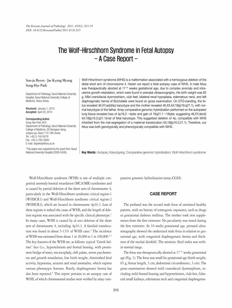
The fetus was therapeutically aborted at 17+0 weeks gestational age (Fig. 1). The fetus was small for gestational age (birth weight, 65 g; femur length, 3 cm; abdominal circumference, 3 cm). The gross examination showed mild craniofacial dysmorphism, in-cluding mild frontal bossing and hypertelorism, club feet, bilat-eral small kidneys, edematous neck and congenital diaphragmat-
The Wolf-Hirschhorn Syndrome in Fetal Autopsy
- A Case Report -
Sun-ju Byeon · Jae Kyung MyungSung-Hye Park
Department of Pathology, Seoul National University Hospital, Seoul National University College of Medicine, Seoul, Korea
Wolf-Hirschhorn syndrome (WHS) is a malformation associated with a hemizygous deletion of the distal short arm of chromosome 4. Herein we report a fetal autopsy case of WHS. A male fetus was therapeutically aborted at 17+0 weeks gestational age, due to complex anomaly and intra-uterine growth retardation, which were found in prenatal ultrasonography. His birth weight was 65 g. Mild craniofacial dysmorphism, club feet, bilateral renal hypoplasia, edematous neck, and left diaphragmatic hernia of Bochdalek were found on gross examination. On GTG-banding, the fe-tus revealed 46,XY,add(4p) karyotype and the mother revealed 46,XX,t(4;18)(p16;q21.1), with nor-mal karyotype of the father. Array comparative genomic hybridization performed on the autopsied lung tissue revealed loss of 4p16.2→4pter and gain of 18q21.1→18qter, suggesting 46,XY,der(4)t(4;18)(p16.2;q21.1)mat of fetal karyotype. This suggested deletion of 4p, compatible with WHS inherited from the mal-segregation of a maternal translocation t(4;18)(p16.2;21.1). Therefore, our fetus was both genotypically and phenotypically compatible with WHS.
Key Words: Autopsy; Karyotyping; Comparative genomic hybridization; Wolf-Hirschhorn syndrome
Received: January 1, 2010Accepted: April 20, 2010
Corresponding AuthorSung-Hye Park, M.D.Department of Pathology, Seoul National University College of Medicine, 28 Yeongeon-dong, Jongno-gu, Seoul 110-799, KoreaTel: +82-2-740-8278Fax: +82-2-765-5600E-mail: [email protected]
*This paper was supported by the grant from Seoul National University Hospital (2009-5426).
The Korean Journal of Pathology 2011; 45(S1): S15-19DOI: 10.4132/KoreanJPathol.2011.45.S1.S15
� Sun-ju�Byeon·Jae�Kyung�Myung·Sung-Hye�ParkS16
ic hernia of Bochdalek type. Microscopic examination showed a low number of nephrons and slightly hypertrophic glomeruli,
with microlithiasis in the left kidney, immature lungs and di-lated subcutaneous lymphatics on the neck, suggesting nuchal hygroma.
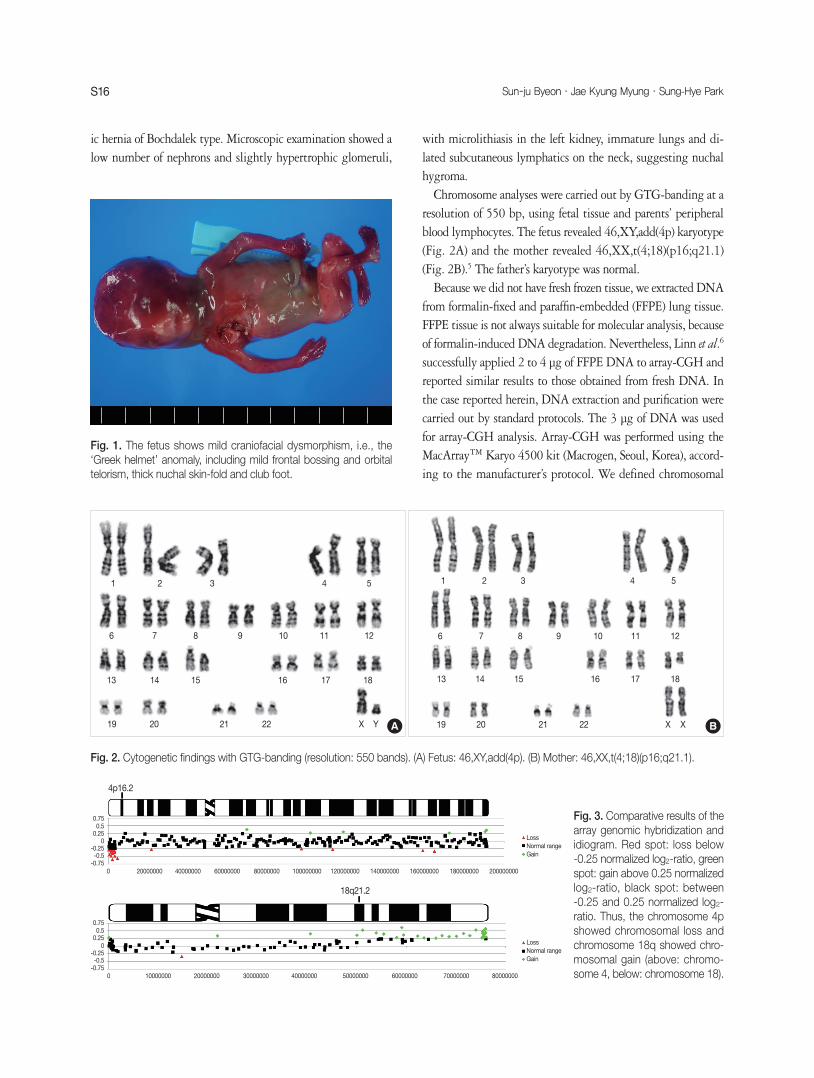
Chromosome analyses were carried out by GTG-banding at a resolution of 550 bp, using fetal tissue and parents’ peripheral blood lymphocytes. The fetus revealed 46,XY,add(4p) karyotype (Fig. 2A) and the mother revealed 46,XX,t(4;18)(p16;q21.1) (Fig. 2B).5 The father’s karyotype was normal.
Because we did not have fresh frozen tissue, we extracted DNA from formalin-fixed and paraffin-embedded (FFPE) lung tissue. FFPE tissue is not always suitable for molecular analysis, because of formalin-induced DNA degradation. Nevertheless, Linn et al.6 successfully applied 2 to 4 μg of FFPE DNA to array-CGH and reported similar results to those obtained from fresh DNA. In the case reported herein, DNA extraction and purification were carried out by standard protocols. The 3 μg of DNA was used for array-CGH analysis. Array-CGH was performed using the MacArrayTM Karyo 4500 kit (Macrogen, Seoul, Korea), accord-ing to the manufacturer’s protocol. We defined chromosomal
Fig. 1. The fetus shows mild craniofacial dysmorphism, i.e., the ‘Greek helmet’ anomaly, including mild frontal bossing and orbital telorism, thick nuchal skin-fold and club foot.
Fig. 2. Cytogenetic findings with GTG-banding (resolution: 550 bands). (A) Fetus: 46,XY,add(4p). (B) Mother: 46,XX,t(4;18)(p16;q21.1).
A B
1 12 23 3
10 107 7
14 1417 17
22 2220 20
9 96 6
13 1316 16
21 2119 19
4 4
11 118 8
15 1518 18
X XXY
5 5
12 12
0.75
4p16.2
18q21.2
0.75
0.25
0.25
0.5
0.5
-0.25
-0.25
-0.5
-0.5
-0.750
0
20000000
10000000
80000000
80000000
LossNormal rangeGain
LossNormal rangeGain
40000000
4000000020000000 30000000 50000000
10000000060000000
60000000 70000000
120000000 160000000140000000 180000000 200000000
-0.75
0
0
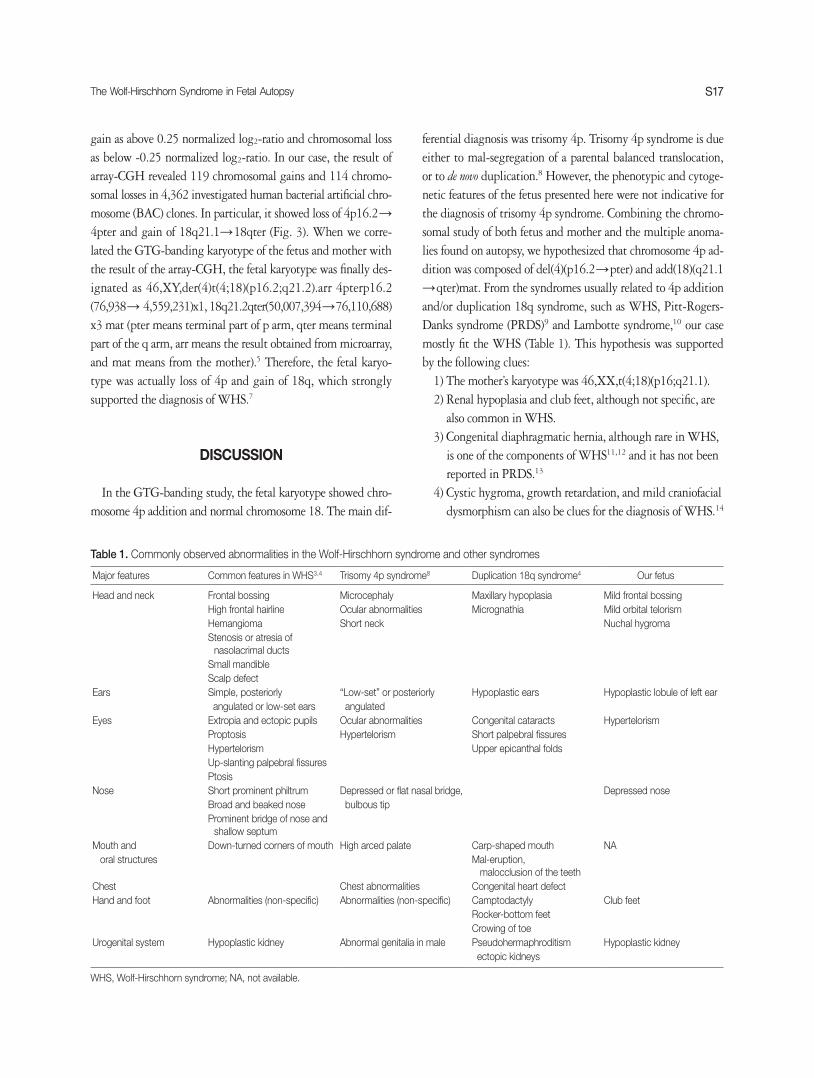
Fig. 3. Comparative results of the array genomic hybridization and idiogram. Red spot: loss below -0.25 normalized log2-ratio, green spot: gain above 0.25 normalized log2-ratio, black spot: between -0.25 and 0.25 normalized log2-ratio. Thus, the chromosome 4p showed chromosomal loss and chromosome 18q showed chro-mosomal gain (above: chromo-some 4, below: chromosome 18).
S17The�Wolf-Hirschhorn�Syndrome�in�Fetal�Autopsy�
gain as above 0.25 normalized log2-ratio and chromosomal loss as below -0.25 normalized log2-ratio. In our case, the result of array-CGH revealed 119 chromosomal gains and 114 chromo-somal losses in 4,362 investigated human bacterial artificial chro-mosome (BAC) clones. In particular, it showed loss of 4p16.2→ 4pter and gain of 18q21.1→18qter (Fig. 3). When we corre-lated the GTG-banding karyotype of the fetus and mother with the result of the array-CGH, the fetal karyotype was finally des-ignated as 46,XY,der(4)t(4;18)(p16.2;q21.2).arr 4pterp16.2 (76,938→ 4,559,231)x1, 18q21.2qter(50,007,394→76,110,688) x3 mat (pter means terminal part of p arm, qter means terminal part of the q arm, arr means the result obtained from microarray, and mat means from the mother).5 Therefore, the fetal karyo-type was actually loss of 4p and gain of 18q, which strongly supported the diagnosis of WHS.7
DISCUSSION
In the GTG-banding study, the fetal karyotype showed chro-mosome 4p addition and normal chromosome 18. The main dif-
ferential diagnosis was trisomy 4p. Trisomy 4p syndrome is due either to mal-segregation of a parental balanced translocation, or to de novo duplication.8 However, the phenotypic and cytoge-netic features of the fetus presented here were not indicative for the diagnosis of trisomy 4p syndrome. Combining the chromo-somal study of both fetus and mother and the multiple anoma-lies found on autopsy, we hypothesized that chromosome 4p ad-dition was composed of del(4)(p16.2→pter) and add(18)(q21.1 →qter)mat. From the syndromes usually related to 4p addition and/or duplication 18q syndrome, such as WHS, Pitt-Rogers-Danks syndrome (PRDS)9 and Lambotte syndrome,10 our case mostly fit the WHS (Table 1). This hypothesis was supported by the following clues:
1) The mother’s karyotype was 46,XX,t(4;18)(p16;q21.1). 2) Renal hypoplasia and club feet, although not specific, are
also common in WHS. 3) Congenital diaphragmatic hernia, although rare in WHS,
is one of the components of WHS11,12 and it has not been reported in PRDS.13
4) Cystic hygroma, growth retardation, and mild craniofacial dysmorphism can also be clues for the diagnosis of WHS.14
Table 1. Commonly observed abnormalities in the Wolf-Hirschhorn syndrome and other syndromes
Major features Common features in WHS3,4 Trisomy 4p syndrome8 Duplication 18q syndrome4 Our fetus
Head and neck Frontal bossing Microcephaly Maxillary hypoplasia Mild frontal bossingHigh frontal hairline Ocular abnormalities Micrognathia Mild orbital telorismHemangioma Short neck Nuchal hygromaS tenosis or atresia of
nasolacrimal ductsSmall mandibleScalp defect
Ears Simple, posteriorly “Low-set” or posteriorly Hypoplastic ears Hypoplastic lobule of left ear angulated or low-set ears angulated
Eyes Extropia and ectopic pupils Ocular abnormalities Congenital cataracts HypertelorismProptosis Hypertelorism Short palpebral fissuresHypertelorism Upper epicanthal foldsUp-slanting palpebral fissuresPtosis
Nose Short prominent philtrum Depressed or flat nasal bridge, Depressed noseBroad and beaked nose bulbous tipP rominent bridge of nose and
shallow septumMouth and Down-turned corners of mouth High arced palate Carp-shaped mouth NA oral structures M al-eruption,
malocclusion of the teethChest Chest abnormalities Congenital heart defectHand and foot Abnormalities (non-specific) Abnormalities (non-specific) Camptodactyly Club feet
Rocker-bottom feetCrowing of toe
Urogenital system Hypoplastic kidney Abnormal genitalia in male Pseudohermaphroditism Hypoplastic kidney ectopic kidneys
WHS, Wolf-Hirschhorn syndrome; NA, not available.
� Sun-ju�Byeon·Jae�Kyung�Myung·Sung-Hye�ParkS18
5) Sporadic case in this family.10
However, the fetus did not show the typical “Greek helmet” face and mental retardation could not be assessed because of too young fetal age (i.e., 17 weeks gestational age). Faced with lim-ited clinical data, we carried out the additional cytogenetic study to reach an accurate diagnosis of WHS. Cytogenetic abnormali-ties are a major cause of MCA/MR syndromes. In many cases, the cytogenetic study on prenatal diagnosis and screening is con-ducted by conventional microscopic analysis, using GTG-band-ing. However, the conventional microscopic cytogenetic study is sometimes unable to detect either microdeletion or microdupli-cation, whereas array-CGH has demonstrated a highly diagnos-tic yield in MCA/MR syndrome patients, including WHS.15,16 In addition to the GTG-band karyotype study, molecular studies can be used to identify deletion of the WHSCR1 and/or WH-SCR2 region on 4p. In many cases, both metaphase fluorescence in situ hybridization and array-CGH have been recently used to check for chromosomal anomaly.17-19 Both methods can be use-ful for identification of 4p16.3 deletion. Nevertheless, the array-CGH study is more informative to determine the length of ei-ther deletion or duplication associated with the clinical pheno-type. The array-CGH was initially developed as a research tool for investigation of the genomic imbalances in cancer, and it is currently used to detect aneuploidies, well-characterized micro-deletion/microduplication syndromes and subtelomeric or other unbalanced chromosomal rearrangements. Therefore, it is now considered as a useful tool for prenatal diagnosis.15,20 Moreover, recently DNA extraction and purification are routinely performed in many laboratories and surgeons are more familiar with them and other methods. The microarray CGH delivered the accurate karyotype of our case and made the diagnosis of WHS.
During the autopsy, it is sometimes difficult to confirm the diagnosis because fresh tissues are not routinely kept at deep freezing temperature. However, with appropriate DNA extrac-tion from the FFPE tissue, array-CGH can be a useful addition-al tool to identify genetic diseases and provide genetic informa-tion in fetal and pediatric autopsy cases.
ACKNOWLEDGEMENTS Thanks to Jeong-Hee Yoon (physical assistant, Department
of Pathology, National Cancer Center, Republic of Korea) for DNA extraction and purification.
REFERENCES
1.ShannonNL,MaltbyEL,RigbyAS,QuarrellOW.Anepidemiolog-icalstudyofWolf-Hirschhornsyndrome:lifeexpectancyandcauseofmortality.JMedGenet2001;38:674-9.
2.LurieIW,LazjukGI,UssovaYI,PresmanEB,GurevichDB.TheWolf-Hirschhornsyndrome.I.Genetics.ClinGenet1980;17:375-84.
3.BattagliaA,FilippiT,CareyJC.UpdateontheclinicalfeaturesandnaturalhistoryofWolf-Hirschhorn(4p-)syndrome:experiencewith87patientsandrecommendationsforroutinehealthsupervision.AmJMedGenetCSeminMedGenet2008;148C:246-51.
4.Gilbert-BarnessE,AllenMIV.Chromosomalabnormalities.In:Gil-bert-BarnessE,KapurRP,OlignyLL,SiebertJR,eds.Potter’spathol-ogyofthefetus,infantandchild.2nded.St.Louis:Mosby,2007;239-42.
5.ShafferLG,SlovakML,CampbellLJ.ISCN2009:anInternationalSystemforHumanCytogeneticNomenclature(2009):recommen-dationsoftheInternationalStandingCommitteeonHumanCyto-geneticNomenclature.Basel:Karger,2009.
6.LinnSC,WestRB,PollackJR,et al.Geneexpressionpatternsandgenecopynumberchangesindermatofibrosarcomaprotuberans.AmJPathol2003;163:2383-95.
7.ZollinoM,MurdoloM,MarangiG,et al.Onthenosologyandpatho-genesisofWolf-Hirschhornsyndrome:genotype-phenotypecorre-lationanalysisof80patientsandliteraturereview.AmJMedGenetCSeminMedGenet2008;148C:257-69.
8.PatelSV,DagnewH,ParekhAJ,et al.Clinicalmanifestationsoftri-somy4psyndrome.EurJPediatr1995;154:425-31.
9.WrightTJ,ClemensM,QuarrellO,AltherrMR.Wolf-HirschhornandPitt-Rogers-Dankssyndromescausedbyoverlapping4pdele-tions.AmJMedGenet1998;75:345-50.
10.HerensC,JamarM,Alvarez-GonzalezML,et al.Privatemultiplecongenitalanomalysyndromesmayresultfromunbalancedsubtletranslocations:t(2q;4p)explainstheLambottesyndrome.AmJMedGenet1997;73:127-31.
11.SergiC,SchulzeBR,HagerHD,et al.Wolf-Hirschhornsyndrome:casereportandreviewofthechromosomalaberrationsassociatedwithdiaphragmaticdefects.Pathologica1998;90:285-93.
12.vanDoorenMF,BrooksAS,HoogeboomAJ,et al.EarlydiagnosisofWolf-Hirschhornsyndrometriggeredbyalife-threateningevent:con-genitaldiaphragmatichernia.AmJMedGenetA2004;127A:194-6.
13.PartingtonMW,FaganK,SoubjakiV,TurnerG.Translocationsin-volving4p16.3inthreefamilies:deletioncausingthePitt-Rogers-Dankssyndromeandduplicationresultinginanewovergrowthsyndrome.JMedGenet1997;34:719-28.
14.BasgulA,KavakZN,AkmanI,BasgulA,GokaslanH,ElciogluN.
S19The�Wolf-Hirschhorn�Syndrome�in�Fetal�Autopsy�
PrenataldiagnosisofWolf-Hirschhornsyndrome(4p-)inassocia-tionwithcongenitaldiaphragmatichernia,cystichygromaandIUGR.ClinExpObstetGynecol2006;33:105-6.
15.deRavelTJ,DevriendtK,FrynsJP,VermeeschJR.What’snewinkaryotyping?Themovetowardsarraycomparativegenomichybri-disation(CGH).EurJPediatr2007;166:637-43.
16.SouthST,WhitbyH,BattagliaA,CareyJC,BrothmanAR.Compre-hensiveanalysisofWolf-HirschhornsyndromeusingarrayCGHindicatesahighprevalenceoftranslocations.EurJHumGenet2008;16:45-52.
17.KimJH,OhPS,NaHY,KimSH,ChoHC.Acaseofmosaicringchro-mosome4withsubtelomeric4pdeletion.KoreanJLabMed2009;
29:77-81.18.ParkHK,KimHJ,HanSH,KimYJ,KimSH.Screeningofsubtelo-mericrearrangementsin100Koreanpediatricpatientswithunex-plainedmentalretardationandanomaliesusingsubtelomericFISH(fluorescenceinsituhybridization).JKoreanMedSci2008;23:573-8.
19.MunSJ,ChoEH,CheyMJ,et al.Recombinantchromosome4withpartial4pdeletionand4qduplicationinheritedfrompaternalperi-centricinversion.KoreanJLabMed2010;30:89-92.
20.RickmanL,FieglerH,Shaw-SmithC,et al.Prenataldetectionofun-balancedchromosomalrearrangementsbyarrayCGH.JMedGenet2006;43:353-61.