the viral f-box protein p0 induces an er-derived autophagy ... · the viral f-box protein p0...
TRANSCRIPT
The viral F-box protein P0 induces an ER-derivedautophagy degradation pathway for the clearanceof membrane-bound AGO1Simon Michaelia, Marion Clavela, Esther Lechnera, Corrado Viottia, Jian Wub, Marieke Duboisa,c, Thibaut Hacquarda,Benoît Derriena,d, Esther Izquierdoe, Maxime Lecorbeillera, Nathalie Bouteillerf, Julia De Ciliaa, Véronique Ziegler-Graffa,Hervé Vaucheretf, Gad Galilib, and Pascal Genschika,1
aInstitut de Biologie Moléculaire des Plantes, Centre National de la Recherche Scientifique, Unité Propre de Recherche 2357, Conventionné avec l’Universitéde Strasbourg, 67084 Strasbourg, France; bDepartment of Plant and Environmental Sciences, Weizmann Institute of Science, 76100 Rehovot, Israel; cVIB-UGent Center for Plant Systems Biology, Ghent University, 9052 Ghent, Belgium; dDNA SCRIPT, 75014 Paris, France; eBiochimie et Physiologie Moléculairedes Plantes, Université Montpellier, CNRS, Institut National de la Recherche Agronomique, SupAgro, 34060 Montpellier, France; and fInstitut Jean-PierreBourgin, UMR1318, Institut National de la Recherche Agronomique, AgroParisTech, CNRS, Université Paris-Saclay, 78000 Versailles, France
Edited by Mark Estelle, University of California San Diego, La Jolla, CA, and approved September 23, 2019 (received for review July 17, 2019)
RNA silencing is a major antiviral defense mechanism in plants andinvertebrates. Plant ARGONAUTE1 (AGO1) is pivotal in RNA silenc-ing, and hence is a major target for counteracting viral suppressorsof RNA-silencing proteins (VSRs). P0 from Turnip yellows virus(TuYV) is a VSR that was previously shown to trigger AGO1 deg-radation via an autophagy-like process. However, the identity ofhost proteins involved and the cellular site at which AGO1 and P0interact were unknown. Here we report that P0 and AGO1 asso-ciate on the endoplasmic reticulum (ER), resulting in their loadinginto ER-associated vesicles that are mobilized to the vacuole in anATG5- and ATG7-dependent manner. We further identified ATG8-Interacting proteins 1 and 2 (ATI1 and ATI2) as proteins that asso-ciate with P0 and interact with AGO1 on the ER up to the vacuole.Notably, ATI1 and ATI2 belong to an endogenous degradationpathway of ER-associated AGO1 that is significantly induced fol-lowing P0 expression. Accordingly, ATI1 and ATI2 deficiency causesa significant increase in posttranscriptional gene silencing (PTGS)activity. Collectively, we identify ATI1 and ATI2 as components ofan ER-associated AGO1 turnover and proper PTGS maintenance andfurther show how the VSR P0 manipulates this pathway.
RNA silencing | autophagy | Arabidopsis
In eukaryotes, gene silencing is essential for development andplays important roles in response to biotic and abiotic stresses,
as well as in epigenetic control of transposable elements. RNAsilencing involves the processing of double-stranded (ds)RNA byDicer-like RNase III enzymes, into small RNAs (sRNAs) of 21to 24 nucleotides in length (1). All types of sRNAs are thenincorporated into a protein complex called RISC (RNA-inducedsilencing complex) that invariably contains a member of thehighly conserved Argonaute (AGO) protein family (2, 3). TheseRISCs are programmed by the bound sRNAs to specifically in-teract with transcripts based on sequence complementarity,resulting in messenger RNA (mRNA) cleavage, translational re-pression, or chromatin modification. An important class of en-dogenous sRNAs is microRNAs (miRNAs) (4, 5), which repressthe expression of one or more target mRNAs and have beenpredicted to control a significant proportion of the transcriptome(6). Important functions for sRNAs have also emerged in thestudy of host−pathogen interactions. In particular, RNA silencingplays a key role in antiviral defense in plants and invertebrates,where populations of sRNAs are produced in infected cells di-rectly by processing dsRNA molecules derived from the viral ge-nome (7, 8). These virus-derived small interfering RNAs (siRNAs)are then incorporated into an antiviral RISC and turned back ontoviral RNAs to trigger their degradation.In the model plant Arabidopsis thaliana (hereafter referred to
as Arabidopsis), the AGO protein family is composed of 10 members
divided into 3 major clades based on their protein similarity (9).Among them, AGO1 plays a central role in both miRNA- andsiRNA-directed silencing (10). Hence, AGO1 loaded with sRNAsmediates endonucleolytic cleavage of target transcripts (11), but afraction of transcripts also evades slicing and instead undergoestranslation repression (12, 13). Moreover, AGO1 plays an im-portant function in the biogenesis and control of secondarysiRNAs (14–16). Finally, besides its regulatory role of endogenousgene expression through miRNAs and endogenous siRNAs, AGO1is paramount in antiviral defense, and, consequently, various ago1mutants exhibit enhanced susceptibility to virus infection (17, 18).However, as a counter defense strategy, viruses have acquiredvarious viral suppressors of RNA silencing (VSRs), which cantarget different steps of the RNA silencing pathway (19, 20).Previous work from our laboratory and others revealed that
the VSR protein P0 from Polerovirus encodes an F-box proteinthat hijacks the host S-phase kinase-associated protein1 (SKP1)-cullin 1 (CUL1)-F-box protein (SCF) ubiquitin-protein ligase(E3) to promote AGO1 degradation (21–24). Although different
Significance
The viral suppressor of RNA silencing P0 is known to targetplant antiviral ARGONAUTE (AGO) proteins for degradation viaan autophagy-related process. Here we utilized P0 to gain in-sight into the cellular degradation dynamics of AGO1, the majorplant effector of RNA silencing. We revealed that P0 targetsendoplasmic reticulum (ER)-associated AGO1 by inducing theformation of ER-related bodies that are delivered to the vacuolewith both P0 and AGO1 as cargos. This process involves ATG8-interacting proteins 1 and 2 (ATI1 and ATI2) that interact withAGO1 and negatively regulate its transgene-silencing activity.Together, our results reveal a layer of ER-bound AGO1 post-translational regulation that is manipulated by P0 to subvertplant antiviral defense.
Author contributions: S.M., M.C., C.V., and P.G. designed research; S.M., M.C., E.L., C.V.,J.W., M.D., T.H., B.D., E.I., M.L., N.B., and H.V. performed research; J.W., J.D.C., V.Z.-G., andG.G. contributed new reagents/analytic tools; S.M., M.C., E.L., C.V., M.D., T.H., B.D., E.I.,M.L., V.Z.-G., H.V., and P.G. analyzed data; and P.G. wrote the paper with theparticipation of S.M.
The authors declare no competing interest.
This article is a PNAS Direct Submission.
This open access article is distributed under Creative Commons Attribution-NonCommercial-NoDerivatives License 4.0 (CC BY-NC-ND).1To whom correspondence may be addressed. Email: [email protected].
This article contains supporting information online at www.pnas.org/lookup/suppl/doi:10.1073/pnas.1912222116/-/DCSupplemental.
First published October 18, 2019.
22872–22883 | PNAS | November 5, 2019 | vol. 116 | no. 45 www.pnas.org/cgi/doi/10.1073/pnas.1912222116
Dow
nloa
ded
by g
uest
on
Mar
ch 2
8, 2
020
Poleroviruses encode P0 proteins that lack sequence similarity,most of them can mediate the decay of AGOs (25, 26). SCF-typeubiquitin E3 ligases promote ubiquitylation of their substrates,which serves as a signal for proteasomal degradation (27, 28).However, inhibition of the proteasome was unable to block P0-mediated degradation of AGO1 (22). Instead, it was shown thatAGO1 degradation was blocked by pharmacological inhibition oftrafficking pathways that lead to the vacuole, including macro-autophagy, and that P0 expression leads to an accumulation ofAGO1 in vacuolar inclusions (29).Macroautophagy (hereafter referred to as autophagy) is an
evolutionary conserved intracellular degradation and recyclingmechanism involving the generation of a specialized doublemembrane termed the phagophore, which likely emanates fromthe endoplasmic reticulum (ER) (30, 31). The phagophore cap-tures cytosolic content (cargo) and then seals to form an auto-phagosome that delivers the cargo to cellular lytic compartments(lysosomes in animals or vacuoles in yeast and plants). Theautophagy process requires more than 40 proteins to function,most of which are annotated as autophagy-related proteins (ATG)(32–34). Under energetically demanding stress, cells induce non-selective autophagy, by which cytosolic content is degraded.However, under specific stress and developmental conditions,subcellular structures are cleared by selective autophagy (34–36), aprocess implying specialized cargo receptors that anchor thecargo to autophagy machinery components. In many cases, cargoreceptors interact with ATG8 proteins, which are known to dec-orate both the inner and outer membranes of the autophagosomeand are involved in autophagosome maturation as well as in theirfusion with the lytic compartment. Frequently, ubiquitylationserves as a signal for the recognition of cargoes destined for se-lective autophagy (36). In plants, only a few selective autophagyreceptors have been identified (34). Among them are neighbor ofBRCA 1 (NBR1), a functional hybrid of mammalian p62 andNBR1 autophagy receptors, which targets ubiquitylated proteinaggregates in plant stress responses (37, 38) and virus particles(39), tryptophan-rich sensory protein (TSPO) that can targetplasma-membrane based aquaporins (40), and RPN10, whichdegrades inactive 26S proteasomes (41). ATG8-interacting 1 and 2(ATI1 and ATI2), on the other hand, are transmembrane proteinsreported to localize to ER- and plastid-derived vesicles that trafficto the vacuole (42, 43). Although ATI1/2 interact with ATG8f andrely on core autophagy machinery to reach the vacuole, ATI1/2decorated bodies are generally distinct from ATG8-decoratedautophagosomes, and their function on the ER is still poorlyunderstood (42).The pivotal role of autophagy during viral infection in plants
has only recently emerged, with reports demonstrating both anti-viral and proviral functions (44, 45). At present, little is knownabout the mechanism of P0-mediated AGO1 protein degradation.The viral F-box protein P0 has been proposed to act upstream ofAGO1 loading (24), as it is very effective in degrading newlysynthesized AGO1 after transient expression in tobacco leaves, butnot endogenous preassembled AGO1 complex. We recently foundthat P0 recognizes the DUF1785 domain of AGO1 to enable itsdegradation (46), but where this occurs at the subcellular level isunknown. Notably, AGO1 protein also decays via a similar path-way in a P0-independent manner, when miRNA production orstability is compromised (29). However, the assumption that theAGO1 protein decays via autophagy in a P0-dependent and in-dependent context mainly relies on the use of pharmacologicaltreatments known to inhibit autophagy, and whether this mecha-nism involves a cargo receptor is presently unknown.Here we took advantage of the VSR P0 of Turnip yellows virus
(TuYV) to induce and monitor Arabidopsis AGO1 degradationdynamics. We report that the ERmembrane is the main site whereAGO1 and P0 encounter each other, forming ER-associatedbodies that contain both proteins and are delivered to the
vacuole. These bodies require functional autophagy to reach thevacuole, yet autophagy deficiency is unable to block P0-mediatedAGO1 degradation. We further identified the ER-localizedproteins ATI1/2 (42, 43) as proteins that mediate the deliveryof both AGO1 and P0 as cargoes en route to the vacuole. ATI1directly interacts with AGO1, and P0 expression is sufficient toinduce a flux of ATI1-decorated vesicles to the vacuole. Defi-ciency in both ATI1/2 does not lead to a marked increase ofAGO1 protein steady-state levels, yet attenuates membrane-associated AGO1 degradation following P0 induction and re-sults in significantly increased sense transgene-mediated post-transcriptional gene silencing (S-PTGS) activity. Together, ourresults suggest that ER-bound AGO1 is targeted for vacuolardegradation and that the VSR P0 evolved to manipulate thispathway.
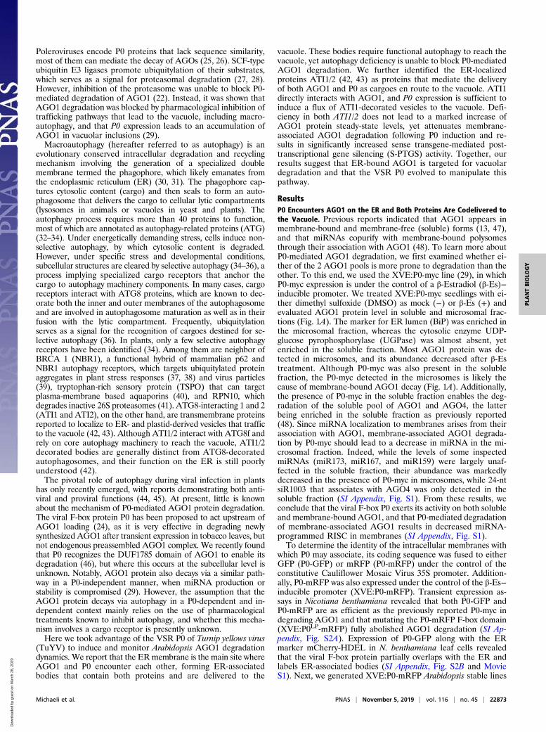
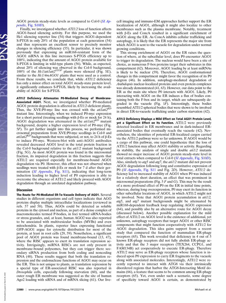
ResultsP0 Encounters AGO1 on the ER and Both Proteins Are Codelivered tothe Vacuole. Previous reports indicated that AGO1 appears inmembrane-bound and membrane-free (soluble) forms (13, 47),and that miRNAs copurify with membrane-bound polysomesthrough their association with AGO1 (48). To learn more aboutP0-mediated AGO1 degradation, we first examined whether ei-ther of the 2 AGO1 pools is more prone to degradation than theother. To this end, we used the XVE:P0-myc line (29), in whichP0-myc expression is under the control of a β-Estradiol (β-Es)−inducible promoter. We treated XVE:P0-myc seedlings with ei-ther dimethyl sulfoxide (DMSO) as mock (−) or β-Es (+) andevaluated AGO1 protein level in soluble and microsomal frac-tions (Fig. 1A). The marker for ER lumen (BiP) was enriched inthe microsomal fraction, whereas the cytosolic enzyme UDP-glucose pyrophosphorylase (UGPase) was almost absent, yetenriched in the soluble fraction. Most AGO1 protein was de-tected in microsomes, and its abundance decreased after β-Estreatment. Although P0-myc was also present in the solublefraction, the P0-myc detected in the microsomes is likely thecause of membrane-bound AGO1 decay (Fig. 1A). Additionally,the presence of P0-myc in the soluble fraction enables the deg-radation of the soluble pool of AGO1 and AGO4, the latterbeing enriched in the soluble fraction as previously reported(48). Since miRNA localization to membranes arises from theirassociation with AGO1, membrane-associated AGO1 degrada-tion by P0-myc should lead to a decrease in miRNA in the mi-crosomal fraction. Indeed, while the levels of some inspectedmiRNAs (miR173, miR167, and miR159) were largely unaf-fected in the soluble fraction, their abundance was markedlydecreased in the presence of P0-myc in microsomes, while 24-ntsiR1003 that associates with AGO4 was only detected in thesoluble fraction (SI Appendix, Fig. S1). From these results, weconclude that the viral F-box P0 exerts its activity on both solubleand membrane-bound AGO1, and that P0-mediated degradationof membrane-associated AGO1 results in decreased miRNA-programmed RISC in membranes (SI Appendix, Fig. S1).To determine the identity of the intracellular membranes with
which P0 may associate, its coding sequence was fused to eitherGFP (P0-GFP) or mRFP (P0-mRFP) under the control of theconstitutive Cauliflower Mosaic Virus 35S promoter. Addition-ally, P0-mRFP was also expressed under the control of the β-Es−inducible promoter (XVE:P0-mRFP). Transient expression as-says in Nicotiana benthamiana revealed that both P0-GFP andP0-mRFP are as efficient as the previously reported P0-myc indegrading AGO1 and that mutating the P0-mRFP F-box domain(XVE:P0LP-mRFP) fully abolished AGO1 degradation (SI Ap-pendix, Fig. S2A). Expression of P0-GFP along with the ERmarker mCherry-HDEL in N. benthamiana leaf cells revealedthat the viral F-box protein partially overlaps with the ER andlabels ER-associated bodies (SI Appendix, Fig. S2B and MovieS1). Next, we generated XVE:P0-mRFP Arabidopsis stable lines
Michaeli et al. PNAS | November 5, 2019 | vol. 116 | no. 45 | 22873
PLANTBIOLO
GY
Dow
nloa
ded
by g
uest
on
Mar
ch 2
8, 2
020
that exhibit efficient P0-mRFP induction following treatmentwith β-Es (SI Appendix, Fig. S2C). Staining root cells that expressP0-mRFP with ER-Tracker (Thermo Fisher Scientific) demon-strated again its partial association with the ER (Fig. 1B). How-ever, in this system, a broader distribution of the signal along theER was observed with the addition of bodies that appear associ-ated with the ER-Tracker signal. These observations suggest thatthe ER might be a site from which membrane-bound AGO1 istargeted for degradation.Previously, it was reported that AGO1 localizes on the ER
membrane (13, 48). We reexamined this by utilizing both fluorescence
and electron microscopy (EM) techniques. First, we transientlyexpressed 35S:GFP-AGO1 in N. benthamiana epidermal leafcells along with the ER marker mCherry-HDEL and focusedon the cytosolic focal planes. This revealed partial associationof AGO1 with the ER in 52% of the examined cells (n = 32) (SIAppendix, Fig. S3 A, Top), while, in other transformed cells, thesignal was cytosolic (SI Appendix, Fig. S3 A, Bottom). To gain insightinto the localization of the endogenous AGO1 and the impactof P0 expression on AGO1 localization at the ultrastructurallevel, we treated Col-0 and XVE:P0-mRFP seedlings with β-Esand concanamycin A (ConcA; an inhibitor of vacuolar degradation),
Fig. 1. P0 and AGO1 colocalize in ER-associated bodies that are destined to the vacuole. (A) Immunoblot analysis of total, pellet (Microsomal), and su-pernatant (Soluble) protein preparations from XVE:P0-myc 7-d-old seedlings grown on Murashige and Skoog (MS) plates with either DMSO (−) or 10 μM β-Es(+). Blots were probed with antibodies raised against Arabidopsis AGO1, AGO4, the c-MYC tag, Luminal binding proteins BiP1/BiP2/BiP3 (BiP), and UGPase.Coomassie blue (CB) staining serves as loading control. (B) CLSM imaging of Arabidopsis root meristematic cells expressing P0-mRFP (red signal) and stainedwith ER-Tracker blue-white DPX (ThermoFisher; green signal). Both the merged image of the signals and an enlarged image of one of the cells are presented.Positions of the nucleus (N) and P0-mRFP labeled bodies (arrowheads) are indicated. (C) (Top andMiddle) TEM imaging of ER (denoted by a dashed red line) in aroot cell that underwent immunogold labeling using @AGO1 antibodies. Arrowheads mark the sites of gold particles in Top. Both Col-0 and the XVE:P0-mRFPline were treated with β-Es and ConcA. (Scale bars: 200 nm.) (Bottom) A graph showing quantification of gold particles associated with the ER, tonoplast (vac.),endosomes (end.), plasma membrane (PM), and plastid + mitochondria (pl./mit.) outer membranes. Quantification was done in samples from β-Es and ConcAtreated (+B+C), or untreated, Col-0 plants. Values represent mean ± SEM. Asterisk (*) denotes statistical significance of gold particle numbers along the ERcompared to the other membranes within the +B+C treated plants, P < 0.05 (Mann–Whitney U test). (D) (Top) CLSM imaging of a tobacco leaf epidermal celltransiently expressing GFP-AGO1 (AGO1), CFP-HDEL (ER), and P0-mRFP (P0). (Bottom) Enlargements of the areas bordered by yellow rectangles in Top. Bodiesexhibiting both P0 and AGO1 signals are highlighted with arrowheads. (E) Immunoblot analysis of total proteins from XVE:P0-mRFP seedlings treated with β-Esfor 0, 8, 24, and 48 h to induce P0-mRFP expression. Blots were probed with specific antibodies as indicated, and CB staining serves as loading control. (F) CLSMimaging of the vacuolar focal plane of root elongation zone cells from an Arabidopsis line harboring pAGO1:GFP-AGO1 and XVE:P0-mRFP and treated with β-Esand ConcA or mock (DMSO) and ConcA. Vacuole lumen is indicated (Vac).
22874 | www.pnas.org/cgi/doi/10.1073/pnas.1912222116 Michaeli et al.
Dow
nloa
ded
by g
uest
on
Mar
ch 2
8, 2
020
and analyzed root cells by immuno-EM (IEM) with the anti-AGO1antibody. We reasoned that using Col-0 plants that were subjectedto the same chemicals as XVE:P0-mRFP plants would allow theassessment of the effect of P0 per se, excluding the possible in-fluence of the drugs. In Col-0 seedlings, the compartment showingthe highest AGO1 labeling density on its limiting membrane wasthe ER (Fig. 1 C, Top and Bottom). In P0-mRFP seedlings, AGO1labeling was detected in few cases on electron-dense structuresadjacent to the ER membrane (Fig. 1 C, Middle and SI Appendix,Fig. S4). Such structures were not detected in the Col-0 samples.This suggests that P0 expression may result in the aggregation ofAGO1 in ER-associated structures. Quantification revealed thatmost of the gold particles were present along the ER compared toall other membranes in both treated and untreated Col-0. How-ever, this enrichment was only statistically significant in thechemically treated seedlings (Mann−Whitney U test, P < 0.05)(Fig. 1 C, Bottom). This might be due to the inhibitory impactof ConcA on vacuolar degradation, resulting in more proteinlingering on the ER membrane. Hence, although AGO1 mayassociate with several membranes and appear also in a solubleform, our results corroborate previous reports pointing towardthe ER as a major site for membrane-bound AGO1 (13, 48). Notably,P0 expression affects the morphology of ER-associated AGO1. Insupport of this, coexpression of GFP-AGO1 and P0-mRFP in N.benthamiana leaves revealed that both proteins exhibited an ER-like expression pattern and colocalized in bodies (SI Appendix, Fig.S3B). To see whether these structures are indeed associated withthe ER, we expressed GFP-AGO1 and P0-mRFP along with anER marker, CFP-HDEL (Fig. 1D). Structures containing P0-mRFP were indeed closely associated with the ER, and somewere also enriched with the GFP-AGO1 signal (Fig. 1D, whitearrowheads). Moreover, time-lapse imaging revealed the motilenature of the P0 and AGO1 labeled bodies (Movie S2). Note thatthe colocalization of GFP-AGO1 and P0-mRFP was only detectedat early time points of their expression, that is, 24 h to 40 h fol-lowing agroinfiltration (hfa). At later time points, that is, 48 hfa to96 hfa, AGO1-labeled foci were no longer detected, probably dueto AGO1 degradation. Moreover, in these transient expression assays,GFP-AGO1 and P0-mRFP were also occasionally detected inthe nuclei (SI Appendix, Fig. S3C).Next, we took advantage of the relative stability of mRFP
to examine whether P0-mRFP is delivered to vacuoles. Totalprotein extraction from XVE:P0-mRFP seedlings at 0, 8, 24,and 48 h following P0-mRFP induction revealed that AGO1protein level decreases as the free-mRFP vacuolar degradationproduct of P0-mRFP accumulates (Fig. 1E), suggesting that P0-mRFP might be delivered to the vacuole along with its tar-get AGO1. To address this, the pAGO1:GFP-AGO1/ago1-27transgenic line (29) was transformed with XVE:P0-mRFP, andlines harboring both constructs were analyzed. Imaging of rootcells in the elongation zone following β-Es and ConcA treatment,compared to ConcA-only treated plants, revealed accumulationof P0-mRFP− and GFP-AGO1−containing bodies within thevacuoles (Fig. 1F). Time-lapse imaging demonstrated randommotion of the bodies, which is typical for vacuole-lumen-residingautophagic bodies (Movie S3). Together, these observations pointtoward the ER as a major site from which P0 induces AGO1degradation and from which it is delivered to the vacuole alongwith AGO1.
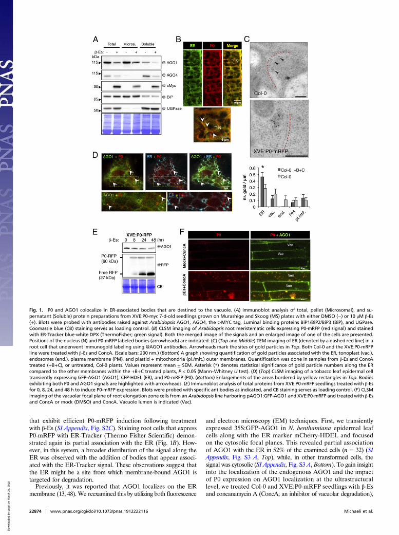
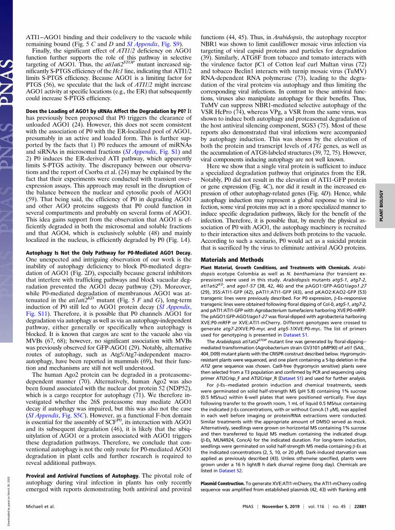
Autophagy Deficiency Impairs P0 Delivery to the Vacuole, yet IsUnable to Block AGO1 Degradation. Next, we examined whetherP0 associates with autophagy components and whether it relieson autophagy for its function. We previously showed thatGFP-AGO1 colocalizes with an autophagosome marker, mRFP-ATG8a, especially following P0 expression or treatment with themembrane-permeable cysteine protease inhibitor E64d in N.benthamiana leaves (29). To determine whether P0 also colocalizes
with autophagosomes, we coexpressed P0-GFP and mRFP-ATG8a in N. benthamiana leaves and imaged them at 40 hfa. Asexpected, the mRFP-ATG8a protein was detected in the cytosol andthe nucleus, as well as in small bodies representing autophagosomes(Fig. 2A). Notably, P0-GFP predominantly colocalized withmRFP-ATG8a in these bodies. To determine whether the deg-radation of P0 bodies is regulated by autophagy, we introducedXVE:P0-mRFP or XVE:P0-myc into atg5-1 and atg7-2, in whichautophagy is inactive (49). First, we utilized XVE:P0-mRFP/atg5-1 and XVE:P0-mRFP/atg7-2 lines and examined theirvacuoles following 18- to 24-h P0-mRFP induction (β-Es) andtreatment with ConcA. While P0-mRFP bodies accumulatedin the vacuoles of control plants (XVE:P0-mRFP; on averageexhibited 59 bodies per 1,000 μm2 of vacuole lumen focal plane),there were almost no bodies detected in XVE:P0-mRFP/atg5-1(1 body per 1,000 μm2) and significantly less P0-mRFP bodies(3.6 bodies per 1,000 μm2) within the vacuoles of XVE:P0-mRFP/atg7-2 (Fig. 2 B and C). Moreover, in cells of the meristematiczone, both autophagy mutants displayed cytosolic structures ofabout 4 μm in diameter that contain P0-mRFP and that wereabsent from wild-type cells (Fig. 2B). These structures weremotile (Movie S4) and may represent aggregated P0-mRFP thataccumulates due to degradation blockage.Given this observation and the fact that the application of
vacuolar-degradation inhibitors such as E64d is sufficient tocounteract the degradative effect of P0-myc on AGO1 (29), wespeculated that genetic disruption of autophagy might achievesimilar results. To test this, we utilized our XVE:P0-myc/atg5-1and XVE:P0-myc/atg7-2 lines and expressed P0-myc in 2 ways: 1)by germinating the plants on β-Es containing media (constitutiveP0 expression) compared to mock (DMSO containing media) or2) by treating seedlings with β-Es or mock 5 d after their ger-mination (induced P0 expression). Strikingly, following P0 ex-pression in both assays (Fig. 2D and SI Appendix, Fig. S5 A andB), none of the autophagy mutant was able to block either AGO1or AGO2 degradation. On the contrary, the AGO1 transcript levelwas significantly increased, ruling out the possibility that the de-cline of protein level is due to reduced transcript level (Fig. 2D).Note that AGO1, but not AGO2, protein level remained stable inthe ago1-57 allele background, used here as a control. This line isinsensitive to P0, as this mutation abrogates the SCF-dependentP0 interaction with AGO1 (46).We next wondered whether the 26S proteasome could take over
P0-mediated AGO1 protein decay if autophagy is compromised.However, when applying Bortezomib, which binds and blocks withhigh affinity and specificity the 26S proteasome (50), AGO1 wasstill degraded by P0 in atg5-1 mutants, while the accumulation ofthe proteasome-sensitive transcription factor EIN3 (51) demon-strated the efficiency of the chemical (SI Appendix, Fig. S5 C,Top). Note that AGO1 transcript level was increased following P0induction, ruling out, again, any negative effect on transcription toexplain AGO1 decay (SI Appendix, Fig. S5 C, Bottom). From theseexperiments, we conclude that P0 can bypass autophagy deficiencyto achieve a reduction in AGO1 protein level even when its de-livery to the vacuole appears to rely on canonical autophagy.
P0 Is Engulfed by ATI1- and ATI2-Decorated Bodies on the ER and IsDelivered with Them to the Vacuole. To get insight into the P0 ER-to-vacuole delivery pathway, we decided to examine the possibleinvolvement of the ER and autophagy-associated ATI1/2. Wewere particularly intrigued by the morphological resemblance (asseen in bright-field imaging) of the P0- and AGO1-containingbodies in the vacuole (SI Appendix, Fig. S6A) and cytosol (SIAppendix, Fig. S6B), with the ER-associated and ATI1-decoratedbodies (ATI-bodies) reported earlier (42). ATI1/2 were shown tointeract with ATG8 proteins and to be delivered on dynamic ER-derived ATI-bodies to the vacuole (42). To examine the possibleinvolvement of ATI1/2 in the process of P0-mediated AGO1
Michaeli et al. PNAS | November 5, 2019 | vol. 116 | no. 45 | 22875
PLANTBIOLO
GY
Dow
nloa
ded
by g
uest
on
Mar
ch 2
8, 2
020
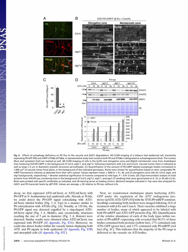
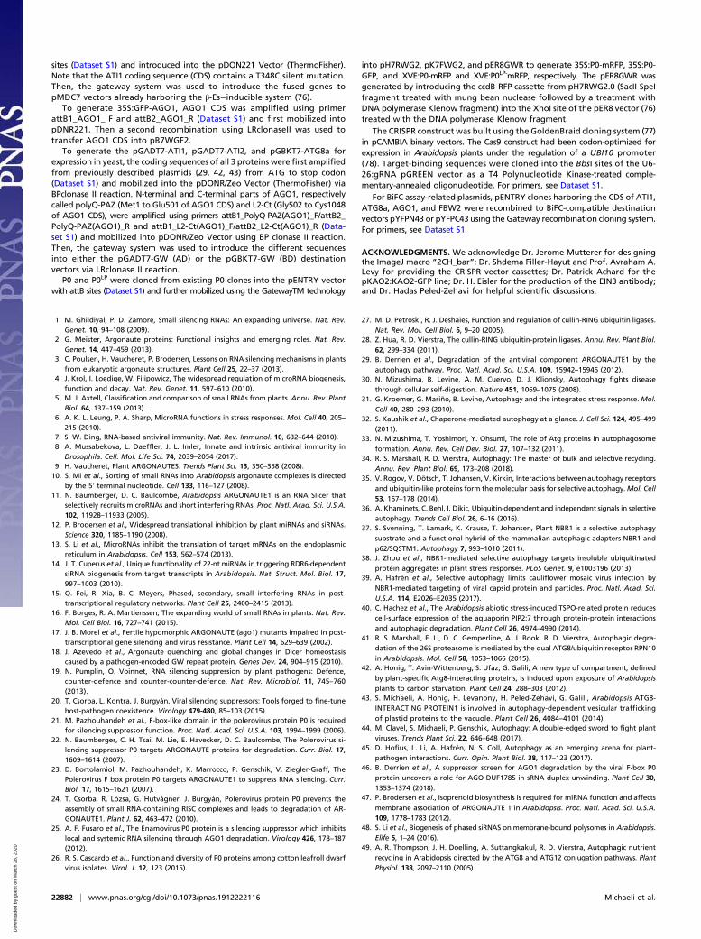
decay, we first expressed ATI1-mCherry or ATI2-mCherry withP0-GFP in N. benthamiana leaf epidermal cells. Already at 30 hfa,we could detect the P0-GFP signal colocalizing with ATI1-mCherry labeled bodies (Fig. 3 A, Top) in a manner similar toP0 colocalization with ATG8a (Fig. 2A). Notably, at 120 hfa, theP0-GFP signal was detected engulfed by a ring-shaped ATI1-mCherry signal (Fig. 3 A, Middle), and, occasionally, structuresreaching the size of 5 μm in diameter (Fig. 3 A, Bottom) weredetected. Similar results were obtained when ATI2-mCherry wasexpressed with P0-GFP (SI Appendix, Fig. S7A). Moreover, wecould also detect bodies within the vacuole lumen displaying bothATI1 and P0 signals in both epidermal (SI Appendix, Fig. S7B)and mesophyll cells (SI Appendix, Fig. S7C).
Next, we transformed Arabidopsis plants harboring ATI1-GFP under the regulation of the ATI1 endogenous pro-moter [pATI1:ATI1-GFP (43)] with the XVE:P0-mRFP construct.Seedlings containing both markers were imaged following 18 h oftreatment with β-Es and ConcA. Their vacuoles exhibited a largenumber of bodies, many of which appeared to be labeled withboth P0-mRFP and ATI1-GFP proteins (Fig. 3B). Quantificationof the relative abundance of each of the body types within vac-uoles of P0-mRFP−expressing cells revealed that 80.5% of thesebodies contained both proteins (yellow bar), 17% contained onlyATI1-GFP (green bar), and 2.5% contained only P0-mRFP (redbar) (Fig. 3C). This indicates that the majority of the P0 cargo isdelivered to the vacuole via ATI-bodies.
Fig. 2. Effects of autophagy deficiency on P0 flux to the vacuole and AGO1 degradation. (A) CLSM imaging of a tobacco leaf epidermal cell, transientlyexpressing P0-GFP (P0) and mRFP-ATG8a (ATG8a). A representative body that contains both P0 and ATG8a is designated as autophagosome (Aut). The nucleus(Nuc) and cytoplasm (Cyt) are marked as well. (B) CLSM imaging of cells in the (Left) root elongation zone and (Right) meristematic zone from Arabidopsislines harboring XVE:P0-mRFP in the background of Col-0, atg5-1, and atg7-2, following treatment with β-Es and ConcA. Vacuole lumen (Vac) is indicated aswell as large (∼5 μm in diameter) cytosolic structures (arrowheads). (C) Quantification of the amount of P0-mRFP labeled autophagic bodies normalized to1,000 μm2 of vacuole lumen focal plane, in the background of the indicated genotypes. Plants were chosen for quantification based on their comparable P0-mRFP fluorescence intensity as detected from their cell’s cytosol. Values represent mean ± SEM (n = 21, 30, and 23 elongation zone cells for Col-0, atg5, andatg7 backgrounds, respectively); * denotes statistical significance of mutants compared to wild type, P < 0.01 (t test). (D) (Top) Immunoblot analysis of totalproteins from XVE:P0-myc containing lines in the background of Col-0, atg7-2, atg5-1, and ago1.57 seedlings that were germinated on 0, 10, or 20 μM of β-Es.Blots were probed with specific antibodies as indicated, and CB staining serves as loading control. (Bottom) Samples indicated in Top were also analyzed forAGO1 and P0 transcript levels by qRT-PCR. Values are average ± SD relative to P0-myc without β-Es.
22876 | www.pnas.org/cgi/doi/10.1073/pnas.1912222116 Michaeli et al.
Dow
nloa
ded
by g
uest
on
Mar
ch 2
8, 2
020
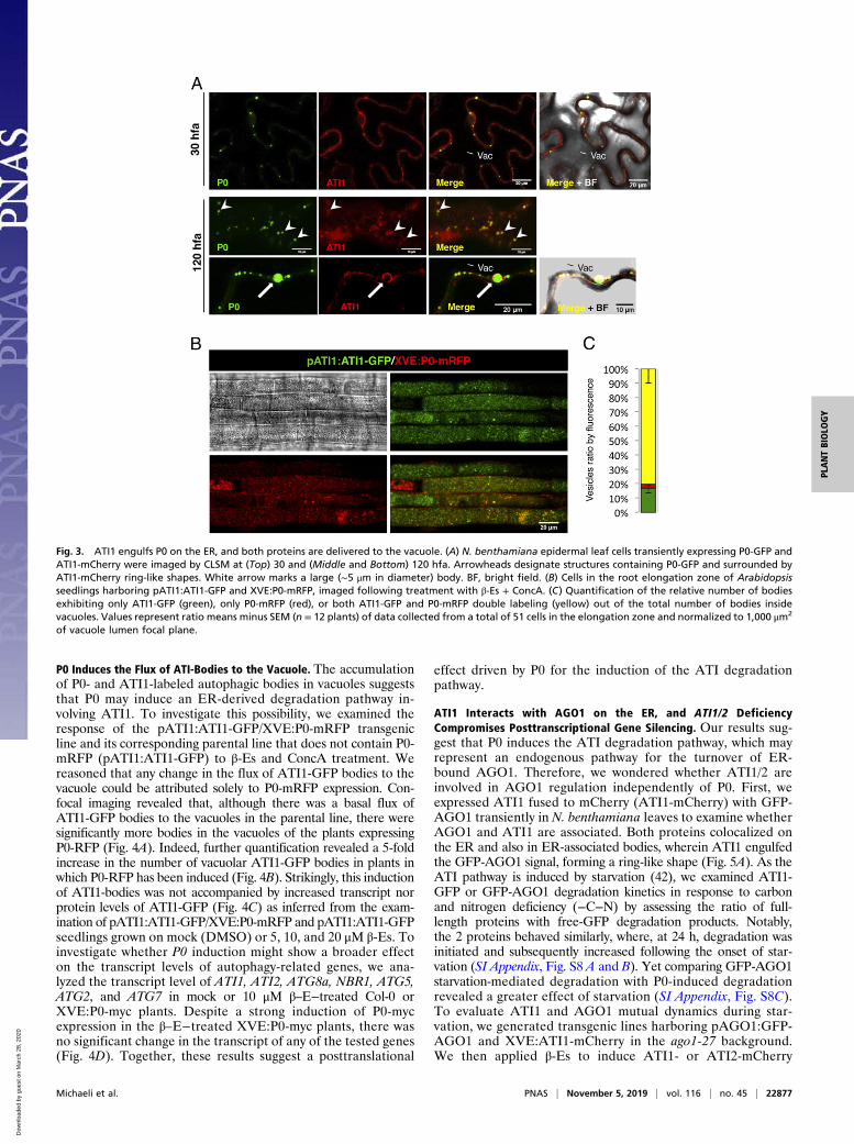
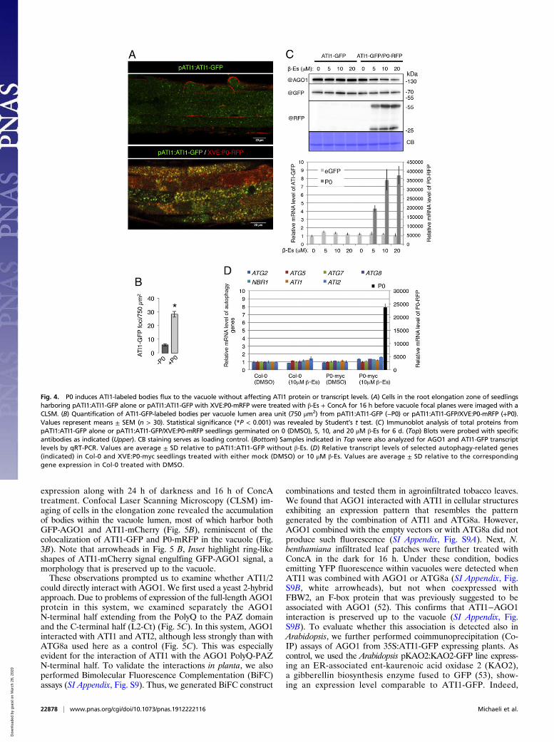
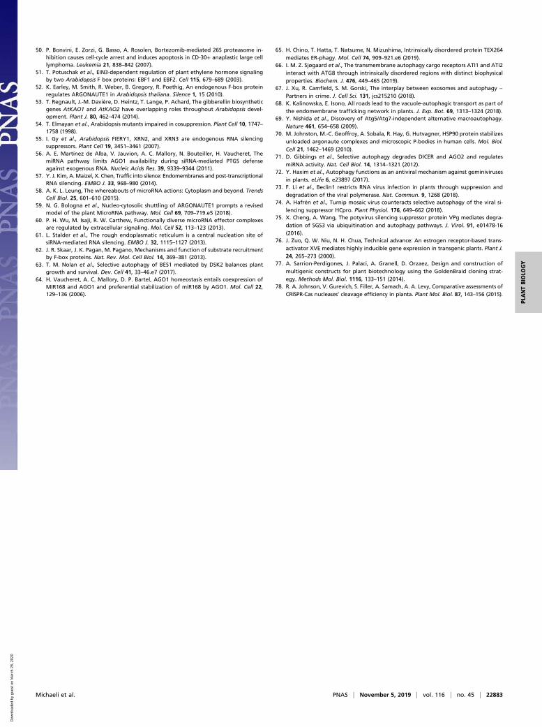
P0 Induces the Flux of ATI-Bodies to the Vacuole. The accumulationof P0- and ATI1-labeled autophagic bodies in vacuoles suggeststhat P0 may induce an ER-derived degradation pathway in-volving ATI1. To investigate this possibility, we examined theresponse of the pATI1:ATI1-GFP/XVE:P0-mRFP transgenicline and its corresponding parental line that does not contain P0-mRFP (pATI1:ATI1-GFP) to β-Es and ConcA treatment. Wereasoned that any change in the flux of ATI1-GFP bodies to thevacuole could be attributed solely to P0-mRFP expression. Con-focal imaging revealed that, although there was a basal flux ofATI1-GFP bodies to the vacuoles in the parental line, there weresignificantly more bodies in the vacuoles of the plants expressingP0-RFP (Fig. 4A). Indeed, further quantification revealed a 5-foldincrease in the number of vacuolar ATI1-GFP bodies in plants inwhich P0-RFP has been induced (Fig. 4B). Strikingly, this inductionof ATI1-bodies was not accompanied by increased transcript norprotein levels of ATI1-GFP (Fig. 4C) as inferred from the exam-ination of pATI1:ATI1-GFP/XVE:P0-mRFP and pATI1:ATI1-GFPseedlings grown on mock (DMSO) or 5, 10, and 20 μM β-Es. Toinvestigate whether P0 induction might show a broader effecton the transcript levels of autophagy-related genes, we ana-lyzed the transcript level of ATI1, ATI2, ATG8a, NBR1, ATG5,ATG2, and ATG7 in mock or 10 μM β–E−treated Col-0 orXVE:P0-myc plants. Despite a strong induction of P0-mycexpression in the β–E−treated XVE:P0-myc plants, there wasno significant change in the transcript of any of the tested genes(Fig. 4D). Together, these results suggest a posttranslational
effect driven by P0 for the induction of the ATI degradationpathway.
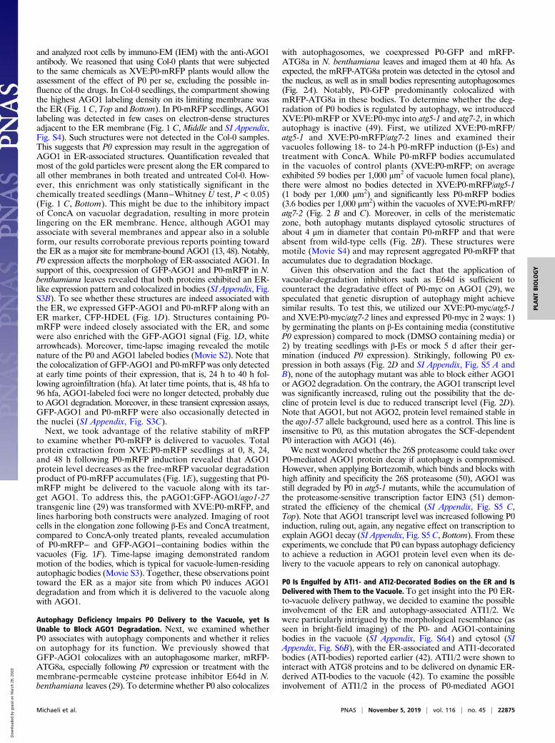
ATI1 Interacts with AGO1 on the ER, and ATI1/2 DeficiencyCompromises Posttranscriptional Gene Silencing. Our results sug-gest that P0 induces the ATI degradation pathway, which mayrepresent an endogenous pathway for the turnover of ER-bound AGO1. Therefore, we wondered whether ATI1/2 areinvolved in AGO1 regulation independently of P0. First, weexpressed ATI1 fused to mCherry (ATI1-mCherry) with GFP-AGO1 transiently in N. benthamiana leaves to examine whetherAGO1 and ATI1 are associated. Both proteins colocalized onthe ER and also in ER-associated bodies, wherein ATI1 engulfedthe GFP-AGO1 signal, forming a ring-like shape (Fig. 5A). As theATI pathway is induced by starvation (42), we examined ATI1-GFP or GFP-AGO1 degradation kinetics in response to carbonand nitrogen deficiency (−C−N) by assessing the ratio of full-length proteins with free-GFP degradation products. Notably,the 2 proteins behaved similarly, where, at 24 h, degradation wasinitiated and subsequently increased following the onset of star-vation (SI Appendix, Fig. S8 A and B). Yet comparing GFP-AGO1starvation-mediated degradation with P0-induced degradationrevealed a greater effect of starvation (SI Appendix, Fig. S8C).To evaluate ATI1 and AGO1 mutual dynamics during star-vation, we generated transgenic lines harboring pAGO1:GFP-AGO1 and XVE:ATI1-mCherry in the ago1-27 background.We then applied β-Es to induce ATI1- or ATI2-mCherry
Fig. 3. ATI1 engulfs P0 on the ER, and both proteins are delivered to the vacuole. (A) N. benthamiana epidermal leaf cells transiently expressing P0-GFP andATI1-mCherry were imaged by CLSM at (Top) 30 and (Middle and Bottom) 120 hfa. Arrowheads designate structures containing P0-GFP and surrounded byATI1-mCherry ring-like shapes. White arrow marks a large (∼5 μm in diameter) body. BF, bright field. (B) Cells in the root elongation zone of Arabidopsisseedlings harboring pATI1:ATI1-GFP and XVE:P0-mRFP, imaged following treatment with β-Es + ConcA. (C) Quantification of the relative number of bodiesexhibiting only ATI1-GFP (green), only P0-mRFP (red), or both ATI1-GFP and P0-mRFP double labeling (yellow) out of the total number of bodies insidevacuoles. Values represent ratio means minus SEM (n = 12 plants) of data collected from a total of 51 cells in the elongation zone and normalized to 1,000 μm2
of vacuole lumen focal plane.
Michaeli et al. PNAS | November 5, 2019 | vol. 116 | no. 45 | 22877
PLANTBIOLO
GY
Dow
nloa
ded
by g
uest
on
Mar
ch 2
8, 2
020
expression along with 24 h of darkness and 16 h of ConcAtreatment. Confocal Laser Scanning Microscopy (CLSM) im-aging of cells in the elongation zone revealed the accumulationof bodies within the vacuole lumen, most of which harbor bothGFP-AGO1 and ATI1-mCherry (Fig. 5B), reminiscent of thecolocalization of ATI1-GFP and P0-mRFP in the vacuole (Fig.3B). Note that arrowheads in Fig. 5 B, Inset highlight ring-likeshapes of ATI1-mCherry signal engulfing GFP-AGO1 signal, amorphology that is preserved up to the vacuole.These observations prompted us to examine whether ATI1/2
could directly interact with AGO1. We first used a yeast 2-hybridapproach. Due to problems of expression of the full-length AGO1protein in this system, we examined separately the AGO1N-terminal half extending from the PolyQ to the PAZ domainand the C-terminal half (L2-Ct) (Fig. 5C). In this system, AGO1interacted with ATI1 and ATI2, although less strongly than withATG8a used here as a control (Fig. 5C). This was especiallyevident for the interaction of ATI1 with the AGO1 PolyQ-PAZN-terminal half. To validate the interactions in planta, we alsoperformed Bimolecular Fluorescence Complementation (BiFC)assays (SI Appendix, Fig. S9). Thus, we generated BiFC construct
combinations and tested them in agroinfiltrated tobacco leaves.We found that AGO1 interacted with ATI1 in cellular structuresexhibiting an expression pattern that resembles the patterngenerated by the combination of ATI1 and ATG8a. However,AGO1 combined with the empty vectors or with ATG8a did notproduce such fluorescence (SI Appendix, Fig. S9A). Next, N.benthamiana infiltrated leaf patches were further treated withConcA in the dark for 16 h. Under these condition, bodiesemitting YFP fluorescence within vacuoles were detected whenATI1 was combined with AGO1 or ATG8a (SI Appendix, Fig.S9B, white arrowheads), but not when coexpressed withFBW2, an F-box protein that was previously suggested to beassociated with AGO1 (52). This confirms that ATI1−AGO1interaction is preserved up to the vacuole (SI Appendix, Fig.S9B). To evaluate whether this association is detected also inArabidopsis, we further performed coimmunoprecipitation (Co-IP) assays of AGO1 from 35S:ATI1-GFP expressing plants. Ascontrol, we used the Arabidopsis pKAO2:KAO2-GFP line express-ing an ER-associated ent-kaurenoic acid oxidase 2 (KAO2),a gibberellin biosynthesis enzyme fused to GFP (53), show-ing an expression level comparable to ATI1-GFP. Indeed,
Fig. 4. P0 induces ATI1-labeled bodies flux to the vacuole without affecting ATI1 protein or transcript levels. (A) Cells in the root elongation zone of seedlingsharboring pATI1:ATI1-GFP alone or pATI1:ATI1-GFP with XVE:P0-mRFP were treated with β-Es + ConcA for 16 h before vacuole focal planes were imaged with aCLSM. (B) Quantification of ATI1-GFP-labeled bodies per vacuole lumen area unit (750 μm2) from pATI1:ATI1-GFP (−P0) or pATI1:ATI1-GFP/XVE:P0-mRFP (+P0).Values represent means ± SEM (n > 30). Statistical significance (*P < 0.001) was revealed by Student’s t test. (C) Immunoblot analysis of total proteins frompATI1:ATI1-GFP alone or pATI1:ATI1-GFP/XVE:P0-mRFP seedlings germinated on 0 (DMSO), 5, 10, and 20 μM β-Es for 6 d. (Top) Blots were probed with specificantibodies as indicated (Upper). CB staining serves as loading control. (Bottom) Samples indicated in Top were also analyzed for AGO1 and ATI1-GFP transcriptlevels by qRT-PCR. Values are average ± SD relative to pATI1:ATI1-GFP without β-Es. (D) Relative transcript levels of selected autophagy-related genes(indicated) in Col-0 and XVE:P0-myc seedlings treated with either mock (DMSO) or 10 μM β-Es. Values are average ± SD relative to the correspondinggene expression in Col-0 treated with DMSO.
22878 | www.pnas.org/cgi/doi/10.1073/pnas.1912222116 Michaeli et al.
Dow
nloa
ded
by g
uest
on
Mar
ch 2
8, 2
020
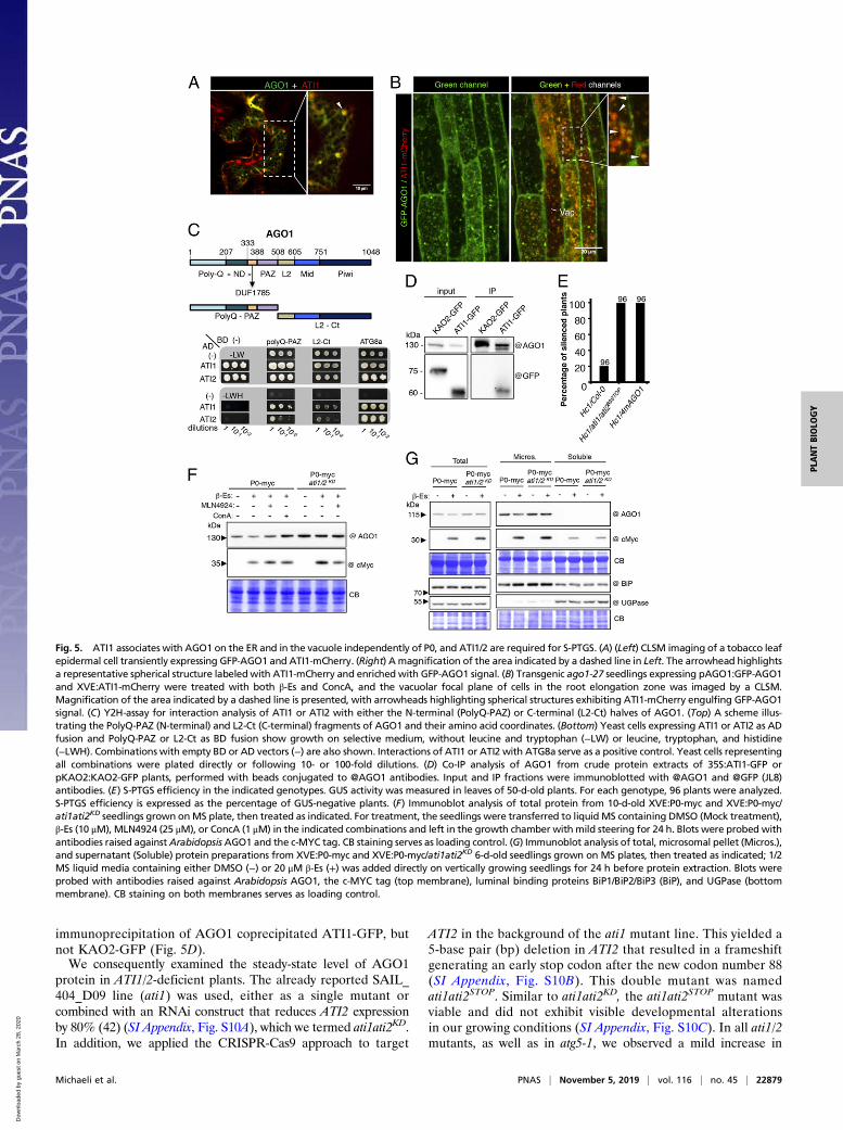
immunoprecipitation of AGO1 coprecipitated ATI1-GFP, butnot KAO2-GFP (Fig. 5D).We consequently examined the steady-state level of AGO1
protein in ATI1/2-deficient plants. The already reported SAIL_404_D09 line (ati1) was used, either as a single mutant orcombined with an RNAi construct that reduces ATI2 expressionby 80% (42) (SI Appendix, Fig. S10A), which we termed ati1ati2KD.In addition, we applied the CRISPR-Cas9 approach to target
ATI2 in the background of the ati1 mutant line. This yielded a5-base pair (bp) deletion in ATI2 that resulted in a frameshiftgenerating an early stop codon after the new codon number 88(SI Appendix, Fig. S10B). This double mutant was namedati1ati2STOP. Similar to ati1ati2KD, the ati1ati2STOP mutant wasviable and did not exhibit visible developmental alterationsin our growing conditions (SI Appendix, Fig. S10C). In all ati1/2mutants, as well as in atg5-1, we observed a mild increase in
Fig. 5. ATI1 associates with AGO1 on the ER and in the vacuole independently of P0, and ATI1/2 are required for S-PTGS. (A) (Left) CLSM imaging of a tobacco leafepidermal cell transiently expressing GFP-AGO1 and ATI1-mCherry. (Right) A magnification of the area indicated by a dashed line in Left. The arrowhead highlightsa representative spherical structure labeledwith ATI1-mCherry and enriched with GFP-AGO1 signal. (B) Transgenic ago1-27 seedlings expressing pAGO1:GFP-AGO1and XVE:ATI1-mCherry were treated with both β-Es and ConcA, and the vacuolar focal plane of cells in the root elongation zone was imaged by a CLSM.Magnification of the area indicated by a dashed line is presented, with arrowheads highlighting spherical structures exhibiting ATI1-mCherry engulfing GFP-AGO1signal. (C) Y2H-assay for interaction analysis of ATI1 or ATI2 with either the N-terminal (PolyQ-PAZ) or C-terminal (L2-Ct) halves of AGO1. (Top) A scheme illus-trating the PolyQ-PAZ (N-terminal) and L2-Ct (C-terminal) fragments of AGO1 and their amino acid coordinates. (Bottom) Yeast cells expressing ATI1 or ATI2 as ADfusion and PolyQ-PAZ or L2-Ct as BD fusion show growth on selective medium, without leucine and tryptophan (−LW) or leucine, tryptophan, and histidine(−LWH). Combinations with empty BD or AD vectors (−) are also shown. Interactions of ATI1 or ATI2 with ATG8a serve as a positive control. Yeast cells representingall combinations were plated directly or following 10- or 100-fold dilutions. (D) Co-IP analysis of AGO1 from crude protein extracts of 35S:ATI1-GFP orpKAO2:KAO2-GFP plants, performed with beads conjugated to @AGO1 antibodies. Input and IP fractions were immunoblotted with @AGO1 and @GFP (JL8)antibodies. (E) S-PTGS efficiency in the indicated genotypes. GUS activity was measured in leaves of 50-d-old plants. For each genotype, 96 plants were analyzed.S-PTGS efficiency is expressed as the percentage of GUS-negative plants. (F) Immunoblot analysis of total protein from 10-d-old XVE:P0-myc and XVE:P0-myc/ati1ati2KD seedlings grown onMS plate, then treated as indicated. For treatment, the seedlings were transferred to liquid MS containing DMSO (Mock treatment),β-Es (10 μM), MLN4924 (25 μM), or ConcA (1 μM) in the indicated combinations and left in the growth chamber with mild steering for 24 h. Blots were probed withantibodies raised againstArabidopsis AGO1 and the c-MYC tag. CB staining serves as loading control. (G) Immunoblot analysis of total, microsomal pellet (Micros.),and supernatant (Soluble) protein preparations from XVE:P0-myc and XVE:P0-myc/ati1ati2KD 6-d-old seedlings grown on MS plates, then treated as indicated; 1/2MS liquid media containing either DMSO (−) or 20 μM β-Es (+) was added directly on vertically growing seedlings for 24 h before protein extraction. Blots wereprobed with antibodies raised against Arabidopsis AGO1, the c-MYC tag (top membrane), luminal binding proteins BiP1/BiP2/BiP3 (BiP), and UGPase (bottommembrane). CB staining on both membranes serves as loading control.
Michaeli et al. PNAS | November 5, 2019 | vol. 116 | no. 45 | 22879
PLANTBIOLO
GY
Dow
nloa
ded
by g
uest
on
Mar
ch 2
8, 2
020
AGO1 protein steady-state levels as compared to Col-0 (SI Ap-pendix, Fig. S10D).Finally, we investigated whether ATI1/2 loss of function affects
AGO1-based silencing activity. For this purpose, we used theHc1 silencing reporter line (54) that triggers AGO1-dependentS-PTGS in only 20% of the population at each generation (54)and thus represents an excellent sensor to precisely monitorchanges in silencing efficiency (55). In particular, it was shownpreviously that expressing an miR168-resistant form of theAGO1 mRNA in this line increases S-PTGS efficiency up to100%, indicating that the amount of AGO1 protein available forS-PTGS is limiting in wild-type plants (56). While, as expected,about 20% of silencing was observed in the Col-0 background,100% of the Hc1/ati1ati2STOP plants were silenced (Fig. 5E),similar to the Hc1/4mAGO1 plants that were used as a control.From these results, we conclude that, while ATI1/2 deficiencyhas only a minor effect on total AGO1 steady-state protein level,it significantly enhances S-PTGS, likely by increasing the avail-ability of AGO1 for S-PTGS.
ATI1/2 Deficiency Attenuates P0-Mediated Decay of Membrane-Associated AGO1. Next, we investigated whether P0-mediatedAGO1 protein degradation is affected in ATI1/2-deficient plants.Thus, the XVE:P0-myc line was crossed with the ati1ati2KD
mutant line. Interestingly, when we induced P0-myc expressionfor a short period (treating seedlings with β-Es or mock for 24 h),AGO1 degradation was attenuated in the ati1ati2KD mutantbackground, despite a higher expression level of P0-myc (Fig.5F). To get further insight into this process, we performed mi-crosomal preparations from XVE:P0-myc seedlings in Col-0 andati1ati2KD backgrounds that were subjected, or not, to 24 h of β-Esto induce P0-myc. Western-blot with anti-AGO1 antibodiesrevealed decreased AGO1 level in the total protein fraction inthe Col-0 background relative to the ati1/2 mutant background(Fig. 5G). As most AGO1 protein was found in the microsomalfraction, its impaired degradation in this fraction suggests thatATI1/2 are required especially for membrane-bound AGO1degradation via P0. However, this effect was not observed whenseedlings were treated with β-Es or mock for 7 d after their ger-mination (SI Appendix, Fig. S11), indicating that long-terminduction leading to higher level of P0 expression is able toovercome the absence of ATI1/2 and likely proceed with AGO1degradation through an unrelated degradation pathway.
DiscussionThe Route for P0-Mediated ER-To-Vacuole Delivery of AGO1. Severalstudies in different organisms and cell types indicate that AGOproteins display multiple intracellular localizations (reviewed inrefs. 57 and 58). Thus, AGOs could be detected as solubleproteins in the cytosol and nucleus, as part of a dense complex ofmacromolecules termed P-bodies, in foci termed siRNA-bodiesor stress granules, and, at least, human AGO2 was also reportedto be associated with multivesicular bodies (MVBs) and evenmitochondria. In plants, reporter lines expressing functionalGFP-AGO1 argue for cytosolic distribution for most of theprotein, at least in root cells (29, 59). Nonetheless, a significantpool of AGO1 protein was found membrane-bound (13, 47),where the RISC appears to exert its translation repression ac-tivity. Intriguingly, miRNA RISCs are not only present inmembrane-bound polysomes, but they can trigger there sec-ondary siRNA production from both coding and noncodingRNA (48). These results suggest that both the translation re-pression and the endonuclease functions of AGO1 may occur onthe ER. This is not unique to plants, as translation repression bya special type of ER-associated RISC was also reported inDrosophila cells, especially following starvation (60), and theouter rough ER membrane was suggested as the site of humanAgo2 loading with siRNA and of mRNA slicing (61). Our live-
cell imaging and immuno-EM approaches further support the ERlocalization of AGO1, although it might also localize to othermembranes such as the plasma membrane. Notably, treatmentwith β-Es and ConcA resulted in a significant enrichment ofAGO1 along the ER. As ConcA inhibits cellular trafficking andautophagy, it is likely that the ER represents the major site fromwhich AGO1 is sent to the vacuole for degradation under normalgrowing conditions.This strong enrichment of AGO1 on the ER raises the ques-
tion of where, at the subcellular level, does P0 encounter AGO1to trigger its degradation. The nucleus would have been a site ofchoice, as numerous F-box proteins target their substrates in thiscompartment (62). Moreover, AGO1 loading by at least miRNAsis likely to be nuclear (59). Therefore, AGO1 conformationalchanges in this compartment might favor the recognition of its P0degron (46). In addition, autophagy-mediated degradation ofArabidopsis nuclear-localized proteins and even protein complexeswas already demonstrated (41, 63). However, our data point to theER as the main site where P0 interacts with AGO1. Likely, P0interacting with AGO1 on the ER induces a flux of bodies con-taining both the F-box and its target, which are subsequently de-graded in the vacuole (Fig. 1F). Interestingly, these bodiesresembled ATI1/2 spherical bodies that were shown to be involvedin direct ER-to-vacuole trafficking under carbon starvation (42).
ATI1/2 Deficiency Displays a Mild Effect on Total AGO1 Protein Level,yet a Significant Effect on Its Function. ATI1/2 were previouslydetected localized in ER membranes and in dark-induced ER-associated bodies that eventually reach the vacuole (42). Nev-ertheless, the identities of potential ER-localized cargos carriedvia the ATI1/2 pathway were so far unknown. If AGO1 would bea cargo of this pathway, one could hypothesize that the loss ofATI1/2 function may affect AGO1 stability or activity. Regardingits stability, the analysis of single and double ati1/2 mutantsshowed no major increase of AGO1 protein steady-state level intotal extracts when compared to Col-0 (SI Appendix, Fig. S10D).Also, similarly to atg5 and atg7, the ati1/2 mutant did not preventAGO1 degradation following long and constitutive expression ofP0 (SI Appendix, Fig. S11 and Fig. 2D). However, ATI1/2 de-ficiency led to increased stability of AGO1 when P0 was inducedfor a relatively short duration, an effect that was prominent inmicrosomal preparations (Fig. 5 F and G). This may be the resultof a more profound effect of P0 on the ER in initial time points,whereas, during long overexpression, P0 may exert its function inother subcellular locations of AGO1, in which ATI1/2 might notbe involved. Note that AGO1 protein accumulation in ati1/2,atg5, and atg7 mutant backgrounds might be attenuated bymiR168-dependent feedback loop regulating AGO1 expression(64), and possibly also by an alternative route for AGO1 decay(discussed below). Another possible explanation for the mildeffect of ATI1/2 on AGO1 level is the existence of additional, yetunknown, autophagy receptors that target AGO1 or other RISCcomponents that might function cooperatively with ATI1/2 forAGO1 degradation. This idea gains support from a recentstudy that compared the function of mammalian ER-phagyreceptors (65). This work revealed that deficiency in 4 out of 5known ER-phagy receptors did not fully abolish ER-phagy ac-tivity and that the 3 major receptors (TEX264, CCPG1, andFAM134B) act cooperatively to execute ER-phagy. ThereforeATI1/2 may serve as ER-phagy receptors whose function is in-duced upon P0 expression to carry ER fragments to the vacuolealong with associated molecules. Interestingly, ATI1/2 were re-cently reported to interact with ATG8 via their intrinsicallydisordered regions that harbor the ATG8-Interacting Motif do-mains (66), a feature that seems to be common among ER-phagyreceptors (65). Yet, even under such a scenario, some degreeof specificity toward AGO1 is certain, as demonstrated by
22880 | www.pnas.org/cgi/doi/10.1073/pnas.1912222116 Michaeli et al.
Dow
nloa
ded
by g
uest
on
Mar
ch 2
8, 2
020
ATI1−AGO1 binding and their codelivery to the vacuole whileremaining bound (Fig. 5 C and D and SI Appendix, Fig. S9).Finally, the significant effect of ATI1/2 deficiency on AGO1
function further supports the role of this pathway in selectivetargeting of AGO1. Thus, the ati1ati2STOP mutant increased sig-nificantly S-PTGS efficiency of theHc1 line, indicating that ATI1/2limits S-PTGS efficiency. Because AGO1 is a limiting factor forPTGS (56), we speculate that the lack of ATI1/2 might increaseAGO1 activity at specific locations (e.g., the ER) that subsequentlycould increase S-PTGS efficiency.
Does the Loading of AGO1 by sRNAs Affect the Degradation by P0? Ithas previously been proposed that P0 triggers the clearance ofunloaded AGO1 (24). However, this does not seem consistentwith the association of P0 with the ER-localized pool of AGO1,presumably in an active and loaded form. This is further sup-ported by the facts that 1) P0 reduces the amount of miRNAsand siRNAs in microsomal fractions (SI Appendix, Fig. S1) and2) P0 induces the ER-derived ATI pathway, which apparentlylimits S-PTGS activity. The discrepancy between our observa-tions and the report of Csorba et al. (24) may be explained by thefact that their experiments were conducted with transient over-expression assays. This approach may result in the disruption ofthe balance between the nuclear and cytosolic pools of AGO1(59). That being said, the efficiency of P0 in degrading AGO1and other AGO proteins suggests that P0 could function inseveral compartments and probably on several forms of AGO1.This idea gains support from the observation that AGO1 is ef-ficiently degraded in both the microsomal and soluble fractionsand that AGO4, which is exclusively soluble (48) and mainlylocalized in the nucleus, is efficiently degraded by P0 (Fig. 1A).
Autophagy Is Not the Only Pathway for P0-Mediated AGO1 Decay.One unexpected and intriguing observation of our work is theinability of autophagy deficiency to block P0-mediated degra-dation of AGO1 (Fig. 2D), especially because general inhibitorsthat interfere with trafficking pathways and block vacuolar deg-radation prevented the AGO1 decay pathway (29). Moreover,while P0-mediated degradation of membranous AGO1 was at-tenuated in the ati1ati2KD mutant (Fig. 5 F and G), long-terminduction of P0 still led to AGO1 protein decay (SI Appendix,Fig. S11). Therefore, it is possible that P0 channels AGO1 fordegradation via autophagy as well as via an autophagy-independentpathway, either generally or specifically when autophagy isblocked. It is known that cargos are sent to the vacuole also viaMVBs (67, 68); however, no significant association with MVBswas previously observed for GFP-AGO1 (29). Notably, alternativeroutes of autophagy, such as Atg5/Atg7-independent macro-autophagy, have been reported in mammals (69), but their func-tion and mechanisms are still not well understood.The human Ago2 protein can be degraded in a proteasome-
dependent manner (70). Alternatively, human Ago2 was alsobeen found associated with the nuclear dot protein 52 (NDP52),which is a cargo receptor for autophagy (71). We therefore in-vestigated whether the 26S proteasome may mediate AGO1decay if autophagy was impaired, but this was also not the case(SI Appendix, Fig. S5C). However, as a functional F-box domainis essential for the assembly of SCFP0, its interaction with AGO1and its subsequent degradation (46), it is likely that the ubiq-uitylation of AGO1 or a protein associated with AGO1 triggersthese degradation pathways. Therefore, we conclude that con-ventional autophagy is not the only route for P0-mediated AGO1degradation in plant cells and further research is required toreveal additional pathways.
Proviral and Antiviral Functions of Autophagy. The pivotal role ofautophagy during viral infection in plants has only recentlyemerged with reports demonstrating both antiviral and proviral
functions (44, 45). Thus, in Arabidopsis, the autophagy receptorNBR1 was shown to limit cauliflower mosaic virus infection viatargeting of viral capsid proteins and particles for degradation(39). Similarly, ATG8F from tobacco and tomato interacts withthe virulence factor βC1 of Cotton leaf curl Multan virus (72)and tobacco Beclin1 interacts with turnip mosaic virus (TuMV)RNA-dependent RNA polymerase (73), leading to the degra-dation of the viral proteins via autophagy and thus limiting thecorresponding viral infections. In contrast to these antiviral func-tions, viruses also manipulate autophagy for their benefits. Thus,TuMV can suppress NBR1-mediated selective autophagy of theVSR HcPro (74), whereas VPg, a VSR from the same virus, wasshown to induce both autophagy and proteasomal degradation ofthe host antiviral silencing component, SGS3 (75). Most of thesereports also demonstrated that viral infections were accompaniedby autophagy induction. This was shown by the elevation ofboth the protein and transcript levels of ATG genes, as well asthe accumulation of ATG8-labeled structures (39, 72, 75). However,viral components inducing autophagy are not well known.Here we show that a single viral protein is sufficient to induce
a specialized degradation pathway that originates from the ER.Notably, P0 did not result in the elevation of ATI1-GFP proteinor gene expression (Fig. 4C), nor did it result in the increased ex-pression of other autophagy-related genes (Fig. 4D). Hence, whileautophagy induction may represent a global response to viral in-fection, some viral proteins may act in a more specialized manner toinduce specific degradation pathways, likely for the benefit of theinfection. Therefore, it is possible that, by merely the physical as-sociation of P0 with AGO1, the autophagy machinery is recruitedto their interaction sites and delivers both proteins to the vacuole.According to such a scenario, P0 would act as a suicidal proteinthat is sacrificed by the virus to eliminate antiviral AGO proteins.
Materials and MethodsPlant Material, Growth Conditions, and Treatments with Chemicals. Arabi-dopsis ecotype Colombia as well as N. benthamiana (for transient ex-pressions) were used in this study. Arabidopsis mutants atg5-1, atg7-2,ati1ati2KD, and ago1-57 (38, 42, 46) and the pAGO1:GFP-AGO1/ago1.27(29), 35S:ATI1-GFP (42), pATI1:ATI1-GFP (43), and pKAO2:KAO2-GFP (53)transgenic lines were previously described. For P0 expression, β-Es–responsivetransgenic lines were obtained following floral dipping of Col-0, atg5-1, atg7-2,and pATI1:ATI1-GFP with Agrobacterium tumefaciens harboring XVE:P0-mRFP.The pAGO1:GFP-AGO1/ago1-27was floral-dipped with agrobacteria harboringXVE:P0-mRFP or XVE:ATI1-mCherry. Different genotypes were crossed togenerate atg7-2/XVE:P0-myc and atg5-1/XVE:P0-myc. The list of primersused for genotyping is presented in Dataset S1.
The Arabidopsis ati1ati2STOP mutant line was generated by floral-dipping−mediated transformation (Agrobacterium strain GV3101 pMP90) of ati1 (SAIL_404_D09) mutant plants with the CRISPR construct described below. Hygromycin-resistant plants were sequenced, and one plant containing a 5-bp deletion in theATI2 gene sequence was chosen. Cas9-free (hygromycin sensitive) plants werethen selected from a T3 population and confirmed by PCR and sequencing usingprimer ATI2Crisp_F and ATI2Crispr_R (Dataset S1) and used for further analysis.
For β-Es−mediated protein induction and chemical treatments, seedswere germinated on solid half-strength MS (pH 5.8) containing 1% sucrose(0.5 MS/suc) within 6-well plates that were positioned vertically. Five daysfollowing transfer to the growth room, 1 mL of liquid 0.5 MS/suc containingthe indicated β-Es concentrations, with or without ConcA (1 μM), was appliedin each well before imaging or protein/RNA extractions were conducted.Similar treatments with the appropriate amount of DMSO served as mock.Alternatively, seedlings were grown on horizontal MS containing 1% sucroseand then transferred to liquid MS medium containing the indicated drugs(β-Es, MLN4924, ConcA) for the indicated duration. For long-term induction,seedlings were germinated on solid half-strength MS media containing β-Es atthe indicated concentrations (2, 5, 10, or 20 μM). Dark-induced starvation wasapplied as previously described (43). Unless otherwise specified, plants weregrown under a 16 h light/8 h dark diurnal regime (long day). Chemicals arelisted in Dataset S2.
Plasmid Construction. To generate XVE:ATI1-mCherry, the ATI1-mCherry codingsequence was amplified from established plasmids (42, 43) with flanking attB
Michaeli et al. PNAS | November 5, 2019 | vol. 116 | no. 45 | 22881
PLANTBIOLO
GY
Dow
nloa
ded
by g
uest
on
Mar
ch 2
8, 2
020
sites (Dataset S1) and introduced into the pDON221 Vector (ThermoFisher).Note that the ATI1 coding sequence (CDS) contains a T348C silent mutation.Then, the gateway system was used to introduce the fused genes topMDC7 vectors already harboring the β-Es−inducible system (76).
To generate 35S:GFP-AGO1, AGO1 CDS was amplified using primerattB1_AGO1_ F and attB2_AGO1_R (Dataset S1) and first mobilized intopDNR221. Then a second recombination using LRclonaseII was used totransfer AGO1 CDS into pB7WGF2.
To generate the pGADT7-ATI1, pGADT7-ATI2, and pGBKT7-ATG8a forexpression in yeast, the coding sequences of all 3 proteins were first amplifiedfrom previously described plasmids (29, 42, 43) from ATG to stop codon(Dataset S1) and mobilized into the pDONR/Zeo Vector (ThermoFisher) viaBPclonase II reaction. N-terminal and C-terminal parts of AGO1, respectivelycalled polyQ-PAZ (Met1 to Glu501 of AGO1 CDS) and L2-Ct (Gly502 to Cys1048of AGO1 CDS), were amplified using primers attB1_PolyQ-PAZ(AGO1)_F/attB2_PolyQ-PAZ(AGO1)_R and attB1_L2-Ct(AGO1)_F/attB2_L2-Ct(AGO1)_R (Data-set S1) and mobilized into pDONR/Zeo Vector using BP clonase II reaction.Then, the gateway system was used to introduce the different sequencesinto either the pGADT7-GW (AD) or the pGBKT7-GW (BD) destinationvectors via LRclonase II reaction.
P0 and P0LP were cloned from existing P0 clones into the pENTRY vectorwith attB sites (Dataset S1) and further mobilized using the GatewayTM technology
into pH7RWG2, pK7FWG2, and pER8GWR to generate 35S:P0-mRFP, 35S:P0-GFP, and XVE:P0-mRFP and XVE:P0LP-mRFP, respectively. The pER8GWR wasgenerated by introducing the ccdB-RFP cassette from pH7RWG2.0 (SacII-SpeIfragment treated with mung bean nuclease followed by a treatment withDNA polymerase Klenow fragment) into the XhoI site of the pER8 vector (76)treated with the DNA polymerase Klenow fragment.
The CRISPR construct was built using the GoldenBraid cloning system (77)in pCAMBIA binary vectors. The Cas9 construct had been codon-optimized forexpression in Arabidopsis plants under the regulation of a UBI10 promoter(78). Target-binding sequences were cloned into the BbsI sites of the U6-26:gRNA pGREEN vector as a T4 Polynucleotide Kinase-treated comple-mentary-annealed oligonucleotide. For primers, see Dataset S1.
For BiFC assay-related plasmids, pENTRY clones harboring the CDS of ATI1,ATG8a, AGO1, and FBW2 were recombined to BiFC-compatible destinationvectors pYFPN43 or pYFPC43 using the Gateway recombination cloning system.For primers, see Dataset S1.
ACKNOWLEDGMENTS. We acknowledge Dr. Jerome Mutterer for designingthe ImageJ macro “2CH_bar”; Dr. Shdema Filler-Hayut and Prof. Avraham A.Levy for providing the CRISPR vector cassettes; Dr. Patrick Achard for thepKAO2:KAO2-GFP line; Dr. H. Eisler for the production of the EIN3 antibody;and Dr. Hadas Peled-Zehavi for helpful scientific discussions.
1. M. Ghildiyal, P. D. Zamore, Small silencing RNAs: An expanding universe. Nat. Rev.Genet. 10, 94–108 (2009).
2. G. Meister, Argonaute proteins: Functional insights and emerging roles. Nat. Rev.Genet. 14, 447–459 (2013).
3. C. Poulsen, H. Vaucheret, P. Brodersen, Lessons on RNA silencing mechanisms in plantsfrom eukaryotic argonaute structures. Plant Cell 25, 22–37 (2013).
4. J. Krol, I. Loedige, W. Filipowicz, The widespread regulation of microRNA biogenesis,function and decay. Nat. Rev. Genet. 11, 597–610 (2010).
5. M. J. Axtell, Classification and comparison of small RNAs from plants. Annu. Rev. PlantBiol. 64, 137–159 (2013).
6. A. K. L. Leung, P. A. Sharp, MicroRNA functions in stress responses. Mol. Cell 40, 205–215 (2010).
7. S. W. Ding, RNA-based antiviral immunity. Nat. Rev. Immunol. 10, 632–644 (2010).8. A. Mussabekova, L. Daeffler, J. L. Imler, Innate and intrinsic antiviral immunity in
Drosophila. Cell. Mol. Life Sci. 74, 2039–2054 (2017).9. H. Vaucheret, Plant ARGONAUTES. Trends Plant Sci. 13, 350–358 (2008).10. S. Mi et al., Sorting of small RNAs into Arabidopsis argonaute complexes is directed
by the 5′ terminal nucleotide. Cell 133, 116–127 (2008).11. N. Baumberger, D. C. Baulcombe, Arabidopsis ARGONAUTE1 is an RNA Slicer that
selectively recruits microRNAs and short interfering RNAs. Proc. Natl. Acad. Sci. U.S.A.102, 11928–11933 (2005).
12. P. Brodersen et al., Widespread translational inhibition by plant miRNAs and siRNAs.Science 320, 1185–1190 (2008).
13. S. Li et al., MicroRNAs inhibit the translation of target mRNAs on the endoplasmicreticulum in Arabidopsis. Cell 153, 562–574 (2013).
14. J. T. Cuperus et al., Unique functionality of 22-nt miRNAs in triggering RDR6-dependentsiRNA biogenesis from target transcripts in Arabidopsis. Nat. Struct. Mol. Biol. 17,997–1003 (2010).
15. Q. Fei, R. Xia, B. C. Meyers, Phased, secondary, small interfering RNAs in post-transcriptional regulatory networks. Plant Cell 25, 2400–2415 (2013).
16. F. Borges, R. A. Martienssen, The expanding world of small RNAs in plants. Nat. Rev.Mol. Cell Biol. 16, 727–741 (2015).
17. J. B. Morel et al., Fertile hypomorphic ARGONAUTE (ago1) mutants impaired in post-transcriptional gene silencing and virus resistance. Plant Cell 14, 629–639 (2002).
18. J. Azevedo et al., Argonaute quenching and global changes in Dicer homeostasiscaused by a pathogen-encoded GW repeat protein. Genes Dev. 24, 904–915 (2010).
19. N. Pumplin, O. Voinnet, RNA silencing suppression by plant pathogens: Defence,counter-defence and counter-counter-defence. Nat. Rev. Microbiol. 11, 745–760(2013).
20. T. Csorba, L. Kontra, J. Burgyán, Viral silencing suppressors: Tools forged to fine-tunehost-pathogen coexistence. Virology 479-480, 85–103 (2015).
21. M. Pazhouhandeh et al., F-box-like domain in the polerovirus protein P0 is requiredfor silencing suppressor function. Proc. Natl. Acad. Sci. U.S.A. 103, 1994–1999 (2006).
22. N. Baumberger, C. H. Tsai, M. Lie, E. Havecker, D. C. Baulcombe, The Polerovirus si-lencing suppressor P0 targets ARGONAUTE proteins for degradation. Curr. Biol. 17,1609–1614 (2007).
23. D. Bortolamiol, M. Pazhouhandeh, K. Marrocco, P. Genschik, V. Ziegler-Graff, ThePolerovirus F box protein P0 targets ARGONAUTE1 to suppress RNA silencing. Curr.Biol. 17, 1615–1621 (2007).
24. T. Csorba, R. Lózsa, G. Hutvágner, J. Burgyán, Polerovirus protein P0 prevents theassembly of small RNA-containing RISC complexes and leads to degradation of AR-GONAUTE1. Plant J. 62, 463–472 (2010).
25. A. F. Fusaro et al., The Enamovirus P0 protein is a silencing suppressor which inhibitslocal and systemic RNA silencing through AGO1 degradation. Virology 426, 178–187(2012).
26. R. S. Cascardo et al., Function and diversity of P0 proteins among cotton leafroll dwarfvirus isolates. Virol. J. 12, 123 (2015).
27. M. D. Petroski, R. J. Deshaies, Function and regulation of cullin-RING ubiquitin ligases.Nat. Rev. Mol. Cell Biol. 6, 9–20 (2005).
28. Z. Hua, R. D. Vierstra, The cullin-RING ubiquitin-protein ligases. Annu. Rev. Plant Biol.62, 299–334 (2011).
29. B. Derrien et al., Degradation of the antiviral component ARGONAUTE1 by theautophagy pathway. Proc. Natl. Acad. Sci. U.S.A. 109, 15942–15946 (2012).
30. N. Mizushima, B. Levine, A. M. Cuervo, D. J. Klionsky, Autophagy fights diseasethrough cellular self-digestion. Nature 451, 1069–1075 (2008).
31. G. Kroemer, G. Mariño, B. Levine, Autophagy and the integrated stress response.Mol.Cell 40, 280–293 (2010).
32. S. Kaushik et al., Chaperone-mediated autophagy at a glance. J. Cell Sci. 124, 495–499(2011).
33. N. Mizushima, T. Yoshimori, Y. Ohsumi, The role of Atg proteins in autophagosomeformation. Annu. Rev. Cell Dev. Biol. 27, 107–132 (2011).
34. R. S. Marshall, R. D. Vierstra, Autophagy: The master of bulk and selective recycling.Annu. Rev. Plant Biol. 69, 173–208 (2018).
35. V. Rogov, V. Dötsch, T. Johansen, V. Kirkin, Interactions between autophagy receptorsand ubiquitin-like proteins form the molecular basis for selective autophagy.Mol. Cell53, 167–178 (2014).
36. A. Khaminets, C. Behl, I. Dikic, Ubiquitin-dependent and independent signals in selectiveautophagy. Trends Cell Biol. 26, 6–16 (2016).
37. S. Svenning, T. Lamark, K. Krause, T. Johansen, Plant NBR1 is a selective autophagysubstrate and a functional hybrid of the mammalian autophagic adapters NBR1 and
p62/SQSTM1. Autophagy 7, 993–1010 (2011).38. J. Zhou et al., NBR1-mediated selective autophagy targets insoluble ubiquitinated
protein aggregates in plant stress responses. PLoS Genet. 9, e1003196 (2013).39. A. Hafrén et al., Selective autophagy limits cauliflower mosaic virus infection by
NBR1-mediated targeting of viral capsid protein and particles. Proc. Natl. Acad. Sci.U.S.A. 114, E2026–E2035 (2017).
40. C. Hachez et al., The Arabidopsis abiotic stress-induced TSPO-related protein reducescell-surface expression of the aquaporin PIP2;7 through protein-protein interactionsand autophagic degradation. Plant Cell 26, 4974–4990 (2014).
41. R. S. Marshall, F. Li, D. C. Gemperline, A. J. Book, R. D. Vierstra, Autophagic degra-dation of the 26S proteasome is mediated by the dual ATG8/ubiquitin receptor RPN10
in Arabidopsis. Mol. Cell 58, 1053–1066 (2015).42. A. Honig, T. Avin-Wittenberg, S. Ufaz, G. Galili, A new type of compartment, defined
by plant-specific Atg8-interacting proteins, is induced upon exposure of Arabidopsisplants to carbon starvation. Plant Cell 24, 288–303 (2012).
43. S. Michaeli, A. Honig, H. Levanony, H. Peled-Zehavi, G. Galili, Arabidopsis ATG8-INTERACTING PROTEIN1 is involved in autophagy-dependent vesicular traffickingof plastid proteins to the vacuole. Plant Cell 26, 4084–4101 (2014).
44. M. Clavel, S. Michaeli, P. Genschik, Autophagy: A double-edged sword to fight plantviruses. Trends Plant Sci. 22, 646–648 (2017).
45. D. Hofius, L. Li, A. Hafrén, N. S. Coll, Autophagy as an emerging arena for plant-pathogen interactions. Curr. Opin. Plant Biol. 38, 117–123 (2017).
46. B. Derrien et al., A suppressor screen for AGO1 degradation by the viral F-box P0protein uncovers a role for AGO DUF1785 in sRNA duplex unwinding. Plant Cell 30,
1353–1374 (2018).47. P. Brodersen et al., Isoprenoid biosynthesis is required for miRNA function and affects
membrane association of ARGONAUTE 1 in Arabidopsis. Proc. Natl. Acad. Sci. U.S.A.109, 1778–1783 (2012).
48. S. Li et al., Biogenesis of phased siRNAS on membrane-bound polysomes in Arabidopsis.Elife 5, 1–24 (2016).
49. A. R. Thompson, J. H. Doelling, A. Suttangkakul, R. D. Vierstra, Autophagic nutrientrecycling in Arabidopsis directed by the ATG8 and ATG12 conjugation pathways. PlantPhysiol. 138, 2097–2110 (2005).
22882 | www.pnas.org/cgi/doi/10.1073/pnas.1912222116 Michaeli et al.
Dow
nloa
ded
by g
uest
on
Mar
ch 2
8, 2
020
50. P. Bonvini, E. Zorzi, G. Basso, A. Rosolen, Bortezomib-mediated 26S proteasome in-hibition causes cell-cycle arrest and induces apoptosis in CD-30+ anaplastic large celllymphoma. Leukemia 21, 838–842 (2007).
51. T. Potuschak et al., EIN3-dependent regulation of plant ethylene hormone signalingby two Arabidopsis F box proteins: EBF1 and EBF2. Cell 115, 679–689 (2003).
52. K. Earley, M. Smith, R. Weber, B. Gregory, R. Poethig, An endogenous F-box proteinregulates ARGONAUTE1 in Arabidopsis thaliana. Silence 1, 15 (2010).
53. T. Regnault, J.-M. Davière, D. Heintz, T. Lange, P. Achard, The gibberellin biosyntheticgenes AtKAO1 and AtKAO2 have overlapping roles throughout Arabidopsis devel-opment. Plant J. 80, 462–474 (2014).
54. T. Elmayan et al., Arabidopsis mutants impaired in cosuppression. Plant Cell 10, 1747–1758 (1998).
55. I. Gy et al., Arabidopsis FIERY1, XRN2, and XRN3 are endogenous RNA silencingsuppressors. Plant Cell 19, 3451–3461 (2007).
56. A. E. Martínez de Alba, V. Jauvion, A. C. Mallory, N. Bouteiller, H. Vaucheret, ThemiRNA pathway limits AGO1 availability during siRNA-mediated PTGS defenseagainst exogenous RNA. Nucleic Acids Res. 39, 9339–9344 (2011).
57. Y. J. Kim, A.Maizel, X. Chen, Traffic into silence: Endomembranes and post-transcriptionalRNA silencing. EMBO J. 33, 968–980 (2014).
58. A. K. L. Leung, The whereabouts of microRNA actions: Cytoplasm and beyond. TrendsCell Biol. 25, 601–610 (2015).
59. N. G. Bologna et al., Nucleo-cytosolic shuttling of ARGONAUTE1 prompts a revisedmodel of the plant MicroRNA pathway. Mol. Cell 69, 709–719.e5 (2018).
60. P. H. Wu, M. Isaji, R. W. Carthew, Functionally diverse microRNA effector complexesare regulated by extracellular signaling. Mol. Cell 52, 113–123 (2013).
61. L. Stalder et al., The rough endoplasmatic reticulum is a central nucleation site ofsiRNA-mediated RNA silencing. EMBO J. 32, 1115–1127 (2013).
62. J. R. Skaar, J. K. Pagan, M. Pagano, Mechanisms and function of substrate recruitmentby F-box proteins. Nat. Rev. Mol. Cell Biol. 14, 369–381 (2013).
63. T. M. Nolan et al., Selective autophagy of BES1 mediated by DSK2 balances plantgrowth and survival. Dev. Cell 41, 33–46.e7 (2017).
64. H. Vaucheret, A. C. Mallory, D. P. Bartel, AGO1 homeostasis entails coexpression ofMIR168 and AGO1 and preferential stabilization of miR168 by AGO1. Mol. Cell 22,129–136 (2006).
65. H. Chino, T. Hatta, T. Natsume, N. Mizushima, Intrinsically disordered protein TEX264mediates ER-phagy. Mol. Cell 74, 909–921.e6 (2019).
66. I. M. Z. Sjøgaard et al., The transmembrane autophagy cargo receptors ATI1 and ATI2interact with ATG8 through intrinsically disordered regions with distinct biophysicalproperties. Biochem. J. 476, 449–465 (2019).
67. J. Xu, R. Camfield, S. M. Gorski, The interplay between exosomes and autophagy −Partners in crime. J. Cell Sci. 131, jcs215210 (2018).
68. K. Kalinowska, E. Isono, All roads lead to the vacuole-autophagic transport as part ofthe endomembrane trafficking network in plants. J. Exp. Bot. 69, 1313–1324 (2018).
69. Y. Nishida et al., Discovery of Atg5/Atg7-independent alternative macroautophagy.Nature 461, 654–658 (2009).
70. M. Johnston, M.-C. Geoffroy, A. Sobala, R. Hay, G. Hutvagner, HSP90 protein stabilizesunloaded argonaute complexes and microscopic P-bodies in human cells. Mol. Biol.Cell 21, 1462–1469 (2010).
71. D. Gibbings et al., Selective autophagy degrades DICER and AGO2 and regulatesmiRNA activity. Nat. Cell Biol. 14, 1314–1321 (2012).
72. Y. Haxim et al., Autophagy functions as an antiviral mechanism against geminivirusesin plants. eLife 6, e23897 (2017).
73. F. Li et al., Beclin1 restricts RNA virus infection in plants through suppression anddegradation of the viral polymerase. Nat. Commun. 9, 1268 (2018).
74. A. Hafrén et al., Turnip mosaic virus counteracts selective autophagy of the viral si-lencing suppressor HCpro. Plant Physiol. 176, 649–662 (2018).
75. X. Cheng, A. Wang, The potyvirus silencing suppressor protein VPg mediates degra-dation of SGS3 via ubiquitination and autophagy pathways. J. Virol. 91, e01478-16(2016).
76. J. Zuo, Q. W. Niu, N. H. Chua, Technical advance: An estrogen receptor-based trans-activator XVE mediates highly inducible gene expression in transgenic plants. Plant J.24, 265–273 (2000).
77. A. Sarrion-Perdigones, J. Palaci, A. Granell, D. Orzaez, Design and construction ofmultigenic constructs for plant biotechnology using the GoldenBraid cloning strat-egy. Methods Mol. Biol. 1116, 133–151 (2014).
78. R. A. Johnson, V. Gurevich, S. Filler, A. Samach, A. A. Levy, Comparative assessments ofCRISPR-Cas nucleases’ cleavage efficiency in planta. Plant Mol. Biol. 87, 143–156 (2015).
Michaeli et al. PNAS | November 5, 2019 | vol. 116 | no. 45 | 22883
PLANTBIOLO
GY
Dow
nloa
ded
by g
uest
on
Mar
ch 2
8, 2
020