the ventral pallidum is critically involved in the development and
TRANSCRIPT

The Ventral Pallidum is Critically Involved in the Developmentand Expression of Morphine-Induced Sensitization
Amanda L Mickiewicz*,1,2, Jeanine E Dallimore1 and T Celeste Napier2
1Department of Pharmacology and Experimental Therapeutics, Stritch School of Medicine, Loyola University Chicago, Maywood, IL, USA;2Department of Pharmacology, Center for Compulsive Behavior and Addiction, Rush University Medical Center, Chicago, IL, USA
Repeated, intermittent exposure to drugs of abuse results in response enhancements to subsequent drug treatments, a phenomenon
referred to as sensitization. As persistent neuronal sensitization may contribute to the long-lasting consequences of drug abuse,
characterizing the neuroanatomical substrates of sensitization is providing insights into addiction. It is known that the ventral tegmental
area (VTA) is necessary for induction, and expression involves the nucleus accumbens (NAc). We reveal here that the ventral pallidum
(VP), a brain region reciprocally innervated by the VTA and the NAc, is a critical mediator of opiate-induced behavioral sensitization.
Blockade of VP m-opioid receptors (via intra-VP CTOP injections) negated the ability of systemic administration of the opiate, morphine
to induce motor sensitization, and for sensitized rats to subsequently express enhanced responding to a morphine challenge. Intra-VP
morphine was sufficient to induce motor sensitization, and this sensitization was expressed following 17 days of withdrawal. Rats with a
treatment history of intra-VP morphine demonstrated cross-sensitization to a challenge injection of systemically administered morphine.
Conversely, repeated systemic treatments of morphine cross-sensitized to an intra-VP morphine challenge. These results indicate that
activation of VP m-opioid receptors is sufficient to evoke behavioral sensitization and that these receptors are necessary for sensitized
responding to systemic morphine. The study pioneers the concept that both development and expression of drug-induced sensitization
are regulated by the VP. Thus, the VP is likely an important contributor to neuronal adaptations that underlie addiction.
Neuropsychopharmacology (2009) 34, 874–886; doi:10.1038/npp.2008.111; published online 30 July 2008
Keywords: morphine; m-opioid receptor; opiate; sensitization; cross-sensitization; ventral pallidum
������������������������������������������������
INTRODUCTION
Repeated intermittent injections of opioids produce anenhancement in motor assessments (sensitization) thatbecome pronounced after a drug-free (withdrawal) periodis imposed between the repeated injections and a challengeinjection (Babbini and Davis, 1972). The neuronal sub-strates that accompany motor sensitization in rats arethought to model some processes that persistently plaguethe drug withdrawn human addict, including the desire tocontinue to engage in drug-taking. This rationale hasspurred considerable interest in ascertaining the neuroana-tomical substrates that underlie drug-induced motorsensitization. Consequently, considerable advancementshave been made toward understanding the temporal vsspatial relationship for anatomical substrates that drive theinduction of behavioral sensitization during repeated drugadministration, and the maintenance and expression of
these behaviors after the drug is withdrawn. A well-acceptedconcept largely derived from studies on psychostimulantsmaintains that glutamatergic inputs to the ventral tegmentalarea (VTA) consist of a critical synapse for induction (forreview, see Wolf, 2002), and this information is transferredby dopaminergic projections to the nucleus accumbens(NAc) where a dopamine–glutamate interaction on mediumspiny neurons participates in the maintenance/expressionof behavioral sensitization (Li et al, 2003). Though not asthoroughly studied as for psychostimulants, aspects of thisconcept have been demonstrated for opioids, where it isclear that the VTA (and not the NAc) is important for theinduction of opioid-induced behavioral sensitization (Joyceand Iversen, 1979; Vezina and Stewart, 1984; Vezina et al,1987).
Activation of the VTA and NAc also engages numerousaffiliated regions before the execution of motor responding.One such structure, the ventral pallidum (VP), regulates theflow of information to and from the NAc and VTA, as wellas inputting the motor outputs of the spinal cord (Zahm,1989; Mogenson and Yang, 1991). Receiving converginginputs from the VTA dopaminergic neurons (Napier et al,1991a; Klitenick et al, 1992; Maslowski-Cobuzzi and Napier,1994; Mitrovic and Napier, 2002) and accumbal opioidergic(and GABAergic) inputs (Zaborszky et al, 1985; Zahm et al,Received 10 May 2008; revised 28 June 2008; accepted 30 June 2008
*Correspondence: AL Mickiewicz, Department of Pharmacology,Center for Compulsive Behavior and Addiction, Rush UniversityMedical Center, 1735 W Harrison Street, Cohn 422, Chicago, IL60612, USA, Tel: + 1 312 563 2417/2416, Fax: + 1 312 563 2403,E-mail: [email protected]
Neuropsychopharmacology (2009) 34, 874–886& 2009 Nature Publishing Group All rights reserved 0893-133X/09 $32.00
www.neuropsychopharmacology.org

1985; Chrobak and Napier, 1993), we predicted that the VPis involved in opiate-induced sensitization. Thus, werevealed that blocking m-opioid receptors within the VP issufficient to block the development of sensitized motorresponding to systemically administered morphine (John-son and Napier, 2000). The present series of fourexperiments further examines the role of the VP ininduction, and expands this work to ascertain its role inexpression of morphine-induced motor sensitization. Toprovide a comparison base for the study series, experiment1 detailed the behavioral profile of the development andpersistence of sensitization after systemic administration ofmorphine in experiments 2–4. Experiment 2 determinedthat activating m-opioid receptors within the VP wassufficient to develop behavioral sensitization, and comparedthe behavioral profile obtained with repeated intra-VPinjections of morphine to that obtained during sensitizationto systemic morphine. Experiment 3 determined that m-opioid receptors in the VP were necessary for the long-termmaintenance, and subsequent expression of sensitizationfollowing a challenge with systemic morphine. Experiment 4revealed that intra-VP morphine injections cross-sensitizedto systemic morphine treatments, and vice versa, to showthat sensitized m-opioid receptor function in the VPcontributes to the behavioral sensitization seen withrepeated systemic injections of morphine.
MATERIALS AND METHODS
Animals
Male Sprague–Dawley rats (Harlan Laboratories Inc.Indianapolis, IN) weighing 225–275 g on arrival werehoused in pairs and acclimated to colony conditions(0700/1900 hours light/dark cycle, temperature maintainedat 23–251C, with access to rat chow and water ad libitum)for at least 1 week before experimentation. The rats werehandled in accord with the procedures established in theGuide for the Care and Use of Laboratory Animals (NationalResearch Council, Washington DC). The specific protocolswere approved by the Loyola University Medical Center,Institutional Animal Care and Use Committee.
Drugs
Systemically administered morphine sulfate (as the salt;Sigma, St Louis, MO; Mallinckrodt, Hazelwood, MO, or theNational Institute on Drug Abuse, Bethesda, MD) wasdissolved in 0.9% NaCl as 10 mg/ml and administered at adose of 5 or 10 mg/kg (i.p.). Controls received an i.p.injection of the 0.9% NaCl solution (saline) as 1 ml/kg.Treatments used for intracerebral injections were preparedas follows: saline (0.5 ml per side), morphine (5 mg per 0.5 mlper side), the m-opioid receptor antagonist D-Phe-Cys-Try-D-Trp-Orn-Thr-Pen-Thr-NH2 (CTOP; Peninsula Labora-tories Inc., Belmont CA; 2.1 mg per 0.5 ml per side). Thisantagonist was selected as the naloxone IC50 /DPDPE IC50
(ie mu/delta ratio) for CTOP is 4800 vs 1300 for anotherpopular analogue, CTAP (Pelton et al, 1986), and this CTOPdose is similar to those used in previous studies demon-strating alterations in motor- and reward-mediated
behaviors in rats (Badiani et al, 1995; Shippenberg andBals-Kubik, 1995; Johnson and Napier, 2000).
General Protocol for Behavioral Sensitization
The behavioral sensitization protocol consisted of fourphases: (1) baseline: rats were given saline i.p. and/or asham VP injection (defined below) for 3–8 days and motoractivity was quantified (described below), the scoreobtained on the last day served as baseline activity. (2)Repeated treatment: 1–3 days later, the five, once-dailyrepeated treatment protocols were initiated. (3) Withdrawal:a 3-, 14-, 17-, or 30-day drug-withdrawal period wasimposed. During the last 2–4 days of withdrawal periodsthat lasted 14 days or more, the rats were again exposed toprocedures that emulated the treatment injection, andmotor activity was quantified (reacclimation). (4) Chal-lenge: At 1 day after the last reacclimation day, the ratsreceived a morphine challenge injection and motor activitywas assessed.
All behavioral measurements were conducted in a dimlylit room with white noise continuously present during thelight cycle of the rats using clear plastic ‘shoeboxes’45� 24� 20 cm (ie standard housing boxes). Motor activitywas quantified by the number of infrared photobeam breaksand tallied by PC computers in 5- or 10-min bins. Duringthe course of the studies, two different monitoring systemswere used. One system was built in house, and Basiconmicrocomputers (Basicon, Beaverton, OR) interphased witha PC counted individual beam breaks (used for experiment 2)from two sets of infrared photocell beams located along thelongitudinal axis of the boxes. Two measures were made: (1)a general activity measure that was the total number ofphotobeam breaks and (2) ‘crossings’, ie the rat transver-sing the length of the box, determined by an interruption ofeither photobeam followed by a beam break at the oppositeend of the cage. The other monitoring system (AppliedConcepts, Ann Arbor, MI) was fitted with five photobeampairs placed along the longitudinal axis (used for experi-ments 1, 3, and 4). The number of beam breaks on thissystem was totaled from the three inside sets of photobeampairs, and as in the other system ‘crossings’ was defined asconsecutive interruption of the two sets on the extreme endsof the boxes.
Specific Experiments
Experiment 1. Profiling morphine-induced behavioralsensitization. A total of 66 rats were used to profilebehaviors, using both automated and observation-basedcollections, in response to a morphine challenge 3-, 14-, and30 days after the last of five saline or morphine pretreat-ments. Observer-ranked categories of motoric patterns andoverall descriptions of the rats’ behaviors were determinedfor a subset of rats mechanically tested on days 1 and 5 ofthe repeated treatments and for the challenge test. To do so,two trained observers assigned a motor score for behaviorsoccurring in 1 min of every 10 min during the 90-min testsession. When there was a discrepancy between the twoobserver numeric scores (which occurred 26% of the time),an averaged score was used. Descriptors for the scores areas follows: 1FAsleep-like; resting in a head-down curled
Ventral pallidum and sensitization to morphineAL Mickiewicz et al
875
Neuropsychopharmacology
position or lying down with eyes closed. 2FInactive,resting quietly; lying down but eyes open, with little or nomovement. 3FSlow active; intermittent grooming, withinfrequent locomotion or rearing/wall climbing. 4FNormalactive; explorative-like behaviors, including periodic sniff-ing, occasional locomotion, rearing/wall climbing. 5FHyperactive; frequent sniffing/licking, locomotion, and/orrearing/wall climbing, typically without a repetitive pattern.6FAbnormal inactive; frozen stance with protruding eyes,piloerection, Straub tail; but devoid of locomotion; appearedto exhibit a heightened state of awareness, (eg hyper-responsive to normal laboratory sounds). 7FAbnormaland restricted motor activity; category 6 behaviors, withlocalized locomotion and/or rearing/wall climbing. 8FAbnormal with occasional motor activityFcategory 6behaviors with infrequent locomotion and/or rearing/wallclimbing that is not localized. 9FIntermittent stereotypy;locomotion typically as infrequent ‘bouts’ or bursts ofmotor activity in between episodes of frequent licking orpaw chewing in between; Straub tail. 10FFast stereotypywith locomotion; fast licking floor/walls or paw chewingwith some locomotion. 11FFast stereotypy; fast, constantstereotypy with prominent licking and/or paw chewing;devoid of locomotion. For experiments 2–4, the rats’behavior was not observationally quantified; however,trained observers qualitatively noted the general motorbehaviors in these rats while were automated collection wasoccurring.
Experiment 2. Verification that activating m-opioidreceptors in the VP is necessary for morphine-inducedbehavioral sensitization. To allow direct injection ofligands into the VP, the animals were fitted with guidecannulae overlying the VP. For surgery (conducted 1 weekafter acclimation to the vivarium), 27 rats were anesthetizedwith Nembutal (50 mg/ml/kg i.p.; Abbott Laboratories,Chicago, IL) and placed into a stereotaxic apparatus (DavidKopf Instruments, Tujunga, CA), with the nosepiece set at3.3 mm below the horizontal. Cannulae (23 gauge, 5-mmwide, 15.6-mm long) were lowered into the brain �0.2 mmposterior to bregma, ±2.25 mm off the midline and�6.3 mm from dural surface. Four screws were set in theskull and the assembly was secured with dental acrylic.
After cannula implantation, the rats were housedindividually and were allowed at least 5 days recoverybefore beginning the experiments. A separate group of ratsserved as the nonimplanted controls (n¼ 10); they weredouble-housed in the same room. Daily, for the next 5 days,the rats were placed into the test box, given a 30-minhabituation period, subjected to a sham VP injectionprocedure (no fluid was injected and injectors that didnot protrude beyond the end of the cannula were used) andan i.p. saline injection. Motor activity was assessed for90 min. The motor scores for the fifth acclimation day wereused as baseline activity. After 1 day, the repeated treatmentprotocol was initiated. The nonimplanted rats wereadministered morphine 10 mg/kg i.p. The implanted ratswere randomly assigned between two treatment groups:intra-VP CTOP (2.1 mg per 0.5 ml per side) + i.p. morphine(10 mg/kg) or VP sham + i.p. morphine (10 mg/kg). After 10days of no treatment, rats were reacclimated for 3 days and
on withdrawal day 14 the rats were subject to a treatmentchallenge either with i.p. morphine or intra-VP CTOP + i.p.morphine.
To allow intracerebral injections, 30-gauge injectors thatextended 1.0 mm beyond the guide cannulae were used. Theopposite end of the injectors were connected, via 0.01-mmTygon microbore tubing (Norton Performance Plastics,Akron, OH), to 10-ml syringes (Hamilton, Reno, NV) held ina CMA/100 microinjection pump (Carnegie Medicin AB,Stockholm, Sweden). All injections were given to conscious,unrestrained rats and delivered over a 5-min period at a rateof 0.1 ml/min. A 1-mm bubble inserted into the Tygontubing upon drug loading was required to move 8–12 mmfor an infusion to be considered successful. Following theinfusion, injectors were left in place for 1 min to allow fordrug diffusion away from the injector tip.
Experiment 3. Demonstration that injections of morphineinto the VP induces behavioral sensitization. To allowdirect injection of ligands into the VP, 57 animals werefitted with guide cannulae overlying the VP as described forexperiment 2. As a site control, eight additional rats wereimplanted with cannula targeted to the NAc where thecannulae were stereotaxically lowered into the brain +1.0 mm posterior to bregma, ±2.25 mm off the midline and�6.0 mm from the dural surface.
Rats with cannulae targeted to the NAc received a singleintra-NAc injection of morphine (5 mg per 0.5 ml per side).Rats with cannulae targeted to the VP were randomlyassigned to intra-VP saline (0.5 ml per side), or intra-VPmorphine (5 mg per 0.5 ml per side) treatment groups. Anintra-VP morphine challenge was given after 3 or 17 days ofwithdrawal (following reacclimation for the latter group).
Experiment 4. Determination of whether there is cross-sensitization between behaviors evoked by intraventralpallidal and systemic morphine treatments. To allowintra-VP drug administration, 37 rats were implanted withbilateral cannulae as described for experiment 2. Inaddition, 26 implanted rats from experiment 3 were alsoused here. To determine whether VP morphine cross-sensitized to systemically administered morphine, twotreatment approaches were used. For one, rats were giveni.p. morphine (10 mg/kg) or i.p. saline (1 ml/kg) for therepeated treatment and intra-VP morphine (5 mg per 0.5 mlper side) for the challenge after 14 days withdrawal. For theother, rats were given intra-VP injections of saline (0.5 mlper side) or morphine (5 mg per 0.5 ml per side) for therepeated treatment and after 3- or 14 days withdrawal (withreacclimation procedures provided for the latter), achallenge of i.p. morphine (5 mg/kg or 10 mg/kg) was given.
Histology
After the completion of behavioral testing, cannulated ratswere deeply anesthetized with chloral hydrate (400 mg/kg,i.p.) and pontamine sky blue (Sigma) was injected into theVP. The brains were removed, fresh frozen using dry ice,and cut in 50-mm coronal sections. Sections mounted onsubbed slides were stained with Pyronin-Y (Sigma). Lightmicroscopy was used to visualize the intracerebral infusion
Ventral pallidum and sensitization to morphineAL Mickiewicz et al
876
Neuropsychopharmacology

site, and the location was agreed on by two treatment-blindobservers and reconstructed on a stereotaxic brain mapmodified from (Paxinos and Watson, 1998).
Statistics
The following within-treatment group comparisons for thefirst and last repeated treatment were used to demonstratedevelopment: paired t-tests for total session counts or atwo-way repeated measures ANOVA for session time vstreatment day, followed by Newman–Keuls test for treat-ment differences at each time interval in a session.Significance was set a priori at po0.025 according toBonferroni’s rule for multiple comparisons of the same data(ie a¼ 0.05 divided by 2). To demonstrate expression,between-treatment groups comparisons of responses to thepost-withdrawal challenge were conducted using a two-wayrepeated measures ANOVA for treatment group vs sessiontime. Post hoc Dunnett’s test was used to determinetreatment difference from control; a post hoc Newman–Keuls test was used to determine treatment differences ateach time interval within a session. Significance was set apriori at po0.05. Data are presented as the mean±SEM.
RESULTS
Experiment 1. Profiling Morphine-Induced BehavioralSensitization
To allow identification of the role of the VP in behavioralsensitization induced by systemically administered mor-phine, it was important to first critically evaluate theresponse profile to the morphine doses used in thisexperimental series. We observed that the acute motoreffects of single 10 mg/kg i.p. injection of morphineincluded an initial period of inactivity with mild catalepsy,followed by activation (Figures 1a–c; refer to day 1 results),as previously reported (Babbini and Davis, 1972). By thefifth once-daily injection of morphine, this profile wasnotably altered (Figures 1a–c; refer to day 5). First, a shortperiod of hyperactivity occurred (captured at the 10-minbin and concluded by 20 min) where the number ofcrossings, total beam breaks, and overall motor score weregreater after treatment on day 5 than day 1. As shown inFigure 1c, a shift occurred from normal exploratorybehavior 10 min after the first morphine dose (ie day 1score of 4±0.7) to stereotypy interrupted with intenselocomotor bursts (day 5 score of 9±0.5). This profile isthought to reflect tolerance to the motor-depressive effectsof morphine (Babbini and Davis, 1972; Babbini et al, 1975;Brady and Holtzman, 1981; Bartoletti et al, 1983; Johnsonand Glick, 1993). Photobeam-detected motor activity for20–60 min post-injection was low for days 1 and 5 (Figures1a and b), but the underlying behaviors differed. Morphineadministration on day 1 induced ‘freezing- hypersensitive-like’ behaviors (score of 7±0.6); whereas after day 5, therats exhibited intermittent stereotypy (score of 9±0.4)(Figure 1c). Photobeam-detected motor behaviors occurringfor the last 30 min of the 90 min post-injection assessmentperiod were enhanced (Figures 1a and b). This locomotorenhancement was strikingly intermittent, and thus was noteasily detected in the 1-min assessment periods used for
observational scoring (compare Figures 1a and b with c). Allbehavioral assessments remained stable with repeated salinetreatments (data not shown). These results show that motorsensitization developed during the repeated morphine
Figure 1 (Experiment 1) Development of motor sensitization tosystemically administered morphine. Data are provided in 10-min bins(data also included in Figure 2); arrow indicates time of i.p. injection. Datawere subjected to two-way repeated measures ANOVA with post hocNewman–Keuls. Left column. Day 1 vs day 5 comparisons. There was nostatistical difference between days 1 and 5 of the saline treatment; thesedata were averaged and are illustrated by the gray dotted line. (a) Beambreaks. There were significant differences between treatment day(F1,28¼ 5.1, p¼ 0.0318), across session time (F8,224¼ 10.4, po0.0001),and a treatment–time interaction occurred (F8,224¼ 3.4, p¼ 0.0011). (b)Crossings. There were differences between treatment days (F1,26¼ 15.1,p¼ 0.0006), across time (F8,208¼ 11.9, po0.0001), with a treatment–timeinteraction (F8,208¼ 4.9, po0.0001). (c) Stereotypy score. Differencesoccurred between treatment days (F1,12¼ 14.0, p¼ 0.0028), across time(F8,96¼ 2.6, p¼ 0.0127), with a treatment–time interaction (F8,96¼ 3.7,p¼ 0.0007). Right column. Expression of motor sensitization after 14 daysof abstinence from repeated systemic treatments of morphine asdetermined by a between-treatment group comparison of the time-related behaviors exhibited to a morphine challenge (10 mg/kg i.p.). (d)Beam breaks. There were no significant differences for treatment group(F1,25¼ 4.0, p¼ 0.0579), differences were significant across session time(F8,200¼ 6.8, po0.0001); there was no treatment group–time interaction(F8,200¼ 1.0, p¼ 0.4666). (e) Crossings. Although no difference occurredfor treatment group (F1,23¼ 2.4, p¼ 0.1381), significance occurred acrosssession time (F8,184¼ 9.2, po0.0001) and for a treatment group–timeinteraction (F8,184¼ 3.6, p¼ 0.0007). (f) Stereotypy score. Significantdifferences occurred between treatment groups (F1,11¼ 24.0,p¼ 0.0005) with no difference across session time (F8,88¼ 1.4,p¼ 0.2235). Nonetheless, a treatment group–time interaction did occur(F8,88¼ 5.5, p¼ 0.0011). *po0.05; **po0.01.
Ventral pallidum and sensitization to morphineAL Mickiewicz et al
877
Neuropsychopharmacology

administration protocol used here. Figures 1d–f show thatthe day 5 behavioral time course was similar to thatobtained with the morphine challenge; therefore, theresponse induced during the repeated treatments persistedlargely intact. Figure 2 shows the persistence of thesebehaviors, with sensitized responding to morphine chal-lenge occurring at 3-, 14-, and 30 days of withdrawal.
To assure that the rats undergoing extended (14 or 30day) withdrawal periods were reacclimated to the testingprocedures during the last 2–4 days before the morphinechallenge, we used the same protocol employed forthe pretreatment baseline. The motor scores obtained forthe last reacclimation day were similar to pretreatmentbaseline levels (paired t-test p-value ranged from 0.11 to0.21; data not shown). Thus, rats in all withdrawal periodgroups were similarly acclimated to the test procedurebefore the challenge was presented. In addition, baselinemotor scores were regained with saline treatments in thetest environment after repeated pairings of morphine,indicating that the pretreatment habituation period
sufficiently acclimated the rats so that the environmentdid not impose cues which contributed to the motor effectsmeasured in the absence of morphine. This suggestion is inagreement with our prior work with 3-day withdrawal(Johnson and Napier, 2000) and it was validated withsubsequent experiments in the current study.
Experiment 2. Verification that l-Opioid Receptors inthe VP are Necessary for Morphine-Induced MotorSensitization
We previously reported that intra-VP injections of theselective m-opioid receptor antagonist, CTOP, before eachdaily i.p. injection of morphine blocked the development ofbehavioral sensitization, and antagonism was not obtainedfrom CTOP injections dorsal to the VP (Johnson andNapier, 2000). We also determined that intra-VP CTOPalone does not alter motor function (Johnson and Napier,2000). Implanted (Figure 3) and nonimplanted rats thatreceived systemic morphine treatments were pooled fordays 1 and 5 (Figures 4a and b). It is noteworthy thatbaseline beam breaks for the single-housed implanted rats(276±50/90 min test period) were similar to the double-housed nonimplanted rats (207±20; Student’s t-test,p40.05), as was the response to 10 mg/kg i.p. morphineon day 1 (227±76 vs 270±43; Student’s t-test, p40.05).Also, sensitization developed between days 1 and 5 for boththe single-housed implanted rats (227±76 vs 386±52;paired t-test, po0.05), and the double-housed nonim-planted rats (270±42 vs 598±58; paired t-test, po0.05).Thus, cannulae implantation and housing conditions didnot appear to impact motor responding to systemicmorphine. The current study corroborated previous find-ings that intra-VP injections of CTOP blocked the develop-ment of sensitization (Figures 4c and d), and extended theprior work to show that sensitized responding to morphinedoes not return even following 14 days of drug withdrawal(Figure 4e and f, filled circles).
We also now reveal that the expression of the sensitizedresponse was blocked when intra-VP injections of CTOPpreceded the acute i.p. morphine challenge. That is,following 14 days of drug abstinence by rats sensitized tomorphine, the ability of a morphine challenge to demon-strate enhanced motor responding is abrogated if the CTOPis injected into the VP 15 min before the morphinechallenge (Figures 4e and f, filled diamonds). The level ofresponding following these treatments was similar to thatobtained following i.p. saline treatments (compare withFigures 1a–c) and following the first injection of intra-VPCTOP + i.p. morphine (compare with Figures 4c and d, day1), and was not to the level obtained following the firsti.p. injection of morphine (compare with Figures 1d and e,day 1). These findings suggest that intra-VP CTOP hindersthe capacity of the rat to demonstrate motor behaviorsengaged by i.p. morphine, whether to a single injection orfollowing development of sensitization.
In summary, m-opioid receptors in the VP were criticallyinvolved in the development of morphine-induced motorsensitization, and a ‘breakthrough’ or ‘recovery’ from thiseffect (due to, for example, an unmasking of extra-pallidalm-opioid receptors that were sensitized by repeated i.p.morphine) did not occur after 3 days (Johnson and Napier,
Figure 2 (Experiment 1) Persistence of motor sensitization induced bysystemic morphine. The total photobeam interruptions tallied for the 90-min test period for the last day of pretreatment acclimation (baseline), thefirst and fifth repeated treatments of saline or morphine (10 mg/kg; pooledacross withdrawal period groups), and following a morphine challenge(10 mg/kg) administered on withdrawal day (W/D) 3, 14, or 30 are shown.Data from the W/D14 group are also shown in Figure 1. Verifying thatsensitization did develop, paired t-tests (**po0.01) between days 1 and 5differed with morphine, for beam breaks (top; t¼�3.473, p¼ 0.001) andcrossings (bottom; t¼�4.381, p¼ 0.000). Persistence of this effect wasdemonstrated with a one-way ANOVA for responses to the morphinechallenge. Treatment history altered the response to morphine challengefor beam breaks (F3,62¼ 4.9, p¼ 0.004) and crossings (F3,61¼ 6.3,p¼ 0.000). A post hoc Dunnett’s test revealed differences betweensaline-pretreated rats (pooled data) and morphine-pretreated rats for eachwithdrawal period group (wpo0.05).
Ventral pallidum and sensitization to morphineAL Mickiewicz et al
878
Neuropsychopharmacology
2000) or 14 days (current study) of withdrawal. In addition,activation of m-opioid receptors in the VP was necessary forthe expression of motor responses induced by the opiate.
Experiment 3. Demonstration that Injections ofMorphine into the VP Induces Behavioral Sensitization
Rats subjected to a single intra-VP injection of morphine(5 mg per 0.5 ml per side) demonstrated increased locomo-tion, characterized by abrupt hopping and bursts oflocomotion, with little stereotypy. This treatment did notproduce the initial catalepsy-like behaviors so prominentwith 10 mg/kg i.p. morphine (qualitative observations).Photobeam assessments revealed the following (compareFigures 5a and b showing intra-VP morphine with Figures1a and b, day 1 showing i.p. morphine): (1) although themaximal score obtained for beam breaks following intra-VPmorphine was analogous to that obtained with 10 mg/kg i.p.morphine, these high scores were obtained much soonerfollowing intra-VP injections. This response profile mayreflect the delay for i.p. administered morphine to reach thebrain, or that the VP, like the VTA (Joyce and Iversen, 1979;Vezina and Stewart, 1984; Kalivas et al, 1985; Vezina et al,1987), may not be involved in the cataleptic effects ofmorphine. (2) VP-treated rats demonstrated less overtlocomotion as indicated by the number of times the ratscross lengthwise in the box. The number of crossingsinduced by the fifth VP morphine injection was slightlyelevated over the number obtained with the first VPinjections, but this effect was delayed, with significantdifferences occurring only at 50 and 70 min post infusion(Figure 5b). Expression of sensitized responding was notobserved 3 days after repeated treatments of morphine(Figures 5c and d). However, 17 days after the repeatedintra-VP treatments, an enhancement of the motoric effectsto a challenge of intra-VP morphine was observed within5 min post infusion, and this persisted for 20 min,corresponding to a 77% change (208±40 vs 368±66;Student’s t-test, po0.05) in motor activity during this timeperiod (Figure 5c, inset). Such rapid enhanced respondingwould be expected to occur subsequent to upregulation ofm-opioid receptor function within the local vicinity of theintracerebrally injected site. To verify that the motor effectsinduced by morphine were not related to the physiochem-ical structure of the alkaloid, we conducted a pilot studywith intra-VP injections of DAMGO, an enkephalin peptideanalogue that is a highly selective, full agonist at the m-opioid receptor. A DAMGO challenge injection (0.15mg per0.5 ml per side) was administered 14 days after the last offive once-daily treatments of DAMGO (0.15 mg) or vehicle.The opioid challenge increased the number of beam breaksby 108% in the 20 min following VP infusion in DAMGO-sensitized rats compared to DAMGO-naive rats (322±76 vs155±24; Student’s t-test, po0.05).
In summary, these findings reveal that repeated activationof m-opioid receptors in the VP were sufficient to inducebehavioral sensitization, as indicated by expression of thebehavior following protracted (but not short-term) with-drawal. As the persistence time frame for expression did notmirror that revealed when i.p. treatments of morphine wereused, additional brain regions likely are involved in theeffects of repeated systemic treatments with morphine.
The profile of the opiate-induced motor effects differedbetween the VP and the NAc (Figure 6), another m-opioidreceptor-expressing limbic region (Moskowitz and Goodman,1984) that is adjacent to the VP (refer to Figure 4). Costall
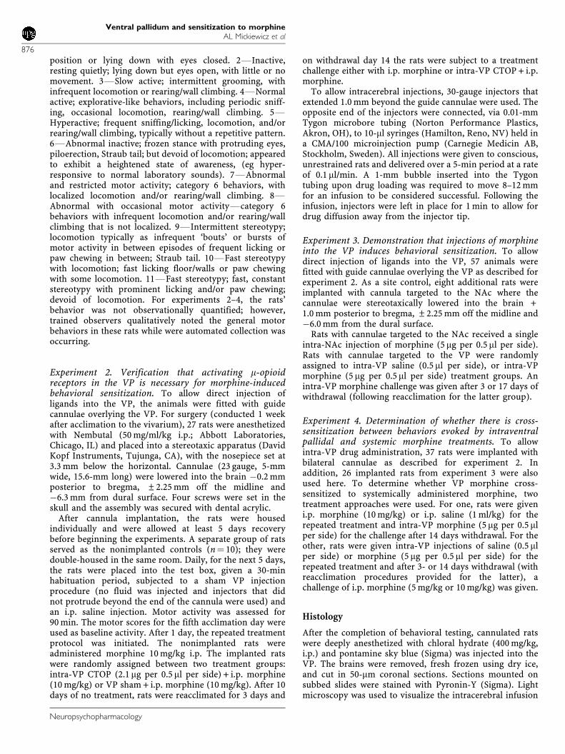
Figure 3 (Experiments 2–4) Injector tip placements within the nucleusaccumbens (NAc) and ventral pallidum (VP). (a) Low magnificationphotomicrograph of a VP coronal section. The left panel shows theunstained slice with pontamine sky blue at the injection site. The middlepanel shows the same slice after Nissl staining, illustrating gliosis. The rightpanel shows the injector tip in the VP under high magnification. Arrowindicates location of the injector tip, filled with pontamine sky blue. Thissection represents tissue with an indwelling cannula for 30 days. (b)Unilateral representations of bilaterally implanted cannulae were collapsedonto five plates redrawn from Paxinos and Watson (1998). The saggitalsection (2.40 mm from midline) demonstrates the area within the NAc andVP sampled, as detailed in coronal sections. Four different coronal sectionsrelative to Bregma are illustrated. Note that injections within the four VPtreatment groups overlapped; ac, anterior commissure; HDB, horizontallimb of the diagonal band; STR, striatum.
Ventral pallidum and sensitization to morphineAL Mickiewicz et al
879
Neuropsychopharmacology

et al (1978) revealed that intra-NAc injections of 3.25-mgmorphine induces a biphasic motor response with an initialdepression that lasts for 2.5 h followed by motor enhance-ments for the next 3 h. This profile predicted ourobservation that for at least 90 min after intra-NAcmorphine injections (5 mg per 0.5 ml per side), the motorscores did not statistically differ from saline controls. Thisis in stark contrast to intra-VP morphine of the same dose,where a more rapid motor activation was observed(Figure 6). The unique profiles exhibited by intra-VP andintra-NAc morphine injections demonstrate that respond-ing obtained with the intra-VP injection protocol employeddid not reflect the drug diffusing into the NAc.
Experiment 4. Determination of Whether there is Cross-Sensitization Between Behaviors Evoked by IntraventralPallidal and Systemic Morphine Treatments
Figures 7a and b show that repeated infusions of morphinedirectly into the VP did not induce sensitized responding toa challenge with systemic morphine (10 mg/kg) following a3-day abstinent period. This is illustrated by the similarity
in motor profile obtained with these rats (ie filled squares inFigure 7) as that observed following a single injection ofmorphine in naive rats (compare with day 1, filleddiamonds, in Figures 1a and b). These findings concurwith those of experiment 3, where it was revealed that theVP did not show evidence of sensitization following a 3 daywithdrawal (see Figures 5c and d), and thus, the VP wouldnot likely contribute to the sensitized behaviors evoked byi.p. morphine at this withdrawal time. Another contributingfactor to these negative outcomes may be the i.p. dose ofmorphine selected, for 10 mg/kg i.p. morphine induced aninitial cataleptic-like effect (see Figure 1c), which may havemasked hyperlocomotion evoked by intra-VP morphine.Thus, we determined that the question of VP morphinecross-sensitization to systemic morphine would be bestaddressed by (1) evaluating a protracted withdrawal timethat better reflects an involvement of the VP, ie at 14 dayswithdrawal and (2) using an i.p. dose of morphine thatincreased locomotion but did not readily evoke catalepsy. Apilot study indicated that 5 mg/kg i.p. morphine met ourcriteria, and as shown in Figures 7c and d, this dose ofmorphine generally increased motor activity in rats that
Figure 4 (Experiment 2) The m-opioid receptor antagonist D-Phe-Cys-Try-D-Trp-Orn-Thr-Pen-Thr-NH2 (CTOP) administered to the ventral pallidum(VP) blocked development and expression of sensitization induced by systemic morphine. Data are provided in 10-min bins; arrow indicates time of i.p.injection. A total of 10 rats included in this study were nonimplanted; all others were implanted with guide cannulae overlying the VP. Data were analyzedwith a two-way repeated measures ANOVA with post hoc Newman–Keuls. Left column. Development of sensitization with intra-VP sham + i.p. morphine,or i.p. morphine alone. Behaviors exhibited on days 1 and 5 of the repeated treatment regimen (a) Beam breaks were significantly different for treatment day(F1,41¼ 14.7, p¼ 0.0004), session time (F8,328¼ 7.4, po0.0001), and treatment day–time interaction occurred (F8,328¼ 4.7, po0.0001). (b) Crossingsshowed significant treatment day (F1,39¼ 8.4, p¼ 0.0061), session time (F8,312¼ 12.5, po0.0001), and treatment–time interaction effects (F8,312¼ 4.7,po0.0001) **po0.01. Middle column. Intra-VP CTOP (2.1 mg per 0.5 ml) preceding each repeated i.p. morphine blocked the development of sensitization.There was no significant treatment day, session time, or treatment day–time interaction effects for beam breaks (c) or crossings (d). Right column. CTOPadministered into the VP either during development, or immediately before a systemic morphine challenge, blocked the expression of sensitization. Forbeam breaks (e), there were significant treatment group (F2,30¼ 12.0, p¼ 0.0001), session time (F8,240¼ 3.4, p¼ 0.001), and treatment group–timeinteraction (F16,240¼ 3.4, po0.0001) effects. For crossings (f), there were significant treatment group (F2,29¼ 4.4, p¼ 0.0213), session time (F8,232¼ 3.5,p¼ 0.0008), and treatment group–time interaction (F16,232¼ 4.4, po0.0001) effects. Specific session time differences between days 1–5 i.p. morphine/i.p.morphine challenge and days 1–5 intra-VP CTOP + i.p. morphine/i.p. morphine challenge; *po0.05; **po0.01. Specific session time differences betweendays 1–5 i.p. morphine/i.p. morphine challenge and days 1–5 i.p. morphine/intra-VP CTOP + i.p. morphine challenge; wwpo0.01. (Note that for experiment 2only, data were collected using locomotor boxes built in house, which resulted in slightly lower scores due to less photocells than the commercial boxesused for experiments 1, 3, and 4.)
Ventral pallidum and sensitization to morphineAL Mickiewicz et al
880
Neuropsychopharmacology
received repeated treatments of intra-VP saline. Theresponse was enhanced following 14 days of drug absti-nence in rats that received repeated treatments with intra-VP morphine (Figure 7c). The number of crossings was notaltered. These results indicate that repeated activation of them-opioid receptors in the VP initiates adaptive processesthat are revealed 2 weeks later upon activation either withmotor enhancing doses of systemically administered orintra-VP injected morphine. We then reasoned that i.p.treatments of morphine should sensitize the VP (aspredicted by our prior biochemical and electrophysiological
evaluations (McDaid et al, 2006)), and this effect may berevealed by an increase in motor activity following intra-VPinjections of the opiate. Figure 8 provides results from theexperiment that evaluated this possibility. Motor responsesto a challenge of intra-VP morphine 14 days after five once-daily treatments of 10 mg/kg i.p. morphine are shown. Bothbeam breaks and crossings were upregulated within the first20 min of the intracerebral injection in rats with a treatmenthistory of i.p. morphine; in contrast to repeated intra-VPmorphine, where only beam breaks were sensitized. Thesedata indicate that i.p. morphine sensitized brain regionswhich regulate locomotion that were not affected withrepeated intra-VP morphine treatments, but which weresubsequently engaged by the intra-VP morphine challenge(refer to Figures 7c and d, 8). In sum, experiment 4demonstrated that at the 14 day withdrawal period, whenintra-VP injections of morphine result in expression of
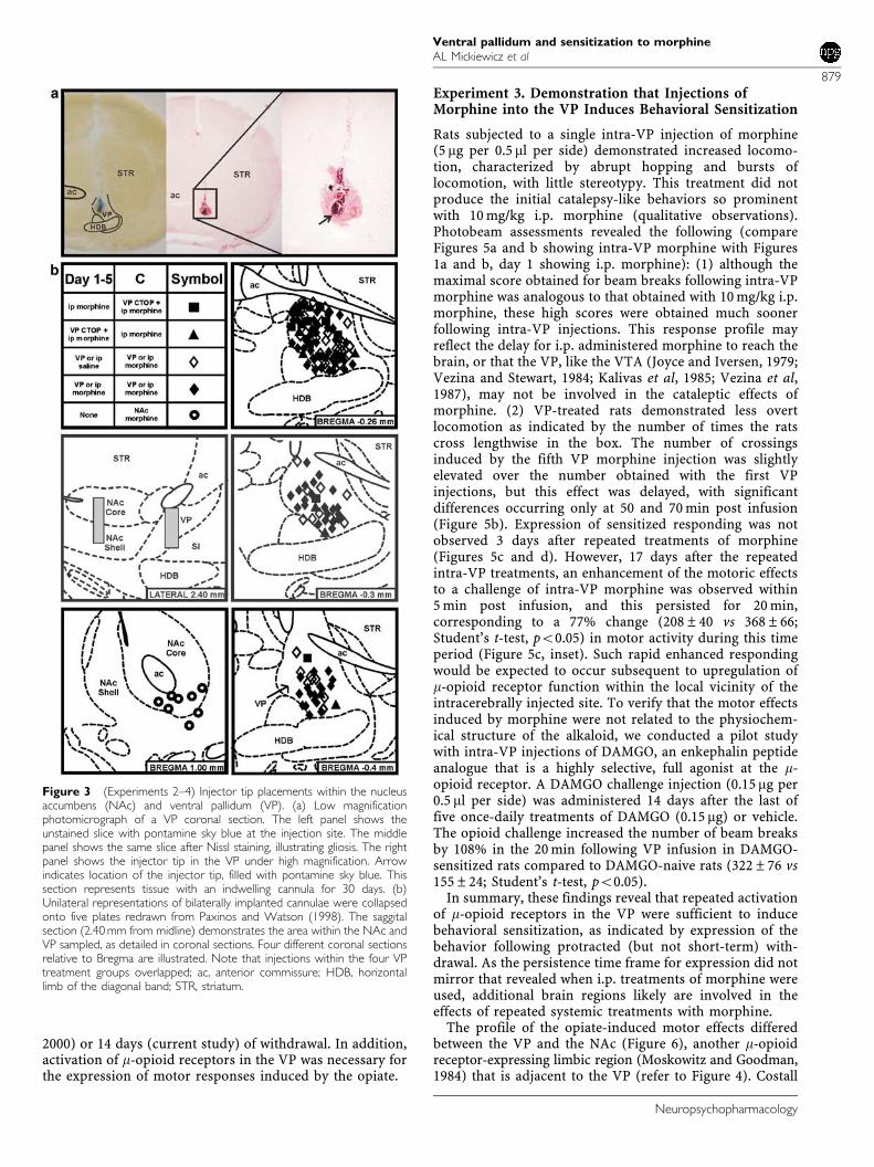
Figure 5 (Experiment 3) Sensitization to intraventral pallidal (VP)morphine injections. Development of sensitization was not revealed duringthe days 1–5 treatment but was demonstrated by expression of sensitizedresponding to a VP morphine challenge. Data are provided in 10-min bins;arrow indicates the time of intra-VP injection. Left column. There was nostatistical difference between days 1 and 5 of the saline treatment; thesedata were averaged and are illustrated by the gray dotted line. Repeatedmorphine treatments (5 mg per 0.5ml per side) revealed no statisticaldifferences between treatment day, across session time, or for a treatmentday–time interaction for beam breaks (a). For crossings (b) there weresignificant treatment day (F1,102¼ 6.8, p¼ 0.0103) and session time(F8,816¼ 4.7, po0.0001) effects, but no treatment day–time interaction(F8,816¼ 1.6, p¼ 0.1221). Right column. Behavioral sensitization wasexpressed following a challenge of intra-VP morphine on withdrawal day17 (WD17), but not WD3. (c) Beam breaks. There was no treatmentgroup effect (F2,31¼ 0.1, p¼ 0.8919), but a significant effect of session time(F8,248¼ 4.9, po0.0001) and a treatment group–time interaction(F16,248¼ 6.4, po0.0001) occurred. Inset. Enhanced resolution of earlybehaviors evoked by intra-VP morphine injections. The boxed portion ofthe beam breaks graph in 5-min bins for the subset of rats where this timeresolution was used (VP saline (n¼ 5), VP morphine W/D3 (n¼ 7), VPmorphine W/D17 (n¼ 9)) is shown. There was a significant treatmentgroup (F2,18¼ 5.2) effect, but no difference across session time (F3,54¼ 1.6)or a treatment group–time interaction (F3,54¼ 1.5). Specific session timedifferences between the VP saline-treated rats and the VP morphine-treated rats only occurred for W/D17. (d) Crossings. No statisticaldifferences occurred between treatment groups, across session time, or fortreatment group–time interactions. *po0.05 and **po0.01.
Figure 6 (Experiment 3) Injection site controls. Different profiles wereobtained when morphine (5mg per 0.5ml per side) was injected into theNAc vs the VP. Data are provided in 10-min bins; arrow indicates time ofintracerebral injection. The injector placement is illustrated in Figure 3; VPdata were also included in Figure 5. A two-way repeated measuresANOVA was performed among treatment groups. (a) Beam breaks. Therewere significant treatment group (F2,25¼ 3.4, p¼ 0.0496) and session time(F8,200¼ 3.6, p¼ 0.007) effects, and a treatment group–time interaction(F16,200¼ 3.5, po0.0001). (b) Crossings. There was no treatment group(F2,25¼ 1.97, p¼ 0.1559) effect; significant differences occurred for sessiontime (F8,200¼ 1.99, p¼ 0.0487) with a treatment group–time interaction(F16,200¼ 2.4, p¼ 0.0027). Specific time differences between intra-VPmorphine and intra-VP saline; *po0.05; **po0.01. Specific timedifferences between intra-VP morphine and intra-NAc morphine;wwpo0.01.
Ventral pallidum and sensitization to morphineAL Mickiewicz et al
881
Neuropsychopharmacology
sensitized motor responding, cross-sensitization betweensystemic and intra-VP injections of morphine can bedemonstrated.
DISCUSSION
The present study revealed four novel findings that modifycurrent concepts of brain circuits underlying drug-inducedbehavioral sensitization: First, selective activation ofm-opioid receptors within the VP was sufficient to developbehavioral sensitization. Second, blockade of VP m-opioidreceptors prevented the development and expression ofsensitization, indicating that these receptors were necessaryfor both phases of the behavior. Third, infusions ofmorphine directly into the VP promoted responding tosystemic morphine administration, and systemic pretreat-ments enhanced responding to intra-VP morphine. Fourth,the effects were independent of environmental context, andthus, pharmacological in nature. These results indicate that
VP m-opioid receptors are critically involved in theneuroadaptive processes that contribute to the developmentand expression of opiate-induced motor sensitization.
Several experimental considerations serve to buttressthese interpretations: (1) Behavioral responding to intracer-ebral injections reflected action within the VP. The motorresponse of acute VP morphine was significantly greaterthan that obtained with similar infusions into the NAc,verifying that motor enhancements following intra-VPinjections were not due to the diffusion of morphine into
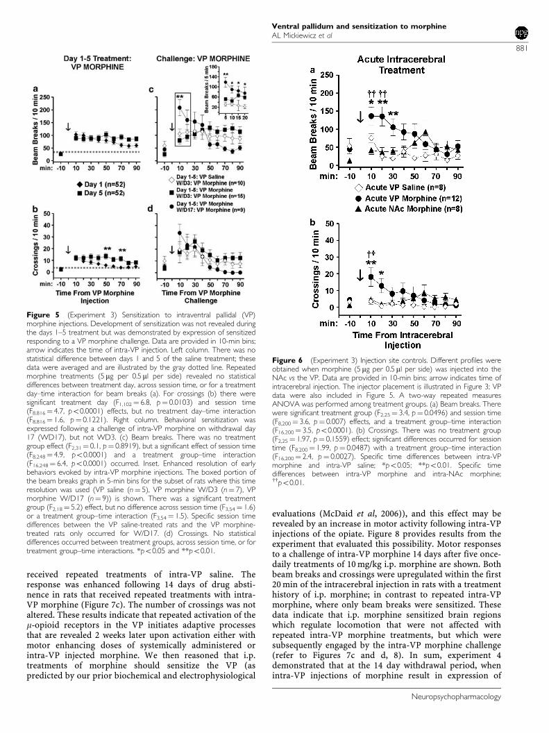
Figure 7 (Experiment 4) Cross-sensitization between intra-VP andsystemic morphine treatments. Morphine was administered systemically as5 mg/kg or 10 mg/kg i.p. following a 3- or 14-day withdrawal period fromrepeated intra-VP treatments of morphine (5 mg per 0.5 ml per side) orsaline (0.5ml per side). Data are provided in 10-min bins; arrow indicatestime of i.p. challenge. Left column. Intra-VP injections of morphine did notcross-sensitize with systemic morphine treatments of 10 mg/kg. Nostatistical differences occurred between the three treatment groups, acrosssession time, or treatment group–time interactions at any time points for(a) beam breaks or (b) crossings. Right column. Intra-VP injections ofmorphine cross-sensitized with a challenge injection of systemic morphinetreatments of 5 mg/kg after 14 days. (c) Beam breaks. There weresignificant treatment group (F1,23¼ 5.8, p¼ 0.0241) and session time(F8,184¼ 5.3, po0.0001) effects, but no treatment group–time interaction(F8,184¼ 0.5, p¼ 0.868). (d) Crossings. There were no significantdifferences between treatment groups, across time, or treatment group–time interactions. Specific time differences; *po0.05.
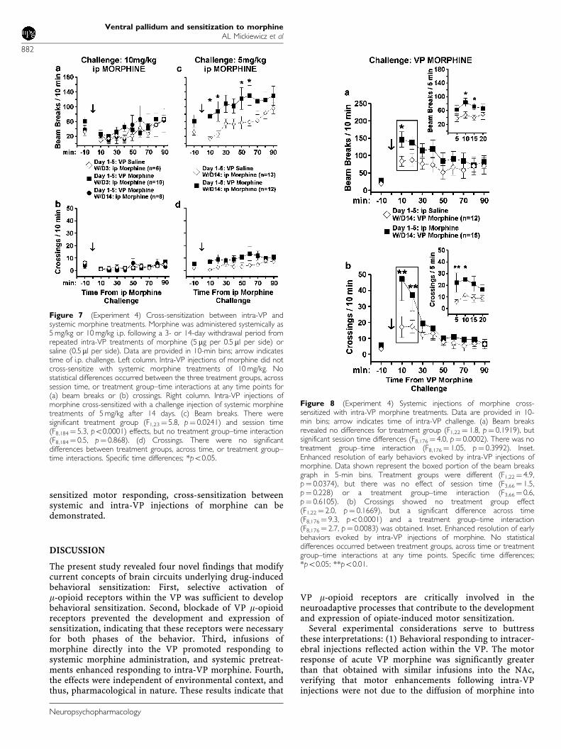
Figure 8 (Experiment 4) Systemic injections of morphine cross-sensitized with intra-VP morphine treatments. Data are provided in 10-min bins; arrow indicates time of intra-VP challenge. (a) Beam breaksrevealed no differences for treatment group (F1,22¼ 1.8, p¼ 0.1919), butsignificant session time differences (F8,176¼ 4.0, p¼ 0.0002). There was notreatment group–time interaction (F8,176¼ 1.05, p¼ 0.3992). Inset.Enhanced resolution of early behaviors evoked by intra-VP injections ofmorphine. Data shown represent the boxed portion of the beam breaksgraph in 5-min bins. Treatment groups were different (F1,22¼ 4.9,p¼ 0.0374), but there was no effect of session time (F3,66¼ 1.5,p¼ 0.228) or a treatment group–time interaction (F3,66¼ 0.6,p¼ 0.6105). (b) Crossings showed no treatment group effect(F1,22¼ 2.0, p¼ 0.1669), but a significant difference across time(F8,176¼ 9.3, po0.0001) and a treatment group–time interaction(F8,176¼ 2.7, p¼ 0.0083) was obtained. Inset. Enhanced resolution of earlybehaviors evoked by intra-VP injections of morphine. No statisticaldifferences occurred between treatment groups, across time or treatmentgroup–time interactions at any time points. Specific time differences;*po0.05; **po0.01.
Ventral pallidum and sensitization to morphineAL Mickiewicz et al
882
Neuropsychopharmacology

this nearby site (Moskowitz and Goodman, 1984).(2) Responding to intra-VP morphine reflected actions atthe m-opioid receptor. The partial agonist alkaloid, mor-phine and the full agonist peptide, DAMGO, induced similarbehavioral effects, and effects to systemic morphine wereblocked by intra-VP injections of the m-opioid antagonist,CTOP. (3) Comparisons of cross-sensitization between i.p.morphine and intra-VP morphine were conducted duringthe time frame (ie 17 days withdrawal) in which sensitiza-tion to morphine was maintained. This claim is substan-tiated by the findings that the i.p. morphine treatmentparadigm used in the current study results in motorsensitization at 3- (Johnson and Napier, 2000), 14- (McDaidet al, 2006), and 30 days of withdrawal (current study). (4)The reacclimation period was sufficient to negate contribu-tion of environmental cues to the morphine-induced motorresponse. When the context in which the animal receives anopiate (ie the unconditioned stimulus) is unique to the drugexperience, the context gains significance with repeateddrug pairings, and the behavioral outcome can reflectstimuli-related effects of context as well as the drug effects(Badiani et al, 2000). To isolate drug effects, an extensivehabituation to the treatment protocol before pairing is usedto remove any contribution of the environmental/procedur-al context to the measured motor response (Johnson andNapier, 2000; current study). (5) Development and expres-sion of morphine-induced sensitization occurred in ratsimplanted with cannulae above the VP. Qualitative assess-ment of gliosis around the injection site (Figure 3a) fromimplanted rats showed no difference between VP morphineand vehicle treatment groups. The opiate-treated ratsdeveloped sensitization (Figures 4a and b) that wasexpressed following a challenge with intra-VP morphine(Figure 5c) or systemic morphine (Figure 7c). Thus, theamount of gliosis present at the injection site was notsufficient to impede opiate-induced sensitization. Collec-tively, these controls support the conclusion that VPm-opioid receptors are important in the development andexpression of opiate-induced motor sensitization.
Insight can be gained about the role of the VP in opioid-induced motor sensitization by comparing the behavioralresponse profile obtained with intra-VP injections ofmorphine to that obtained with injections into other brainregions, and to systemic morphine treatments. Acutesystemic administration of moderate morphine dosesinduces a biphasic motor response consisting of an initialdepression of motor activity followed by hypermotility(Babbini and Davis, 1972; current study). Acute intra-NAcinjection of morphine in doses similar to that used in thecurrent experiment also produces motor depression/cata-lepsy followed by hypermotility (Costall et al, 1978). Incontrast, acute administration of opioids into the VTAresults only in hypermotility (Kelley et al, 1980; Joyce andIversen, 1979; Kalivas et al, 1983) as do acute intra-VPopioid injection (Baud et al, 1988; Austin and Kalivas, 1990;Napier, 1992; Alesdatter and Kalivas, 1993; Churchill andKalivas, 1999; Johnson et al, 1996; current study). Withrepeated systemic administration of opiates, the initialmotor depression is attenuated (likely reflecting tolerance tothe cataleptic effects of morphine) and overall activity isrobustly increased (ie sensitized) (Babbini and Davis, 1972;Babbini et al, 1975; Brady and Holtzman, 1981; Bartoletti
et al, 1983; Johnson and Glick, 1993; current study).Multiple infusions of opioids into the NAc (Vezina et al,1987; Cunningham et al, 1997), VTA (Joyce and Iversen,1979; Vezina and Stewart, 1984; Kalivas et al, 1985; Vezinaet al, 1987) and VP (present study), all result inhyperlocomotion, but as discussed below, the temporalnature of this effect differs among the regions.
Motor sensitization can be induced (developed) in theVTA, and it can be expressed with a VTA opioid challengefollowing short-term withdrawal (Kalivas et al, 1985; Vezinaet al, 1987). This transient phenomenon is transferred to theNAc that mediates the persistence of this behavior (forreview, see Vanderschuren and Kalivas, 2000). The VP isunique in that it is involved in both phases. We previouslyrevealed that the VP is involved in the development ofmorphine-induced motor sensitization (Johnson and Na-pier, 2000). The present results confirmed this finding andare the first to demonstrate that this region also is involvedin expression of this behavior. We report that activation ofm-opioid receptors in the VP, similar to the VTA, wassufficient to induce a sensitized behavioral response. Butthe VP distinguishes itself from the VTA in that theprogressive increase in motor activity seen during repeatedintra-VTA opioids was not obtained during repeated intra-VP treatment. We also reveal that a sensitized motorresponse is not expressed upon intra-VP morphinechallenge after 3 days withdrawal, a response that isobtained with opioid activation of the VTA. In theserespects, the behavioral response elicited from repeatedintra-VP morphine is similar to intra-NAc infusions ofmorphine or DAMGO, where sensitization is not expressedfollowing short-term withdrawal (Vezina et al, 1987). Inaddition, we reveal here that a sensitized motor responsecan be expressed upon intra-VP morphine challenge afterlong-term (17 days) withdrawal. These results suggest theVP is sufficient for the initiation of enduring adaptationsthat result in the expression of opioid-induced sensitization.
A commonly accepted theory of behavioral sensitizationis that the mesolimbic dopamine system mediates thisbehavior. Dopamine is a moderately abundant neurotrans-mitter in the VP (Napier and Potter, 1989; Zaborszky et al,1993), and it has widespread influence on VP function(Maslowski and Napier, 1991; Napier et al, 1991b). VPopioids regulate local dopamine transmission (for review,see Napier and Mitrovic, 1999), and dopamine receptors inthe VP are obligatory for acute motor function mediated byintra-VP m-opioid receptors (Napier, 1992). Locally appliedmorphine typically enhances VP neuronal activity (Johnsonand Napier, 1997; McDaid et al, 2006), reflecting aninhibition of GABAergic inputs from the NAc (Chrobakand Napier, 1993; Johnson and Napier, 1997) and/ordopaminergic inputs from the VTA (Mitrovic and Napier,2002). In turn, the VP sends GABAergic outputs to the NAc(Hakan et al, 1992) and VTA (Fonnum et al, 1978; Kalivaset al, 1993) that regulate the number of VTA neurons thatare able to exist in an active, spiking mode (Floresco et al,2003). The VTA provides dopaminergic inputs to the NAc(Fallon and Moore, 1978; Haglund et al, 1979; Swanson,1982) and VP (Napier et al, 1991a; Klitenick et al, 1992;Mitrovic and Napier, 2002). Dopamine overflow in the NAccore occurs during expression of morphine-inducedsensitization following long-term withdrawal (Cadoni and
Ventral pallidum and sensitization to morphineAL Mickiewicz et al
883
Neuropsychopharmacology
Di Chiara, 1999) and intra-VP morphine increases dopa-mine neurotransmission in the NAc (Anagnostakis andSpyraki, 1994). In light of this literature, the presentfindings indicate that sensitization-induced changes in VPm-opioid receptor function reduces GABAergic inhibition ofVP GABAergic control over the VTA, leading to anincreased number of active dopamine neurons in the VTAand increasing dopamine concentration and receptoractivation in the NAc and VP. Like the NAc (Costall andNaylor, 1975) dopamine receptor activation in VP (Napierand Chrobak, 1992) enhances motor function. Thus, thecurrent findings reveal a means by which the VPcontributes to the sensitization process.
It is important to note that there is a dopamine-independent component of opioid-mediated behaviors.Acute injections of opioid agonists into the NAc increasemotor activity that is not blocked by neuroleptics or6-OHDA-induced lesions of the NAc (Pert and Sivit, 1977;Kalivas et al, 1983). Studies of opiate-motivated rewardreveal that lesions of the NAc do not affect self-adminis-tration of morphine (Dworkin et al, 1988). Sensitizationinduced by chronic systemic administration of morphine isattenuated, but not prevented by lesions of the NAc or VTA(Bunney et al, 1984) or by antagonism of VP dopaminereceptors (Johnson and Napier, 2000). One potential sitethat may be included in the neural adaptations engenderedby chronic opiate exposure is the mediodorsal thalamus.This region receives projections from the VP (Young et al,1984; Vives and Mogenson, 1985) and connects to the motorsystems (Groenewegen, 1988; Berendse et al, 1992).Inactivation of the mediodorsal thalamus by procaineprevents the motor responses to acute intra-VP opioidtreatment, and acute injections of opioids into themediodorsal thalamus result in motor activation thatremains intact with inhibition of the VTA (Klitenick andKalivas, 1994). Thus, sensitization-induced enhancementsin VP m-opioid receptor function could also reduceGABAergic inhibition of VP GABAergic influences on themedial dorsal nucleus, and this disinhibition would bepredicted to increase thalamic excitatory outputs tohindbrain motor systems. Additional studies are neededto verify whether the mediodorsal thalamic nucleus isinvolved in sensitization processes.
Glutamate transmission is also involved in opiate-induced sensitization. In the VP, ionotropic glutamatereceptors are functionally upregulated 14 days after asensitizing regimen of morphine (McDaid et al, 2006), andintra-VP injections of glutamatergic antagonists in mor-phine-sensitized rats block the expression of motorsensitization to a subsequent challenge of i.p. morphine(Dallimore et al, 2006). Thus, a tonic activation of VPglutamatergic receptors appears important to the enduringeffects of repeated morphine exposure. Glutamatergicinputs to the VP arise largely from the basolateral amygdala(Russchen and Price, 1984; Fuller et al, 1987) and medialsubthalamic nucleus (Kita and Kitai, 1987; Smith andParent, 1988). The subthalamic nucleus is regulated bym-opioid agonists (Shen and Johnson, 2002), but it is notknown if these become sensitized with repeated activation.However, the basolateral amydgala demonstrates enhance-ments in receptor-mediated signaling cascades (ie MAPK)in morphine-sensitized rats (Eitan et al, 2003). Thus, it is
possible that a sensitized glutamatergic drive to the VP isassociated with morphine-induced behavioral sensitization.
In summary, the current study revealed that activation ofm-opioid receptor in the VP are sufficient and necessary toinduce a sensitized locomotor response to morphine andthat cross-sensitization occurs between morphine treat-ments given systemically and directly into the VP. As wefurther elucidate the brain regions that underlie theneuroadaptations associated with repeated drug exposure,we are learning that the major circuit may not be confinedto the VTA-NAc corridor, and redundant systems areinvolved in the various phases of drug-induced sensitiza-tion. We propose that the VP is one of the criticalneuroanatomical substrates mediating drug-induced sensi-tization.
DISCLOSURE/CONFLICT OF INTEREST
The author(s) declare that, except for income received fromthe primary employer, no financial support or compensa-tion has been received from any individual or corporateentity over the past 3 years for research or professionalservice and there are no personal financial holdings thatcould be perceived as constituting a potential conflict ofinterest.
ACKNOWLEDGEMENTS
This work was supported by Loyola University ChicagoGraduate School, and United States Public Health GrantsDA05255 to TCN and DA019783 to ALM and TCN.
REFERENCES
Alesdatter JE, Kalivas PW (1993). Inhibition of mu opioid-inducedmotor activity in the ventral pallidum by D1 receptor blockade.Behav Pharmacol 4: 645–651.
Anagnostakis Y, Spyraki C (1994). Effect of morphine applied byintrapallidal microdialysis on the release of dopamine in thenucleus accumbens. Brain Res Bull 34: 275–282.
Austin MC, Kalivas PW (1990). Enkephalinergic and GABAergicmodulation of motor activity in the ventral pallidum. J Phar-macol Exp Ther 252: 1370–1377.
Babbini M, Davis WM (1972). Time–dose relationships forlocomotor activity effects of morphine after acute or repeatedtreatment. Br J Pharmacol 46: 213–224.
Babbini M, Gaiardi M, Bartoletti M (1975). Persistence of chronicmorphine effects upon activity in rats 8 months after ceasing thetreatment 32. Neuropharmacology 14: 611–614.
Badiani A, Leone P, Stewart J (1995). Intra-VTA injections of themu-opioid antagonist CTOP enhance locomotor activity. BrainRes 690: 112–116.
Badiani A, Oates MM, Robinson TE (2000). Modulation ofmorphine sensitization in the rat by contextual stimuli.Psychopharmacology (Berl) 151: 273–282.
Bartoletti M, Gaiardi M, Gubellini G, Bacchi A, Babbini M (1983).Long-term sensitization to the excitatory effects of morphine. Amotility study in post-dependent rats. Neuropharmacology 22:1193–1196.
Baud P, Mayo W, Le Moal M, Simon H (1988). Locomotorhyperactivity in the rat after infusion of muscimol and [D-Ala2]Met-enkephalin into the nucleus basalis magnocellularis.Possible interaction with cortical cholinergic projections. BrainRes 452: 203–211.
Ventral pallidum and sensitization to morphineAL Mickiewicz et al
884
Neuropsychopharmacology
Berendse HW, Galis-de Graaf Y, Groenewegen HJ (1992).Topographical organization and relationship with ventral striatalcompartments of prefrontal corticostriatal projections in the rat.J Comp Neurol 316: 314–347.
Brady LS, Holtzman SG (1981). Locomotor activity in morphine-dependent and post-dependent rats. Pharmacol Biochem Behav14: 361–370.
Bunney WC, Massar VJ, Pert A (1984). Chronic morphine-inducedhyperactivity in rats is altered by nucleus accumbens and ventraltegmental lesions. Psychopharmacology (Berl) 82: 318–321.
Cadoni C, Di Chiara G (1999). Reciprocal changes in dopamineresponsiveness in the nucleus accumbens shell and core and inthe dorsal caudate-putamen in rats sensitized to morphine.Neuroscience 90: 447–455.
Chrobak JJ, Napier TC (1993). Opioid and GABA modulation ofaccumbens-evoked ventral pallidal activity. J Neural Trans 93:123–143.
Churchill L, Kalivas PW (1999). The involvement of themediodorsal nucleus of the thalamus and the midbrainextrapyramidal area in locomotion elicited from the ventralpallidum. Behav Brain Res 104: 63–71.
Costall B, Fortune DH, Naylor RJ (1978). The induction ofcatalepsy and hyperactivity by morphine administered directlyinto the nucleus accumbens of rats. Eur J Pharmacol 49: 49–64.
Costall B, Naylor RJ (1975). The behavioural effects of dopamineapplied intracerebrally to areas of the mesolimbic system. Eur JPharmacol 32: 87–92.
Cunningham ST, Finn M, Kelley AE (1997). Sensitization of thelocomotor response to psychostimulants after repeated opiateexposure: role of the nucleus accumbens. Neuropsychopharma-cology 16: 147–155.
Dallimore JE, Mickiewicz AL, Napier TC (2006). Intra-ventralpallidal glutamate antagonists block expression of morphine-induced place preference. Behav Neurosci 120: 1103–1114.
Dworkin SI, Guerin GF, Co C, Goeders NE, Smith JE (1988). Lackof an effect of 6-hydroxydopamine lesions of the nucleusaccumbens on intravenous morphine self-administration. Phar-macol Biochem Behav 30: 1051–1057.
Eitan S, Bryant CD, Saliminejad N, Yang YC, Vojdani E, Keith Jr Det al. (2003). Brain region-specific mechanisms for acutemorphine-induced mitogen-activated protein kinase modulationand distinct patterns of activation during analgesic tolerance andlocomotor sensitization. J Neurosci 23: 8360–8369.
Fallon JH, Moore RY (1978). Catecholamine innervation of thebasal forebrain. IV. Topography of the dopamine projectionto the basal forebrain and neostriatum. J Comp Neurol 180:545–580.
Floresco SB, West AR, Ash B, Moore H, Grace AA (2003). Afferentmodulation of dopamine neuron firing differentially regulatestonic and phasic dopamine transmission. Nat Neurosci 6:968–973.
Fonnum F, Gottesfeld Z, Grofova I (1978). Distribution ofglutamate decarboxylase, choline acetyltransferase and aromaticamino acid decarboxylase in the basal ganglia of normal andoperated rats. Evidence for striatopallidal, striatoentopeduncularand striatonigral GABAergic fibers. Brain Res 143: 125–138.
Fuller TA, Russchen FT, Price JL (1987). Sources of presumptiveglutamergic/aspartergic afferents to the rat ventral striatopallidalregion. J Comp Neurol 258: 317–338.
Groenewegen HJ (1988). Organization of the afferent connectionsof the mediodorsal thalamic nucleus in the rat, related to themediodorsal-prefrontal topography. Neuroscience 24: 379–431.
Haglund L, Kohler C, Ross SB, Kelder D (1979). Forebrainprojections of the ventral tegmentum as studied by axonaltransport of [3H]dopamine in the rat. Neurosci Lett 12:301–306.
Hakan RL, Berg GI, Henriksen SJ (1992). Electrophysiologicalevidence for reciprocal connectivity between the nucleus
accumbens septi and ventral pallidal region. Brain Res 581:344–350.
Johnson DW, Glick SD (1993). Dopamine release and metabolismin nucleus accumbens and striatum of morphine-tolerant andnontolerant rats. Pharmacol Biochem Behav 46: 341–347.
Johnson K, Churchill L, Klitenick MA, Hooks MS, Kalivas PW(1996). Involvement of the ventral tegmental area in locomotionelicited from the nucleus accumbens or ventral pallidum.J Pharmacol Exp Ther 277: 1122–1131.
Johnson PI, Napier TC (1997). Morphine modulation of GABA-and glutamate-induced changes of ventral pallidal neuronalactivity. Neuroscience 77: 187–197.
Johnson PI, Napier TC (2000). Ventral pallidal injections of a muantagonist block the development of behavioral sensitization tosystemic morphine. Synapse 38: 61–70.
Joyce EM, Iversen SD (1979). The effect of morphine appliedlocally to mesencephalic dopamine cell bodies on spontaneousmotor activity in the rat. Neurosci Lett 14: 207–212.
Kalivas PW, Churchill L, Klitenick MA (1993). GABA andenkephalin projection from the nucleus accumbens andventral pallidum to the ventral tegmental area. Neuroscience57: 1047–1060.
Kalivas PW, Taylor S, Miller JS (1985). Sensitization to repeatedenkephalin administration into the ventral tegmental area of therat. I. Behavioral characterization. J Pharmacol Exp Ther 235:537–543.
Kalivas PW, Widerlov E, Stanley D, Breese G, Prange Jr AJ (1983).Enkephalin action on the mesolimbic system: a dopamine-dependent and a dopamine-independent increase in locomotoractivity. J Pharmacol Exp Ther 227: 229–237.
Kelley AE, Stinus L, Iversen SD (1980). Interactions between D-ala-met-enkephalin, A10 dopaminergic neurones, and sponta-neous behaviour in the rat. Behav Brain Res 1: 3–24.
Kita H, Kitai ST (1987). Efferent projections of the subthalamicnucleus in the rat: light and electron microscopic analysis withthe PHA-L method. J Comp Neurol 260: 435–452.
Klitenick MA, Deutch AY, Churchill L, Kalivas PW (1992).Topography and functional role of dopaminergic projectionsfrom the ventral mesencephalic tegmentum to the ventralpallidum. Neuroscience 50: 371–386.
Klitenick MA, Kalivas PW (1994). Behavioral and neurochemicalstudies of opioid effects in the pedunculopontine nucleusand mediodorsal thalamus. J Pharmacol Exp Ther 269:437–448.
Li Y, Kolb B, Robinson TE (2003). The location of persistentamphetamine-induced changes in the density of dendriticspines on medium spiny neurons in the nucleus accumbensand caudate-putamen. Neuropsychopharmacology 28: 1082–1085.
Maslowski RJ, Napier TC (1991). Dopamine D1 and D2 receptoragonists induce opposite changes in the firing rate of ventralpallidal neurons. Eur J Pharmacol 200: 103–112.
Maslowski-Cobuzzi RJ, Napier TC (1994). Activation of dopami-nergic neurons modulates ventral pallidal responses evoked byamygdala stimulation. Neuroscience 62: 1103–1120.
McDaid J, Dallimore JE, Mackie A, Napier TC (2006). Changes inaccumbal and ventral pallidal pCREB and deltaFosB inmorphine-sensitized rats: correlations with receptor-evokedelectrophysiological measures in the ventral pallidum. Neuro-psychopharmacology 31: 1212–1226.
Mitrovic I, Napier TC (2002). Mu and kappa opioid agonistsmodulate ventral tegmental area input to the ventral pallidum.Eur J Neurosci 15: 257–268.
Mogenson GJ, Yang CR (1991). The contribution of basal forebrainto limbic-motor integration and the mediation of motivation toaction. In: Napier TC, Kalivas PW, Hanin I (eds). The BasalForebrain: Anatomy to Function. Plenum Press: New York andLondon. pp 267–290.
Ventral pallidum and sensitization to morphineAL Mickiewicz et al
885
Neuropsychopharmacology

Moskowitz AS, Goodman RR (1984). Light microscopic autoradio-graphic localization of mu and delta opioid binding sites in themouse central nervous system. J Neuroscience 4: 1331–1342.
Napier TC (1992). Dopamine receptors in the ventral pallidumregulate circling induced by opioids injected into the ventralpallidum. Neuropharmacology 31: 1127–1136.
Napier TC, Chrobak JJ (1992). Evaluations of ventral pallidaldopamine receptor activation in behaving rats. Neuroreport 3:609–611.
Napier TC, Mitrovic I (1999). Opioid modulation of ventral pallidalinputs. Ann NY Acad Sci 877: 176–201.
Napier TC, Muench MB, Maslowski RJ, Battaglia G (1991a). Isdopamine a neurotransmitter within the ventral pallidum/substantia innominata. Adv Exp Med Biol 295: 183–195.
Napier TC, Potter PE (1989). Dopamine in the rat ventral pallidum/substantia innominata: biochemical and electrophysiologicalstudies. Neuropharmacology 28: 757–760.
Napier TC, Simson PE, Givens BS (1991b). Dopamine electro-physiology of ventral pallidal/substantia innominata neurons:comparison with the dorsal globus pallidus. J Pharmacol ExpTher 258: 249–262.
Paxinos G, Watson C (1998). The Rat Brain in StereotaxicCoordinates. Academic Press: New York.
Pelton JT, Kazmierski W, Gulya K, Yamamura HI, Hruby VJ(1986). Design and synthesis of conformationally constrainedsomatostatin analogues with high potency and specificity for muopioid receptors. J Med Chem 29: 2370–2375.
Pert A, Sivit C (1977). Neuroanatomical focus for morphine andenkephalin-induced hypermotility. Nature 265: 645–647.
Russchen FT, Price JL (1984). Amygdalostriatal projections in therat. Topographical organization and fiber morphology shownusing the lectin PHA-L as an anterograde tracer. Neurosci Lett47: 15–22.
Shen KZ, Johnson SW (2002). Presynaptic modulation of synaptictransmission by opioid receptor in rat subthalamic nucleus invitro. J Physiol 541: 219–230.
Shippenberg TS, Bals-Kubik R (1995). Involvement of themesolimbic dopamine system in mediating the aversive effectsof opioid antagonists in the rat. Behav Pharmacol 6: 99–106.
Smith Y, Parent A (1988). Neurons of the subthalamic nucleus inprimates display glutamate but not GABA immunoreactivity.Brain Res 453: 353–356.
Swanson LW (1982). The projections of the ventral tegmental areaand adjacent regions: a combined fluorescent retrograde tracerand immunofluorescence study in the rat. Brain Res Bull 9:321–353.
Vanderschuren LJ, Kalivas PW (2000). Alterations in dopaminergicand glutamatergic transmission in the induction and expressionof behavioral sensitization: a critical review of preclinicalstudies. Psychopharmacology (Berl) 151: 99–120.
Vezina P, Kalivas PW, Stewart J (1987). Sensitization occurs to thelocomotor effects of morphine and the specific u opioid receptoragonist, DAGO, administered repeatedly to the ventraltegmental area but not to the nucleus accumbens. Brain Res417: 51–58.
Vezina P, Stewart J (1984). Conditioning and place-specificsensitization of increases in activity induced by morphine inthe VTA. Pharmacol Biochem Behav 20: 925–934.
Vives F, Mogenson GJ (1985). Electrophysiological evidence thatthe mediodorsal nucleus of the thalamus is a relay between theventral pallidum and the medial prefrontal cortex in the rat.Brain Res 344: 329–337.
Wolf ME (2002). Addiction: making the connection betweenbehavioral changes and neuronal plasticity in specific pathways.Mol Interv 2: 146–157.
Young WS, Alheld GF, Heimer L (1984). The ventral pallidalprojection to the mediodorsal thalamus: a study with fluorescentretrograde tracers and immunohistofluorescence. J Neuroscience4: 1626–1638.
Zaborszky L, Alheid GF, Heimer L (1985). Mapping of transmitter-specific connections: simultaneous demonstration of antero-grade degeneration and changes in the immunostaining patternsinduced by lesions. J Neurosci Methods 14: 255–266.
Zaborszky L, Cullinan WE, Luine VN (1993). Catecholaminergic–cholinergic interaction in the basal forebrain. In: Cuello AC (ed).Progress in Brain Research. Elsevier Science Publishing Co. Inc:NY, Vol. 98, pp 31–49.
Zahm DS (1989). The ventral striatopallidal parts of the basalganglia in the ratFII. Compartmentation of ventral pallidalefferents. Neuroscience 30: 33–50.
Zahm DS, Zaborszky L, Alones VE, Heimer L (1985). Evidence forthe coexistence of glutamate decarboxylase and met-enkephalinimmunoreactivities in axon terminals of rat ventral pallidum.Brain Res 325: 317–321.
Ventral pallidum and sensitization to morphineAL Mickiewicz et al
886
Neuropsychopharmacology