the value of 3d visualization operative planning system in
TRANSCRIPT

RESEARCH ARTICLE Open Access
The value of 3D visualization operativeplanning system in ultrasound-guidedpercutaneous microwave ablation for largehepatic hemangiomas: a clinicalcomparative studyXin Li†, Chao An†, Fangyi Liu, Zhigang Cheng, Zhiyu Han, Xiaoling Yu, Linan Dong, Jie Yu and Ping Liang*
Abstract
Background: To evaluate the value of a three dimension (3D)visualization operative planning system in ultrasound-guided percutaneous microwave ablation (US-PMWA) for large hepatic hemangiomas (LHHs).
Methods: Fifty-eight patients with LHHs were divided into 3D and 2D groups. The therapeutic efficacy was assessed bycontrast-enhanced imaging during follow-up. Hepatic and renal function were examined. The complete ablation, tumorvolume shrinkage, and complication rates were analyzed.
Results: The ablation time and energy of the 3D group were lower than those of the 2D group (1152.0 ± 403.9 s vs.1379.7 ± 375.8 s and 87,407.2.9 ± 50,387.0 J vs. 117,775.8 ± 46,245.6 J, P = 0.031 and 0.021, respectively). The 3D grouphad a higher complete ablation rate than the 2D group (97.7 ± 2.4% vs. 94.5 ± 3.7%, P < 0.001). The incidence ofhemoglobinuria after ablation in the 3D group was lower than that in the 2D group (32.0% vs. 57.6%, P = 0.047).The levels of alanine aminotransferase (ALT), aspartate aminotransferase (AST), alkaline phosphatase (ALP), andcreatinine (Cre) after ablation in the 3D group were lower than those in the 2D group (126.7 ± 56.4 U/L vs. 210.9 ± 96.2 U/L,P< 0.001; 141.0 ± 60.8 U/L vs. 211.4 ± 90.0 U/L, P= 0.001; 57.3 ± 17.6 U/L vs. 80.8 ± 41.9 U/L, P= 0.010; and 66.6 ± 16.6mmol/Lvs. 84.5 ± 39.6 mmol/L, P = 0.037, respectively). There were no significant differences in antenna insertion and thevolume reduction rate between the groups. One patient developed acute kidney injury shortly after ablation inthe 2D group and recovered after hemodialysis. No other severe complications occurred during the follow-up period.
Conclusions: The 3D visualization operative planning system has a relatively high clinical application value inproviding scientific, reasonable, quantifiable, and individualized therapy for LHHs by US-PMWA.
Keywords: 3D visualization operative planning system, Microwave ablation, Large hepatic hemangiomas
BackgroundHepatic hemangioma is the most frequently encounteredsolid benign liver neoplasm, with an incidence of 0.4 to20% in the general population and accounting for 73%of all benign liver lesions [1–3]. There is no controversyconcerning the management of incidentally identifiedand asymptomatic hepatic hemangiomas, which require
no intervention or further observation if there are noadverse complications or effects on adjacent organs.Only large hepatic hemangiomas (LHHs) that lead tosymptoms such as abdominal pain or distension, anemia,portal hypertension, obstructive jaundice, spontaneous ortraumatic rupture, coagulopathy or serious psychologicaleffects may require intervention [4–9]. However, thedefinition of LHHs varies in the literature. Some authorsreport hemangioma > 4 cm as large [7, 10, 11], while avery small number of authors define those > 10 cm asLHH [12–14]. However, in most reports in the literature,
© The Author(s). 2019 Open Access This article is distributed under the terms of the Creative Commons Attribution 4.0International License (http://creativecommons.org/licenses/by/4.0/), which permits unrestricted use, distribution, andreproduction in any medium, provided you give appropriate credit to the original author(s) and the source, provide a link tothe Creative Commons license, and indicate if changes were made. The Creative Commons Public Domain Dedication waiver(http://creativecommons.org/publicdomain/zero/1.0/) applies to the data made available in this article, unless otherwise stated.
* Correspondence: [email protected]†Xin Li and Chao An contributed equally to this work.Department of Interventional Ultrasound, Chinese PLA General Hospital, 28Fuxing Road, Beijing 100853, China
Li et al. BMC Cancer (2019) 19:550 https://doi.org/10.1186/s12885-019-5682-5
LHHs are defined as being ≥5 cm and warranting therapywhen they lead to clinical symptoms or the risk oflife-threatening spontaneous rupture and hemorrhage[5, 7, 15]. In this study, LHH was defined as a hemangiomawith a maximum diameter ≥ 5 cm.Surgical resection and surgical enucleation are the
traditional modalities for the treatment of LHHs [9, 13,16–18]. However, other therapies, including liver trans-plantation, transcatheter arterial embolization [19–22],radiation therapy [23, 24], thermal ablation (radio-frequency and microwave ablation) [3, 8, 11, 25–27],and medications (bevacizumab, sorafenib, cisplatin andbleomycin) [28, 29], have also been reported in themanagement of LHHs. In some cases, operative treatmentof LHHs remains a challenge for surgeons because ofmassive intraoperative blood loss and difficulty in control-ling bleeding [30, 31]. Compared with other ablation tech-nique, the benefits of microwave ablation, which has ahigher thermal efficiency, include fewer limitations due tothe heat-sink effect and tissue charring and the achieve-ment of a larger ablation volume in a shorter time,with better clinical effects [11, 32–34]. Therefore,microwave ablation is a better choice for the treat-ment of symptomatic LHHs.Importantly, LHHs often compress surrounding vital
structures, such as the gastric outlet, hepatic vein, portalvein, hepatic artery, bile duct, or inferior vena cava,which could increase the difficulty and risk of treatment[35–37]. Methods to protect surrounding organs andsimultaneously achieve complete ablation are important.Moreover, hemoglobinuria and acute renal failure occur-ring after ablation have been reported in the literature,including cases requiring hemodialysis [10, 38]. There-fore, how to achieve scientific, objective, quantifiable,and individualized operative planning is a key issueconcerning ablation. Operative planning is typically per-formed based on the spatial awareness and experience ofoperators’ “human brain” using two-dimensional (2D)imaging data, which are highly subjective and abstract.The operators’ evaluation is inconsistent with the actualsituation in some cases and may lead to incompleteablation or major complications [39]. A three dimensional(3D) visualization operation planning system not onlydisplays the location and spatial relationship of the tumorwith the surrounding structures, quantifies the tumor sizeand volume, predicates the time-temperature profile, andimproves the safety and effectiveness of ablation but alsoenables planning of the implantation route and accuratepositioning of the ablation antenna [40–42]. The purposeof this study was to explore the clinical value of a 3Dvisualization operative planning system in ultrasound-guided percutaneous microwave ablation (US-PMWA) forlarge hepatic hemangiomas compared to the 2D operativeplanning system.
MethodsPatients and study designThis single-center, retrospective study protocol wasapproved by the Ethics Committee of the Chinese PLAGeneral Hospital (Beijing, China) and was conducted inaccordance with the principles of the Declaration ofHelsinki. From January 2011 to August 2018, 58 patients(43 females, 15 males; mean age 43.5 ± 8.2 years) with58 LHHs (mean maximum diameter 7.4 ± 1.5 cm, range5–12.6 cm) were divided into two groups. The 3D groupincluded 25 patients who underwent US-PMWA with theaid of the self-developed 3D visualization operative plan-ning system. The 2D group included 33 patients whounderwent US-PMWA using the conventional 2D imageoperative planning methods.The inclusion criteria for US-PMWA were as follows:
(1) definite diagnosis of LHH ≥5 cm based on the typicalenhancement pattern on contrast-enhanced computedtomography (CT), magnetic resonance imaging (MRI),or contrast-enhanced ultrasound (CEUS); (2) clinicalsymptoms typically caused by LHHs present for at least1 year, including abdominal pain, nausea, vomiting,abdominal fullness, and serious mental burden [10, 11];(3) diagnoses of LHH pathologically proven by ultrasound(US)-guided core needle biopsy before ablation; and (4)prothrombin time < 25 s; prothrombin activity > 40%; andplatelet count > 60 cells × 109/L. The exclusion criteriawere: (1) severe cardiopulmonary disease; (2) serious renalfailure (creatinine above the normal level); (3) severehepatic failure (aminotransferase and bilirubin greaterthan the twice the normal level). Written informedconsent was obtained from all participating patients.
3D visualization operative planning system, US-PMWA,and auxiliary techniques3D visualization operative planning systemAll patients with LHH underwent conventional ultra-sound, CEUS, and/or CT/MRI to delineate the targettumor within 7 days before ablation. A desktop com-puter (Lenovo) with an Intel Core i5 processor for anempirical study in our department was used to perform3D visualization preoperative planning. A series of CTor MRI data (0.625 mm- or 2.5 mm-thick slices) of theLHHs before MWA were converted to DICOM formatand then imported into the 3D visualization treatmentplatform (Hokai Company, Zhuhai, China). Our grouporiginally developed the 3D visualization platform soft-ware that has novel functions as follows: 1) rapidsegmentation in the target (within 2min); 2) volumecalculation in the target; 3) simulation of the thermal field;4) interactive pre-ablative planned strategy by manualoperation; and 5) assessment of ablative effect by tumormapping. The graphical user interface displayed thereal-time simulation 2D ultrasound guided planning and
Li et al. BMC Cancer (2019) 19:550 Page 2 of 9

the 3D visualization planning, as well as the planning pathfrom the transverse, coronal, and sagittal planes. Theapplication of 3D visualization technology enables aradiologist to perform various operations on the 3Dimage, such as free movement, rotation, and scaling, todevelop a puncture plan by seeing more intuitively. Theliver mass with a 5-mm tumor-free margin, vessels, andsurrounding vital structures was segmented and re-constructed, which can be demonstrated via stereo displayin the 3D visualization.The 3D operation planning system of US-PMWA
should abide by the following principles: (1) confor-mational ablation was applied while covering the entiretumor only; (2) avoid ablation of vital structures, parti-cularly the secondary biliary duct, arteries and veins, orintestinal tract; and (3) minimize the distance of electrodeinsertion trajectories while avoiding puncture pathwaysvia critical structures. The planning system was designedto iterate these goals until a reasonable and feasible planwas achieved [39].
Us-PMWAThe ablations were performed in our institution, and allpatients were hospitalized. The microwave system(KY-2000, Kangyou Medical, Nanjing, China) compriseda MW generator, flexible coaxial cable, and cooled-shaftantenna. A 2450MHz or 915MHzMW system wasused. The 2450MHzMW system consisted of threeindependent MW generators, three flexible coaxial cables,and three water-pumping machines that can simultan-eously drive three 15-gauge cooled-shaft antennae (1.1 cmantenna tip). The 915MHzMW system consisted of twoindependent MW generators, two flexible coaxial cables,and two water-pumping machines that can simultaneouslydrive two 15-gauge cooled-shaft antennae (2.2 cm antennatip). The MW system generators were capable of pro-ducing 1-100W of power output. The 15-G cooled-shaftantenna was coated with Teflon to prevent adhesion withdual channels inside the antenna shaft, through whichdistilled water continuously circulated via a peristalticpump that could cool the shaft to prevent overheating.The patient was unconscious, under intravenous
anesthesia (propofol, 6-12mg/kg/h; ketamine, 1-2 mg/kg),during ablation in the operating room. All treatmentswere carried out by two operators with more than 5 yearsof ablation experience according to the operative plan.If the tumors and tumor-feeding arteries were not wellvisualized with conventional US, CEUS-guided ablationwith SonoVue (Bracco, Milan, Italy) was performed.US-guided biopsy was performed using an 18-G cuttingneedle (C. R. Bard, Japan) before ablation. First, thetumor-feeding artery of the hemangioma was ablatedwith higher power (60-80W) until no blood flow orhyperenhancement was visible on color Doppler flow
imaging (CDFI) and CEUS. An 18-G percutaneoustranshepatic cholangiography (PTC) needle (Hakko,Japan) was inserted into the hemangioma to simulta-neously aspirate the blood from the blood sinus of thehemangioma. Two antennae were subsequently implantedin the proper location of the LHH according to the 3Dvisualization operative planning system. The hyperechoicarea of ablation was monitored by greyscale ultrasound,and the endpoint of ablation was determined. The anten-nae tracks were routinely cauterized to avoid bleedingduring withdrawal.
Auxiliary techniquesAccording to the preoperative contrast-enhanced CT orMRI imaging or 3D visualization operative planning systemevaluation, hydrodissection and thermal monitoring tech-niques were applied for LHHs abutting vital structures toavoid thermal injury. After application of a local anesthetic(1% lidocaine), a 16-G intravenous catheter (BD Angiocath;Sandy, UT, USA) was inserted into the peritoneal cavitybetween the edge of the liver and the abutting gastrointes-tinal tract using US guidance. A sufficient amount of chillednormal saline was delivered until a separation of greaterthan 0.5 cm was achieved and maintained during the pro-cedure. Then, 21-G thermocouple needles were equippedon the MWA system, which are easily visualized by US dur-ing ablation, for LHHs abutting vital structures. Based onour experimental evidence, the temperature cut-off of abla-tion was set at 60 °C. If the measured temperature reached60 °C, ablation was immediately terminated and activatedagain after the temperature decreased to 50 °C [10, 11].
Therapeutic effect evaluation and follow-upThe therapeutic effect was assessed by ultrasound at 1,3, 6, and 12months for 1 year and then at 6-monthintervals after ablation during the follow-up period. If arecurrence tumor was detected by more than 40–50% ofthe ablation zone, contrast-enhanced imaging was per-formed. Technical effectiveness was defined as 90–100%of the hemangiomas ablated on contrast enhancedimaging within 3 days after ablation. The complete ablationwas evaluated via an enhancement on contrast-enhancedimaging and calculated by the post 3D planning system(Fig. 1a-l). The complete ablation rate and tumor volumeshrinkage rate were analyzed. Complications were classi-fied according to the Society of Interventional RadiologyClassification system for Complications by Outcome [43].The hepatic and renal function and routine urine testresults were examined before and 1 day after ablation.Once abnormal results were detected, medical treatmentwas provided until resolution occurred. The numberof antenna insertions, tumor volume, ablation time,ablation energy, and aspiration blood volume wererecorded and analyzed.
Li et al. BMC Cancer (2019) 19:550 Page 3 of 9

Statistical analysisData were analyzed using SPSS 21.0 for Windows (SPSSInc., Chicago, IL, USA). Continuous data are expressed asthe mean ± standard deviation (SD). The paired t-test or x2
test was used to compare values between two groups. Thecomparison of continuous variables between two groupswas performed via Student’s t-test or the Mann-WhitneyU-test. Changes in the hepatic and renal function beforeand after ablation were compared using paired t-tests.Pearson chi-squared analysis or Fisher exact tests wereperformed to compare the categorical variables. P values< 0.05 were considered statistically significant.
ResultsBasic characteristics of patients, tumors, and ablationsTable 1 reports all patient, LHH, and ablation data.Among 58 LHHs, 11 lesions were located in the left lobecompressing the gastric outlet; 47 lesions were locatedin the right lobe, with 10 lesions abutting the intestinaltract. Hydrodissection and thermal monitoring tech-niques were applied for 21 patients with a 100% one-time success rate. All patients successfully underwent asingle ablation according to 2D or 3D pre-operative plan-ning. The aspiration blood volume was 108.5 ± 79.2ml(14-359ml), the ablation energy was 104,685.9 ± 49,997.8 J(3000-226,400 J), the ablation time was 1281.6 ± 401.2 s
Fig. 1 Images of a 28-year-old woman who underwent US-PMWAfor larger hepatic cavernous hemangioma (LHH) (10.4 cmx6.9cmx8.4 cm) assisted by a 3D visualization operative planning system.a Preoperative contrast-enhanced MRI showed the LHH withperipheral nodular hyperenhancement in the artery phase in theright lobe accompanied by two feeding arteries (white arrows).b-c Preoperative contrast-enhanced ultrasound imaging alsoshowed the LHH with peripheral nodular hyperenhancement inthe artery phase in the right lobe (white arrow) companying withtwo feeding arteries (yellow arrow). d-f 3D visualization operativeplanning system showed the location and relationship with the tumorand the surrounding organs and the feed arteries stereoscopically(yellow arrow), quantized the volume of the liver and LHH (liver:1396.94 ml; LHH:197 ml), projected the number and the pathway ofthe ablation antenna implantation, the ablation time and energy,simulated the thermal field, and provided the location of the aspirationblood needle, which is at the center of the LHH. g-i The ablationprocedure was performed according to the panning. First, the arterieswere ablated by higher power (g) and blood was aspirated from thesinus (h). Two antennas were subsequently implanted in the LHHaccording to planning with two insertion and eight ablation points (i)(yellow arrow). j-l After ablation, the CEUS image immediately afterablation showed no enhancement in the ablation zone with a shrunksize (j). In the MRI image 3 days after ablation, the tumor residualnuclear and ablation zones were clearly demonstrated, and theresidual nuclear zone had largely shrunk (k). The post-operative 3Dvisualization system safely showed the tumor residual nuclear andablation zones and the surrounding organs; the volumes of thetumor residual nuclear and ablation zones and the reduction rateof the volume were also calculated (58.6 ml, 131.0 ml and 70.3%) (l)(yellow and white arrows)
Li et al. BMC Cancer (2019) 19:550 Page 4 of 9

(480–2230 s), the antenna insertion was 4.4 ± 1.6 (2–9),the complete ablation rate was 95.9 ± 3.5% (90–100%), and the reduction rate of the tumor volume was52.1 ± 8.4% (35.2–76.7%).
Changes in hepatic and renal function before and afterablationThe preoperative characteristics and postoperative findingsare reported in Table 2. The values of alanine aminotrans-ferase (ALT), aspartate aminotransferase (AST), totalbilirubin (TB), direct bilirubin (DBIL), and creatinine (Cre)after ablation were higher than the values before ablation,with significant differences ((22.8 ± 16.9U/L (6.8–84.1) vs.174.6 ± 91.1U/L (37–543.9), P < 0.001; 19.0 ± 7.0U/L
(10.6–40.0) vs. 181.0 ± 85.7 U/L (32.0–479.9), P < 0.001;12.3 ± 4.6 U/L (5.4–26.6) vs. 30.5 ± 14.3 U/L (3.8–74.1),P < 0.001; 4.9 ± 1.4 U/L (2.3–7.8) vs. 9.6 ± 8.1 U/L (1.2–59.2), P < 0.001; and 59.6 ± 13.0mmol/L (34.5–92.9) vs.76.1 ± 32.7mmol/L (38.2–204.0), P = 0.010, respectively).Although the indexes of alkaline phosphatase (ALP),gamma-glutamyl transpeptidase (GGT), and blood ureanitrogen (BUN) after ablation were higher than thevalues before ablation, no significant differences wereidentified (64.3 ± 16.5 U/L (39.4–98.8) vs. 70.7 ± 35.4 U/L(19.4–178.9), P = 0.179; 24.2 ± 13.6 U/L(6.7–80.2) vs.33.0 ± 19.2 U/L (1.9–92.8), P = 0.150; and 4.5 ± 1.2 μmol/L(2.6–7.9) vs. 5.6 ± 4.5 μmol/L (1.3–26.4), P = 0.146,respectively). All data suggested that the effects ofUS-PMWA for LHH on hepatic and renal functions weretransient and resolved rapidly with medical treatment.While the incidence rate of hemoglobinuria was 46.5%(27/58), only 1 patient developed acute renal failure; thispatient recovered after hemodialysis. No other severecomplications occurred during the perioperative andfollow-up periods.
Comparison of clinical characteristics between the 3Dplanning group and 2D planning groupThe characteristics of the 3D and 2D groups beforeablation are shown in Table 3. There were no significantstatistical differences between the groups in terms of age,mean maximal tumor diameter, tumor volume, andaspiration blood volume (43.8 ± 8.8 years (28–57) vs.43.2 ± 7.9 years (31–60); 7.5 ± 1.7 cm (5–12.5) vs. 7.2 ± 1.3cm (5.1–10.8); 148.2 ± 89.1ml (26.8–301.1) vs. 128.4 ±71.4ml (39.8–375.8); and 108.4 ± 84.9ml (14–284) vs.108.5 ± 75.9ml (21–359); P = 0.766, 0.451, 0.352, and0.996, respectively). No significant differences were identi-fied in hepatic and renal function as measured by ALT,AST, ALP, GGT, Cre, BUN, STB, and DBIL before abla-tion (20.3 ± 15.2 U/L (6.9–84.0) vs. 24.7 ± 18.0 U/L (9.0–107.5); 19.2 ± 6.5 U/L (11.8–38.6) vs. 18.9 ± 7.4 U/L (10.8–34.6); 63.0 ± 18.1 U/L (40.4–98.8) vs. 65.2 ± 15.4 U/L (39.8–94.3); 23.5 ± 16.6 mmol/L (8.6–38.1) vs. 24.8 ± 11.0mmol/L (6.7–48.2); 63.0 ± 8.9μmol/L (49.9–78.7) vs. 57.2 ±15.1μmol/L (37.6–85.9); 4.7 ± 1.2mmol/L (2.8–7.8) vs. 4.2 ±1.0mmol/L (2.6–6.4); 12.3 ± 3.6 U/L (6.4–20.1) vs.12.4 ± 5.3 U/L (5.4–26.3); and 4.5 ± 1.3 U/L (2.3–7.5)vs. 5.2 ± 1.5 U/L (2.9–7.9); P = 0.330, 0.872, 0.618, 0.709,0.092, 0.055, 0.923, and 0.070, respectively). Thus, thebaseline data of the two groups showed no differences,and the outcomes were comparable.Table 3 reports the results after ablation in the 3D and
2D groups. The ablation time and energy of the 3Dgroup were lower than those of the 2D group (1152.0 ±403.9 s (600–1890) vs. 1379.7 ± 375.8 s (810–2230) and87,407.2.9 ± 50,387.0 J (3000–226,400) vs. 117,775.8 ±46,245.6 J (6000–200,800); P = 0.031 and 0.021,
Table 1 The characteristics of patients, tumors and ablations ofLHHs patients
Parameters Date
Patients (No.) 58
Male/Female (No.) 15/43
Age (years) 43.5±8.2(28-60)
Tumors (No.) 58
Mean max diameter (cm) 7.4±1.5(5-12.6)
Volume of tumor (ml) 136.9±79.4(26.8-375.9)
Location (right/left) 47/11
Hydrodissection (ml) 21
Thermal monitoring 21
Aspiration blood volume (ml) 108.5±79.2(14-359)
Ablation energy (J) 104685.9±49997.8(3000-226400)
Ablation time (s) 1281.6±401.2(480-2230)
Antenna insertion (No.) 4.4±1.6(2-9)
Complete ablation rate (%) 95.9±3.5(90-100)
Reduction rate of volume (%) 52.1±8.4(35.2-76.7)
Rate of hemoglobinuria (%) 46.5(27/58)
Table 2 The changes of liver and renal function before andafter ablation in LHHs patients
Parameters Pre-operative Post-operative P value
ALT(U/L) 22.8±16.9(6.8-84.1) 174.6±91.1(37-543.9) <0.001*
AST(U/L) 19.0±7.0(10.6-40.0) 181.0±85.7(32.0-479.9) <0.001*
ALP (U/L) 64.3±16.5(39.4-98.8) 70.7±35.4(19.4-178.9) 0.179
GGT (U/L) 24.2±13.6(6.7-80.2) 33.0±19.2(1.9-92.8) 0.150
Cre (umol/L) 59.6±13.0(34.5-92.9) 76.1±32.7(38.2-204.0) 0.010*
BUN (mmol/L) 4.5±1.2(2.6-7.9) 5.6±4.5(1.3-26.4) 0.146
STB (U/L) 12.3±4.6(5.4-26.6) 30.5±14.3(3.8-74.1) <0.001*
DBIL (U/L) 4.9±1.4(2.3-7.8) 9.6±8.1(1.2-59.2) <0.001*
ALT alanine aminotransferase, AST aspartate aminotransferase, ALP alkalinephosphatase, GGT gamma-glutamyl transpeptidase, Cre creatinine, BUN bloodurea nitrogen, STB total bilirubin, DBIL direct bilirubin; *-pre: index ofpreablation; *-post: index of postablation
Li et al. BMC Cancer (2019) 19:550 Page 5 of 9
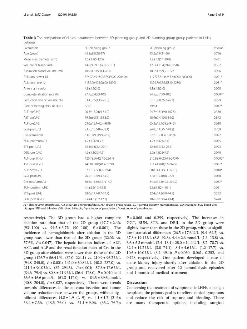
respectively). The 3D group had a higher completeablation rate than that of the 2D group (97.7 ± 2.4%(93–100) vs. 94.5 ± 3.7% (90–100), P < 0.001). Theincidence of hemoglobinuria after ablation in the 3Dgroup was lower than that of the 2D group (32.0% vs.57.6%, P = 0.047). The hepatic function indices of ALT,AST, and ALP and the renal function index of Cre in the3D group after ablation were lower than those of the 2Dgroup (126.7 ± 56.4 U/L (37.0–224.1) vs. 210.9 ± 96.2 U/L(94.8–345.0), P < 0.001; 141.0 ± 60.8 U/L (40.2–237.0) vs.211.4 ± 90.0 U/L (32–294.5), P = 0.001; 57.3 ± 17.6 U/L(34.6–79.4) vs. 80.8 ± 41.9 U/L (36.4–178.0), P = 0.010; and66.6 ± 16.6 μmol/L (51.3–117.0) vs. 84.5 ± 39.6 μmol/L(40.8–204.0), P = 0.037, respectively). There were trendstowards differences in the antenna insertion and tumorvolume reduction rate between the groups, without sig-nificant differences (4.8 ± 1.9 (2–9) vs. 4.1 ± 1.2 (2–6);53.4 ± 7.5% (43.5–76.0) vs. 51.1 ± 9.0% (35.2–76.7);
P = 0.068 and 0.299, respectively). The increases inGGT, BUN, STB, and DBIL in the 3D group wereslightly lower than those in the 2D group, without signifi-cant statistical differences (26.5 ± 17.6 U/L (9.4–64.2) vs.37.8 ± 19.1 U/L (8.8–92.8), 4.4 ± 2.6 mmol/L (1.3–13.8) vs.6.6 ± 5.3mmol/L (2.4–18.1); 28.0 ± 14.4 U/L (8.7–70.7) vs.32.4 ± 14.2 U/L (3.8–74.1); 8.4 ± 4.6 U/L (1.2–17.7) vs.10.6 ± 10.0 U/L (3.4–49.4); P = 0.060, 0.061, 0.252, and0.428, respectively). One patient developed a case ofacute kidney injury shortly after ablation in the 2Dgroup and recovered after 12 hemodialysis episodesand 1 month of medical treatment.
DiscussionConcerning the treatment of symptomatic LHHs, a benignneoplasm, the primary goal is to relieve clinical symptomsand reduce the risk of rupture and bleeding. Thereare many therapeutic options, including surgical
Table 3 The comparison of clinical parameters between 3D planning group and 2D planning group group patients in LHHspatients
Parameters 3D planning group 2D planning group P value
Age (years) 43.8±8.8(28-57) 43.2±7.9(31-60) 0.766
Mean max diameter (cm) 7.5±1.7(5-12.5) 7.2±1.3(5.1-10.8) 0.451
Volume of tumor (ml) 148.2±89.1 (26.8-301.1) 128.4±71.4(39.8-375.8) 0.352
Aspiration blood volume (ml) 108.4±84.9 (14-284) 108.5±75.9(21-359) 0.996
Ablation power (J) 87407.2.9±50387.0(3000-226400) 117775.8±46245.6(6000-200800) 0.021*
Ablation time (s) 1152.0±403.9(600-1890) 1379.7±375.8(810-2230) 0.031*
Antenna insertion 4.8±1.9(2-9) 4.1±1.2(2-6) 0.068
Complete ablation rate (%) 97.7±2.4(93-100) 94.5±3.7(90-100) 0.0004*
Reduction rate of volume (%) 53.4±7.5(43.5-76.0) 51.1±9.0(35.2-76.7) 0.299
Case of hemoglobinuria (No.) 8/17 19/14 0.047*
ALT-pre(U/L) 20.3±15.2(6.9-84.0) 24.7±18.0(9.0-107.5) 0.330
AST-pre(U/L) 19.2±6.5(11.8-38.6) 18.9±7.4(10.8-34.6) 0.872
ALP-pre(U/L) 63.0±18.1(40.4-98.8) 65.2±15.4(39.8-94.3) 0.618
GGT-pre(U/L) 23.5±16.6(8.6-38.1) 24.8±11.0(6.7-48.2) 0.709
Cre-pre(umol/L) 63.0±8.9 (49.9-78.7) 57.2±15.1(37.6-85.9) 0.092
BUN-pre(mmol/L) 4.7±1.2(2.8-7.8) 4.2±1.0(2.6-6.4) 0.055
STB-pre (U/L) 12.3±3.6(6.4-20.1) 12.4±5.3(5.4-26.3) 0.923
DBIL-pre (U/L) 4.5±1.3(2.3-7.5) 5.2±1.5(2.9-7.9) 0.070
ALT-post (U/L) 126.7±56.4(37.0-224.1) 210.9±96.2(94.8-345.0) 0.0002*
AST-post (U/L) 141.0±60.8(40.2-237.0) 211.4±90.0(32-294.5) 0.001*
ALP-post(U/L) 57.3±17.6(34.6-79.4) 80.8±41.9(36.4-178.0) 0.010*
GGT-post(U/L) 26.5±17.6(9.4-64.2) 37.8±19.1(8.8-92.8) 0.060
Cre-post(umol/L) 66.6±16.6(51.3-117.0) 84.5±39.6(40.8-204.0) 0.037*
BUN-post(mmol/L) 4.4±2.6(1.3-13.8) 6.6±5.3(2.4-18.1) 0.061
STB-post (U/L) 28.0±14.4(8.7-70.7) 32.4±14.2(3.8-74.1) 0.252
DBIL-post (U/L) 8.4±4.6 (1.2-17.7) 10.6±10.0(3.4-49.4) 0.428
ALT alanine aminotransferase, AST aspartate aminotransferase, ALP alkaline phosphatase, GGT gamma-glutamyl transpeptidase, Cre creatinine, BUN blood ureanitrogen, STB total bilirubin, DBIL direct bilirubin; *-pre: index of preablation; *-post: index of postablation
Li et al. BMC Cancer (2019) 19:550 Page 6 of 9

resection, transcatheter arterial embolization, thermalablation, steroid treatment, radiation therapy, hepaticarterial ligation, and thermal ablation. Image-guidedthermal ablation has been broadly applied clinically due toits advantages of minimal invasion, safety, convenience,efficacy, tolerability, and a shorter recovery time. Hepatichemangiomas are benign tumors of the liver that consistof clusters of blood-filled cavities (sinusoids), lined byendothelial cells and fed by the hepatic artery [44].The typical cause of hemangioma formation is abnor-
mal vascular development during embryonic develop-ment due to a lack of smooth muscle tissue in abnormalblood vessels, leading to slow blood flow, blood stag-nation, and slow heat dissipation [1]. MWA has theadvantages of a broader thermal range and a more evenspread, rapid heating speed, and reduced influence ofcarbonization and blood-flow perfusion; furthermore,multiple antennas can conduct ablation simultaneouslywithout interference, exerting a synergistic effect, thusachieving a broader ablation range and shorter ablationtime. Thus, MWA is the most suitable choice [11, 45].The complex association between LHH and the sur-
rounding vital structures could increase the difficulty andrisk of treatment, prolong the operation time, increaseablation energy requirements, and potentially lead todamage to veins, bile ducts, or the intestinal tract andincomplete ablation [46–50]. It has been reported, basedon a multivariate analysis, that the ablation time was anindependent risk factor associated with hemoglobinuria[10, 45]. In this comparative study, the data showed thatthe ablation time and energy of the 3D group were lowerthan those of the 2D group. The 3D group had a highercomplete ablation rate than the 2D group. The incidenceof hemoglobinuria after ablation in the 3D group waslower than that of the 2D group. The effects on hepaticand renal function in the 3D group were milder thanthose in the 2D group. These results indicate that the 3Dpreoperative planning system provided valuable anato-mical information regarding the LHHs and surroundingorgans, displayed stereoscopically.The calculation of the tumor volume, programming of
the antenna implantation, and precise prediction of thethermal field were also well planned, enabling a safe, pre-cise, and successful ablation. The influence on renal func-tion was reduced and the incidence of hemoglobinuriawas lower with the 3D visualization operative planningsystem. The results indicated that the LHHs were ablated,rather than normal liver tissue. Conformal ablation wasachieved with combination therapy. There was no thermaldamage to the veins, bile ducts, and intestinal tract attri-buted to the auxiliary techniques, such as hydrodissectionand thermal monitoring techniques [11, 51].In ablation therapy, the tumor volume reduction rate
is primarily dependent on the percentage of tumor
ablation. The edge of LHHs is moved farther from highrisk organs due to volume reduction after the ablationprocess, which leads to the relief of clinical symptoms.While incomplete ablation can occur and hepatic andrenal function can be markedly affected, “eccentric abla-tion therapy” can be achieved in some cases. Althoughno significant differences were identified, the volumereduction rate of the 3D visualization operative planninggroup was slightly higher. These data further suggest theimportance of the 3D visualization operative planningsystem in LHH ablation.Serious complications should not occur in the treat-
ment of benign lesions, including LHHs. One case ofacute kidney injury shortly after ablation, with recoveryafter 12 hemodialysis treatments and 1month of medicaltreatment, occurred in the 2D group. For this patient,the LHH maximum diameter was 8.6 cm and the volumewas 295.29 ml, which was not the largest LHH includedin this study. The ablation time was 1980 s and theablation energy was 326,400 J; both of these variables arerisk factors for complications. The ALT, AST, and Crelevels after ablation were 489 U/L, 1063.1 U/L, and227 μmol/L, respectively, which were markedly increasedfrom the pre-treatment values (17.3 U/L, 20.8 U/L, and75.3 μmol/L, respectively), which indicates that moreliver tissue was ablated, as verified by imaging. However,more treatments administered to patients may increasemental stress. Scientific, objective, quantifiable, andindividualized operative planning are key issues for safeand effective ablation.Although this study was a comparative study, there are
several limitations. First, this study was a retrospective in-vestigation; thus, a prospective, randomized, controlledtrial would provide more scientifically valid data. Second,the study was performed in a single institution with a rela-tively small sample. The results should be confirmed byanother cohort or a prospective, multicenter study with alarger sample size. Third, with the development andincorporation of applied mathematics, physics, and medi-cine, a more quantifiable, predictable, and controllable 3Dvisualization operative planning system may be developedthat will provide more benefits for patients and operators.
ConclusionThe 3D visualization operation planing system providedmore spatial imaging information and data on the relation-ship of the tumor with the surrounding structures; it alsoquantified the ablation techniques and the thermal field,providing scientific, objective, reasonable, quantifiable, andindividualized operative planning for US-PMWA of LHHs.These factors are key issues for safe and effective ablation.Compared with the 2D group, the 3D group showed ahigher complete ablation rate and a comparable reductionvolume rate, with less effects on hepatic and renal function
Li et al. BMC Cancer (2019) 19:550 Page 7 of 9

and a lower incidence of hemoglobinuria. Therefore,the 3D visualization operative treatment planningsystem has a relatively high clinical application valuefor use with US-PMWA for LHHs.
Abbreviations2D: Two-dimensional; 3D: Three dimensional; ALP: Alkaline phosphatase;ALT: Alanine aminotransferase; AST: Aspartate aminotransferase; BUN: Bloodurea nitrogen; CDFI: Color Doppler flow imaging; CEUS: Contrast-enhancedultrasound; Cre: Creatinine; CT: Computed tomography; DBIL: Direct bilirubin;GGT: Gamma-glutamyl transpeptidase; LHHs: Large hepatic hemangiomas;MRI: Magnetic resonance imaging; PTC: Percutaneous transhepaticcholangiography; SD: Standard deviation; TB: Total bilirubin; US: Ultrasound;US-PMWA: Ultrasound-guided percutaneous microwave ablation
AcknowledgementsNot applicable.
FundingThis work was supported by the National Key R&D Program of China (No.2017YFC0112000), three grants from the National Scientific FoundationCommittee of China (No. 81430039, 81627803 and 81801723), and the ClinicalResearch Support Foundation of the Chinese PLA General Hospital (No. 2017FC-CXYY-3005). The funders had no role in the design and conduct of the study;collection, analysis, and interpretation of the data; preparation, review, orapproval of the manuscript; and decision to submit the manuscript forpublication.
Availability of data and materialsThe datasets generated and/or analyzed during the current study will beavailable on reasonable request by contacting Dr. Xin Li ([email protected]).
Authors’ contributionsXL participated in the data analysis and drafted the manuscript. PL conceivedthe study and carried out the editorial support for this manuscript. PL, JY,ZGC.,ZYH, FYL, XLY, CA and LND participated in the ablation procedure. XL, FYL,CA and LND participated in the 3D visualization operative planning, and XL andCA participated in image collection. All authors have read and approved thefinal manuscript.
Ethics approval and consent to participateThis retrospective study protocol was approved by the Ethics Committee ofthe Chinese PLA General Hospital (Beijing, China) and was conducted inaccordance with the principles of the Declaration of Helsinki. Consent toparticipate was obtained from every participant.
Consent for publicationNot applicable.
Competing interestsThe authors declare that they have no competing interests.
Publisher’s NoteSpringer Nature remains neutral with regard to jurisdictional claims inpublished maps and institutional affiliations.
Received: 2 December 2018 Accepted: 7 May 2019
References1. Mergo PJ, Ros PR. Benign lesions of the liver. Radiol Clin N Am.
1998;36:319–31.2. Belli L, De Carlis L, Beati C, Rondinara G, Sansalone V, Brambilla G. Surgical
treatment of symptomatic giant hemangiomas of the liver. Surg GynecolObstet. 1992;174:474–8.
3. Gao J, Fan RF, Yang JY, Cui Y, Ji JS, Ma KS, et al. Radiofrequency ablation forhepatic hemangiomas: a consensus from a Chinese panel of experts. WorldJ Gastroenterol. 2017;23:7077–86.
4. Tang L, Zhou WP. Education and imaging. Hepatobiliary and pancreatic:large cavernous hemangioma with obstructive jaundice. J GastroenterolHepatol. 2009;24:930.
5. Srinivasa S, Lee WG, Aldameh A, Koea JB. HPB (Oxford). 2015;17:872–80.6. Watzke HH, Linkesch W, Hay U. Giant hemangioma of the liver
(Kasabach-Merritt syndrome): successful suppression of intravascularcoagulation permitting surgical removal. J Clin Gastroenterol. 1989;11:347–50.
7. Schnelldorfer T, Ware AL, Smoot R, Schleck CD, Harmsen WS, Nagorney DM.Management of giant hemangioma of the liver: resection versusobservation. J Am Coll Surg. 2010;211:724–30.
8. Tang XY, Wang Z, Wang T, Cui D, Zhai B. Efficacy, safety and feasibility ofultrasound-guided percutaneous microwave ablation for large hepatichemangioma. J Dig Dis. 2015;16:525–30.
9. Mocchegiani F, Vincenzi P, Coletta M, Agostini A, Marzioni M, Baroni GS, etal. Prevalence and clinical outcome of hepatic haemangioma with specificreference to the risk of rupture: a large retrospective cross-sectional study.Dig Liver Dis. 2016;48:309–14.
10. Liu F, Yu X, Cheng Z, Han Z, Dou J, Yu J, et al. Risk factors for hemoglobinuriaafter ultrasonography-guided percutaneous microwave ablation for largehepatic cavernous hemangiomas. Oncotarget. 2018;9:25708–13.
11. Liu F, Yu X, Liang P, Cheng Z, Han Z, Yu J. Ultrasonography-guidedpercutaneous microwave ablation for large hepatic cavernoushaemangiomas. Int J Hyperthermia. 2018;34:1061–6.
12. van Tilborg AA, Nielsen K, Scheffer HJ, van den Tol P, van Waesberghe JH,Sietses C, et al. Bipolar radiofrequency ablation for symptomatic giant (>10cm) hepatic cavernous haemangiomas: initial clinical experience. Clin Radiol.2013;68:e9–e14.
13. Toro A, Mahfouz AE, Ardiri A, Malaguarnera M, Malaguarnera G, Loria F, etal. What is changing in indications and treatment of hepatic hemangiomas.A review. Ann Hepatol. 2014;13:327–39.
14. Di Carlo I, Koshy R, Al Mudares S, Ardiri A, Bertino G, Toro A. Giantcavernous liver hemangiomas: is it the time to change the sizecategories? Hepatobiliary & pancreatic diseases international : HBPD INT,vol. 15; 2016. p. 21–9.
15. Hoekstra LT, Bieze M, Erdogan D, Roelofs JJ, Beuers UH, van Gulik TM.Management of giant liver hemangiomas: an update. Expert RevGastroenterol Hepatol. 2013;7:263–8.
16. Miura JT, Amini A, Schmocker R, Nichols S, Sukato D, Winslow ER, et al.Surgical management of hepatic hemangiomas: a multi-institutionalexperience. HPB (Oxford). 2014;16:924–8.
17. Li M, Zhang C, Zhang T, Wang L, Ding Y, Niu Z, et al. Outcome usingselective hemihepatic vascular occlusion and Pringle maneuver forhepatic resection of liver cavernous hemangioma. World J Surg Oncol.2015;13:267.
18. Zhang W, Huang ZY, Ke CS, Wu C, Zhang ZW, Zhang BX, et al. Surgicaltreatment of Giant liver hemangioma larger than 10 cm: a single Center'sexperience with 86 patients. Medicine. 2015;94:e1420.
19. Giavroglou C, Economou H, Ioannidis I. Arterial embolization of gianthepatic hemangiomas. Cardiovasc Intervent Radiol. 2003;26:92–6.
20. Huang XQ, Huang ZQ, Duan WD, Zhou NX, Feng YQ. Severe biliarycomplications after hepatic artery embolization. World J Gastroenterol.2002;8:119–23.
21. Firouznia K, Ghanaati H, Alavian SM, Nassiri Toosi M, Ebrahimi Daryani N,Jalali AH, et al. Management of liver hemangioma using trans-catheterarterial embolization. Hepat Mon. 2014;14:e25788.
22. Bailey J, Di Carlo S, Blackwell J, Gomez D. Same day arterial embolisationfollowed by hepatic resection for treatment of giant haemangioma. BMJCase Rep. 2016. https://doi.org/10.1136/bcr-2015-213259.
23. Park WC, Rhillips R. The role of radiation therapy in the management ofhemangiomas of the liver. JAMA. 1970;212:1496–8.
24. Gaspar L, Mascarenhas F, da Costa MS, Dias JS, Afonso JG, Silvestre ME.Radiation therapy in the unresectable cavernous hemangioma of the liver.Radiother Oncol. 1993;29:45–50.
25. Zagoria RJ, Roth TJ, Levine EA, Kavanagh PV. Radiofrequency ablation of asymptomatic hepatic cavernous hemangioma. AJR Am J Roentgenol.2004;182:210–2.
26. Park SY, Tak WY, Jung MK, Jeon SW, Cho CM, Kweon YO, et al.Symptomatic-enlarging hepatic hemangiomas are effectively treated bypercutaneous ultrasonography-guided radiofrequency ablation. J Hepatol.2011;54:559–65.
Li et al. BMC Cancer (2019) 19:550 Page 8 of 9

27. Gao J, Ji JS, Ding XM, Ke S, Xin ZH, Ning CM, et al. Laparoscopicradiofrequency ablation for large subcapsular hepatic hemangiomas:technical and clinical outcomes. PLoS One. 2016;11:e0149755.
28. Mahajan D, Miller C, Hirose K, McCullough A, Yerian L. Incidental reductionin the size of liver hemangioma following use of VEGF inhibitorbevacizumab. J Hepatol. 2008;49:867–70.
29. Yamashita S, Okita K, Harada K, Hirano A, Kimura T, Kato A, et al. Giantcavernous hepatic hemangioma shrunk by use of sorafenib. Clin JGastroenterol. 2013;6:55–62.
30. Aslan A, Meyer Zu Vilsendorf A, Kleine M, Bredt M, Bektas H. AdultKasabach-Merritt syndrome due to hepatic Giant hemangioma. Case RepGastroenterol. 2009;3:306–12.
31. Vagefi PA, Klein I, Gelb B, Hameed B, Moff SL, Simko JP, et al.Emergent orthotopic liver transplantation for hemorrhage from a giantcavernous hepatic hemangioma: case report and review. J GastrointestSurg. 2011;15:209–14.
32. Wright AS, Sampson LA, Warner TF, Mahvi DM, Lee FT Jr.Radiofrequency versus microwave ablation in a hepatic porcine model.Radiology. 2005;236:132–9.
33. Yu J, Yu XL, Han ZY, Cheng ZG, Liu FY, Zhai HY, et al. Percutaneouscooled-probe microwave versus radiofrequency ablation in early-stagehepatocellular carcinoma: a phase III randomised controlled trial. Gut.2017;66:1172–3.
34. Ziemlewicz TJ, Wells SA, Lubner MA, Musat AI, Hinshaw JL, Cohn AR, et al.Microwave ablation of giant hepatic cavernous hemangiomas. CardiovascIntervent Radiol. 2014;37:1299–305.
35. Aydin C, Akbulut S, Kutluturk K, Kahraman A, Kayaalp C, Yilmaz S. Gianthepatic hemangioma presenting as gastric outlet obstruction. Int Surg.2013;98:19–23.
36. Koszka AJ, Ferreira FG, de Aquino CG, Ribeiro MA, Gallo AS, Aranzana EM,et al. Resection of a rapid-growing 40-cm giant liver hemangioma. World JHepatol. 2010;2:292–4.
37. Zhou JX, Huang JW, Wu H, Zeng Y. Successful liver resection in a gianthemangioma with intestinal obstruction after embolization. World JGastroenterol. 2013;19:2974–8.
38. van Tilborg A, Dresselaars HF, Scheffer HJ, Nielsen K, Sietses C, van den TolPM, et al. RF ablation of Giant hemangiomas inducing acute renal failure: areport of two cases. Cardiovasc Intervent Radiol. 2016;39:1644–8.
39. Liu F, Liang P, Yu X, Lu T, Cheng Z, Lei C, et al. A three-dimensionalvisualisation preoperative treatment planning system in microwaveablation for liver cancer: a preliminary clinical application. Int JHyperthermia. 2013;29:671–7.
40. Schumann C, Bieberstein J, Braunewell S, Niethammer M, Peitgen HO.Visualization support for the planning of hepatic needle placement. Int JComput Assist Radiol Surg. 2012;7:191–7.
41. Wu W, Xue Y, Wang D, Li X, Xue J, Duan S, et al. Application of 3D imagingin the real-time US-CT fusion navigation for minimal invasive tumor therapy.Int J Comput Assist Radiol Surg. 2015;10:1651–8.
42. Li X, Yu J, Liang P, Yu X, Cheng Z, Han Z, et al. Combination therapy ofthree-dimensional (3D) visualisation operative treatment planning systemand US-guided percutaneous microwave ablation in larger renal cellcarcinomas (D >/= 4 cm): preliminary results. Int J Hyperthermia. 2017;33(3):271-7.
43. Sacks D, McClenny TE, Cardella JF, Lewis CA. Society of InterventionalRadiology clinical practice guidelines. J Vasc Interv Radiol. 2003;14:S199–202.
44. Bajenaru N, Balaban V, Savulescu F, Campeanu I, Patrascu T. Hepatichemangioma -review. J Med Life. 2015;8(Spec Issue):4–11.
45. Wang Z, Tang X, Qi X, Shi Y, Chi J, Li P, et al. Feasibility, safety, and efficacyof ultrasound-guided percutaneous microwave ablation for giant hepatichemangioma. Int J Hyperthermia. 2018;35(1):246-52.
46. Groeschl RT, Riggle KM, Quebbeman EJ, Christians KK, Turaga KK, Tsai S,et al. Hepatectomy for hemangioma; safe, but is it successful? Hepato-gastroenterology. 2014;61:2009–13.
47. Gao J, Ke S, Ding XM, Zhou YM, Qian XJ, Sun WB. Radiofrequency ablationfor large hepatic hemangiomas: initial experience and lessons. Surgery.2013;153:78–85.
48. Choi J, Lee YJ, Hwang DW, Chon SH, Nagpal A, Park KM. Surgical treatment ofgiant hepatic hemangiomas: technical point of view. Am Surg. 2011;77:48–54.
49. Ho HY, Wu TH, Yu MC, Lee WC, Chao TC, Chen MF. Surgical managementof giant hepatic hemangiomas: complications and review of the literature.Chang Gung Med J. 2012;35:70–8.
50. Zenzen W, Perez-Atayde AR, Elisofon SA, Kim HB, Alomari AI. Hepatic failurein a rapidly involuting congenital hemangioma of the liver: failure ofembolotherapy. Pediatr Radiol. 2009;39:1118–23.
51. Zhang D, Liang P, Yu X, Cheng Z, Han Z, Yu J, et al. The value ofartificial pleural effusion for percutaneous microwave ablation of livertumour in the hepatic dome: a retrospective case-control study. Int Jhyperthermia. 2013;29:663–70.
Li et al. BMC Cancer (2019) 19:550 Page 9 of 9