the use of osteopathic manipulation in a clinic and home ... · cage motion; and a five-question...
TRANSCRIPT

The Use of Osteopathic Manipulation in a Clinic and Home Setting to Address Pulmonary Distress as Related to Asthma in Southwest Virginia
Macy Little Latter
Dissertation submitted to the Faculty of the Virginia Polytechnic Institute and State University
In partial fulfillment of the requirements for the degree of
Doctor of Philosophy
In
Education, Curriculum and Instruction
Dean Sutphin, Chair Kerry Redican David Harden Billie Lepczyk
January 30, 2009
Blacksburg, Virginia
Keywords: asthma, osteopathic manipulative treatment, Southwest Virginia
Copyright 2008, Macy Little Latter
The Use of Osteopathic Manipulation in a Clinic and Home Setting to Address
Pulmonary Distress as Related to Asthma in Southwest Virginia
Macy Little Latter
ABSTRACT
Osteopathic Manipulative Therapy (OMT) is underutilized in addressing lung
function and symptoms in asthma patients. The objective of this study was to
determine if a single session of OMT can improve lung function and symptoms in
patients suffering from asthma, and if patients can be taught a self-administered
home OMT protocol to control their symptoms, in order to develop a protocol by
which physicians can apply OMT to address lung disease in patients. This was a
purposive randomized controlled quasi-experimental study which took place in family
practice, pulmonology, and asthma specialist offices in southwest Virginia. The
intervention was a ten-minute semi-individualized OMT protocol and a self-
administered home OMT education session.
Variable baseline, within-subject study design was utilized, allowing each
person to serve as his or her own control. Pre and posttest measurements included:
participant spirometry FEV1, FVC, and PEF; thoracic excursion upper and lower rib
cage motion; and a five-question rating scale to determine current asthma symptoms.
A ten-minute OMT session included an individualized thoracic and rib screening and
treatment, suboccipital release, diaphragm release, and thoracic pump. Comparison
between pre- and post-OMT lung function and symptoms portrayed change. For the
second part of the study, the participants were divided into two groups with group
two receiving a ten-minute home OMT education session and a handout of the home
iii
OMT techniques. All participants returned two weeks later for a follow up lung
function assessment.
Statistically significant (p<.05) improvements after initial OMT were
documented for 8 of 10 measurements. Only two spirometry values, FEV1 and PEF, did
not significantly improve. The group who participated in the home OMT education
session had statistically significant improvements in 3 of 10 measurements, including
the upper and lower thoracic excursion measurements and the overall asthma
symptoms rating.
With a simple, easy to repeat, 10 minute semi-individualized OMT session,
researchers demonstrated improved lung function and symptoms in this group of
participants in Southwest Virginia. The addition of a home OMT education session was
demonstrated to be at least partially beneficial. Future studies should expand on this
pilot study with the researchers recommending using a larger patient population
including patients with lower pre-treatment spirometry values in order to accurately
monitor potential for change.
iv
Dedication
This thesis is dedicated to VCOM, which was gracious enough to give me the
opportunity of a lifetime to explore international community medicine and allow me
to expand my knowledge and perspectives as a physician. Also, I would like to thank
my husband and family. Without their unwavering support and unending patience, I
would not have accomplished all that I have.

v
Acknowledgements
I would like to thank each of my participants for volunteering to help with this
study. I would also like to thank my committee: Dr. Dean Sutphin, Dr. David Harden,
Dr Billie Lepczyk and Dr. Kerry Redican, for their support and interest. Also, I would
like to acknowledge Dr. Stephen Werre for his statistical aid. In addition, I would like
to acknowledge the financial support of the Edward Via Virginia College of
Osteopathic Medicine in completing this study and for supporting the Global Health
Leadership Fellowship that enabled me to pursue the PhD and complete the thesis
research of which these findings were part.
vi
Table of Contents
Abstract ……………………………………………………………………………………………………………………ii Dedication………………………………………………………………………………………………………………..iv Acknowledgements …………………………………………………………………………………………………..v Table of Contents …………………………………………………………………………………………………….vi Chapter 1: Introduction …………………………………………………………………………………………….1
Background of Problem ………………………………………………………………………………….1 Statement of Problem …………………….…………………………………………………………….4 Purpose of Study …………………..………………………………………………………………………4 Research Objectives ….….……………………………………………………………………………..4 Research Hypotheses ….……….……………………………………………………………………….5 Definition of Terms …..………………………………………………………………………………….6 Limitations of Study ….…………………………………………………………………………………..7 Significance of the Study ….…………………………………………………………………………..7
Chapter 2: Literature Review …………………………………………………………………………………..9
Asthma ….……………………………………………………………………………………………………….9 Asthma in Southwest Virginia ….………………………………………………………………….15 Osteopathic Manipulation ….………………………………………………………………………..16 Previous Research on the Use of Various Types of Manipulation for Asthma ………………………………………………………………….19 Problems in the Literature ….………………………………………………………………………22
Chapter 3: Methodology ………………………………………………………………………………………….23
Population ….………………………………………………………………………………………………..23 Sample ….….………………………………………………………………………………………………….23

vii
Procedure …………………………………………………………………………………………………….24
Design …………………………………………………………………………………………………24 Recruitment and Criteria .………………………………………………………………….26 First Visit …………………………………………………………………………………………….27 Patient Survey ……………………………………………………………………………..28 Thoracic Excursion ……………………………………………………………………….28 Spirometry ……………………………………………………………………………………29 OMT Session ………………………………………………………………………………….30 Individualized Thorax and Rib Treatment ……………………….31 Diaphragm Release ……………………………………………………………33 Occipito-Atlantal Joint …………………………………………………….34 Thoracic Lymphatic Pump ……………………………………………….34 Division into Groups …………………………………………………………………………..35 Home OMT education session ……………………………………………………..35 Suboccipital Release ……………………………………………………………………35 Rib Raising …………………………………………………………………………………..36 Pectoral Traction ………………………………………………………………………..36
Two week follow up……………………………………………………………………..37 Instrumentation….…………………………………………………………………………………..37 Data Analysis ….………………………………………………………………………………………39
Chapter 4: Results ………………………………………………………………………………………………………..41 Objective 1 ….……….…………………………………………………………………………………………..45 Spirometry ………………………………………………………………………………………………45
viii
Thoracic Excursion …………………………………………………………………………………46 Patient Symptoms ………………………………………………………………………………….47 Objective 2 ….…………………………………………………………………………………………………….47 Spirometry ………………………………………………………………………………………………48 Thoracic Excursion …………………………………………………………………………………48 Patient Symptoms ………………………………………………………………………………….50 Mini Asthma Quality of Life Questionnaire (MAQLQ) …………………………….51 Home OMT Group Follow Up Questionnaire ………………………………………….53 Objective 3 ….…………………………………………………………………………………………………..55 Objective 4 ….…………………………………………………………………………………………………..59 Chapter 5: Summary and Conclusion …………………………………………………………………………..60 Summary ….……………..………………………………………………………………………………………..60 Conclusion ….……………………………………………………………………………………………………..61 Initial pre and post OMT assessment ……………………………………………………..61 Use of Home OMT ……………………………………………………………………………………62 Environmental Impact …………………………………………………………………………….62 Effect of Age and Gender ……………………………………………………………………….62 Recommendations for Future Research ….……………………………………………….…….. 64 Recommendations for Future Practice ….…………………………………………….…………..64 References …………………………………………………………………………………………………………………….67 Appendix A Mini AQLQ ………………………………………………………………………………………………………….74 Appendix B
ix
Patient Questionnaire ………………………………………………………………………………………76 Appendix C Pre-OMT Survey ……………………………………………………………………………………………….79 Appendix D Post-OMT Survey ………………………………………………………………………………………………80 Appendix E Follow-up Survey ……………………………………………………………………………………………..81 Appendix F Patient Home OMT Handout …………………………………………………………………………….82
x
List of Tables
Table 1. Pre and Post-OMT Spirometry Value Statistics……………………………………………….45 Table 2. Pre and Post-OMT Thoracic Excursion Values…………………………………………………46 Table 3. Pre and Post-OMT Asthma Symptoms…………………………………………………………….47 Table 4. Paired Sample t-Test Results for Spirometry at Baseline and Follow Up……..48
Table 5. Paired Sample t-Test Results for Upper and Lower Rib Thoracic Excursion Measurements.………………………………………………………………………………………………………………50 Table 6. Upper Ribs Comparison of the Interaction Between Gender and Home OMT Sliced by Gender…………………………………………………………………………………………………………..50 Table 7. Upper Ribs Comparison of the Interaction Between Gender and Home OMT Sliced by Home OMT …………………………………………………………………………………………………….50 Table 8. Wilcoxin 2-Sided Test for the Rating Scale …………………………………………………..51 Table 9. Chi-Squared Test to Determine Effect of Home OMT on MID Scores from the MAQLQ ………………………………………………………………………………………………………………………….52
xi
List of Figures
Figure 1. Prevalence of Asthma in the United States and Virginia …………………………….3 Figure 2. Classification of Asthma ……………………………………………………………………………….12 Figure 3. Study Design ………………………………………………………………………………………………….25 Figure 4. Gender Classification of Participants….…………………………………………………………43 Figure 5. Ethnicity Classification of Participants …………………………………………………………43 Figure 6. Histogram of Age of Participants ………………………………………………………………….44 Figure 7. Classification of Asthma Participants …………………………………………………………..44 Figure 8. Home OMT Follow Up Indicators ………………………………………………………………….53 Figure 9. Patient Questionnaire Responses …………………………………………………………………56

1
Chapter 1
Introduction
This first chapter will serve to introduce the problem which was chosen to be
studied. The purpose, objectives, hypothesis, and limitations will be reviewed in
order to introduce the research and create a better understanding for the reader.
Background of the problem Lung disease is any disorder in which the lungs do not function properly. Lung
disease has been classified into three main types: obstructive, restrictive, and
defective in the transfer of oxygen from air to blood. Despite these classifications,
most lung disease is actually a combination of the above. Some of the most common
lung diseases include asthma, chronic bronchitis, chronic obstructive lung disease
(COPD), emphysema and interstitial lung disease (Kaufman, 2006). In this project, I
focused on the obstructive lung disease asthma. Obstructive disease is characterized
by an increase in the resistance to airflow due to a partial or complete obstruction of
the trachea, bronchi, or bronchioles. The individual disorders of emphysema, chronic
bronchitis, asthma, and bronchiectasis have distinct clinical characteristics (Kumar,
Abbas, & Fausto, 2005, p. 717).
Asthma is a chronic inflammatory disease which affects more than 20 million
Americans (American Lung Association, 2005). This disease manifests as a reversible
bronchospasm of smooth muscle which develops further into bronchial edema, viscous
secretions, mucous plugging, and atelectasis (Camargo, 2006). About 4-5% of adults
and 10% of children have been estimated to have symptoms of asthma (Kasper et al.,
2005). These attacks, which are triggered by allergens, exercise, infections, weather

2
changes, or other irritants, range from mild shortness of breath and coughing to
wheezing, chest pain, and even hospitalization. This disease poses a significant
burden on the United States. Asthma causes around 4000 deaths and costs the United
States $6-$11 billion each year. Prescriptions drugs are the most significant medical
expenditure, reaching a total of $5 billion a year (American Lung Association, 2005).
In addition, asthmatic children miss 12.8 million days of school each year, while
adults miss 24.5 million workdays (Scariati, Roberge, & Dye, 2006).
Asthma is characterized by narrowing of the bronchial airways and contraction
of bronchial smooth muscle due to inflammation. In addition, there are antigen-
antibody interactions that set off a metabolic cascade, which results in the formation
of potent smooth muscle contractors such as leukotrienes. Bronchomotor tone is
further controlled by an intricate balance between the adrenergic and cholinergic
nervous systems with beta-receptor blockage in the lungs leading to
bronchoconstriction (Camargo, 2006; Kasper et al., 2005; Kumar et al., 2005).
This obstructive lung disease is prevalent in southwest Virginia. In Virginia, one
in sixteen children suffers from asthma and it is the leading cause of hospitalization.
This disease affects low-income and minority patients the most, who are prevalent in
southwest Virginia (CHIP of Virginia, 2002). The 2007 American Lung Association State
of the Air Report shows that the particle pollution levels in the southwest counties in
Virginia have increased over the years (American Lung Association, 2007). The CDC’s
Behavioral Risk Factor Surveillance System reported that, while the overall incidence
of asthma in adults has dropped in the United States since 2004, the incidence in
Virginia has actually risen ("Asthma,") as has the economic burden (Virginia Asthma

3
Coalition, 2004) (Figure 1). There were over 9,000 hospital discharges with a primary
diagnosis of asthma in Virginia in 2004, with the total charges reaching $96 million
(Virginia Asthma Coalition, 2004).
Figure 1. Prevalence of Asthma in the United States and Virginia
There are multiple pharmacologic medications designed to alleviate these lung
diseases today. While these treatments have been effective in controlling asthma,
there is another option for relieving patients with this condition. This option is
osteopathic manipulative treatment (OMT). There is evidence in the literature,
dating back from the beginning of osteopathy in 1874, that OMT has a role in
controlling lung disease (Allen, 1971). The foundation of osteopathy is the intricate
reciprocal relationship between the body’s structure and it’s function, along with the
belief that the body contains within it an ability to heal itself (Ward, 2003). By

4
applying this concept to patients with asthma, osteopathic physicians can address
asthma in a unique way. In addition, there are no studies regarding home OMT
treatment methods for patients with lung disease. These home treatments may be a
way to address the issue of access to care in rural southwest Virginia and give patients
another option for treating their lung condition.
Statement of the problem
There is a high prevalence of asthma in southwest Virginia and there are multiple
osteopathic manipulative techniques (OMT) which are thought to improve lung
function. However, the studies regarding the effectiveness of using osteopathic
manipulative treatment (OMT) as an office treatment or a home treatment to address
obstructive lung disease are inadequate.
Purpose of Study
The purpose of this study was to determine the viability of OMT treatment for
asthma and the effectiveness of home OMT education, in order to implement a
program to improve asthma control.
Research Objectives
1. Determine the relationship of OMT on lung function as demonstrated by
basic spirometry, thoracic excursions, and subjective patient symptoms.
5
2. Compare lung function measures of asthma patients using self-administered
home treatment as supplemental to doctor-administered OMT treatment with
those of asthma patients who only receive doctor-administered OMT.
3. Determine the relationship among selected environmental factors such as
job type, pets, and smoking habits with demographics characteristics of
patients in this study.
4. Determine the relationship of age and gender on the use of OMT for asthma.
Research Hypotheses
1. A researcher-developed protocol of Osteopathic Manipulative Treatment,
including screening and treatment of the thoracic and rib cage, diaphragm
release, suboccipital release, and thoracic lymphatic pump, will
significantly (p<.05) improve the spirometry values, thoracic excursion
measurements, and symptoms in the selected group of participants suffering
from asthma.
2. A researcher-developed educational session designed to teach asthma
patients home OMT will significantly improve the spirometry, thoracic
excursion measurements, and symptoms in those participants over a two-
week time period, as compared to a group of asthma patients who do not do
home OMT.
3. Differences in age and gender will be significantly related to response to
OMT techniques for asthma.
6
4. There will be a significant relationship between environmental factors of
job type, pets, and smoking habits and asthma symptoms in the chosen
population.
Definition of Terms
For the purpose of my study, I have defined the following:
Asthma – A reversible obstructive lung disease characterized as a chronic
inflammatory disorder of the airways that causes recurrent episodes of
wheezing, breathlessness, chest tightness, and cough.
Environmental factors –Selected factors which may act as intervening variables and
have an affect on the patient’s asthma. The factors which will be studied in
this project are work environment, home environment, smoking, exercise, and
allergies.
Osteopathic manipulative therapy (OMT) – “The therapeutic application of manually
guided forces by an osteopathic physician to improve physiologic function
and/or support homeostasis that have been altered by somatic dysfunction.”
(Ward, 2003)
Spirometry – A test to measure how much air one can move in and out of their lungs
and how quickly this can be done.
Thoracic Excursion – The difference between thoracic circumference at the end of
forced inspiration and thoracic circumference at the end of forced expiration.
7
Limitations of Study
There are several limitations to this study. One of the most limiting factors is
the number of subjects to which I had access. Due to time and financial constraints, I
was only able to gather data from 32 subjects. Another limitation inherent in this
study is reliance on patient compliance and effort. The results of the spirometry test
are dependent upon the effort that the patient displays when doing the test. If the
patient does not adequately try to perform the test as specified, then the results
regarding lung function are inaccurate. Another part of the study which relies on the
patient’s compliance and effort is the request to perform home OMT maneuvers. In
order to obtain correct results on the benefit of home OMT, the patient is required to
actually perform the home OMT sessions as prescribed. Researchers did not make
home visits to ensure that the patients were doing the techniques or that they were
doing them correctly. Another limitation to this study is the two-week time period
between clinic visits. During this time period, the researcher had no control over
what the patient did and there are multiple variables which may have had an affect
on patients’ asthma and skewed the results of the study. These are all limitations
over which the researcher had no control.
Significance of the Study
This project has the potential to significantly impact people in southwest
Virginia living with an obstructive lung disease. If the primary researcher finds that

8
OMT improves lung function, as demonstrated by thoracic excursion, spirometry
values, and a subjective improvement, then OMT may be a beneficial addition to the
treatment regime of asthma patients. In addition, this project has the potential to
financially impact the inhabitants of southwest Virginia living with an obstructive lung
disease. The primary researcher will be teaching half of the subjects a home
treatment regime which may enable them to control their symptoms without spending
additional money on medications or physician visits. Another significant part of this
project is the study of the longevity of OMT directed towards lung function. It will be
useful to determine how long the treatments last, so that the treatment plans for
these patients can be better validated. Lastly, this project has the potential to
increase the use of OMT in southwest Virginia by increasing the awareness of its utility
in asthma and encouraging physicians to utilize it.
A review of literature was utilized to determine previous work in the area,
review methodological approaches and develop a strong conceptual grounding and
theoretical base to guide the study and demonstrate its importance.

9
Chapter 2
Literature Review
In this chapter, the literature pertaining to asthma, asthma in Virginia, and the
use of manipulation will all be explored. This will provide a background understanding
for the reader and a foundation for the remaining chapters on the project itself.
Asthma
Asthma is a chronic inflammatory disease of the lungs (Goroll & Jr, 1995, p.
304; Kasper et al., 2005, p. 668; Kumar et al., 2005, p. 723). This inflammation
increases the lung’s responsiveness to stimuli, which results in episodic, reversible
obstruction of the lower airways. The severity ranges from mild limitation of the
patient’s activity to severe and even life-threatening symptoms (Kasper et al., 2005,
p. 668).
There is a complex pathophysiology associated with asthma which involves
bronchial hyperresponsiveness, airway inflammation, and intermittent airway
obstruction (Morris, 2007). Airway inflammation is the underlying disorder in asthma.
There are several characteristics which lead to this inflammation, including mast cell
degranulation, eosinophilic infiltration, recruitment and proliferation of T cells, and
endothelial activation. Once this happens, the normal epithelial lining of the airways
is denuded and goblet cells begin to proliferate. This edema causes sensitization of
the bronchial walls and results in hyperresponsiveness to stimuli. This
hyperresponsiveness results in airway obstruction (Goroll & Jr, 1995, p. 304).
The etiology of airway hyperresponsiveness is not clear; however, there are
several triggers for airway reactivity, such as allergenic, pharmacologic,
10
environmental, occupational, infectious, emotional, and exercise-related (Kasper et
al., 2005, p. 668). Airway obstruction results from inflammation, acute
bronchoconstriction, mucous plugging, airway edema, and remodeling. The early
asthmatic response and bronchoconstriction results from an immunoglobulin E
mediated cascade which follows an allergen exposure. The airway edema occurs
anywhere from 6-24 hours after this allergen exposure. Mucous plugging is formed
from exudates of serum proteins and cellular debris which can take weeks to resolve.
Longstanding inflammation causes airway remodeling and structural changes, which
may affect the reversibility of asthma (Morris, 2007).
In addition to these pathways of asthma, there is a neurogenic role as well.
The bronchial smooth muscles have autonomic innervation which either directly or
indirectly affects their function. Parasympathetics cause bronchial constriction while
sympathetics cause bronchial relaxation. Both bronchial irritants and emotional
stress appear to trigger vagal reflexes and stimulate bronchospasm (Goroll & Jr, 1995,
p. 305).
Though the cause of asthma is unclear, there are several triggers which have
been identified. There seems to be a genetic link in the sense that if someone in the
family has asthma, it is likely that other family members will have it. Also, there are
environmental triggers such as air pollution, smoke, and allergens. Some of the most
common allergen triggers for asthma include animal dander, cockroaches, dust mites,
mold, and pollen ("Lung Diseases: Asthma," 2006). The strongest allergens which lead
to sensitization are dust mites and cockroach antigens which explains why there is a
high prevalence of this disease in inner-city residents (Goroll & Jr, 1995, p. 305).
11
Other irritants which may act as triggers are cold air, strong odors, and scented
products. There are other potential causes, including gastroesophageal reflux,
infections, medicines such as aspirin and beta-blockers, and sulfites in foods (Lung
Diseases: Asthma, 2006).
Asthma is prevalent in industrialized nations and affects an estimated 5-10% of
the population. About 5 million of those affected are children younger than six years.
There has been a recent rise in the prevalence and morbidity associated with asthma.
Suspected causes for this increase are environmental allergens, increased air
pollution, urbanization, and passive smoking. Asthma occurs in people of all races,
but in the United States, there is a higher percentage of this disease in African
Americans. In early childhood, the ratio of boys with asthma to girls is 2:1; however,
after puberty the ratio is 1:1, and into adulthood it shifts to 1:2 (Morris, 2007). The
gender differences have been studied and found to be similar in research studies
(Berhane et al., 2000).
The clinical presentation of asthma includes wheezing, dyspnea, cough, and
chest tightness. Although wheezing is the most common presentation, if the
obstruction involves the smaller airways, then wheezing may not be present. The
physical examination may vary from individual to individual. Between acute episodes,
there may be no abnormal findings on exam. If the asthma is associated with
allergies, there may be indication of this such as ocular shiners, a transverse crease
on the nose, allergic rhinitis, and conjunctival inflammation. The anterior-posterior
diameter of the chest may be increased due to hyperinflation of the lungs which may
lead to abdominal breathing patterns. During an acute asthma attack, the physical
12
exam will be different. The respiratory rate will likely be increased, there will likely
be a wheeze or crackles heard on auscultation of the lungs, and the oxygen saturation
will be decreased (Sharma, 2006).
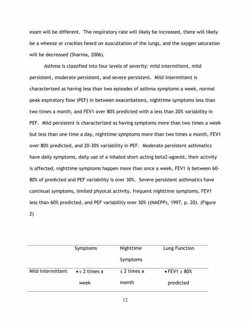
Asthma is classified into four levels of severity: mild intermittent, mild
persistent, moderate persistent, and severe persistent. Mild intermittent is
characterized as having less than two episodes of asthma symptoms a week, normal
peak expiratory flow (PEF) in between exacerbations, nighttime symptoms less than
two times a month, and FEV1 over 80% predicted with a less than 20% variability in
PEF. Mild persistent is characterized as having symptoms more than two times a week
but less than one time a day, nighttime symptoms more than two times a month, FEV1
over 80% predicted, and 20-30% variability in PEF. Moderate persistent asthmatics
have daily symptoms, daily use of a inhaled short-acting beta2-agonist, their activity
is affected, nighttime symptoms happen more than once a week, FEV1 is between 60-
80% of predicted and PEF variability is over 30%. Severe persistent asthmatics have
continual symptoms, limited physical activity, frequent nighttime symptoms, FEV1
less than 60% predicted, and PEF variability over 30% ((NAEPP), 1997, p. 20). (Figure
2)
Symptoms Nighttime
Symptoms
Lung Function
Mild Intermittent • ≤ 2 times a
week
≤ 2 times a
month
• FEV1 ≥ 80%
predicted

13
• PEF variability <
20%
Mild Persistent • 2 times a week
but < 1 time a
day
• Activity may be
affected
> 2 times a
month
• FEV1 ≥ 80%
predicted
• PEF variability
20-30%
Moderate
Persistent
• Daily symptoms
• Daily use of
inhaled short-
acting beta2-
agonist
• Activity
affected
> 1 time a week • FEV1 60-80%
predicted
• PEF variability
>30%
Severe Persistent • Continual
symptoms
• Limited physical
activity
Frequent • FEV1 <60%
predicted
• PEF variability >
30%
Figure 2. Classification of asthma
Aside from clinical diagnoses, there are a few tests which can be performed to
diagnose asthma. The National Asthma Education and Prevention Program

14
recommends the use of spirometry to diagnose and follow asthma ((NAEPP), 1997).
An obstructive disorder such as asthma presents with a normal forced vital capacity
and a reduced FEV1. Also associated with asthma is a ≥ 12% or 200mL improvement in
FEV1 after inhaling a short-acting bronchodilator (Sharma, 2006). Peak flow meters
are another way of testing and monitoring asthma. Although it is a less reliable test,
patients may use peak flow monitoring at home to assess their current asthma status
(Morris, 2007). Laboratory tests are not indicated to diagnose asthma, but can be
used to rule out other possible diagnoses.
Asthma carries a certain level of morbidity and mortality. The continuous
inflammation may cause airway remodeling which results in further exacerbations of
asthma. Overall, there is a 0.1% mortality each year from this disease. Despite
advancements in therapy, the current mortality associated with asthma has risen over
the last decades(American Lung Association, 2005). The morbidity associated with
asthma is largely a diminished quality of life. Activity can be affected by the severity
of asthma. It is estimated that about 100 million work and school days are lost each
year because of this disease. In addition, more than 1.8 million emergency room
visits are due to asthma (Morris, 2007). This places a large financial burden on
everyone involved. The estimated direct medical cost to our nation, with prescription
drugs accounting for the largest part, is around $11.5 billion, while the indirect costs,
such as loss of productivity, add another $4.6 billion (American Lung Association,
2005).
The treatment of asthma may involve many avenues. The most successful
treatment is the removal of irritating agents and triggers. The next line of treatment
15
is one of two pharmacologic routes: inhibit smooth muscle contraction and reverse
inflammation (Kasper et al., 2005, p. 669). Pharmacologic treatment varies
depending on the severity of the disease, with each classification having different
recommended treatments. To inhibit smooth muscle contraction, beta-adrenergic
agonists, anticholinergics, and methylxanthines are used. To reverse or prevent
inflammation, glucocorticoids, mast cell stabilizing agents, and leukotriene inhibitors
are used. Treatment begins with a short-acting beta2 agonist, then a low-dose
inhaled corticosteroid. The next step is to add a long-acting beta2 agonist and
further medications to address the inflammation (Kasper et al., 2005, p. 669) .
Asthma in Southwest Virginia
Rural southwest Virginia is not immune to the perils of asthma. In fact, the
prevalence for areas of southwest Virginia was higher than those for the United
States in a study conducted in 2004. In Roanoke, the rate per 10,000 was 20.2 while
the same rate in the United States was 17.0 (Virginia Asthma Coalition). Of the
southwestern counties evaluated in a 2007 report from the American Lung
Association, Wythe County had a 8.67% prevalence, Salem had a 8.65% prevalence,
Roanoke County had a 8.62% prevalence, and Bristol had a 8.61% prevalence. Of
those ranked, Salem, Roanoke, and Bristol all scored a C on the particle pollution
scale (American Lung Association, 2007).
One toll of asthma is significant financial burden. In Virginia, there were 9,460
hospital visits, totaling $96 million, in 2004. This is a significant amount of money

16
when considering the rural population. There are other risks besides financial when
dealing with asthma. In Virginia, from 1999 to 2004, there were 680 asthma deaths
(Asthma in Virginia: A Comprehensive Data Report, 2006). Death from asthma is rare
and preventable, and this number demonstrates a lack of access to care or a problem
in self-management of asthma which needs to be addressed.
A study conducted in 2001 analyzed the difference between urban and rural
populations with asthma. It was found that in the rural areas, up to 95% of the study
participants had primary care physicians taking care of their asthma treatment (Kuo
& Craig, 2001). Therefore, it is important that these rural family practice physicians
have methods to empower and treat their patients living with asthma.
Due to the increasing incidence and the economic burden of asthma, it is
reasonable to begin to think about other options for asthma control. One other option
for improving asthma control is to utilize osteopathic manipulation. The following
literature review will serve to inform the reader as to why this treatment was chosen
to further research.
Osteopathic Manipulation
Osteopathic manipulative treatment (OMT) is manual therapy delivered to
alleviate somatic dysfunctions and improve physiologic function. The founder of
osteopathy, Andrew Taylor Still, began teaching these methods in the first
osteopathic school in 1892 and techniques have been expanded since then (Ward,
2003, p. 21). The principles and theory of OMT allow it to be utilized for a variety of
17
conditions and disease states. There are several characteristics which make it a good
choice for addressing various lung diseases, such as asthma. With the use of
manipulative treatment, patients could have improved function of their lungs,
enhanced healing of the airways, and improved quality of life with or without the
concomitant use of standard pharmacologic treatment.
The autonomic nervous system plays a role in asthma through its innervations
to the airways (Raji, 2005). The respiratory system receives innervation from a large
pulmonary plexus which sits near the pulmonary artery. The lateral horn of thoracic
spinal segments 2-7 send sympathetic neurons to this plexus. Most of these nerves
target the glandular tissue which surrounds the bronchi as opposed to targeting the
musculature like other nerves do. When stimulated, these beta-adrenergic receptors
of the sympathetic nervous system cause bronchial dilation and viscous secretions.
The parasympathetic innervations to the airways occur via the Vagus nerve. The
pulmonary branches of this nerve terminate on bronchial smooth muscle, mucosal
glands, and blood vessels. Parasympathetic stimulation causes bronchoconstriction,
vasodilation, and hypersecretion of serous fluid (Blumenfeld, 2002, p. 230). By
understanding the nervous system and the lungs, OMT techniques have been
developed to address this area of concern. There are several techniques which are
thought to balance the parasympathetics and sympathetics, thus improving the
symptoms of asthma.
Thoracic structure also plays a role in lung function and asthma. The thoracic
cage must be able to move unimpeded in order to maintain sufficient arterial supply,
venous and lymphatic drainage, and response to the neural regulatory system. The
18
ribs, surrounding musculature, and diaphragm all influence the physiology of the lungs
(Ward, 2003, p. 506). Many OMT techniques address structure as it relates to
function. In treating asthma, there are many structures which can be manipulated to
improve their function. OMT can be directed towards the rib cage, the musculature,
and the diaphragm to improve their mobility and thus improve the symptoms of
asthma.
In addition to neural and structural influences, there are lymphatic influences
on respiration. Osteopathic physicians have long been interested in the role of
lymphatics in maintaining health. Proper lymphatic drainage encourages proper
tissue activity and metabolism and aids in immunologic function. Effective lymphatic
drainage of the lungs is normally achieved by contraction of the diaphragm and
thoracic cage movement during respiration. However, in patients with asthma, both
of these mechanisms may be compromised, leading to impaired lymphatic drainage.
This impairment results in reduced healing time, a diminished ability to fight
infection, and decreased ability for medications to reach their target tissues. There
are several OMT techniques which address the problem of lymphatics: rib raising,
diaphragm release, and lymphatic pumps (Sevarese et al, 2003, p. 125).
An article written by two osteopathic physicians describes the benefit that OMT
can have on treating respiratory problems. After a 69-year-old man was admitted to
the hospital with respiratory failure, the physicians prescribed treatment which
combined OMT with standard patient care to apply the many benefits of OMT to
address this problem. In the end, the authors describe respiration as “a dynamic
orchestration involving coordinated reflex neural activity; abdominal, diaphragmatic,

19
and other muscular contractions; motions of fascial planes; and the movement of
more than 146 joints” (Stretanski & Kaiser, 2001, p. 448). These physicians had found
that the altered structure of this patient’s thoracic cage resulted in inefficient
function of his respiratory system. With appropriate osteopathic correction, this
man’s respiration improved dramatically.
Previous Research on the Use of Various Types of Manipulation for Asthma
While osteopathic manipulation is frequently used to address asthma in
practice, there are limited studies which prove its effectiveness. In fact, there are
very few research papers which evaluate any use of OMT which makes its use for
asthma both difficult and important to study. Some researchers have studied the use
of manipulation with asthma, chronic obstructive pulmonary disease, and other
respiratory problems such as pneumonia and post-surgery lung function. The
chiropractic community has put together several studies regarding the use of their
manipulation on asthmatics, which is similar to osteopathic manipulation on some
levels. For those reasons, the chiropractic literature will be used here to provide
some background and review for this study which uses osteopathic manipulation.
The literature in the chiropractic research is inconsistent. Some studies reveal
that chiropractic manipulation benefits patients with asthma, and some studies
demonstrate that there is no benefit. This may be due to the variability across
techniques, patients, and chiropractors. One of the largest studies in the chiropractic
literature was conducted with 420 participants in Australia. This study was designed
20
to explore what effects chiropractic manipulation had on symptoms, depression and
anxiety, and level of immunity in asthma patients. Dr.Hayek found that the group
undergoing the manipulation had a significant improvement in asthma symptoms,
depression, and anxiety. In addition, it was found that the treatment group had
increases in IgA and a decrease in cortisol, which suggests increased immunological
capacities caused by manipulation. This would benefit the patient by warding off
asthma attacks and reducing the incidence of airway infections (Ali et al., 2002).
Another chiropractic study of children with asthma demonstrated varying
results. This study demonstrated that objective measurements of lung function did
not change with the administration of chiropractic manipulation. However, there was
a 20% reduction in bronchodilator use, 10-28% improvement in quality of life scores,
and a 39% reduction of the asthma severity ratings. Overall, there was a 50-75%
improvement rating (Bronfort, Evans, Kubic, & Filkin, 2001). This study demonstrates
a subjective, but not an objective improvement.
There were two large chiropractic studies conducted which both demonstrated
no statistically significant benefit associated with manipulating patients with asthma
(Balon et al., 1998; Nielsen, Bronfort, Bendix, Madsen, & Weeke, 1995). However,
both trials demonstrated an improvement in patient-perceived asthma symptoms,
with Nielsen’s study showing a patient-rated asthma severity decreasing by 34%
(Nielsen et al., 1995). A retrospective literature review study found that, overall, the
chiropractic literature involving the use of manipulation for asthma patients
demonstrated an increase in subjective measures, but not objective measures (Balon
& Mior, 2004).

21
Within the osteopathic literature, there are similar displays of variation, but
the results tend to be more favorable for the benefits of OMT. Just as the
chiropractic community studied manipulating children with asthma, so did the
osteopathic profession. In one study, conducted by Guiney, Chou, Vianna, &
Lovenheim (2005), OMT was shown to have a beneficial effect on pediatric asthma
patients. It was found that, after OMT treatment, there was a statistically significant
improvement in peak expiratory flow rates (Guiney et al., 2005).
Another well-known osteopathic study was conducted to quantify the effects of
OMT on patients with chronic asthma. In this study, ten subjects were used in a
pretest-posttest crossover design. Respiratory excursion, peak expiratory flow rates,
and subjective measures of asthma symptoms were monitored. The results
demonstrated that respiratory excursion significantly increased after the
administration of OMT, while peak expiratory flow rates and subjective asthma
symptoms did not improve (Bockenhauer, Julliard, Lo, Huang, & Sheth, 2002). These
results are encouraging, because previous research and educational literature
demonstrates the importance of thoracic cage movement to facilitate breathing;
however, the peak expiratory flow and symptoms data is disappointing. There are,
however, several critiques to this particular study. First of all, all subjects were
treated for the same dysfunction, whether or not they had that dysfunction, which is
not how OMT is practiced in the clinical setting. Also, there was a very low power in
this study, which only had ten participants. Another critique focused on the choice of
objective measures. While peak flow meters are a useful monitoring device, it has

22
been shown that spirometry is a more credible means of measuring pulmonary
function (Cali, 2002).
Problems in the Literature
As is evident from the above studies, there is a gap in the literature pertaining
to the use of osteopathic manipulation to treat asthma. There are only a handful of
studies conducted on this topic and the results are inconsistent. Most of the studies
seem to agree that there is a patient-perceived improvement regardless of objective
measures of improvement. I hope to add to these studies with my own research on
the use of OMT to treat asthma. Using some of the techniques and methods from the
above research, I attempted to solidify and answer the question of its benefit in this
population. In addition, I sought to answer a question which had not been asked in
past research: Can patients be taught home OMT techniques which benefit their
symptoms? Many osteopathic physicians teach their patients things they can do at
home to help (Paulus, 2005), but there is no research to further explore this practice.
If it proves beneficial, this research may provide a viable solution to the problem of
access to care, financial resources, and empowerment of asthmatic patients in rural
southwest Virginia.

23
Chapter 3
Methodology
This chapter will be used to explain the methodology used in this research
project. The population, sample, study design, instrumentation, and data analysis will
be explained.
Population
The population was asthma patients in treatment in clinics in Giles, Blacksburg,
and Christiansburg during the time period of October to November 2007. Purposive
sampling was used to select from the population of people age 6-56 at the three
clinics.
Sample
From the population of asthma patients, as previously defined, a sample of 32
participants was enrolled in the study using a well defined process approved by IRB.
Physician clinics in Giles, Blacksburg, and Christiansburg were surveyed for their
prevalence of patients with asthma. The physicians were asked to hand out
information sheets to any patient that met the criteria set and invite them to
participate in the study. The participants were given an outline of the dates and
asked to come to the clinic at those times. At that time, the informed consents were
signed and the participants were randomly assigned to either the group receiving only
24
OMT in the office (group 1) or the group who will be trained in home manipulation
(group 2). Thus, the criteria for the purposive sample selection were: attendance at
the clinic during the time period, access to information and invitation to participate
in the study.
Procedure
Design
This is a quasi-experimental research study to determine relationships between
OMT and lung function. The independent variables were the OMT delivered to the
subject and research-selected environmental factors based on the related literature,
while the dependent variable is lung function as measured by spirometry, thoracic
excursion, and subjective assessment. A variable baseline, within-subject study
design was utilized. With this design, patient baseline symptoms and lung function
were assessed at a point in time. Then, a treatment was introduced and the
measurements continued. This allowed for the measured variables to be compared
before and after the treatment, in order to determine if the treatment had an effect.
The baseline measurement occurred at different times for each subject,to control for
the effect of time on treatment. This particular design has been used by many
osteopathic researchers in the past and has been shown to be a valid means of
designing a study (Ward, 2003, p. 1180). The steps of the project can be visualized in
Figure 3.

25
Initial Visit to Clinic
OMT Session
Lung Assessment
Group 1
Group 2
Learn how to do home OMT techniques
Lung Assessment
Return to clinic in 2 weeks
Lung Assessment
No additional education
Lung Assessment: 1. Spirometry 2. Thoracic
Excursion 3. Patient Survey

26
Figure 3. Study Design
For part of the study, each participant served as his or her own control. This
was due to the uniqueness and individuality of the disease process. Each person’s
lung function is unique and should not be compared to others. However, the changes
in lung function can be compared. A second phase of the study compared the two
groups; one received home treatment and one did not.
Recruitment and Criteria
The first step was to recruit participants. This was accomplished through five
rural clinics in southwest Virginia whose locations included: Christiansburg, Giles, and
Blacksburg. Two months before the study began, these clinics were given fliers
advertising the study, to place in their waiting rooms. In addition, the physicians
were given the inclusion and exclusion criteria and were asked to introduce the study
to any of their patients who matched the criteria. All interested patients either left
contact information with the clinics for the primary researcher to contact them, or
they were given contact information and asked to contact the primary researcher for
further information. Based on this initial contact with the primary researcher, the
patient was officially enrolled in the study. The sample size was not adequate at this
stage. To supplement the initial sample, the primary researcher used the physicians
and their records to select patients who met the criteria and they were contacted and
asked to participate. The combined strategy of self-selection and active solicitation
provided a cross section of the patients across the geographical clinic service area.
27
The inclusion criteria included participants aged 6 to 56 years, diagnosed with
mild intermittent, mild persistent, moderate persistent, or severe persistent asthma
and participants who had the diagnosis for more than three months. My exclusion
criteria included patients with congestive heart failure, pneumonia in the past three
months, an active infection, an oral steroid burst in the last three months,
hospitalization in the last three months, and OMT treatment in the last three months.
These criteria were selected based on previous research (Bockenhauer et al., 2002;
Guiney et al., 2005).
First Visit
After the patients had been recruited, they came to the specified clinic on a
specified start date. Each participant was coded and recorded in the code book to
ensure confidentiality throughout the study. The participants were given the
informed consent and allowed a chance to ask questions about the study. Afterwards,
the participants were randomly assigned to either group 1 or 2 on an alternating
basis. If a participant expressed that they did not have anyone who was able to
administer the home OMT treatment, then they were automatically placed in the no-
home-treatment group (group 1). Each participant was also given a questionnaire
regarding their demographics, disease state, medications, medical history, and
environmental exposure (Appendix B). The patients were given an asthma quality of
life questionnaire (AQLQ) to determine their current quality of life as related to
asthma. This questionnaire was developed in the UK and has been utilized in several
studies concerning asthma. A shortened version called the Mini-AQLQ was used for
this study (Appendix A). This tool was developed to shorten the amount of time a
28
patient has to spend filling out a survey and has been found to be a good
measurement of asthma quality of life (Juniper, Guyatt, Cox, Ferrie, & King, 1999).
Once the above coding and administration of the questionnaire was finished,
the study began with a pre-manipulation lung function assessment. This included a
pre-OMT survey regarding the participants’ current perception of their lungs and lung
function (Appendix C). Also, thoracic excursion was measured. The last pre-OMT
assessment was a pulmonary function measurement using a portable spirometer.
Patient Survey
The pre-OMT survey consisted of questions designed to identify the patient’s
perceived current lung function. There were questions asking the participant to rate
their lung function, score how well they can breathe, and describe their symptoms.
More specific information on the questionnaire is included in the instrumentation
section of this Thesis.
Thoracic Excursion
The thoracic excursion was measured using the cloth tape technique discussed
by Bockenhauer, Chen, Julliard & Weedon (2007). At peak inhalation, the participant
was asked to hold his/her breath while the researcher used a cloth tape to measure
the circumference of the upper and lower chest wall. The upper thoracic excursion
measurement was taken at the level of the fifth thoracic spinous process and the
third intercostals space at the mid-clavicular line. The lower thoracic excursion
measurement was taken at the level of the tenth thoracic spinous process and the
xiphoid process (Bockenhauer et al.,2007). The patient was asked to hold his/her
breath at peak exhalation for these measurements to be repeated. The upper and
29
lower thoraric measurements in inhalation and exhalation were repeated and
averaged. Then, the total thoracic excursion was calculated by taking the difference
in the inhalation and exhalation measurements.
Spirometry
The spirometry test for pulmonary function was taken using a Brentwood
portable spirometer. Each patient was entered into the program database on a
laptop before beginning. The patient was given verbal directions on how to perform
the test. The participant was given a nose clip and a mouthpiece. The patient’s
number was recorded on the mouthpiece and they used the same one throughout the
duration of the study. With the nose clip in place, the participant stood up straight
and took a deep breath in. Then, they placed the mouthpiece in their mouth being
sure to create a tight seal around it before blowing out as hard as they could and as
long as they could. As they performed the test, participants received instructions
based on a study conducted in 1985. Researchers studied the influence of various
instructions given during spirometry. They examined the instructions to blow hard,
blow fast, and blow long. It was found that by instructing the patient to blow harder,
the most stable pattern of results was obtained (Harm, Marion, Creer, & Kotses,
1985). At the end of exhalation, they removed the mouthpiece and breathed
normally. The participant was asked to repeat this test for a total of three trials.
The researcher examined the results to ensure that all values were similar, and then
the best of the three was recorded in the diagnostics program on the laptop. The
values examined were forced vital capacity (FVC), forced expiratory volume in 1
second (FEV1), and peak expiratory flow (PEF).
30
Spirometry is an easy, noninvasive test which can be easily performed in a
primary care office setting. There are no adverse side effects documented from the
use of spirometry aside from minor discomfort (Ferguson, Enright, Buist, & Higgins,
2000, p. 1151). Office spirometry is recommended for patients with respiratory
symptoms such as dyspnea, wheezing, and chronic cough, to detect airway
obstruction due to asthma or COPD (Ferguson et al., 2000, p. 1146). It is also
recommended to help diagnose and track the progression of asthma (NAEPP 1997, p.
17) Spirometry was chosen over the use of peak flow meters for several reasons. First
of all, peak expiratory flow alone is insensitive to obstruction of the smaller airways.
Also, results from a peak flow meter are highly dependent on the patient’s effort,
which can be better monitored using spirometry. (Ferguson et al., 2000, p. 1153).
Another disadvantage of using peak flow meters is their inter- and intra - subject
variability (Gardner, Crapo, Jackson, & Jensen, 1992). For all of these reasons,
spirometry was chosen as the form of measuring lung function in this study.
OMT Session
After the pre-OMT measurements were taken, a five- to ten-minute OMT
session was performed. This time frame was chosen to accurately reflect the typical
clinical situation in a busy rural family practice clinic in southwest Virginia. The OMT
techniques were chosen for their effectiveness in addressing pulmonary dysfunction,
their ease of delivery, and their comfort to the patient. The techniques chosen were:
an individualized thoracic/rib treatment with muscle energy or facilitated positional
release, a diaphragm release, a myofascial technique for the occipito-atlantal joint,
and a thoracic lymphatic pump.

31
Individualized Thorax and Rib Treatment
In order to effectively treat a patient with OMT, it is important to locate a
dysfunction and treat it. It would not be in accordance with the osteopathic
philosophy for this study to pick out specific treatments to perform on every
participant. Instead, the participant must be screened to discern his or her specific
needs. As stated by Bockenhauer, “a protocol that permits individualization of
therapy to address each subject’s particular somatic dysfunctions would be more
appropriate” (Bockenhauer et al., 2002, p. 374). Therefore, in this study, the
participants were screened using the Stiles technique for upper thoracic (T1-6) and
rib lesions. These areas were chosen because the sympathetic innervations to the
lungs is derived from spinal nerves coming from T1-6 and because of the mechanical
restriction that rib dysfunctions can create on breathing.
The Stiles screening technique allows the physician to determine the area of
greatest restriction in the patient. By identifying and treating this key lesion, the
patient gets the greatest benefit. While doing the screen, the physician is palpating
for end-feel. A dysfunction is found when a hard end-feel is palpated. The area of
greatest restriction (AGR) is the area that has the hardest end-feel (Grentz, Yonts,
Stiles, Broadwater, & Jones, 2004, p. 15). To screen, the physician places their left
hand on the patient’s left shoulder while the patient is seated on a manipulation
table. The physician’s right thumb and thenar eminence is placed over the left
articular column. Through the left hand, the physician introduces slight flexion or
extension, sidebending and left rotation while translating anterior/medial with the
right thenar eminence and thumb. This springing motion is repeated as the physician
32
moves his or her hand down the thoracic spine. These steps are then repeated on the
right side of the patient (Grentz et al., 2004, p. 16). The physician notes the AGR in
the thoracic region and then determines if this indicates a thoracic vertebra or rib
dysfunction. If it is a thoracic vertebra, then the restriction will be localized to one
to two segments and the hardness of the end-feel will decrease as the physician
palpates laterally. If it is a rib lesion, the hard end-feel will worsen as the physician
palpates laterally (Grentz et al., 2004, p. 17).
Once the AGR is found in the thoracic region, the dysfunction can be
appropriately corrected with muscle energy or facilitated positional release. Muscle
energy is a direct technique in which the physician localizes the muscles surrounding
the involved joint and then positions the joint into its area of restriction. The patient
is then instructed to move that portion of their body away from the restriction for 3
to 5 seconds while the physician resists using isometric counterforce. The patient
then relaxes, allowing the involved muscle to relax. The physician then moves the
joint further into the restricted barrier and the process is repeated. After 3-4 rounds,
the treatment is complete (Dowling, 1997, p. 86). Facilitated positional release (FPR)
is an indirect myofascial release which normalizes hypertonic muscle tissue. In this
technique, the physician will place the appropriate region of dysfunction into a
position of ease and shorten the muscles involved. A facilitating force such as
compression, torsion, or a combination of these is applied. The physician holds the
patient in this position for 3-4 seconds and then releases (Shiowitz, 1997, p. 91).
If AGR is found in the ribs, patients are screened to find the key rib lesion. To
do this, the patient will be supine on the treatment table. The physician places their

33
monitoring fingers over two to three ribs and monitors movement while the patient
takes deep breaths. When a rib or group of ribs does not move properly during
inspiration or expiration, then there is dysfunction. Once the dysfunctional rib or ribs
are found, the physician will treat the key rib with muscle energy or facilitated
positional release. For a group of inspired ribs, the key rib is the bottom rib; for a
group of expired ribs, the key rib is the top one (Grentz et al., 2004, p. 177).
Diaphragm Release
A direct soft tissue technique is used to address the diaphragm. The diaphragm
has several attachments in the thorax. An area that has been noted to cause
problems with respiration is the arcuate ligaments. The ligaments are thickenings of
the thoracolumbar fascia and thus connect the thoracic region and the lumbar region
– a prime area for dysfunction. This ligament passes over the psoas major muscle and
arches from the crus of the diaphragm to the transverse process of the first lumbar
vertebrae. In addition, part of this ligament passes over the quadratus lumborum
muscle and attaches to the 12th rib. In order to address this area, the patient lies
supine on the treatment table while the physician stands on the right side of the
patient, facing the head of the table. The physician places his/her right hand
underneath the patient’s left lower ribs and his/her left hand underneath the
patient’s right lower ribs. The physician’s middle three fingers contacted the space
lateral to the vertebral column in between the 11th and 12th ribs. The physician then
exerts a gentle superior, lateral, and caudal force. This position is held for several
seconds until the physician palpates a release and relaxation of the structures

34
underneath the finger pads. The physician then repositions, takes up more slack in
the tissues, and holds this position until release is palpated again (Harden, 2007).
Occipito-Atlantal Joint
The next region which needs to be addressed is the occipito-atlantal (OA) joint.
This region is closely linked with the Vagus nerve, which controls the parasympathetic
responses of respiration. This area is treated with a passive myofascial technique.
The patient lies supine on the treatment table with the physician standing at the head
of the table. The physician cups his/her palms underneath the patient’s head and
places the fingers in the occipital sulcus bilaterally. Traction is slowly applied and
maintained for a few seconds before being slowly released. This is repeated several
times to ensure a proper linear stretch and release of hypertonic muscles in this
region (Spinaris & DiGiovanna, 1997, p. 111).
Thoracic Lymphatic Pump
The last OMT technique which is used is the lymphatic pump. This technique is
performed after the somatic dysfunctions found in the OA region, thoracic vertebra,
rib cage, and diaphragm are addressed. This technique allows free movement of
lymphatic fluid which is important in asthma patients. The patient is asked to lie
supine on the treatment table with the physician at the head of the table. The
physician places his/her thenar eminences on the patients chest just below the
clavicles, with the fingers spreading out over the rib cage. The patient is asked to
take a deep breath and exhale fully. During exhalation, the physician increases the
pressure on the rib cage, exaggerating the motion. As the patient inhales, the

35
physician releases the pressure, and then reapplies it when the patient exhales. This
cycle is repeated for 5-10 respirations (Kimberly, 2000, p. 429).
After the OMT, the thoracic excursion and spirometry measurements were
repeated. The patient was also asked to fill out a survey regarding their estimation of
lung function immediately after the OMT session (Appendix D). These results were
compared to the pre-OMT results.
Division into Groups
The patients were randomly assigned to one of two groups on an alternating
basis. Both groups underwent the procedure described above. However, group 2
received additional training in home OMT maneuvers which were to be administered
by a spouse, family member, or friend.
Home OMT education session
The group receiving home OMT instructions were given an instruction sheet
(Appendix F) describing the techniques and the application of them. The instruction
sheet was written and tested on non-participants who were not familiar with OMT to
ensure that the participants in the study would be able to understand the
instructions. Each participant also received personal demonstration by the primary
researcher to ensure that they understood the procedures. This group was then asked
to perform the techniques at home three to four times a week and keep track of their
progress. The techniques taught were three simple, low risk, soft tissue techniques;
suboccipital release, pectoral traction, and rib raising.
Suboccipital Release

36
The suboccipital release is a soft tissue technique performed on the
suboccipital muscles. Here, the patient is supine on a treatment table or floor with
the operator sitting at the head of the table. The operator places his or her finger
pads palm up beneath the patient’s suboccipital muscles. The operator then slowly
and gently applies force towards the ceiling and towards themselves to stretch the
muscles. This position is held until the underlying muscles begin to relax. This action
may be repeated until maximal response is obtained (Kimberly, 2000, p. 37; Nicholas
& Nicholas, 2007, p. 86).
Rib Raising
This technique is performed with the patient lying supine on a treatment table
or floor. The operator is seated on either side of the patient. The operator’s hands
are placed palm up underneath the patient’s back. The pads of the fingers should be
on the paravertebral musculature. The operator then exerts a gentle lateral and
upward force to engage the soft tissue. This stretch is held for several seconds and
then released. The operator then moves down the spine and repeats this maneuver.
Once the operator has done this along the rib cage of the patient, he or she switches
sides and repeats the technique (Nicholas & Nicholas, 2007, p. 104).
Pectoral Traction
This technique is again performed with patient lying supine on the treatment
table or floor. The operator is at the head of the patient. The operator places
his/her hand over the pectoralis muscle and grasps the inferior margins called the
anterior axillary fold between his/her fingers and palms. Next, the operator leans
backwards and pulls the pectoralis muscle medially and superiorly. The patient is
37
then told to take in a deep breath and let it all out while the operator continues to
hold this traction. The patient completes two to three respiratory cycles before the
operator gently releases his/her hold (Kimberly, 2000, p. 62).
Two Week Follow-Up
Both groups of participants were then asked to return to the clinic two weeks
after their initial visit. The patients were asked to fill out the Mini-AQLQ again to
assess their asthma quality of life. Also, the patients were given a follow-up
questionnaire to ask about their participation in the study and their views on its
effectiveness (Appendix E). Then, thoracic excursion and spirometry was performed
to determine their pulmonary function after two weeks. At follow up, 30 out of the
32 participants returned.
Instrumentation
In order to implement this study, several instruments were used. There was a
questionnaire, pre and post-OMT survey, thoracic excursion measurement,
spirometer, and a 2 week follow up survey. The appropriate reliability and validity of
these instruments will be further discussed here.
The thoracic excursion method created by Bockenhauer (2007) has been
assessed for reliability. It was found to be a reliable and useful instrument in the
clinical setting for evaluating thoracic excursion. This method was shown to be most
reliable when used to measure changes which are expected to be greater than 0.6

38
cm. Also, his study showed that better reliability is met if measurements are
repeated and averaged, which was done in this protocol (Bockenhauer et al., 2007, p.
195). An additional aspect which improved the reliability and validity of this
instrument was that there was only one person taking the measurements. Therefore,
inter-rater reliability is not a concern (McKenzie, Neiger, & Smeltzer, 2005, p. 103).
There have been many studies to validate the use of spirometry to evaluate
lung function. However, many of these studies have determined the accuracy and
precision of hospital-based instruments which are operated by trained technicians.
Based on review by experts in this field, it was determined that this type of
spirometry is acceptable for the purpose of detecting airway obstruction.
Additional strategies were employed to assure valid results based on studies
that showed certain procedures, if not addressed, could limit validity. (Studies
conducted on spirometry results obtained from outpatient clinicsyielded poor results.)
In one study, less than one third of the sessions obtained two acceptable
measurements, and most of the measurements lasted less than the required six
seconds (Eaton et al., 1999, p. 421). This same study demonstrated that a two-hour
spirometry training workshop resulted in the nurses obtaining better test sessions
(Eaton et al., 1999, p. 422). With this knowledge, and in order to obtain valid
spirometry values, the researcher studied the technique of administering the test,
worked with a pulmonologist to ensure correct technique, and practiced several times
before beginning the study.
The pre and post OMT surveys, as well as the follow up survey, were examined
for reliability. Internal consistency is the method used to ensure that these

39
instruments were reliable. This was established with a Crombach alpha coefficient of
.834.
The validity of these instruments was ensured with content validity testing.
With this method, a panel of experts reviewed each survey to ensure that each
question was appropriate (McKenzie et al., 2005, p. 104) and addressed all the
research objectives which the instrument was intended to measure.
The asthma quality of life questionnaire (AQLQ) has been validated by many
previous studies and has been found to be a valid instrument for assessing asthma
symptoms (Juniper et al., 1999; Juniper et al., 1992; Juniper, Guyatt, Ferrie, &
Griffith, 1993). The panel of experts for this study also concluded that the AQLQ was
valid.
Data Analysis
In order to analyze the data, both SPSS (SPSS Graduate Pack 14.0 for Windows)
and SAS systems were used. The pre- and post-OMT spirometry values, two thoracic
excursion values, and the five patient symptom questionnaire answers were compared
and analyzed. Each set of values was tested for normality using a Q-Q plot before
further statistical tests were run. If the data was normal, then a paired-sample t-test
was conducted to evaluate the impact of OMT on the participants’ lung function and
symptom scores on each of the instruments. An Eta-Squared test was then performed
on all significant findings to determine the magnitude of the effect of OMT on scores.
40
If the data were skewed, a Wilcoxon 1-sample test or signed rank test was performed
to test the significance of the median.
To analyze the results of the AQLQ, the two surveys were scored separately.
To score the survey, all 15 of the responses were added and then that number was
divided by 15. This gave a score between 1 and 7. A score of 7 means the patient has
no impairments due to their asthma and a score of 1 means there is significant
impairment. After the degree of impairment was determined for each participant at
the beginning and at the end of the study, the results were compared. The designers
of the AQLQ found the minimal important difference (MID), meaning “the smallest
difference in score which patients perceive as beneficial and would mandate, in the
absence of troublesome side effects and excessive cost, a change in the patient’s
management” (Juniper, 1991, p. 28). The MID for the AQLQ is close to 0.5 on the 7
point scale. This means that a change of 0.5 or more reflects a significant change
(Juniper, Guyatt, Willan, & Griffith, 1994).
In order to analyze the level of improvement of asthma that home OMT
treatments had for the participants, a bivariate data analysis called the t-test was
utilized (McKenzie et al., 2005, p. 333). If the data was found to be normal, then a
paired-sample t-test was conducted on the initial and follow-up values to determine if
there was a difference between group 1 and group 2. An Eta squared test was
performed on all significant (p<.05) values to determine the magnitude of effect that
the intervention had on the scores. If the data was skewed, then a Wilcoxon 1-sample
test or signed rank test was performed to test the significance of the median values.

41
Chapter Four
Results
In this chapter, the final results of the study will be displayed. They will be
organized into the four specific objectives.
The purpose of this study was to determine the viability of OMT treatment with
asthma and the effectiveness of home OMT education, in order to implement a
program to improve asthma control. The principle investigator studied the use of OMT
on 32 asthma patients in Blacksburg, Christiansburg, and Giles by designing and
implementing a protocol involving delivering OMT to all participants and testing pre-
and post-OMT lung function. Lung function was measured by spirometry, thoracic
excursion, and a five question patient symptom rating scale.
In addition to determining the immediate effect of OMT on asthma symptoms,
the investigator placed the participants into two groups -- one who received
education on home OMT techniques, and one who did not -- in order to determine the
effectiveness of including a home OMT education session in basic patient treatment.
All but two participants then returned for a two week follow up lung assessment. The
investigator analyzed the data using both SPSS and SAS systems. After each set of
values was tested for normality, a paired sample t-test was conducted to evaluate the
impact of OMT on the participants’ lung function and symptom scores on each of the
instruments. An Eta squared test was also performed on all significant findings to
determine the magnitude of the effect that OMT had on scores. If the data were
skewed, a t-test could not be used, so a Wilcoxon 1-sample test or signed rank test
was performed to test the significance of the median value.

42
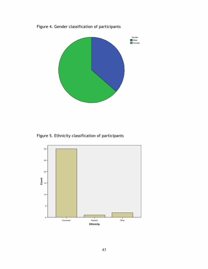
The demographics of the sample are important in interpreting the overall
results of the data analysis. The principle investigator was able to recruit a total of
32 participants for this study, of which 30 returned for follow up. There were 10
male and 22 female participants (see Figure 4). Of these participants, the ethnicity
was primarily Caucasian with one Hispanic participant and two who classified
themselves as others (see Figure 5). The age of the participants varied from age 6 to
56 with a mean age of 30 (see Figure 6). The participants had a wide range of
classification of asthma. There were 11 patients with mild intermittent, eight with
mild persistent, nine with moderate persistent, and three with severe persistent
asthma (see Figure 7). Ten of the participants had been hospitalized for asthma-
related complications in the past. Also of interest is that five of the participants had
tried either chiropractic or osteopathic manipulation for relief of their asthma
symptoms.
43
Figure 4. Gender classification of participants
FemaleMale
Gender
Figure 5. Ethnicity classification of participants
OtherHispanicCaucasian
Ethnicity
30
25
20
15
10
5
0
Count

44
Figure 6. Histogram of age of participants
6050403020100
Age
10
8
6
4
2
0
Frequency
Mean =30.12 Std. Dev. =13.727
N =33
Histogram of Age
Figure 7. Classification of asthma participants
Severe PersistentModerate PersistentMild PersistentMild Intermittent
class of asthma
12
10
8
6
4
2
0
Count

45
The results of the data analysis provided findings for each of the research
objectives as shown below.
Objective 1. Determine the relationship of OMT on lung function as demonstrated by
basic spirometry, thoracic excursions, and subjective patient symptoms.
The relationship of OMT on lung function for each of the instruments was
assessed separately for each of the 32 participants.
Spirometry
Analysis of the spirometry data (FEV1, FVC, and PEF) demonstrates normality. The
paired-sample t-test showed a significant (p<.05) increase between the pre and post-
OMT FVC values, but the change in FEV1 or PEF values were not significant. Results
are displayed in Table 1. Although all 32 participants carried a diagnosis of asthma
and suffered symptoms, only 1 had a FVC value of less than 84%, 5 had an FEV1 value
of less than 80%, and 5 had a PEF of less than 80% before OMT, which was the result of
physician care over a period of time before this research began. Therefore, there was
limited potential for improvement in the spirometry values. Participants who had
lower starting spirometry values showed the largest improvement ranging from a 15%
to a 34% improvement after the OMT session.
Table 1. Pre and Post-OMT Spirometry Value Statistics
Mean Standard Deviation
T(31) P (Eta Square)
Pre FVC
Post FVC
98.32
101.29
13.32
12.57
2.49 .019 (.167)
Pre FEV1 93.78 15.64 .289 .774

46
Post FEV1 94.22 14.57
Pre PEF
Post PEF
92.33
91.23
20.18
18.39
.629 .534
N=32, p<.05
Thoracic Excursion
Analysis of the upper and lower rib cage excursion demonstrates normality.
The paired-sample t-test for the upper ribs shows a statistically significant increase
(p<.05) in upper rib thoracic excursion from pre-OMT (M=.89, SD=.55) to post-OMT
(M=1.22, SD=.61, t(31)=5.51, p=<.0001) with an eta squared statistic of .49. The
analysis demonstrated an equally statistically significant increase (p<.05) in thoracic
excursion of the lower ribs from pre-OMT (M=1.18, SD=.75) to post-OMT (M=1.77,
SD=.89, t(31)=11.04, p=<.0001) with an eta squared statistic of .79. Results are
displayed in Table 2.
Table 2. Pre and Post-OMT Thoracic Excursion Measurements
Mean Standard Deviation
T(31) P Eta squared
Pre Upper Ribs
Post Upper Ribs
.89
1.22
.55
.66
5.511 <.0001 .49
Post Upper Ribs
Post Lower Ribs
1.18
1.78
.74
.89
11.04 <.0001 .99
N=32, p<.05

47
Patient Symptoms
Analysis of the five patient symptom questions demonstrated skewness,
therefore a Wilcoxon 1-sample test was performed. The analysis of all five of the
questions: current shortness of breath, chest tightness, wheezing, ability to take a
deep breath, and overall asthma symptoms from pre to post-OMT showed statistically
significant (p<.05) improvement in the medians as seen in Table 3.
Table 3. Pre and Post-OMT Asthma Symptoms
Symptom (scale of 1 to 10)
Pre OMT Median (St Dev)
Post OMT Median (St Dev)
Difference in Median
P using Wilcoxon 1 sample test
Shortness of Breath
9.0 (2.21) 10.0 (1.49) 1.0 .0012
Chest Tightness
8.0 (1.91) 9.0 (1.60) 1.0 <.0001
Wheezing 10.0 (2.18) 10.0 (1.06) 0.0 (difference in mean 0.758)
.0239
Ability to Take a Deep Breath
8.0 (1.67) 9.0 (1.22) 1.0 <.0001
Overall Asthma Symptoms
8.0 (2.04) 9.0 (1.17) 1.0 <.0001
N=32, p<.05
Objective 2. Compare lung function measures of asthma patients using self-
administered home treatment as supplemental to doctor administered OMT
treatment with asthma patients who only receive doctor administered OMT.
Participants were divided into two groups; 16 were in the control with no home
OMT, and 14 were in the home OMT education group. The difference in lung function
measures were again analyzed separately for each of the instruments used. In

48
addition, scores from the Mini AQLQ were analyzed and compared for each group to
determine if adding home OMT improved the participants’ quality of life as related to
asthma.
Spirometry
Analysis of the three spirometry values, FEV1, FVC, and PEF, demonstrated
normality. The paired sample t-test did not show significant improvement from
baseline to follow up for any of the three values. Results are displayed in Table 4.
Table 4. Paired sample t-test results for spirometry at baseline and follow up
Mean Standard
Deviation
T(29) P
Baseline FVC
Follow Up FVC
98.32
99.92
13.32
14.06
0.965 .3433
Baseline FEV1
Follow Up FEV1
93.78
94.66
15.64
14.71
.578 .5681
Baseline PEF
Follow Up PEF
92.33
92.2
20.18
17.87
.458 .6512
N=30, p<.05
Thoracic Excursion
The values obtained for upper and lower thoracic excursion were found to be
normal. The paired sample t-test for the upper ribs shows a statistically significant
(p<.05) improvement in the upper rib thoracic excursion from the initial visit (M=.89,

49
SD=.55) to the follow up visit (M=1.05, SD=.53, t(29)=2.51, p=.0177) with an Eta
squared statistic of .18. (See Table 5).
To further explore this improvement, an ANOVA test was used to find the least
squares means value when factoring in the difference in the two groups (home OMT
and control) and the genders of the participants. The Type III Test of Fixed Effects
demonstrated an interaction between gender and the two groups. Therefore, to
determine the interaction between home OMT and gender, the data were evaluated
using the slice option of the Glimmix procedure to get a difference in the least
squares means. Both genders had statistically significant improvements (p<.05) in the
upper rib thoracic excursion value from the initial visit to the follow up visit (Gender
1 p=<.0001, Gender 2 p=.0238) as displayed in table 6. The group that underwent the
home OMT protocol had a statistically significant change from baseline (p=.0008) as
compared to the group that did not do home OMT (p=.8474). (See table 7).
The paired sample t-test for the lower ribs shows a statistically significant
(p<.05) improvement in lower rib thoracic excursion from the initial visit (M=1.18,
SD=.75) to the follow up visit (M=1.38, SD=.78, t(29)=3.63, p=.0011) with an Eta
squared statistic of .31. See table 5. In order to further delineate this improvement,
an ANOVA test was used to get a least squares means value and test the effect of self-
administered home OMT and gender on the change in lower rib thoracic excursion
from initial to follow up visit. The group who participated in the home OMT protocol
had statistically significant (p<.05) improvement in lower rib thoracic excursion as
compared to the group who did not do home OMT (p=.0045). There was no significant
difference between males and females (p=.1383).

50
Table 5. Paired sample t-test results for upper and lower rib thoracic excursion measurements Mean Standard
Deviation T(29) P Eta
squared Baseline Upper Ribs FU Upper Ribs
.89 1.05
.55
.53 2.51 .0177 .18
Baseline Lower Ribs FU Lower Ribs
1.18 1.38
.75
.78 3.63 .0011 .31
N=30, p<.05
Table 6. Upper ribs comparison of the interaction between gender and home OMT sliced by gender Control Home OMT Difference
in Means P
Male -0.04167 0.9167 -.9583 <.0001
Female -0.01500 0.2636 -.2786 .0238
N=30, p<.05
Table 7. Upper ribs comparison of the interaction between gender and home OMT sliced by home OMT Male Mean Female Mean Difference in
Means p
Control -0.04167 -0.01500 -0.02667 0.8474
Home OMT 0.9167 0.2636 0.6530 0.0008
N=30, p<.05
Patient Symptoms
Analysis of the data obtained from the ranking scale demonstrated skewness.
Therefore, the data were analyzed using a Wilcoxin 1-sample test which
demonstrated no statistically significant (p<.05) improvements in the median values
for the rating of shortness of breath, chest tightness, wheezing, or ability to take a

51
deep breath from baseline to follow up. There was a significant improvement in the
rating of overall asthma symptoms from baseline (M=8, SD=2.04) to follow up (M=9.0,
SD=1.47, p=.0386). (See Table 8).
Table 8. Wilcoxin 2 sided test for the rating scale.
Symptom Baseline Median (St Dev)
Follow Up Median (St Dev)
Difference in Median
P using Wilcoxin 2-sided test
Shortness of Breath
9.0 (2.21) 9.0 (1.80) 0 .487
Chest Tightness
8.0 (1.91) 9.0 (2.04) 1.0 .735
Wheezing 10.0 (2.18) 10.0 (1.52) 0 .848
Ability to take a deep breath
8.0 (1.67) 10.0 (1.51) 2 .3485
Overall Asthma Symptoms
8.0 (2.04) 9.0 (1.47) 1 .0386
N=30, p<.05
Mini Asthma Quality of Life Questionnaire (MAQLQ)
This questionnaire was answered by all participants at the initial visit and again
at the follow up visit. It was scored according to the protocol designed by the
researchers who invented the questionnaire which was described in Chapter three of
this Thesis.
The scores for the MAQLQ obtained are broken up into different categories.
There is an overall score and separate scores for environmental, emotion, activity,
and symptoms of asthma. Each category is scored on a scale of one to seven. The
best score is a 7, which means that the patient has no impairments in quality of life
due to his or her asthma, and the lowest score is a 1, which means that his or her

52
quality of life is severely impaired by asthma. Each patient was scored in each of the
categories and given an overall score for the initial visit and the follow up visit. Next,
the Minimal Important Difference (MID) was obtained for each participant. The MID is
defined as “the smallest difference in score which patients perceive as beneficial and
would mandate, in the absence of troublesome side effects and excessive costs, a
change in the patient’s management” (Juniper et al., 1994). For this questionnaire,
the MID has been found to be 0.5. Therefore, if the difference in the environmental
score in patient 1 went from a 4 initially to a 4.5 at follow up, then whatever
intervention was added to this patient’s asthma management during those 2 weeks
would be considered beneficial enough to keep it as a permanent asthma
management technique for that patient.
After scoring all participant responses and determining the MID, a Chi-Squared
analysis was run. The number of patients who had a significant change in their scores
was compared between the control and home OMT group and were separated using a
Fisher’s exact test. Then, each score was tested in a model using logistical regression
to determine the relationship and effect home OMT had on the scores. None of the
categories on the MAQLQ significantly changed due to the addition of home OMT to
regular asthma treatment.
Table 9. Chi-Squared test to determine affect of Home OMT on MID scores from the
MAQLQ
Category Group MID <0.5 (not
significant)
MID > 0.5
(significant)
Chi square
value
Overall Control 5/16 11/16 0.8731

53
Home OMT 4/14 10/14
Symptoms Control
Home OMT
7/16
4/14
9/16
10/14
0.3055
Activity Control
Home OMT
10/16
8/14
6/16
6/14
0.8232
Emotional Control
Home OMT
6/16
3/14
10/16
11/14
0.2272
Environmental Control
Home OMT
8/16
7/14
8/16
7/14
1.0
N=30, p<.05
Home OMT group follow up questionnaire
Those in the home OMT group answered questions regarding the usefulness and
perceived benefit of the home treatments. When asked how many times a week they
performed the home OMT, two participants answered 1 time a week, six participants
answered 2-3 times a week, and seven answered that they did the home OMT more
than 2-3 times a week. When asked how easy it was to do the home OMT techniques
on a scale from zero to ten with zero being not very and ten being very, one
participant rated a 7, three rated an 8, two rated a 9, and nine rated a 10. When
asked what effect the participants thought the home OMT had on their asthma
symptoms, two answered a lot of improvement, ten answered some improvement,
one answered not very much improvement, and two answered no change. (See Figure
8).

54
Figure 8. Home-OMT Follow Up Indicators
Question Asked Answers Frequency
Frequency of OMT use Zero times
1 time a week
2-3 times a week
More than 2-3 times a week
0
2
5
7
Ease of home OMT
(0 – not easy, 10 – very easy)
0
1
2
3
4
5
6
7
8
9
10
0
0
0
0
0
0
0
1
2
2
9
Perceived effect A lot of improvement
Some improvement
Not very much improvement
No change
Symptoms worsened
2
9
1
2
0

55
N=14
Objective 3. Determine the relationship among selected environmental factors with
demographics of patients in this study.
In order to explore this objective, each participant filled out an initial
questionnaire which asked about environmental factors, living conditions, and other
participant characteristics (See Appendix B)
Overall, there were 20 students, three teachers, two customer service workers,
one hair stylist, one cleaner, one homemaker, one office manager, one machinist,
one banker, and one horse handler. There were no current smokers enrolled in the
study, so the impact of smoking on asthma symptoms could not be explored. Only four
of the participants said they had no allergies. Otherwise, 25 had allergies to dust, 26
to pollen, 21 to cats, 17 to dogs, 18 to trees, 13 to flowers, eight to mold, one to
feathers, one to hay, and one to cockroaches. Fourteen of the participants did not
own a pet. Eleven had cats, 11 had dogs, two had horses, one had a sheep, three had
cows, two had birds, and three had fish. Thirty of the participants are receiving
active treatment from their physician for asthma while two were not. The majority
of people saw either allergists (11) or family practice (10) physicians for their asthma,
while nine saw a pulmonologist, and one saw an internist. As for medication use, all
but two participants use an albuterol inhaler regularly. Fourteen use an inhaled
steroid, thirteen use a leukotriene inhibitor (Singulair), six use a home nebulizer, and
five use various other allergy medications. Overall, the participants had been
diagnosed with asthma anywhere from .5 to 30 years prior to enrolling in the study.

56
Ten had been hospitalized before for asthma or related complications. Five
participants answered that they had had either osteopathic or chiropractic
manipulation for their asthma in the past. In total, 21 of the participants exercise at
least three to four times a week (see Figure 11).
Figure 9. Patient Questionnaire Responses
Question Response Answer
Occupation Student
Teacher
Customer Service
Hair Stylist
Cleaner
Homemaker
Office Manager
Machinist
Banking
Horse Handler
20
3
2
1
1
1
1
1
1
1
Smoke Yes
No
0
32
Allergies to: Dust
Pollen
Cats
Dogs
25
26
21
17

57
Trees
Flowers
None
Other
18
13
4
Mole (8), Feather (1), Hay
(1), cockroaches (1)
Pets in or around house: Cats
Dogs
Horses
Sheep
Cows
Chickens
Birds
None
Other
11
11
2
1
3
0
2
14
Fish (3), Hamster (1)
Duration of Asthma
diagnosis
0-5 yrs
6-10 yrs
11-15 yrs
16-20 yrs
21-25 yrs
26-30 yrs
> 30 yrs
6
6
5
4
5
4
2
Treated for Asthma Yes
No
30
2
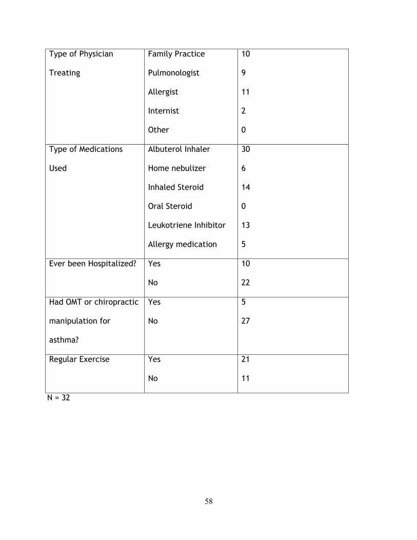
58
Type of Physician
Treating
Family Practice
Pulmonologist
Allergist
Internist
Other
10
9
11
2
0
Type of Medications
Used
Albuterol Inhaler
Home nebulizer
Inhaled Steroid
Oral Steroid
Leukotriene Inhibitor
Allergy medication
30
6
14
0
13
5
Ever been Hospitalized? Yes
No
10
22
Had OMT or chiropractic
manipulation for
asthma?
Yes
No
5
27
Regular Exercise Yes
No
21
11
N = 32

59
Objective 4. Determine the relationship of age and gender on the use of OMT for
asthma.
Due to the uneven distribution of ages and limited number of participants, the
data gathered was not sufficient to draw conclusions about the relationship of age on
the use of OMT for asthma.
However, the researchers did use the data to begin exploring the relationship
of gender on the use of OMT for asthma. There were no differences found in
spirometry results, lower rib thoracic excursion, or MAQLQ based on gender. As
discussed in the findings from Objective 2, there are indications that gender had an
effect on the upper rib thoracic excursion data. Overall, more in depth analysis and a
larger population is needed to draw further conclusions.

60
Chapter Five
Summary, Conclusions, and Recommendations
This final chapter serves to discuss the summary of the results, the conclusions
which can be drawn from this study, and recommendations both for future practice as
well as future studies.
Summary
The purpose of this study was to determine the viability of OMT treatment for
asthma and the effectiveness of home OMT education in order to implement a
program to improve asthma control. The following objectives were addressed:
1. Determine the relationship of OMT on lung function as demonstrated by
basic spirometry, thoracic excursions, and subjective patient symptoms.
2. Compare lung function measures of asthma patients using self-administered
home treatment as supplemental to doctor administered OMT treatment with
asthma patients who only receive doctor administered OMT.
3. Determine the relationship among selected environmental factors with
demographics of patients in this study.
4. Determine the relationship of age and gender on the use of OMT for asthma.
Chapter Four demonstrated the results of each of these objectives while
Chapter Five builds on the findings to develop conclusions on the viability of OMT
treatment on asthma symptoms, as well as the effectiveness home OMT education in
improving asthma control.

61
Conclusions
Initial pre and post OMT lung assessment
Statistically significant (p<.05) improvements were documented for 8 out of 10
measurements, which included upper and lower rib thoracic excursion, patient ratings
of ability to take a deep breath, shortness of breath, wheezing, tightness in the chest,
and overall asthma symptoms, and spirometric FVC value. To strengthen the
significance, further statistics in the form of the Eta-Squared analysis demonstrated
that OMT had a large effect on the changes. Statistical significance was not
determined for 2 of the 10 tests, including spirometric FEV1 and PEF values. One
potential explanation for the spirometry data being not significant is that the patient
population which was recruited was already receiving quality care and their asthma
symptoms were already well controlled in regards to pulmonary function, as was
demonstrated by the data in chapter four regarding the participant spirometry values.
The fact that the patient subjective symptoms improved after OMT, but the
objective spirometric values did not improve is an interesting finding. There are
several possible explanations for this. Perhaps the patients felt better because their
musculoskeletal restrictions were alleviated, giving improvement in the thoracic
excursion measurements, but the physiology and functioning of the lungs themselves
were not affected which is why the spirometry values did not change. Another
explanation is that the subjects enjoyed the hands-on approach and it is a placebo
effect, meaning the patients feel better but there is no justifiable physiologic reason
behind it.

62
Use of Home OMT
The use of home OMT demonstrated statistically significant (p<.05)
improvements at the two week follow up in three of the ten measurements including
upper and lower thoracic excursion and the overall asthma symptom rating. The
majority of the participants found the home OMT sessions to be easy to perform and
beneficial. There were no significant (p<.05) improvements from baseline in seven out
of ten measurements including the three spirometry values, FEV1, PEF, and FVC, and
the patient symptoms of ability to take a deep breath, chest tightness, wheezing,
shortness of breath.
Environmental impact
The majority of the participants were students. None of the asthmatics in this
study were smokers. The majority of the participants had allergies, most commonly
to cats, dogs, trees, dust, and pollen. Half of the participants had pets in or around
the house with the majority owning cats and dogs. All but two of the participants
were actively being treated by a physician for their asthma. The majority of the
participants relied on an allergist or a family practitioner for the treatment of their
asthma symptoms. The majority of the participants exercise regularly. Only five of
the participants had ever had osteopathic or chiropractic manipulation to try to
alleviate asthma symptoms.
Effect of age and gender
The data was not sufficient to draw conclusions regarding the impact that age
or gender may have on the benefit of OMT for asthma symptoms.
Hypothesis test:

63
1. A researcher developed protocol of Osteopathic Manipulative
Treatment, including screening and treatment of the thoracic and
rib cage, diaphragm release, suboccipital release, and thoracic
lymphatic pump will significantly (p<.05) improve the spirometry
values, thoracic excursion measurements, and symptoms in the
selected group of participants suffering from asthma.
This hypothesis is accepted at the .05 level.
2. A researcher-developed educational session designed to teach
asthma patients home OMT will significantly improve the
spirometry, thoracic excursion measurements, and symptoms in
those participants over a two week time period as compared to a
group of asthma patients who do not do home OMT.
This hypothesis failed to be accepted due to the equivocal findings. There is
sufficient evidence to demonstrate that teaching asthma home OMT techniques
to alleviate asthma symptoms is beneficial; however, another investigation is
needed to further explore this.
3. There will be a significant relationship between environmental
factors and asthma symptoms in the chosen population.
This hypothesis failed to be accepted due to a lack of adequate
information to draw a conclusion. Further investigation is needed.
4. Differences in age and gender will be significantly related to
response to OMT techniques for asthma.
64
This hypothesis failed to be accepted due to the limitations previously
discussed. Further investigation is needed to delineate this relationship.
Recommendations for Future Research
Based on the findings and conclusions, recommendations were made for future
research and related areas of study:
1) In future studies, the limited time series of two weeks could be expanded to
a longer time series design, first to validate the two week results from this study, and
second, to document change at subsequent two week intervals depending on time
constraints and resources.
2) While this pilot study had enough participants to draw statistically
significant conclusions, for future studies or replications of this study, this
investigator recommends a larger participant size which will allow for a more widely
distributed age range, ethnicity, and asthma classification for further investigation
into variability of response to OMT for asthma symptoms.
3) Although the education session for the home OMT group in this study was
sufficient enough to teach participants the skills necessary, making and distributing
video or DVD instructions to this group would allow the researcher to strengthen the
education session and further ensure proper technique at home.
4) In future research, adding exclusion criteria in the protocol which would
require researchers to recruit a sample with lower baseline spirometry lung function
values would allow for a clearer investigation of the effects of OMT on these values.
65
5) The home OMT education session could be strengthened by lengthening the
time that participants practice home OMT techniques with the instructor and having
weekly follow ups to ensure correct techniques are being used.
6) This pilot study attempted to recruit participants from rural areas and teach
them how to do OMT techniques at home to alleviate asthma symptoms; however,
future research may extend further into rural communities to evaluate the
effectiveness of this education session in more rural and possibly less educated areas.
7) This pilot study explored a possible OMT session which could be utilized in a
regular office visit to further aid in alleviating asthma symptoms; however, future
studies could progress and add to this an actual physician visit to determine the
overall ease of incorporation and perceived benefit from both the physician and the
patient.
Recommendations for Practice
Due to the overall positive findings regarding the use of OMT for asthma
symptoms, it is recommended that these treatments be regarded as viable options for
asthma patients. The protocol was a simple, semi-individualized way to address
asthma symptoms and create an immediate improvement in lung function.
Throughout the study, although the participants were not asked to comment,
the principle investigator received many comments about OMT. Many times,
immediately following the sessions, the participants would get up off of the OMT
table and exclaim how good they felt and how amazed they were at their ability to
66
breathe deeply. These were unsolicited comments which go to support the use of
these treatments for asthma patients.
Regardless of what the numbers of the measurements say, if the patient feels
that his or her lung function has increased and that asthma symptoms are improved,
then that is enough to show physicians that this is a successful way to treat their
patients. I strongly recommend that any physician who is treating asthma study and
learn the techniques used in this study and incorporate them into regular treatment
protocol for asthmatics.
67
References
Ali, S., Hayek, R., Holland, R., McKelvy, S., Boyce, K., & Curson, P. (2002). Effect of
chiropractic treatment on the endocrine and immune system in asthmatic
patients. Paper presented at the 2002 International Conference on Spinal
Manipulation, Des Moines, IA.
Allen, T. W. (1971). Management of acute exacerbations of chronic obstructive
pulmonary disease. The Journal of the American Osteopath Association, 71(4),
330-.
American Lung Association (2005). Trends in asthma morbidity and mortality:
Epidemiology and statistics unit.
American Lung Association, (2007). American Lung Association State of the Air Report:
2007. Retrieved April 15, 2007, from
http://www.kintera.org/site/c.ivKTL8MWIwG/b.1162277/apps/nl/content2.as
p?content_id=9ACA8944-41BB-41BD-9C1D-89AA3FE35D66¬oc=1
Asthma. Retrieved April 12 2007, from http://www.breatheroanoke.org/asthma.htm
Asthma in Virginia: A Comprehensive Data Report, 2006.). Richmond, VA: Virginia
Department of Health.
Balon, J., Aker, P. D., Crowther, E. R., Danielson, C., Cox, P. G., O'Shaughnessy, D.,
et al. (1998). A Comparison of Active and Simulated Chiropractic Manipulation
as Adjunctive Treatment for Childhood Asthma. New England Journal of
Medicine, 339(15), 1013-1020.
Balon, J. W., & Mior, S. A. (2004). Chiropractic care in asthma and allergy. Annals of
Allergy, Asthma and Immunology, 93, S55-S60.

68
Berhane, K., McConnell, R. O. B., Gilliland, F., Islam, T., James Gauderman, W.,
Avol, E., et al. (2000). Sex-specific Effects of Asthma on Pulmonary Function in
Children. American Journal of Respiratory and Critical Care Medicine, 162(5),
1723-1730.
Blumenfeld, H., (2002). Neuroanatomy Through Clinical Cases. Sunderland, MA:
Sinauer Associates, Inc.
Bockenhauer, S. E., Chen, H., Julliard, K. N., & Weedon, J. (2007). Measuring
Thoracic Excursion: Reliability of the Cloth Tape Measure Technique. Journal of
the American Osteopath Association, 107(5), 191-196.
Bockenhauer, S. E., Julliard, K. N., Lo, K. S., Huang, E., & Sheth, A. M. (2002).
Quantifiable effects of osteopathic manipulative techniques on patients with
chronic asthma. Journal of the American Osteopath Association, 102(7), 371-
375.
Bronfort, G., Evans, R., Kubic, P., & Filkin, P. (2001). Chronic Pediatric Asthma and
Chiropractic Spinal Manipulation: A Prospective Clinical Series and Randomized
Clinical Pilot Study. Journal of Manipulative Physiologic Therapy, 24(6), 369-
377.
Cali, G. (2002). Parameters of asthma and manipulation study questioned. Journal of
the American Osteopathic Association, 102(11), 573.
Camargo, C. A. (2006). Asthma [Electronic Version]. E-medicine. Retrieved April 17,
2007 from www.emedicine.com/emerg/topic43.htm.

69
CHIP of Virginia, (2002). Fighting the Asthma Epidemic [Electronic Version]. GROW, 6.
Retrieved April 17, 2007 from
http://www.chipofvirginia.org/pdf/GrowFall02.pdf.
Dowling, D. J. (1997). Muscle Energy Techniques. In E. L. DiGiovanna & S. Shiowitz
(Eds.), An Osteopathic Approach to Diagnosis and Treatment (2 ed.). New York:
Lippincott Williams & Wilkins.
Eaton, T., Withy, S., Garrett, J. E., Mercer, J., Whitlock, R. M. L., & Rea, H. H.
(1999). Spirometry in Primary Care Practice: The Importance of Quality
Assurance and the Impact of Spirometry Workshops. Chest, 116(2), 416-423.
Ferguson, G. T., Enright, P. L., Buist, A. S., & Higgins, M. W. (2000). Office
Spirometry for Lung Health Assessment in Adults: A Consensus Statement From
the National Lung Health Education Program. Chest, 117(4), 1146-1161.
Gardner, R. M., Crapo, R. O., Jackson, B. R., & Jensen, R. L. (1992). Evaluation of
accuracy and reproducibility of peak flow meters at 1,400 m. Chest, 101(4),
948-952.
Goroll, A. H., & Jr, A. G. M. (1995). Primary Care Medicine: Office Evaluation and
Management of the Adult Patient (4th ed.). New York: Lippincott Williams &
Wilkins.
Grentz, L. C., Yonts, R. C., Stiles, E. G., Broadwater, J. M., & Jones, D. W. (Eds.).
(2004). Pocket OMT Manual (2 ed.). Pikeville, KY: Mountain Medicine Publisher.

70
Guiney, P. A., Chou, R., Vianna, A., & Lovenheim, J. (2005). Effects of Osteopathic
Manipulative Treatment on Pediatric Patients With Asthma: A Randomized
Controlled Trial. Journal of the American Osteopath Association, 105(1), 7-12.
Harden, D. (2007). Diaphragm Release Technique. In M. Latter (Ed.). Blacksburg, VA.
Harm, D., Marion, R., Creer, T., & Kotses, H. (1985). Effects of instructions on
pulmonary function values. Journal of Asthma, 22(6), 289-294.
Juniper, E. (1991). Background, Administration and Analysis. QOL Technologies Ltd.
Juniper, E., Guyatt, G., Cox, F. M., Ferrie, P., & King, D. R. (1999). Development and
validation of the Mini Asthma Quality of Life Questionnaire. The European
Respiratory Journal, 14, 32-38.
Juniper, E., Guyatt, G., Epstein, R., Ferrie, P., Jaeschke, R., & Hiller, T. (1992).
Evaluation of impairment of health related quality of life in asthma:
development of a questionnaire for use in clinical trials. Thorax, 47(2), 76-83.
Juniper, E., Guyatt, G., Ferrie, P., & Griffith, L. (1993). Measuring Quality of Life in
Asthma. American Review of Respiratory Disease, 147, 832-838.
Juniper, E., Guyatt, G., Willan, A., & Griffith, L. (1994). Determining a Minimal
Important Change in a Disease-Specific Quality of Life Questionnaire. Journal
of Clinical Epidemiology, 47(1), 81-87.
Kasper, D. L., Braunwald, E., Fauci, A. S., Hauser, S. L., Longo, D. L., & Jameson, J.
L. (Eds.). (2005). Harrison's Manual of Medicine (16th ed.). New York: McGraw-
Hill Medical Publishing Division.
Kaufman, D. A. (2006). Lung Disease [Electronic Version]. Medline Plus. Retrieved July
1, 2007 from http://www.nlm.nih.gov/medlineplus/ency/article/000066.htm.
71
Kimberly, P. E. (2000). Outline of Osteopathic Manipulative Procedures. Marceline,
Missouri: Walsworth Publishing Company.
Kumar, V., Abbas, A. K., & Fausto, N. (Eds.). (2005). Robbins and Cotran Pathologic
Basis of Disease (7th ed.). Philadelphia: El Sevier Saunders.
Kuo, A., & Craig, T. (2001). A retrospective study of risk factors for repeated
admissions for asthma in rural/suburban university hospital. Journal of the
American Osteopathic Association, 101(5), 14-17.
Lung Diseases: Asthma. (2006). Diseases and Conditions Index Retrieved July 24,
2007, from
http://www.nhlbi.nih.gov/health/dci/Diseases/Asthma/Asthma_WhatIs.html
McKenzie, J. F., Neiger, B. L., & Smeltzer, J. L. (2005). Planning, Implementing, and
Evaluating Health Promotion Programs: A Primer. New York: Pearson Education
Inc.
Morris, M. J. (2007). Asthma. E-Medicine Retrieved July 24, 2007, from
http://www.emedicine.com/med/topic177.htm
National Asthma Education and Prevention Program (NAEPP). (1997). Expert panel
report 2: guidelines for the diagnosis and management of asthma. Bethesda,
MD: National Institutes of Health.
Nicholas, A. S., & Nicholas, E. A. (2007). Atlas of Osteopathic Techniques. New York:
Lippincott Williams & Wilkins.
Nielsen, N. H., Bronfort, G., Bendix, T., Madsen, F., & Weeke, B. (1995). Chronic
asthma and chiropractic spinal manipulation: a randomized clinical trial.
Clinical & Experimental Allergy, 25(1), 80-88.
72
Paulus, S. (2005). Hearing the Call of Osteopathy. Inter Linea: Exploring the
Philosophy of Osteopathy Retrieved July 25, 2007, from
http://www.interlinea.org/hands/hearing_the_call_of_osteopathy.html
Raji, M. (2005). The Nervous System as Potential Targets for Asthma
Treatments.[Electronic Version]. American Journal of Respiratory Cell and
Molecular Biology. Vol 33, pp. 211 Retrieved January 25, 2008 from
http://ajrcmb.atsjournals.org/cgi/content/full/33/2/211.
Scariati, P. D., Roberge, L., & Dye, T. (2006). Beating Asthma: A Community-Based
Asthma Education Initiative. Journal of the American Osteopath Association,
106(1), 16-22.
Sevarese R.G., Capobianco, J., Cox, J. (2003). OMT Review: A Comprehensive Review
in Osteopathic Medicine. New York: Lippincott Williams & Wilkins.
Sharma, G. (2006). Asthma. E-Medicine Retrieved July 24, 2007, from
http://www.emedicine.com/ped/topic152.htm
Shiowitz, S. (1997). Facilitated Positional Release. In E. L. DiGiovanna & S. Shiowitz
(Eds.), An Osteopathic Approach to Diagnosis and Treatment. New York:
Lippincott Williams & Wilkins.
Spinaris, T., & DiGiovanna, E. (1997). Treatment of the Cervical Spine. In E. L.
Digiovanna & S. Shiowitz (Eds.), An Osteopathic Approach to Diagnosis and
Treatment (2 ed.). New York: Lippincott Williams & Wilkins.
Stretanski, M. F., & Kaiser, G. (2001). Osteopathic philosophy and emergent
treatment in acute respiratory failure. Journal of the American Osteopathic
Association, 101(8), 447-449.
73
Virginia Asthma Coalition Asthma Data and Statistics. Retrieved July 1, 2007 from
http://www.virginiaasthmacoalition.org/AsthmaData.htm
Virginia Asthma Coalition (2007). Asthma Toll in Virginia. Retrieved May 6, 2007,
from
http://www.virginiaasthmacoalition.org/Docs/Asthma%20Toll%20in%20VA%20Fa
ct%20Sheet.pdf
Ward, R. C. (Ed.). (2003). Foundations for Osteopathic Medicine (2nd ed.). New York:
Lippincott Williams & Wilkins.

74
Appendix A

75

76
Appendix B
Patient Questionnaire
Demographics
Gender: ____ Male ____ Female
Age ____
Ethnicity: ___ Caucasian ___ African American ___ Hispanic ___ Asian ___ Other
Town of Residence: ______________________
Town of this Clinic: ______________________
Environmental
Occupation: ____________________________ Years in Current Occupation: ____
Do you smoke? If no, skip next two questions.
a. Yes
b. No
How long have you been a smoker? __________
How many packs a day do you smoke? ___________
Do you have allergies to any of the following? (check all that apply)
a. Dust
b. Pollen
c. Cats

77
d. Dogs
e. Trees
f. Flowers
g. Other _______________________
h. None
What types of pets are in or around the house?
a. Cats
b. Dogs
c. Horses
d. Sheep
e. Cows
f. Chickens
g. Birds
h. Other ___________
i. None
Medical
How long have you been diagnosed with asthma? _________
How often do you experience symptoms of asthma?
a. less than 2 times a week
b. 2 times a week
c. More than 2 times a week
d. Daily
e. Continual
How often do you have nighttime symptoms of asthma?
a. less than 2 times a month
b. more than 2 times a month
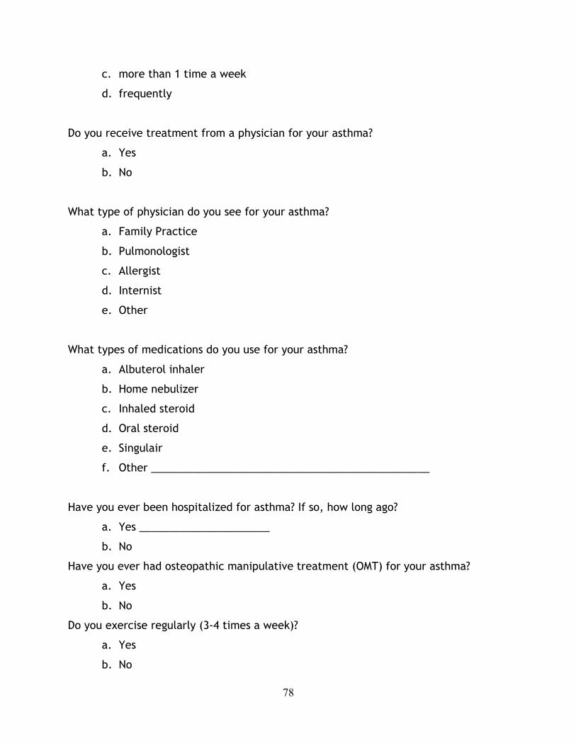
78
c. more than 1 time a week
d. frequently
Do you receive treatment from a physician for your asthma?
a. Yes
b. No
What type of physician do you see for your asthma?
a. Family Practice
b. Pulmonologist
c. Allergist
d. Internist
e. Other
What types of medications do you use for your asthma?
a. Albuterol inhaler
b. Home nebulizer
c. Inhaled steroid
d. Oral steroid
e. Singulair
f. Other _______________________________________________
Have you ever been hospitalized for asthma? If so, how long ago?
a. Yes ______________________
b. No
Have you ever had osteopathic manipulative treatment (OMT) for your asthma?
a. Yes
b. No
Do you exercise regularly (3-4 times a week)?
a. Yes
b. No
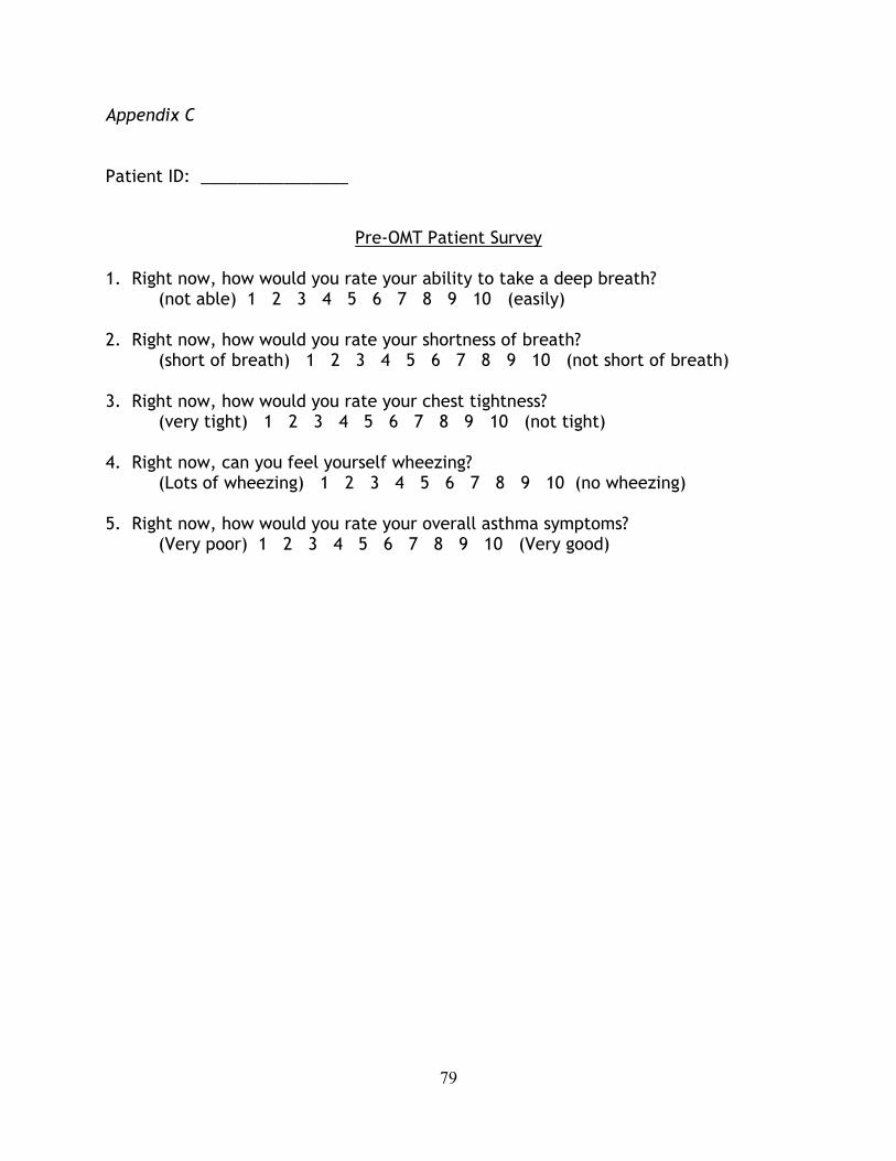
79
Appendix C
Patient ID: ________________
Pre-OMT Patient Survey
1. Right now, how would you rate your ability to take a deep breath? (not able) 1 2 3 4 5 6 7 8 9 10 (easily) 2. Right now, how would you rate your shortness of breath? (short of breath) 1 2 3 4 5 6 7 8 9 10 (not short of breath) 3. Right now, how would you rate your chest tightness? (very tight) 1 2 3 4 5 6 7 8 9 10 (not tight) 4. Right now, can you feel yourself wheezing? (Lots of wheezing) 1 2 3 4 5 6 7 8 9 10 (no wheezing) 5. Right now, how would you rate your overall asthma symptoms? (Very poor) 1 2 3 4 5 6 7 8 9 10 (Very good)

80
Appendix D Patient ID: ________________
Post-OMT Patient Survey
1. Right now, how would you rate your ability to take a deep breath? (not able) 1 2 3 4 5 6 7 8 9 10 (easily) 2. Right now, how would you rate your shortness of breath? (short of breath) 1 2 3 4 5 6 7 8 9 10 (not short of breath) 3. Right now, how would you rate your chest tightness? (very tight) 1 2 3 4 5 6 7 8 9 10 (not tight) 4. Right now, can you feel yourself wheezing? (Lots of wheezing) 1 2 3 4 5 6 7 8 9 10 (no wheezing) 5. Right now, how would you rate your overall asthma symptoms? (Very poor) 1 2 3 4 5 6 7 8 9 10 (Very good)
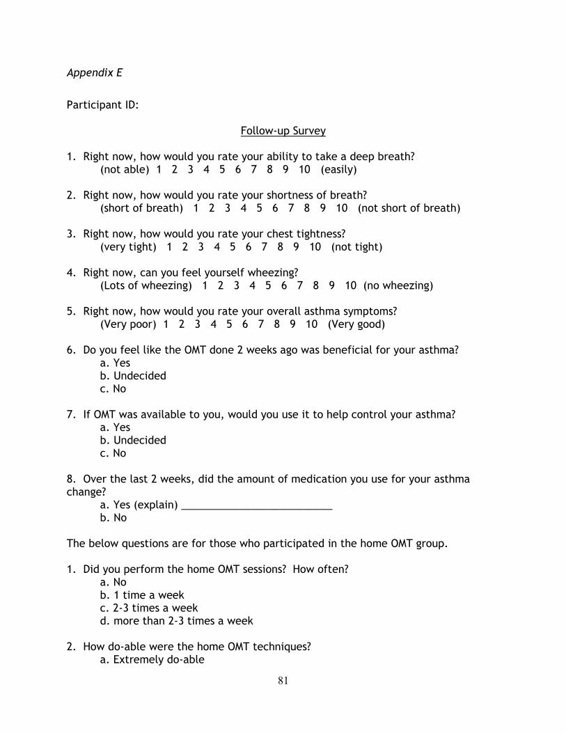
81
Appendix E
Participant ID:
Follow-up Survey 1. Right now, how would you rate your ability to take a deep breath? (not able) 1 2 3 4 5 6 7 8 9 10 (easily) 2. Right now, how would you rate your shortness of breath? (short of breath) 1 2 3 4 5 6 7 8 9 10 (not short of breath) 3. Right now, how would you rate your chest tightness? (very tight) 1 2 3 4 5 6 7 8 9 10 (not tight) 4. Right now, can you feel yourself wheezing? (Lots of wheezing) 1 2 3 4 5 6 7 8 9 10 (no wheezing) 5. Right now, how would you rate your overall asthma symptoms? (Very poor) 1 2 3 4 5 6 7 8 9 10 (Very good) 6. Do you feel like the OMT done 2 weeks ago was beneficial for your asthma? a. Yes b. Undecided c. No 7. If OMT was available to you, would you use it to help control your asthma? a. Yes b. Undecided c. No 8. Over the last 2 weeks, did the amount of medication you use for your asthma change? a. Yes (explain) __________________________ b. No The below questions are for those who participated in the home OMT group. 1. Did you perform the home OMT sessions? How often? a. No b. 1 time a week c. 2-3 times a week d. more than 2-3 times a week 2. How do-able were the home OMT techniques? a. Extremely do-able
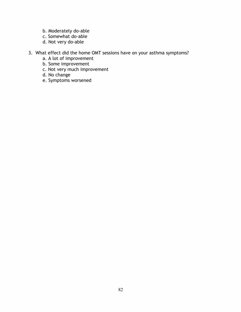
82
b. Moderately do-able c. Somewhat do-able d. Not very do-able 3. What effect did the home OMT sessions have on your asthma symptoms? a. A lot of improvement b. Some improvement c. Not very much improvement d. No change e. Symptoms worsened
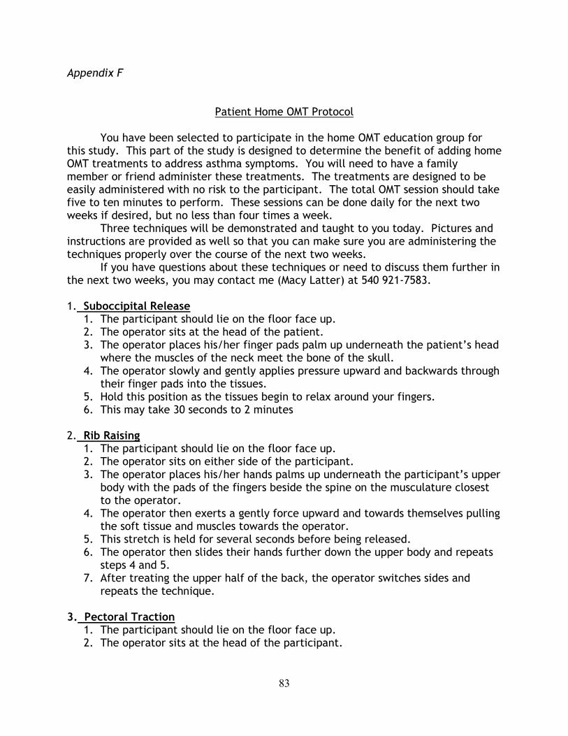
83
Appendix F
Patient Home OMT Protocol You have been selected to participate in the home OMT education group for this study. This part of the study is designed to determine the benefit of adding home OMT treatments to address asthma symptoms. You will need to have a family member or friend administer these treatments. The treatments are designed to be easily administered with no risk to the participant. The total OMT session should take five to ten minutes to perform. These sessions can be done daily for the next two weeks if desired, but no less than four times a week. Three techniques will be demonstrated and taught to you today. Pictures and instructions are provided as well so that you can make sure you are administering the techniques properly over the course of the next two weeks. If you have questions about these techniques or need to discuss them further in the next two weeks, you may contact me (Macy Latter) at 540 921-7583. 1. Suboccipital Release
1. The participant should lie on the floor face up. 2. The operator sits at the head of the patient. 3. The operator places his/her finger pads palm up underneath the patient’s head
where the muscles of the neck meet the bone of the skull. 4. The operator slowly and gently applies pressure upward and backwards through
their finger pads into the tissues. 5. Hold this position as the tissues begin to relax around your fingers. 6. This may take 30 seconds to 2 minutes
2. Rib Raising
1. The participant should lie on the floor face up. 2. The operator sits on either side of the participant. 3. The operator places his/her hands palms up underneath the participant’s upper
body with the pads of the fingers beside the spine on the musculature closest to the operator.
4. The operator then exerts a gently force upward and towards themselves pulling the soft tissue and muscles towards the operator.
5. This stretch is held for several seconds before being released. 6. The operator then slides their hands further down the upper body and repeats
steps 4 and 5. 7. After treating the upper half of the back, the operator switches sides and
repeats the technique. 3. Pectoral Traction
1. The participant should lie on the floor face up. 2. The operator sits at the head of the participant.
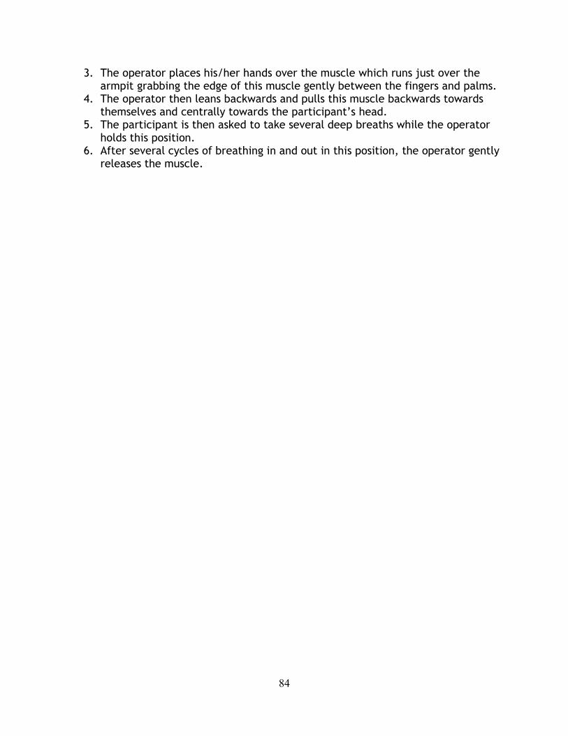
84
3. The operator places his/her hands over the muscle which runs just over the armpit grabbing the edge of this muscle gently between the fingers and palms.
4. The operator then leans backwards and pulls this muscle backwards towards themselves and centrally towards the participant’s head.
5. The participant is then asked to take several deep breaths while the operator holds this position.
6. After several cycles of breathing in and out in this position, the operator gently releases the muscle.