the urinary system - lamission.edu 25 urinary.pdf · the urinary system •kidneys, ureters,...
TRANSCRIPT

26-1
The Urinary System
• Kidneys, ureters, urinary
bladder & urethra
• Urine flows from each
kidney, down its ureter to
the bladder and to the
outside via the urethra
• Filter the blood and return
most of water and solutes
to the bloodstream

26-2
Overview of Kidney Functions
• Regulation of blood ionic composition
– Na+, K+, Ca+2, Cl- and phosphate ions
• Regulation of blood pH, osmolarity & glucose
• Regulation of blood volume
– conserving or eliminating water
• Regulation of blood pressure
• Release of erythropoietin & calcitriol
• Excretion of wastes & foreign substances

26-3
Internal Anatomy of the Kidneys
• Parenchyma of kidney
– renal cortex = superficial layer of kidney
– renal medulla
• inner portion consisting of 8-18 cone-shaped renal
pyramids separated by renal columns
• renal papilla point toward center of kidney
• Drainage system fills renal sinus cavity
– cuplike structure (minor calyces) collect urine
from the papillary ducts of the papilla
– minor & major calyces empty into the renal pelvis
which empties into the ureter
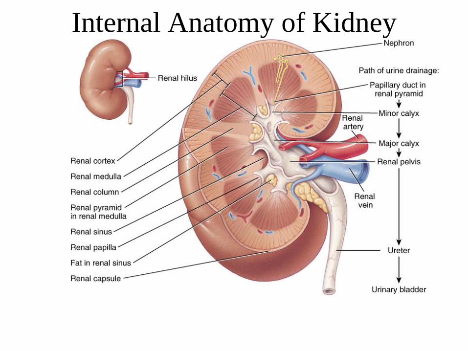
Internal Anatomy of Kidney

26-5
Human Kidney

26-6
Blood & Nerve Supply of Kidney
• Abundantly supplied with blood vessels
– receive 25% of resting cardiac output via renal arteries
• Functions of different capillary beds
– glomerular capillaries where filtration of blood occurs
– peritubular capillaries that carry away reabsorbed
substances from filtrate (renal cortex)
– vasa recta supplies nutrients to medulla
• Sympathetic vasomotor nerves regulate blood
flow by altering arterioles

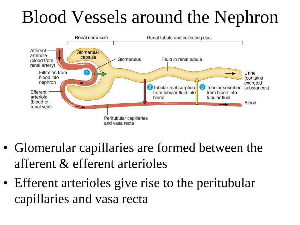
Blood Vessels around the Nephron
• Glomerular capillaries are formed between the
afferent & efferent arterioles
• Efferent arterioles give rise to the peritubular
capillaries and vasa recta
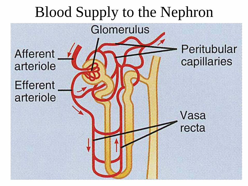
Blood Supply to the Nephron

26-10
The Nephron
• Kidney has over 1 million nephrons composed
of a corpuscle and tubule
• Renal corpuscle = site of plasma filtration
– glomerulus is capillaries where filtration occurs
– glomerular (Bowman’s) capsule is double-walled
epithelial cup that collects filtrate
• Renal tubule
– proximal convoluted tubule
– loop of Henle dips down into medulla
– distal convoluted tubule
• Collecting ducts and papillary ducts drain
urine to the renal pelvis and ureter

Cortical Nephron
• 80-85% of nephrons are cortical nephrons
• Renal corpuscles are in outer cortex and loops of Henle lie
mainly in cortex

Juxtamedullary Nephron
• 15-20% of nephrons are juxtamedullary nephrons
• Renal corpuscles close to medulla and long loops of Henle extend into
deepest medulla enabling excretion of dilute or concentrated urine
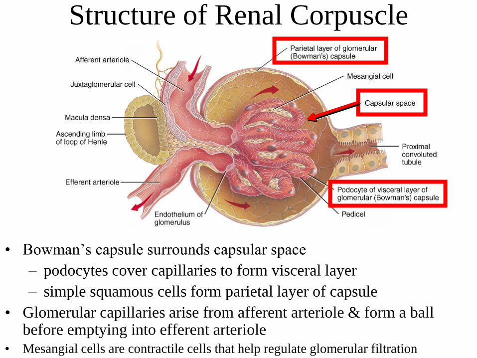
Structure of Renal Corpuscle
• Bowman’s capsule surrounds capsular space
– podocytes cover capillaries to form visceral layer
– simple squamous cells form parietal layer of capsule
• Glomerular capillaries arise from afferent arteriole & form a ball before emptying into efferent arteriole
• Mesangial cells are contractile cells that help regulate glomerular filtration

26-14
Juxtaglomerular Apparatus
• Structure where afferent arteriole makes contact with ascending
limb of loop of Henle
– macula densa is thickened part of ascending limb
– juxtaglomerular cells are modified muscle cells in arteriole
– Functions to help regulate blood pressure within kidneys
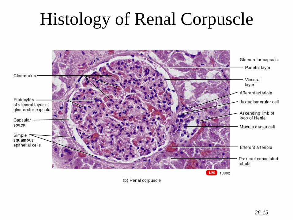
26-15
Histology of Renal Corpuscle

26-16
Number of Nephrons
• Remains constant from birth
– any increase in size of kidney is size increase of
individual nephrons
• If injured, no replacement occurs
• Dysfunction is not evident until function
declines by 25% of normal (other nephrons
handle the extra work)
• Removal of one kidney causes enlargement
of the remaining until it can filter at 80% of
normal rate of 2 kidneys

Overview of Renal Physiology
• Glomerular filtration of plasma
• Tubular reabsorption
• Tubular secretion

Anatomy of Ureters
• 10 to 12 in long
• Varies in diameter from 1-10 mm
• Extends from renal pelvis to
bladder
• Retroperitoneal
• Enters posterior wall of bladder
• Physiological valve only
– bladder wall compresses ureteral
openings as it expands during filling
– flow results from peristalsis, gravity
& hydrostatic pressure

Location of Urinary Bladder
• Posterior to pubic symphysis
• In females is anterior to vagina & inferior to uterus
• In males lies anterior to rectum

Anatomy of Urinary Bladder
• Hollow, distensible muscular organ with capacity of 700 - 800 mL
• Trigone is smooth flat area bordered by 2 ureteral openings and one
urethral opening

Micturition Reflex• Micturition or urination (voiding)
• Stretch receptors signal spinal cord and brain
– when volume exceeds 200-400 mL
• Impulses sent to micturition center in sacral spinal cord
(S2 and S3) & reflex is triggered
– parasympathetic fibers cause detrusor muscle to contract,
external & internal sphincter muscles to relax
• Filling causes a sensation of fullness that initiates a
desire to urinate before the reflex actually occurs
– conscious control of external sphincter
– cerebral cortex can initiate micturition or delay its occurrence
for a limited period of time

26-22
Anatomy of the Urethra• Females
– length of 1.5 in., orifice between clitoris & vagina
– histology
• transitional changing to nonkeratinized stratified
squamous epithelium, lamina propria with elastic fibers &
circular smooth muscle
• Males
– tube passes through prostate, UG diaphragm & penis
– 3 regions of urethra
• prostatic urethra, membranous urethra & spongy urethra
• circular smooth muscle forms internal urethral sphincter &
UG diaphragm forms external urethral sphincter

26-23
Urinary Incontinence
• Lack of voluntary control over micturition
– normal in 2 or 3 year olds because neurons to
sphincter muscle is not developed
• Stress incontinence in adults
– caused by increases in abdominal pressure that
result in leaking of urine from the bladder
• coughing, sneezing, laughing, exercising, walking
– injury to the nerves, loss of bladder flexibility,
or damage to the sphincter