the unique physiology of solid tumors: opportunities (and … · of both human and animal tumors...
TRANSCRIPT

(CANCER RESEARCH 58. 1408-1416. April I. 1998]
Review
The Unique Physiology of Solid Tumors: Opportunities (and Problems) forCancer Therapy1
J. Martin Brown2 and Amato J. Giaccia
Department of Radiation Oncologo, Stanford University School of Medicine, Stanford, California 94305-5468
Abstract
The physiology of solid tumors differs from that of normal tissues in anumber of important aspects, the majority of which stem from differencesbetween the two vasculatures. Compared with the regular, ordered vas-
culature of normal tissues, blood vessels in tumors are often highlyabnormal, distended capillaries with leaky walls and sluggish flow. Tumorgrowth also requires continuous new vessel growth, or angiogenesis. Thesephysiological differences can be problems for cancer treatment; for example, hypoxia in solid tumors leads to resistance to radiotherapy and tosome anticancer drugs. However, these differences can also be exploitedfor selective cancer treatment. Here we review four such areas that areunder active investigation: (a) hypoxia-selective cytotoxins take advantage
of the unique low oxygen tension in the majority of human solid tumors.Tirapazamine, a drug in the final stages of clinical trials, is one of the morepromising of these agents; (ft) leaky tumor blood vessels can be exploitedusing liposomes that have been sterically stabilized to have a long intra-vascular half-life, allowing them to selectively accumulate in solid tumors;
(c) the tumor microenvironment is a stimulus to angiogenenesis, andinhibition of angiogenesis can be a powerful anticancer therapy not susceptible to acquired drug resistance; and (</) we discuss attempts to usegene therapy activated either by the low oxygen environment or bynecrotic regions of tumors.
Introduction
Tumor Physiology: A Relatively Unexplored Target for CancerTherapy. Nonsurgical methods of cancer treatment, primarily radiation therapy and chemotherapy, rely almost exclusively on agents thatkill cells. The main problem with these current treatments, however,is that they do not, in general, have specificity for cancer cells. In thecase of radiation therapy, a degree of specificity is achieved bylocalizing the radiation to the tumor and its immediate surroundingnormal tissue. For anticancer drugs, it is primarily the rapid proliferation of many of the cancer cells that makes them more sensitive tocell killing than their normal cellular counterparts. However, bothmodalities are limited by their cytotoxic effects on normal cells. In thecase of radiotherapy, normal tissue surrounding the tumor limits theradiation dose, whereas for anticancer drugs, it is usually the killing ofrapidly dividing normal cells, such as those in the bone marrow, hairfollicles, and epithelial cells lining the gastrointestinal tract, that limitthe dose that can be given.
To achieve greater efficacy with present day treatments, investigators are attempting to exploit differences between normal and malignant cells at the cellular and molecular level. However, there is asecond critical difference between normal and malignant tissues thathas the potential for exploitation to produce more specific anticancer
Received 12/31/97; revised 2/6/98; accepted 2/17/98.The costs of publication of this article were defrayed in pan by the payment of page
charges. This article must therefore be hereby marked advertisement in accordance with18 U.S.C. Section 1734 solely to indicate this fact.
' Supported by USPHS Grants CA 15201. CA 67166 (to J. M. B.). and CA 64489 (to
A.J. G.).2 To whom requests for reprints should be addressed, at Division of Radiation Biology
(CBRL-GK103). Department of Radiation Oncology. Stanford University School ofMedicine, Stanford. CA 94305-5468. Phone: (650)723-5881; Fax: (650)723-7382.
therapy. As this review will detail, the physiology of solid tumors atthe microenvironmental level is sufficiently different from that of thenormal tissues from which they arise to provide a unique and selectivetarget for cancer treatment. To date, targeting tumor physiology foranticancer therapy has received considerably less attention than approaches based on the cellular and molecular differences betweentransformed and untransformed cells. Table 1 lists the principal differences in physiology between normal and malignant tissues that canbe exploited (and can also be a problem) in cancer treatment.
The Vasculature: Basis for the Unique Physiology of SolidTumors. The underlying differences between the physiology of normal and tumor tissues stems from the tumor vasculature. This iscomposed of two types of vessels: the existing vessels in normaltissues into which the tumor has invaded; and tumor microvesselsarising from neovascularization resulting from increased expressionof proangiogenic factors produced by tumor cells. Both types ofvessels develop structural and physiological abnormalities that havebecome a hallmark of the tumor microvasculature. Although earlystudies of tumor blood flow described marked heterogeneity and oftensluggish flow (1), it was the later studies of vascular casting techniques and window chamber preparations that identified the structuralbasis for these flow inhomogeneities (2-4). These studies showed thattumor blood vessels are highly irregular, tortuous, have arterio-venous
shunts, blind ends, lack smooth muscle or enervation, and haveincomplete endothelial linings and basement membranes (Fig. 1). Asa result, blood flow is often sluggish, highly irregular, and the vesselsmuch "leakier" than those in normal tissues. These characteristics of
tumor vasculature lead to the physiological differences described inthe following sections that present both problems and opportunitiesfor treatment of solid tumors.
Hypoxia-selective Cytotoxins
Tumor Hypoxia. The pioneering work of Gray et al. (5) demonstrated that the sensitivity to radiation damage of cells and tissuesdepended on the presence of oxygen at the time of irradiation. Thehistológica! studies of human lung adenocarcinomas by Thomlinsonand Gray (6) provided a mechanism by which cells could be hypoxicin tumors. They postulated that because of their unrestrained growth,tumor cells would be forced away from vessels beyond the effectivediffusion distance of oxygen in respiring tissue, thereby becominghypoxic and eventually necrotic. Given typical values for intracapil-
lary oxygen tensions and oxygen consumption rates, the oxygendiffusion distance would be approximately 150 ju.ni (Fig. 2).
Fig. 2 shows two important further consequences of reducingoxygen concentration: (a) the fraction of proliferating cells and/or therate of cell proliferation decreases as a function of distance from thevasculature (7, 8), a phenomenon that, at least in vitro, is largely, orwholly, the result of decreasing oxygen levels (9). An importantconsequence of this hypoxia-induced inhibition of proliferation is that
because most anticancer drugs are primarily effective against rapidlydividing cells, their effectiveness would be expected to fall off as a
1408
on June 3, 2020. © 1998 American Association for Cancer Research. cancerres.aacrjournals.org Downloaded from

EXPLOITING TUMOR PHYSIOLOGY
Table 1 Phvsioiogical characteristics of malignant tissues that can potentially be exploited for cancer therapy, how these differ from those of normal tissues, and how thesecharacteristics may also be detrimental to therapy
CharacteristicsMicrovasculature
Oxygénation
NecrosisNormal
tissueDeveloped
with ordered,regulatedflowHeterogenous,
but rarelyhypoxicregionsNot
presentTumorConstant
new vessel growth; vesselsleaky, tortuous, often sluggishand irregular flow
Highly heterogeneous with hypoxicregionscommonPresentDetrimental
aspects fortherapyPoor
delivery of some therapeutic agentsdue to irregular flow and highinterstitial pressure
Reduces tumor sensitivity to radiationand anticancer drugs; predisposes toincreased malignancy (e.g.. metastasis)
Not known if anyMethod
of exploiting fortherapyAntiangiogenic
agentsStealthliposomesSelective
cytoloxins; gene therapytargeted byhypoxiaGene
therapy targeted to necrosis
function of distance from blood vessels. This has been shown experimentally (10); and (/?) because hypoxic cells must be the ones mostdistant from blood vessels, they will be exposed to lower concentrations of drug than those adjacent to blood vessels, primarily as a resultof the metabolism of such agents through successive cellular layers.
Thus, tumor hypoxia would be expected to be an important factorleading to resistance to radiotherapy (because of hypoxia, per se,affecting cellular radiation sensitivity) and to chemotherapy (becauseof lower proliferation and lower drug concentrations in the hypoxiccells). The consequent reduction of cell kill to anticancer treatment asa function of distance from tumor blood vessels is shown in Fig. 2.
What is the evidence for this model? Hypoxia is a common featureof both human and animal tumors (11. 12). The vast majority ofhuman solid cancers have median pO2 levels lower than their normaltissue of origin (12, 13). In animal tumors, it can be shown that thesehypoxic cells are also viable and contribute to the resistance oftransplanted tumors to both radiation (14) and to some anticancerdrugs (15, 16). In human tumors, there is direct evidence frommeasurements of oxygen levels that hypoxia contributes to resistanceto radiotherapy (17-19). Similar studies have not been performed with
chemotherapy, although the evidence of a strong correlation betweenthe response of head and neck cancers to chemotherapy and toradiotherapy implicates hypoxia as a cause of drug resistance (20, 21).
Hypoxia in solid tumors, however, has an important consequence inaddition to conferring a direct resistance to radiation and chemother
apy. Graeber et al. (22) showed recently that low oxygen levelscaused apoptosis in minimally transformed mouse embryo fibroblustsand that this apoptosis depended to a large extent on wild-type p53.
They further showed, using these same cells growing as solid tumorsin immune-deprived mice, that apoptosis colocalized with hypoxicregions in tumors derived from p53 wild-type mice. In tumors derivedfrom p53 -/— cells, there was much less apoptosis and no colocal-
ization with tumor hypoxia. These findings provide evidence thathypoxia, by selecting for mutant p53. might predispose tumors to amore malignant phenotype. Clinical data support this conclusion.Studies both with soft tissue sarcomas (23) and with carcinoma of thecervix (24) have shown that hypoxic tumors are more likely to bemetas tatic.
The model shown in Fig. 2 of hypoxic cells occurring at thediffusion distance of oxygen is the classic model of tumor hypoxiagenerally attributed to Thomlinson and Gray (6). However, we andothers have proposed that tumor hypoxia can occur in a second way,by temporary obstruction or cessation of tumor blood flow-the so-
called acute hypoxia model (25, 26). Definitive evidence for this typeof acute hypoxia arising from fluctuating blood flow has come fromelegant studies with transplanted tumors in mice using diffusion-
limited fluorescent dyes (27, 28). Because fluctuating blood flow hasalso been demonstrated in human tumors (29, 30), it is likely that thistype of hypoxia is also present in human tumors. The consequences ofacute hypoxia will be similar to those of the diffusion-limited hypoxia.
Normal Tumor
Temporary Hypoxia/"^i-\ s\\ i if* •¿�*»»***"Sk.
Occlusion
BlindEnds
BreakinVesselWall
AVShuntFig. 1. Diagram showing the principal differences between the vasculature of normal and malignant tissues. Whereas normal tissues have relatively uniform and well-ordered blood
vessels that are sufficiently close together to oxygenate all of the tissue, blood vessels in tumors are tortuous, have incomplete vessel walls, have sluggish and irregular blood flow,
and have regions of hypoxia between the vessels.
1409
on June 3, 2020. © 1998 American Association for Cancer Research. cancerres.aacrjournals.org Downloaded from

EXPLOITING TUMOR PHYSIOLOGY
Fig. 2. A diagrammatical representation of aportion of a tumor cord surrounding a capillary. Asthe oxygen concentration decreases with increasingdistances from the capillary, both cell proliferationrates and drug concentration (Cone.) also decrease.The hypoxic environment also can select againstcells expressing wild-type p53. Shown on the right
is how the level of cell kill to radiation and to manyanticancer (CHEM.) drugs decreases with increasing distance from the capillary. It also shows howa drug with specificity for killing hypoxic cellswould give a complementary profile of cell killcompared with radiation and chemotherapy, andthat the combined toxicity would be greater thanthat achievable by either agent alone. Redrawnfrom Brown and Sum (40).
RADIATION/ CHEM.DRUG
•¿�- - -|COMBINED]
O 50 100 150
Distance from Capillary (Jim)
Any cells surrounding a closed blood vessel will be resistant toradiation killing because of their lack of oxygen at the time ofradiation and will be exposed to lower levels of anticancer drugs thanthose surrounding blood vessels with normal flow. This would beexpected to lead to inhomogeneities in response to anticancer agents,as has been observed in experimental tumors (31).
Hypoxia-selective Cytotoxins. Can the low oxygen levels in tu
mors be turned from a disadvantage to an advantage in cancer treatment? Such a possibility was proposed over 20 years ago by Lin et ai.(32), who reasoned that compounds based on the quinone structure ofmitomycin C might be more active in hypoxic tumors. It was knownat that time that mitomycin C required metabolic reduction of thebenzoquinone ring to produce the cytotoxic bifunctional alkylatingagent. Lin et al. (32) reasoned that a lower oxidation reduction (redox)potential for tumor tissue relative to most normal tissues could increase reductive activation of these quinone derivatives in tumors.Although this was not the correct mechanism for the increased cyto-
toxicity of mitomycin C and certain analogues toward hypoxic cells(much lower levels of hypoxia are needed to change cellular redoxpotential), these studies were important in suggesting the potential ofhypoxia-activated drugs and led to the concept of selectively killingthe hypoxic cells in solid tumors (33-37).
It is important to note that specifically killing the hypoxic cells intumors has greater therapeutic potential than oxygenating the cells orchemically sensitizing them to radiation or chemotherapy. Not only isthe killing tumor specific (hypoxia is tumor specific), but the cellskilled are the ones resistant to conventional therapy. This principle of"complementary cytotoxicity" is illustrated in Fig. 2. The combined
killing of two agents with complementary cytotoxicity is potentiallymuch greater than that of two agents acting on the same cell population. The other major advantage of hypoxia-selective cytotoxins is
their potential for providing enhancement to the killing of standardanticancer drugs. This cannot be done by temporarily oxygenating thehypoxic tumor cells.
There are presently three different classes of hypoxia-specific cy
totoxins that are in use clinically or are being developed for clinicaluse. They are the quinone antibiotics, the nitroimidazoles, and thebenzotriazine di-N-oxides. In the quinone class, the three principal
agents of current clinical interest are mitomycin C, porfiromycin, andE09. All are structurally similar and require reductive metabolism foractivity. Each is converted on reductive metabolism to a bifunctionalalkylating agent and probably produce their major cytotoxic activitythrough the formation of DNA interstrand cross-links. Reviews of the
mechanism of action, pharmacology, and preclinical and clinicalactivities of these drugs have been published (38-40).
Mitomycin C, justifiably considered to be the prototype bioreduc-
tive drug (37), was introduced into the clinic in 1958 and has demonstrated efficacy toward a number of different tumors in combinationwith other chemotherapy drugs and with radiation. However, itsselective toxicity toward hypoxic cells is modest, with values forhypoxic cytotoxicity ratios (the ratio of drug concentration to produceequal cell kill for aerobic and hypoxic cells) of 1 (no preferentialtoxicity) to approximately 5 (37, 41-43). However, based on this
activity, mitomycin C has been combined with radiotherapy in tworandomized trials of head and neck cancer (44, 45), the pooled resultsof which gave a statistically significant disease-free survival benefit.
Whether this promising finding is the result of preferential cytotoxicity of mitomycin C toward hypoxic cells or to cytotoxicity to bothaerobic and hypoxic cells is, however, open to debate (40). Nonetheless, encouraged by this success, the Yale group is now testingporfiromycin, a drug that has a somewhat greater hypoxic-selective
toxicity than mitomycin C.The third drug in this series, E09, is a much more efficient substrate
for DT-diaphorase than either mitomycin C or porfiromycin and
shows high toxicity to both aerobic and hypoxic cells in cells withhigh DT-diaphorase levels. Cells with low DT-diaphorase levels are
much less susceptible to killing by E09 under aerobic conditions butshow a high (up to 50-fold) preferential toxicity toward hypoxic cells.
However, the pharmacokinetics of this agent work against its clinicalutility, and Phase I clinical studies have shown little activity of thisdrug (46).
A second class of bioreductive agents is the nitroimidazoles, the firsttwo of which, metronidazole and misonidazole, were extensively testedas hypoxic radiosensitizing agents (47). Further drug development byAdams et al. (48) produced a compound, RSU1069 (l(2-nitro-l-imida-zolyl)-3-(l-aziridinyl)-2-propanol), which has been shown to be a highly
efficient cytotoxic agent with activity both in vitro and in vivo (48, 49).RSU1069 has a hypoxic cytotoxicity ratio of some 10-100 for different
cell lines in vitro, and it, or its prodrug, RB6145, has shown excellentactivity with mouse tumor models when combined with irradiation oragents that induce hypoxia (50). Unfortunately, however, clinical testingof RB6145 has been aborted due to irreversible cytotoxicity towardretinal cells (51).
TPZ3 is the first, and thus far, only representative of a third class of
hypoxia-selective cytotoxins. This drug has a high selective hypoxiccytotoxicity (20-300 for different cell lines) and maintains its differ-
3 The abbreviations used are: TPZ. tirapazamine; RES, reticular endothelial system;
VEGF, vascular endothelial growth factor; TNF, tumor necrosis factor; HRE, hypoxia-responsive element; 5-FC, 5-fluorocytosine.
1410
on June 3, 2020. © 1998 American Association for Cancer Research. cancerres.aacrjournals.org Downloaded from

EXPLOITING TUMOR PHYSIOLOGY
Fig. 3. A schematic showing the metabolism of TPZ to its active free radical moietycausing preferential toxicity to hypoxic cells by producing DNA damage. The postulatedclose association of the reducÃaseenzyme(s) to DNA is shown, ssb, single-strand break;dsb. double-strand break.
ential toxicity relative to aerobic cells at oxygen concentrations —¿�10-
fold higher than do other bioreductive drugs (52). This could be animportant reason for its excellent efficacy in preclinical models, bothwith radiation and some anticancer drugs, because cells at "intermediate" oxygen levels may be more important than the extremely
hypoxic cells in governing tumor response to fractionated irradiation(53).
The mechanism of the selective toxicity of TPZ is illustrated in Fig.3. The toxic species has been identified as the radical formed by the1-electron reduction of TPZ, with DNA double-strand breaks, leading
to chromosome breaks as the principal mechanism of cell killingunder hypoxia (54). Recent work has shown that the activating en-
zyme(s) leading to DNA damage is located in the cell nucleus,probably associated with the nuclear matrix (55, 56). Under aerobicconditions, oxygen can remove the additional electron from the TPZradical, thereby back-oxidizing it to the nontoxic parent with the
concomitant production of Superoxide radical (57). In preclinicaltesting, TPZ has shown itself to be effective in sensitizing tumors tofractionated radiation (58) with no increase of sensitivity of normaltissues (59). TPZ is also effective in increasing tumor cell killingwithout increasing the toxicity of a number of commonly used anti-
cancer drugs, particularly cisplatin and carboplatin (60, 61). Thispotentiation can be quite large, up to the equivalent of giving fivetimes more cisplatin to the tumor for no increase in the systemictoxicity of cisplatin (60, 62).
Based on these preclinical studies, Phase I clinical trials wereinitiated in 1992, and at this time, Phase II trials with radiation (63),with chemotherapy (64), as well as two large Phase III trials combining TPZ with cisplatin in stage IIIB and IV nonsmall cell lung cancer,have been completed. The results of these trials will be available inmid 1998.
In summary, TPZ is the first drug introduced into the clinic as aspecific hypoxia-activated drug. The potential of targeting drug cyto-
toxicity and/or anticancer drug potentiation to the hypoxic cells insolid tumors is an exciting one and exploits what is clearly a difference between the physiology of tumors and normal tissues. Furtherdrug development of new hypoxia-selective cytotoxins is clearly
warranted, because it is unlikely that TPZ is the optimum hypoxia-selective cytotoxin.
Liposome Delivery
Tumor vessels are often leaky, demonstrating increased permeability to circulating large molecules, even up to sizes of 400 nm indiameter (65-67). This leakiness of the vasculature results from an
incomplete or missing endothelial cell lining and basement membrane(65, 68). Increased permeability of tumor vasculature has allowedspecific targeting of anticancer drugs to tumors using small liposomesof ~100-nm diameter that have been specially modified to remain in
the circulation for long periods of time.The discovery that drugs could be encapsulated in fatty vesicles or
liposomes in the 1960s led to high enthusiasm that this system wouldfill the role of the "magic bullet" for drug delivery. However, animal
experiments soon proved these early expectations to be misguided,because liposomes, when injected i.V., are rapidly recognized andremoved by the RES. An elegant solution to this problem was foundby coating lipids with carbohydrate groups to have them resembleerythrocytes (69-71). These developments gave rise to today's steri-cally stabilized, or Stealth'"1, liposome which is coated with polyeth
ylene glycol to provide a steric barrier to recognition and destructionby the RES (72). Such liposomes with encapsulated epirubicin ordoxorubicin exhibit improved antitumor efficacy compared with conventional liposomes or to free drug (73). Stealth liposomes remain inthe blood circulation for extended periods of time, with most of theliposomes remaining in the blood and only 10-15% ending up in the
liver, a major improvement over conventional liposomes, the majorityof which are rapidly captured by the RES in the liver (74).
Despite the impressive preclinical activity of both doxorubicin andvincristine encapsulated in sterically stabilized (Stealth) liposomes,none of the studies to date have attempted to determine the extent towhich the increased antitumor activity is the result of increased tumorconcentrations of liposomes carrying the drug or to prolonged exposure time of the tumor cells to drug. Presumably, both are important,although the benefit of prolonged exposure time is very likely to bedependent on tumor proliferation rate and the drug in question.
Clinical studies of doxorubicin encapsulated in Stealth liposomes(Doxil) began a number of years ago, and the results to date confirmthe prolonged circulation time (plasma tl/2 of 45 h compared with~ 10 h for free doxorubicin) and increased concentrations of free drug
in pleural effusion of a variety of tumors in those patients treated withdoxil compared with free doxorubicin (75). These Phase I/II studieshave also identified a novel toxicity of doxorubicin in Stealth liposomes, i.e., a prolonged desquamating painful dermatitis primarilyaffecting hands and feet, possibly due to liposome-mediated deposi
tion of drug in the skin. However, the results to date for a variety ofsolid tumors support the preclinical findings that increased antitumorefficacy will be likely with doxorubicin encapsulated in Stealth liposomes versus free drug.
Despite the obvious advantage provided by leaky blood vesselsallowing high tumor concentrations of Stealth liposomes, it has to berecognized that the distribution of the liposomes is highly heterogeneous and concentrated in the perivascular interstitial regions (67, 76).Presumably, the antitumor activity of these heterogeneous deposits isthe result of a slow release of drug throughout the tumor from thesehot spots of high drug concentrations.
1411
on June 3, 2020. © 1998 American Association for Cancer Research. cancerres.aacrjournals.org Downloaded from

EXPLOITING TUMOR PHYSIOLOGY
Antiangiogenesis
In the early 1970s, Folkman (77, 78) introduced what was then thecontroversial hypothesis that the growth of solid tumors was absolutely dependent upon new blood vessel formation, or angiogenesis,developing from outside the growing tumor mass. He also suggestedthat, this being the case, therapy aimed specifically at the angiogenicprocess or "antiangiogenesis" could be effective if tumor growth is
angiogenesis dependent. Although slow to gain momentum in themidst of the promise for selective therapies based on the identificationand functional characterization of tumor oncogenes and suppressorgenes, work on antiangiogenesis as a therapy for solid tumors hasaccelerated enormously in the past 5 years, with several lead compounds that now show great promise in preclinical testing. Because acomprehensive review of the literature in this field is beyond thescope of the present review, we will focus instead on the mostpromising targets and particularly those controlled by the tumormicroenvironment.
At first sight, the large number of angiogenic factors that have beenimplicated in tumor vascularization, including basic and acidic fibro-
blast growth factor, VEGF, transforming growth factors a and ß,TNF-a, interleukin 8, and angiogenin, to name a few of the more
important (79, 80), would make it seem unlikely that to target a singleor a few angiogenic factors would be successful. Based on the rapiditywith which tumor cells can adapt and select for mutants resistant tocommon anticancer drugs, it would seem probable that inhibition ofone or more of these proangiogenic factors would cause the tumors toswitch their angiogenic dependent growth to other cytokines. However, strategies aimed at one of these factors, VEGF and/or its twohigh-infinity receptors expressed in endothelial cells, flt-1 and KDR/Flk-1, have been remarkably promising (81-83). Indeed, VEGF is
rapidly emerging as the dominant angiogenic factor in solid tumordevelopment.
In addition to its potent and specific vascular endothelial mitogenicactivity, VEGF also increases vascular permeability. Indeed, it wasoriginally discovered as a tumor-secreted protein that rendered mi-
crovasculature hyperpermeable and was named vascular permeabilityfactor (84). One possible reason for the major importance of VEGF asan angiogenic agent is that it is the only one of the angiogenic factorsthat also produces vascular permeability, and there is a considerablebody of evidence suggesting that the microvascular hyperpermeabilityis an essential factor in angiogenesis favoring the migration of endothelial cells through the extravascular matrix (85).
Numerous strategies are presently being used to inhibit VEGFactivity in tumors. They include antisense VEGF mRNA, monoclonalantibodies, and farnesyltransferase inhibitors. A proof of concept ofthis approach is that monoclonal antibodies against VEGF have beenshown to inhibit the growth of human tumors in nude mice with aconcomitant reduction in vascularity, although the same antibodiesproduced no effect on the growth of the tumor cells in vitro (86).
A variation on the strategy of inhibiting VEGF (or any angiogenicfactor) is to inhibit its receptor. The proof of principle of the effectiveness of this was shown by Millauer et al. (81), who demonstratedthat infection of the vasculature surrounding implanted glioblastomacells with viruses expressing a dominant negative mutant form ofFlk-1 suppressed glioblastoma tumor growth. Also, the tumors that
arose had a large central necrosis surrounded by a thin layer of tumorcells with no invasion of the tumor by vasculature. Recently, highpotency small molecule inhibitors of the Flk-1 receptor for VEGF
have been isolated and shown to inhibit angiogenesis (82), to markedly inhibit the growth of s.c. tumors, and to prevent metastatic spread(83). These data provide evidence that VEGF expression can have aprofound influence on tumor growth and metastatic spread.
As with many biological processes, there are natural antagonists toangiogenesis, such as angiostatin and endostatin, that could be used inconjunction with inhibitors of angiogenic factors or their receptors(87). Angiostatin is a peptide formed from the cleavage of plasmin-
ogen, which, when secreted by tumors, inhibits the growth of métastases in the same host (87). It acts by selectively inhibiting endothelialcells to respond to angiogenic signals, and when given to mice bearingtransplanted murine or human tumors, causes marked regression ofthe tumors to microscopic dormant foci (88-90). Recently, a second
natural inhibitor of angiogenesis, endostatin, with similar potent activity against established transplanted tumors has been described (91).
Although angiogenesis is a highly attractive tumor-specific target
for therapy, it is nonetheless a normal process occurring in embryonicand placental development, wound healing, ovulation, and chronicinflammation (92). The signal linking these different processes could,in many instances, be hypoxia. Intuitively, this makes sense. A cellthat is low in oxygen responds to this stress by secreting a cytokinethat will increase blood vessel growth toward that cell. In tumors,there is compelling evidence that hypoxia stimulates angiogenesis byincreasing VEGF production. One of the most elegant early demonstrations of this came from the work of Shweiki et al. (93), whoshowed that in human glioblastomas, VEGF message was highlyexpressed adjacent to areas of focal necrosis (where hypoxia would beexpected) and that new capillary bundles were localized alongsideVEGF-producing cells. Further evidence that tumor hypoxia is an
important signal for angiogenesis, and a determinant of tumor growth,is provided by the recent work of Maxwell et al. (94), who showedthat tumors growing from cells deficient in the hypoxia-activatedtranscription factor, hypoxia-inducible factor 1, did not show increased VEGF induction adjacent to necrosis, were poorly vascular-ized, and grew much slower than tumors with competent hypoxia-
inducible factor 1.Because VEGF can be induced in normal cells by hypoxia (95, 96),
the question arises as to the relative contributions to tumor angiogenesis from the microenvironment (i.e., hypoxia) and from malignancyper se (97). Mazure et al. (98) have shown recently that it is probablyboth: oncogenic transformation (at least by certain oncogenes) andhypoxia act synergistically to induce VEGF. Therefore, the use ofagents that inhibit oncogenic ras activity may act as antitumor agentsin part by inhibiting signaling pathways in the angiogenic activationcascade.
Antiangiogenesis therapy has several important advantages overstandard anticancer treatment: (a) it targets a process that, under mostcircumstances, is tumor specific and therefore likely to have littlesystemic toxicity; (b) it has the advantage that with an endothelial celltarget, there is not the problem of the drug having to reach tumor cells,often many cell layers from the vasculature; and (c) one of the mostimportant advantages of antiangiogenic therapy is that the geneticstability of endothelial cells (as opposed to tumor cells) should prevent the development of drug resistance on repeated administration ofthe agent. This was first proposed by Kerbel (99) and recently dramatically demonstrated by Boehm et al. (100), who induced multipleregressions in transplanted mouse tumors by repeated administrationof endostatin. This concept is illustrated in Fig. 4.
In summary, antiangiogenetic strategies are now showing a greatdeal of promise in murine tumor models. The responses of suchtransplanted tumors to inhibitors of VEGF or its receptor Flk-1, or to
the natural antiangiogenetic peptides, angiostatin and endostatin, areextremely impressive. We believe it would not be overly optimistic toconclude that if human spontaneous tumors are as dependent oncontinued angiogenesis as are the more rapidly growing transplantedtumors in rodents (and this is a major unknown), then these strategiescould constitute a very important advance in cancer therapy.
1412
on June 3, 2020. © 1998 American Association for Cancer Research. cancerres.aacrjournals.org Downloaded from

EXPLOITING TUMOR PHYSIOLOGY
Anti-tumor therapy
I I I \
Therapyterminated
Regression of drug-
sensitive tumor cells ( )('Response')
Anti-tumor therapy
MM
Overgrowth of drug-
resistant tumor cellsubpopulations (•)
('Relapse')
No response('Resistance')
Anti-tumor angiogenesis Therapytherapy directed against terminated
endothelial cells
B MM
Repeat therapy
MM
Regession of tumor bloodvessels and tumor mass;
induction of dormancy ('Response')
Regrowth ofdormant tumor
Regression of bloodvessels and tumormass ('Resistance')
Fig. 4. The mechanism by which resistance to many standard anticancer drugs develops by outgrowth of drug-resistant tumor cell subpopulations, contrasted with the lack ofdevelopment of resistant tumors when antiangiogenesis therapy is directed against endothelial cells [redrawn from Kerbel ( 112)j.
Gene Therapy
Activated by Hypoxie Stress Response. The newest direction forexploiting tumor physiology is aimed toward the evolving field ofgene therapy. In this novel approach to anticancer therapy, geneticmaterial is transferred into cells by a variety of techniques with theultimate goal of selective killing of cancer cells and the sparing ofnormal cells. At present, this selective killing of tumor cells can beachieved either by controlling vector delivery and activity or bytransgene expression. Previous studies by Hallaban et al. (101) havetested the feasibility of intratumoral control of transgene expressionusing a radiation-inducible promotor ligated to the TNF-a gene. Theywere able to demonstrate radiation increased TNF-a production in-tratumorally after a 50-Gy single dose of ionizing radiation. Furthermore, the radiation-induced increase in TNF-a production resulted inincreased tumor cell killing. These experiments on radiation-inducible
gene expression establish the proof in principle that a strategy tocontrol gene expression by stress-responsive promotors is feasible in
vivo.A direct test of the ability of the low oxygen conditions to selec
tively control gene expression and increase cell kill was performed byDachs et al. (102), who linked a HRE from the mouse phosphoglyc-erate kinase-1 gene to the bacterial cytosine deaminase-encoding gene
and stably transfected it into HT 1080 cells. To test the hypothesis thatonly under low oxygen conditions would the expression of the bacterial cytosine deaminase gene be increased, the HT 1080 cells thatwere stably transfected with the HRE-cytosine deaminase gene construct were exposed either to 5-fluorouracil or to 5-FC under aerobicand hypoxic conditions. Only hypoxic cells were sensitive to 5-FC,the inactive prodrug, which requires enzymatic conversion to 5-flu
orouracil by cytosine deaminase to be cytotoxic. The increased sensitivity of HT 1080 transfectants to 5-FC suggested that in vitro HREs
could be used to control the expression of a prodrug activatingenzyme. At present, we do not have functional data on the ability ofHREs to control the activation of nontoxic prodrugs into a tumoricidalform in vivo. However, the ability of HREs to induce the activity ofa reporter gene has been evaluated intratumorally, and HREs seem tobe able to transcriptionally regulate gene expression under low oxygen conditions in vivo (102).
There are numerous implications of these studies for cancer genetherapy: (a) the use of HREs will provide a selective means ofcontrolling gene transcription in a wide variety of solid tumors basedon the lower oxygen levels of tumors compared with normal tissues;(b) the use of enzymes under the control of an HRE adds a safeguard(compared with expression of a toxic substance), because increasedexpression of the prodrug activating enzyme is itself nontoxic; (c) theexpanding list of inactive cytotoxic prodrugs and prodrug activatingenzymes increases the possibility of finding the most efficaciouscombinations for different tumor types: and (cf) continued research onthe regulation of gene induction by hypoxia should offer new tran-
scriptional regulatory elements as well as transcriptional stabilizingelements that will increase the dynamic range and specificity oftranscriptional responses to a low oxygen environment. Potentiallythis approach, therefore, would allow the activation of a nontoxicprodrug to its toxic metabolite selectively in solid tumors. However,as with most forms of gene therapy, targeting the constructs to thetumor remains a major hurdle.
Targeted by Tumor Necrosis. It has been known for severaldecades that certain species of anaerobic bacteria of the genus Clos-
triclium can selectively germinate and grow in the hypoxic/necroticregions of solid tumors after i.v. injection of spores. This was firstdramatically demonstrated by Malmgren and Flanagan (103) with C.tetani, the causative agent of tetanus. Mice, when injected i.v. with
spores of this bacteria, remained healthy unless they had tumors, inwhich case death by tetanus resulted within 48 h. This was caused bygermination of the bacteria in the tumors and release of toxins sys-
temically. Mose et al. (104) later isolated a nonpathogenic strain of C.sporogenes (later renamed C. oncolyticum), which germinated intumors after i.v. injection of the spores, causing tumor lysis andshrinkage of the tumors (104). Extensive preclinical testing wasfollowed by clinical trials of this agent, particularly with patients withglioblastoma who received injections of up to IO10 C. oncolyticum
spores (105, 106). Lysis was demonstrated in the tumors, with noevidence of clostridial germination or tissue destruction in the surrounding normal tissue. With the exception of mild to moderate fever,the patient suffered no ill effects from the injection of these organisms. However, no clinical benefit was demonstrated, presumably
1413
on June 3, 2020. © 1998 American Association for Cancer Research. cancerres.aacrjournals.org Downloaded from
EXPLOITING TUMOR PHYSIOLOGY
Clostridia Plasmid withgene for prodrugactivating enzyme
o o o °O O o °
O O ° o
TransformedClostridia asspores
Intravenous injection of sporesof transformed Clostridia
Spores germinate only in thetumor producing prodrugactivating enzyme selectively inthe hypoxic and necrotic areas
Injected non toxic prodrugconverted to toxic drugonly in the tumor
Active chemotherapy drugdiffuses from the hypoxic /necrotic regions
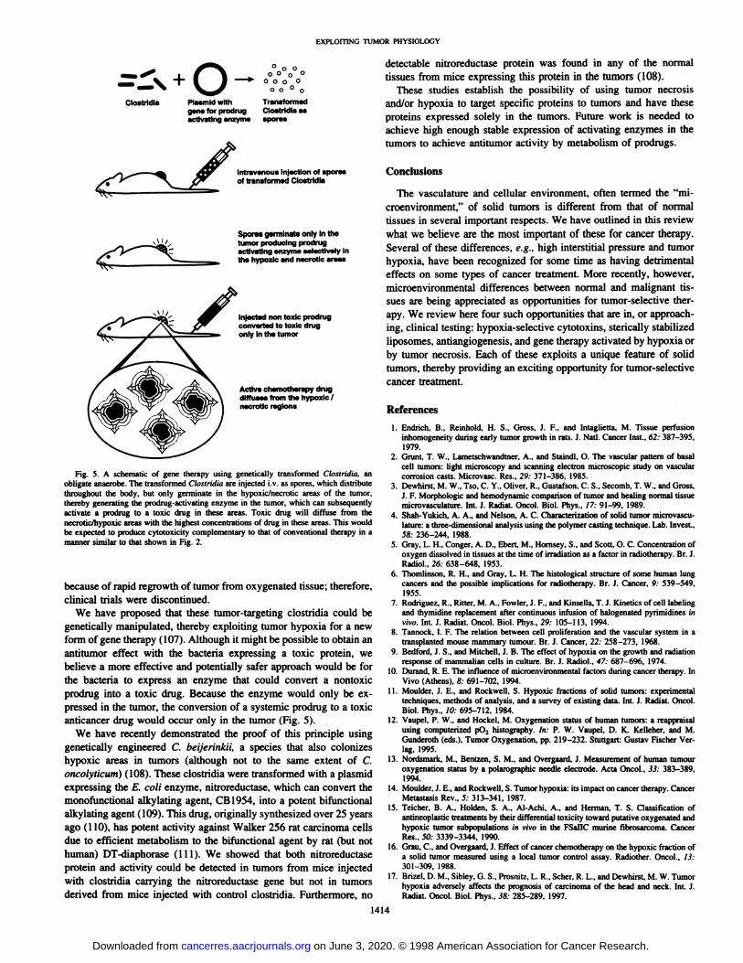
Fig. 5. A schematic of gene therapy using genetically transformed Clostriilia. anobligate anaerobe. The transformed Clostridia are injected i.V. as spores, which distributethroughout the body, but only germinate in the hypoxic/necrotic areas of the tumor,thereby generating the prodrug-activating enzyme in (he tumor, which can subsequentlyactivate a prodrug to a toxic drug in these areas. Toxic drug will diffuse from thenecrolic/hypoxic areas with the highest concentrations of drug in these areas. This wouldbe expected to produce cytotoxicity complementary to thai of conventional therapy in amanner similar to that shown in Fig. 2.
because of rapid regrowth of tumor from oxygenated tissue; therefore,clinical trials were discontinued.
We have proposed that these tumor-targeting Clostridia could be
genetically manipulated, thereby exploiting tumor hypoxia for a newform of gene therapy (107). Although it might be possible to obtain anantitumor effect with the bacteria expressing a toxic protein, webelieve a more effective and potentially safer approach would be forthe bacteria to express an enzyme that could convert a nontoxicprodrug into a toxic drug. Because the enzyme would only be expressed in the tumor, the conversion of a systemic prodrug to a toxicanticancer drug would occur only in the tumor (Fig. 5).
We have recently demonstrated the proof of this principle usinggenetically engineered C. beijerinkii, a species that also colonizeshypoxic areas in tumors (although not to the same extent of C.oncolyticum) (108). These clostridia were transformed with a plasmidexpressing the E. coli enzyme, nitroreductase, which can convert themonofunctional alkylating agent, CB1954, into a potent bifunctionalalkylating agent (109). This drug, originally synthesized over 25 yearsago (110), has potent activity against Walker 256 rat carcinoma cellsdue to efficient metabolism to the bifunctional agent by rat (but nothuman) DT-diaphorase (111). We showed that both nitroreductase
protein and activity could be detected in tumors from mice injectedwith clostridia carrying the nitroreductase gene but not in tumorsderived from mice injected with control clostridia. Furthermore, no
detectable nitroreductase protein was found in any of the normaltissues from mice expressing this protein in the tumors (108).
These studies establish the possibility of using tumor necrosisand/or hypoxia to target specific proteins to tumors and have theseproteins expressed solely in the tumors. Future work is needed toachieve high enough stable expression of activating enzymes in thetumors to achieve antitumor activity by metabolism of prodrugs.
Conclusions
The vasculature and cellular environment, often termed the "mi-croenvironment," of solid tumors is different from that of normal
tissues in several important respects. We have outlined in this reviewwhat we believe are the most important of these for cancer therapy.Several of these differences, e.g., high interstitial pressure and tumorhypoxia, have been recognized for some time as having detrimentaleffects on some types of cancer treatment. More recently, however,microenvironmental differences between normal and malignant tissues are being appreciated as opportunities for tumor-selective ther
apy. We review here four such opportunities that are in, or approaching, clinical testing: hypoxia-selective cytotoxins, sterically stabilized
liposomes, antiangiogenesis, and gene therapy activated by hypoxia orby tumor necrosis. Each of these exploits a unique feature of solidtumors, thereby providing an exciting opportunity for tumor-selective
cancer treatment.
References
10
12
13.
14.
16.
Endrich, B., Reinhold, H. S., Gross, J. F., and ¡maglietta, M. Tissue perfusioninhomogeneity during early tumor growth in rats. J. Nati. Cancer Inst., 62: 387-395.
1979.Grunt. T. W., Lametschwandtner. A., and Staindl. O. The vascular pattern of basalcell tumors: light microscopy and scanning electron microscopic study on vascularcorrosion casts. Microvasc. Res., 29: 371-386, 1985.Dewhirst, M. W., Tso, C. Y., Oliver, R., Gustafson, C. S., Secomb, T. W„and Gross,J. F. Morphologic and hemodynamic comparison of tumor and healing normal tissuemicrovasculature. Int. J. Radial. Oncol. Biol. Phys., 17: 91-99. 1989.Shah-Yukich, A. A., and Nelson, A. C. Characterization of solid tumor microvasculature: a three-dimensional analysis using the polymer casting technique. Lab. Invest.,58: 236-244, 1988.Gray, L. H., Conger, A. D., Eben, M., Hornsey, S., and Scott, O. C. Concentration ofoxygen dissolved in tissues at the time of irradiation as a factor in radiotherapy. Br. J.Radiol., 26: 638-648. 1953.
Thomlinson, R. H.. and Gray, L. H. The histológica! structure of some human lungcancers and the possible implications for radiotherapy. Br. J. Cancer, 9. 539-549,
1955.Rodriguez, R., Ritter. M. A., Fowler, J. F., and Kinsella, T. J. Kinetics of cell labelingand thymidine replacement after continuous infusion of halogenaled pyrimidines invivo. Int. J. Radial. Oncol. Biol. Phys., 29: 105-113, 1994.Tannock. I. F. The relation between cell proliferation and the vascular system in atransplanted mouse mammary tumour. Br. J. Cancer, 22: 258-273, 1968.
Bedford, J. S., and Mitchell. J. B. The effect of hypoxia on the growth and radiationresponse of mammalian cells in culture. Br. J. Radiol., 47: 687-696, 1974.
Durand, R. E. The influence of microenvironmental factors during cancer therapy. InVivo (Athens), 8: 691-702, 1994.
Moulder, J. E., and Rockwell, S. Hypoxic fractions of solid tumors: experimentaltechniques, methods of analysis, and a survey of existing data. Int. J. Radiât.Oncol.Biol. Phys., 10: 695-712. 1984.Vaupel. P. W.. and Hockel, M. Oxygénationstatus of human tumors: a reappraisalusing computerized pO2 histography. In: P. W. Vaupel, D. K. Kelleher, and M.Gunderoth (eds.). Tumor Oxygénation,pp. 219-232. Stuttgart: Gustav Fischer Verlag. 1995.Nordsmark, M.. Bentzen, S. M., and Overgaard, J. Measurement of human tumouroxygénationstatus by a polarographic needle electrode. Acta Oncol., 33: 383-389,1994.Moulder, J. E., and Rockwell, S. Tumor hypoxia: its impact on cancer therapy. CancerMetastasis Rev., 5.- 313-341, 1987.Teicher, B. A., Holden, S. A., AI-Achi, A., and Herman. T. S. Classification ofantineoplastic treatments by their differential loxicity toward putative oxygenated andhypoxic tumor subpopulations in vivo in the FSallC murine fibrosarcoma. CancerRes., 50: 3339-3344, 1990.
Grau, C., and Overgaard, J. Effect of cancer chemotherapy on the hypoxic fraction ofa solid tumor measured using a local tumor control assay. Radiother. Oncol., 13:301-309, 1988.
Brizel. D. M., Sibley. G. S., Prosnitz, L. R., Scher, R. L., and Dewhirst, M. W. Tumorhypoxia adversely affects the prognosis of carcinoma of the head and neck. Int. J.Radial. Oncol. Biol. Phys., 38: 285-289. 1997.
1414
on June 3, 2020. © 1998 American Association for Cancer Research. cancerres.aacrjournals.org Downloaded from

EXPLOITING TUMOR PHYSIOLOGY
18. Gatenby, R. A., Kessler, H. B., Rosenblum. J. S., Coia. L. R.. Moldofsky. P. J., and 46.Hartz, W. H. Oxygen distribution in squamous cell carcinoma métastasesand itsrelationship to outcome of radiation therapy. Int. J. Radial. Oncol. Biol. Phys., 14:831-838, 1988.
19. Nordsmark, M., Overgaard, M., and Overgaard, J. Pretreatment oxygénationpredicts 47.radiation response in advanced squamous cell carcinoma of the head and neck.Radiother. Oncol., 41: 31-40, 1996.
20. Ensley, J. F., Jacobs, J. R., Weaver, A., Kinzie, J., Crissman, J.. Kish, J. A., 48.Cummings, G., and Al-Sarraf. M. Correlation between response to cisplatinum-
combination chemotherapy and subsequent radiotherapy in previously untreatedpatients with advanced squamous cell cancers of the head and neck. Cancer (Phila.). 49.54: 811-814, 1983.
21. Jaulerry. C.. Rodriguez. J., Brunin, F., Jouve, M., Mosseti, V., Point, D., Pontvert. D.. 50.Validire, P., Zafrani. B., Blaszka, B., Asselain, B., Pouillart, P., and Brugere,J. Induction chemotherapy in advanced head and neck tumors: results of two randomized trials. Int. J. Radial. Oncol. Biol. Phys., 23: 483-489, 1992. 51.
22. Graeber, T. G., Osmanian, C., Jacks, T., Housman, D. E., Koch, C. J., Lowe, S. W..and Giaccia, A. J. Hypoxia-medialed selection of cells with diminished apoptoticpotential in solid tumours. Nature (Lond.), 379: 88-91, 1996. 52.
23. Brizel, D. M., Scully, S. P., Harrelson, J. M., Layfield. L. J., Bean, J. M., Prosnitz,L. R., and Dewhirst, M. W. Tumor oxygénationpredicts for the likelihood of distant 53.métastasesin human soft tissue sarcoma. Cancer Res., 56: 941-943, 1996.
24. Hockel, M., Schlenger. K., Aral, B., Milze, M., Schaffer, U., and Vaupel, P. Association between tumor hypoxia and malignant progression in advanced cancer of the 54.uterine cervix. Cancer Res., 56: 4509-4515, 1996.
25. Sutherland, R. M., and Franko, A. J. On the nature of the radiobiologically hypoxic 55.fraction in tumors. Int. J. Radial. Oncol. Biol. Phys., 6: 117-120, 1980.
26. Brown, J. M. Evidence for acutely hypoxic cells in mouse tumours, and a possible 56.mechanism of reoxygenation. Br. J. Radiol., 52: 650-656, 1979.
27. Chaplin, D. J., Durand, R. E., and Olive, P. L. Acute hypoxia in tumors: implicationsfor modifiers of radiation effects. Int. J. Radial. Oncol. Biol. Phys., 12: 1279-1282, 57.
1986.28. Trotter, M. J., Chaplin, D. J., and Olive, P. L. Use of a carbocyanine dye as a marker
of functional vasculature in murine tumours. Br. J. Cancer, 59; 706-709, 1989. 58.
29. Kotelnikov, V. M., Coon, J. S., Taylor, S., Hutchinson, J., Panje. W., Caldarelli,D. D., LaFollette, S., and Preisler, H. D. in vivo labelling with halogenated pyrimi-dines of squamous cell carcinomas and adjacent non-involved mucosa of head and 59.neck region. Cell Proliferation, 28: 497-509, 1995.
30. Hill, S. A., Pigott, K. H., Saunders, M. I., Powell, M. E., Arnold, S., Obeid, A.. Ward,G., Leahy, M., Hoskin, P. J., and Chaplin, D. J. Microregional blood flow in murine 60.and human tumours assessed using laser Doppler microprobes. Br. J. Cancer, 74(Suppl. 27): s260-s263, 1996. 61.
31. Simpson-Herren, L., Noker, P. E., and Wagoner, S. D. Variability of tumor responseto chemotherapy. II. Contribution of tumor heterogeneity. Cancer Chemother. Phar-macol., 22: 131-136, 1988. 62.
32. Lin, A. J., Cosby, L. A., Shansky. C. W., and Sartorelli, A. C. Potential bioreductivealkylating agents. 1. Benzoquinone derivatives. J. Med. Chem., ¡5:1247-1252, 1972.
33. Zeman. E. M., Brown, J. M., Lemmon, M. J., Hirst. V. K.. and Lee, W. W. SR-4233: 63.
a new bioreductive agent with high selective toxicity for hypoxic mammalian cells.Int. J. Radiât.Oncol. Biol. Phys., 12: 1239-1242, 1986.
34. Wilson, W. R., Moselen. J. W., Cliffe, S., Denny, W. A., and Ware, D. C. Exploitingtumor hypoxia through bioreductive release of diffusible cytoloxins: the cobalt(III)- 64.nitrogen mustard complex SN 24771. Int. J. Radial. Oncol. Biol. Phys., 29: 323-327.
1994.35. Kennedy. K. A.. Teicher, B. A., Rockwell, S., and Sartorelli, A. C. The hypoxic tumor
cell: a target for selective cancer chemotherapy. Biochem. Pharmacol., 29: 1-8, 1980. 65.
36. Sartorelli, A. C. Therapeutic attack of hypoxic cells of solid tumors. Cancer Res.. 48:775-778, 1988.
37. Rockwell. S., Kennedy, K. A., and Sartorelli. A. C. Mitomycin-C as a protolype 66.bioreductive alkylaling agent: I'Mvitro studies of metabolism and cytotoxicity. Int. J.
Radial. Oncol. Biol. Phys., 8: 753-755, 1982. 67.
38. Rockwell, S., Sartorelli, A. C., Tomasz. M., and Kennedy. K. A. Cellular pharmacology of quinone bioreductive alkylating agents. Cancer Metastasis Rev.. 12: 165-
176, 1993. 68.39. Raulh. A. M., Marshall, R. S., and Kuehl, B. L. Cellular approaches to bioreductive
drug mechanisms. Cancer Metastasis Rev., 12: 153-164, 1993. 69.40. Brown, J. M., and Sum, B. G. Hypoxia-specific cytotoxins in cancer therapy. Semin.
Radiât.Oncol., 6: 22-36, 1996. 70.
41. Fracasso, P. M., and Sartorelli. A. C. Cytotoxicity, and DNA lesions produced bymitomycin C and porfiromycin in hypoxic and aerobic EMT6 and Chinese hamster 71.ovary cells. Cancer Res., 46: 3939-3944, 1986.
42. Rauth. A. M., Mohindra, J. K.. and Tannock, I. F. Activity of mitomycin C for aerobicand hypoxic cells in vitro and in vivo. Cancer Res.. 43: 4154-4158. 1983. 72.
43. Ludwig, C. U„Peng, Y. M., Beaudry, J. N., and Salmon, S. E. Cytotoxicity ofmitomycin C on clonogenic human carcinoma cells is not enhanced by hypoxia.Cancer Chemother. Pharmacol., 12: 146-150, 1984. 73.
44. Haffty, B. G., Son, Y. H., Sasaki, C. T., Papac, R., Fischer, D., Rockwell, S.,Sartorelli, A., and Fischer, J. J. Mitomycin C as an adjunct to postoperative radiationtherapy in squamous cell carcinoma of the head and neck: results from two randomized clinical trials [see comments]. Int. J. Radiât.Oncol. Biol. Phys., 27: 241-250, 74.
1993.45. Weissberg, J. B., Son, Y. H., Papac, R. J., Sasaki, C., Fischer, D. B., Lawrence, R.. 75.
Rockwell, S., Sartorelli. A. C., and Fischer, J. J. Randomized clinical trial ofmitomycin C as an adjunct to radiotherapy in head and neck cancer. Int. J. Radial.Oncol. Biol. Phys., 17: 3-9, 1989.
1415
Schellens, J. H., Planting, A. S.. van Acker, B. A., Loos, W. J., de Boer-Dennert. M.,van der Burg. M. E.. Koier, I., Krediet, R. T., Stoter, G., and Verweij, J. Phase I andpharmacologie study of the novel indoloquinone bioreductive alkylating cytotoxicdrug E09. J. Nati. Cancer Inst., 86: 906-912, 1994.
Dische, S.. Saunders. M. I., Anderson, P., Stratford. M. R.. and Minchinton. A.Clinical experience with nitroimidazoles as radiosensitizers. Ini. J. Radial. Oncol.Biol. Phys., 8: 335-338. 1982.Adams, G. E., Ahmed, I., Sheldon, P. W.. and Stratford. I. J. RSU 1069, a 2-nitroi-midazole containing an alkylating group: high efficiency as a radio- and chemosen-sitizer in vitro and in vivo. Int. J. Radiât.Oncol. Biol. Phys.. 10: 1653-1656, 1984.Hill. R. P., Gulyas, S., and Whitmore. G. F. Studies of the in vivo and in vitrocytotoxicily of the drug RSU-1069. Br. J. Cancer, 53: 743-751, 1986.Bremner, J. C. M. Assessing the bioreductive effectiveness of the nitroimidazoleRSU 1069 and its prodrug RB6145: with particular reference to in vivo methods ofevaluation. Cancer Metastasis Rev., 12: 177-193, 1993.Breider, M. A.. Pilcher. G. D.. Graziano. M. J.. and Gough. A. W. Retinal degeneration in rats induced by CI-1010, a 2-nitroimidazole radiosensitizer. Toxicol. Pathol..in press, 1998.Koch, C. J. Unusual oxygen concentration dependence of toxicity of SR-4233. ahypoxic cell toxin. Cancer Res., 53: 3992-3997. 1993.Wouters, B. G., and Brown. J. M. Cells at intermediate oxygen levels can be moreimportant than the "hypoxic fraction" in determining tumor response to fractionated
radiotherapy. Radial. Res., 147: 541-550, 1997.
Brown, J. M. SR 4233 (tirapazamine): a new anticancer drug exploiting hypoxia insolid tumours. Br. J. Cancer, 67: 1163-1170, 1993.
Evans, J. E., and Brown, J. M. The hypoxic toxicity of tirapazamine results formintranuclear reduction to the drug. Proc. Am. Assoc. Cancer Res., 38: 246, 1997.Delahoussaye. Y. M.. Wouters, B. G., Evans, J. E., and Brown. J. M. Intranuclearmetabolism of tirapazamine by matrix-associated reductases. Proc. Am. Assoc. Can
cer Res.. 38: 163, 1997.Lloyd. R. V., Duling. D. R.. Rumyantseva. G. V., Mason, R. P., and Bridson, P. K.Microsomal reduction of 3-amino-1.2.4-benzolriazine 1,4-dioxide to a free radical.Mol. Pharmacol., 40: 440-445, 1991.Brown, J. M.. and Lemmon, M. J. Potentiation by the hypoxic cylotoxin SR 4233 ofcell killing produced by fractionated irradiation of mouse tumors. Cancer Res.. 50:7745-7749, 1990.
Brown. J. M.. and Lemmon, M. J. Tumor hypoxia can be exploited to preferentiallysensitize tumors to fractionated irradiation. Int. J. Radiât.Oncol. Biol. Phys., 20:457-461, 1991.Done, M. J., and Brown. J. M. Tumor-specific, schedule-dependent interactionbetween tirapazamine (SR 4233) and cisplatin. Cancer Res., 53: 4633-4636, 1993.Done. M. J., and Brown. J. M. Modification of the antitumor activity of chemolher-
apeutic drugs by the hypoxic cytotoxic agent tirapazamine. Cancer Chemother.Pharmacol., 39: 361-366, 1997.
Done, M. J.. and Brown. J. M. Poieniiation of the anticancer effect of cisplatin by thehypoxic cytotoxin tirapazamine. In: P. W. Vaupel. D. K. Kelleher, and M. Gunderoth(eds.). Tumor Oxygénation,pp. 125-135. Stuttgart: Fischer-Verlag. 1995.Lee. D-J., Trotti, A., S., S., Rostock, R., Fisher, C., von Roemeling, R., Harvey, E..
and Groves, E. A phase II trial of radiotherapy with concurrent tirapazamine, ahypoxic cytototoxin, for advanced head and neck carcinomas. Int. J. Radiât.Oncol.Biol. Phys., in press, 1997.Treat, J., Haynes, B., Johnson, E., Belani, C., Greenberg, R., Rodriquez, R., Drobbins,P., Miller, W. J., Meehan. L., and von Roemeling, R. Tirapazamine with cisplatin: aphase II trial in advanced stage non-small cell lung cancer (NSCLC). Proc. Am. Soc.Clin. Oncol. Annu. Meet., 16: 1633, 1997.Dvorak, H. F., Nagy. J. A.. Dvorak. J. T., and Dvorak, A. M. Identification andcharacterization of the blood vessels of solid lumors that are leaky to circulatingmacromolecules. Am. J. Pathol.. 133: 95-109. 1988.
Dvorak. H. F. Leaky lumor vessels: consequences for tumor stroma generation andfor solid lumor therapy. Prog. Clin. Biol. Res., 354A: 317-330, 1990.
Yuan, F., Leunig, M., Huang. S. K., Berk, D. A., Papahadjopoulos. D., and Jain, R. K.Microvascular permeability and interstitial penetration of sterically stabilized(Stealth) liposomes in a human tumor xenograft. Cancer Res., 54: 3352-3356, 1994.
Jain, R. K. Transport of molecules across tumor vasculature. Cancer Metastasis Rev.,6: 559-593, 1987.
Allen, T. M., and Chonn. A. Large unilamellar liposomes with low uptake into thereticuloendolhelial system. FEES Lett.. 223: 42-46, 1987.Papahadjopoulos. D., and Gabizon, A. Targeting of liposomes lo lumor cells in vivo.Ann. NY Acad. Sci., 507: 64-74, 1987.Gabizon, A., and Papahadjopoulos. D. Liposome formulations with prolonged circulation time in blood and enhanced uptake by tumors. Proc. Nati. Acad. Sci. USA, 8.5:6949-6953, 1988.Lasic, D. D., Martin, F. J.. Gabizon. A.. Huang, S. K., and Papahadjopoulos. D.Sterically stabilized liposomes: a hypothesis on the molecular origin of the extendedcirculation times. Biochim. Biophys. Acta. /070: 187-192, 1991.
Papahadjopoulos. D.. Allen. T. M.. Gabizon. A.. Mayhew, E., Matthay, K., Huang,S. K., Lee, K. D., Woodle, M. C., Lasic, D. D.. Redemann, C., et al. Slericallystabilized liposomes: improvements in pharmacokinetics and antitumor therapeuticefficacy. Proc. Nati. Acad. Sci. USA, Ä8:11460-11464, 1991.Woodle, M. C., and Lasic. D. D. Slerically stabilized liposomes. Biochim. Biophys.Acta. 1I13: 171-199, 1992.Gabizon. A.. Catane, R., Uziely, B., Kaufman, B.. Safra. T.. Cohen. R., Martin, F..Huang, A., and Barenholz, Y. Prolonged circulation time and enhanced accumulationin malignant exúdales of doxorubicin encapsulated in polyethylene-glycol coatedliposomes. Cancer Res.. 54: 987-992, 1994.
on June 3, 2020. © 1998 American Association for Cancer Research. cancerres.aacrjournals.org Downloaded from

EXPLOITING TUMOR PHYSIOLOGY
76. Wu. N. Z., Da, D.. Rudoll, T. L., Needham, D.. Whorton, A. R.. and Dewhirst. M. W.Increased microvascular permeability contributes to preferential accumulation ofStealth liposomes in tumor tissue. Cancer Res., 53: 3765-3770, 1993.
77. Folkman, I. Anti-angiogenesis: new concept for therapy of solid tumors. Ann. Surg.,175: 409-416, 1972.
78. Folkman, J. Tumor angiogenesis: therapeutic implications. N. Engl. J. Med., 285:1182-1186, 1971.
79. Folkman, J.. and Klagsburn, M. Angiogenic factors. Science (Washington DC), 2.J5:442-447, 1987.
XO. Harris. A. L., Zhang. H., Moghaddam, A., Fox, S.. Scott, P., Pattison, A., Gatter, K.,Stratford, I., and Bicknell, R. Breast cancer angiogenesis-new approaches to therapy
via antiangiogenesis, hypoxic activated drugs, and vascular targeting. Breast CancerRes. Treat., 38: 97-108. 1996.
81. Millauer. B.. Shawver. L. K., Plate. K. H., Risau, W., and Ullrich, A. Glioblastomagrowth inhibited in w'w; by a dominant-negative Flk-1 mutant. Nature (Lond.), 367:
576-579, 1994.
82. Strawn, L. M., McMahon, G., App. H., Schreck. R.. Kuchler, W. R., Longhi, M. P.,Hui, T. H., Tang, C. Levitzki, A., Gazit, A., Chen, I., Ken, G., Orfi, L., Risau, W.,Flamme, 1., Ullrich. A., Hirth. K. P., and Shawver, L. K. Flk-l as a target for tumorgrowth inhibition. Cancer Res.. 50: 3540-3545, 19%.
83. Fong, T. A. T., McMahon. G.. Kim, Y.. Sun, L.. Tang. C., Chen, J., Sutton. B.,Schreck, R., Smolich. B., Jacobs. J.. and Shawver, L. K. Small molecule inhibitors ofFlk-1 suppress subcutaneous growth of multiple tumor types, inhibit angiogenesis,
and produce attenuation of metastasis. Proc. Am. Assoc. Cancer Res., 38: #1789.1997.
84. Senger, D. R.. Galli, S. J., Dvorak, A. M.. Perruzzi, C. A., Harvey, V. S., and Dvorak,H. F. Tumor cells secrete a vascular permeability factor that promotes accumulationof ascites fluid. Science (Washington DC), 219: 983-985. 1983.
85. Dvorak, H. F., Detmar, M., Claffey, K. P., Nagy, J. A., van de Water, L.. and Senger,D. R. Vascular permeability factor/vascular endothelial growth factor: an importantmediator of angiogenesis in malignancy and inflammation. Int. Arch. Allergy Immu-nol., 107: 233-235, 1995.
86. Kim. K. J., Li, B., Winer, J.. Armanini, M., Gillett, N., Phillips, H. S., and Ferrara, N.Inhibition of vascular endothelial growth factor-induced angiogenesis suppressestumour growth in vim. Nature (Lond.), 362: 841-844, 1993.
87. O'Reilly, M. S., Holmgren, L.. Shing, Y., Chen, C., Rosemhal, R. A., Moses. M..
Lane. W. S., Cao, Y.. Sage, E. H., and Folkman, J. Angiostatin: a novel angiogenesisinhibitor that mediates the suppression of métastasesby a Lewis lung carcinoma. Cell,79: 315-328, 1994.
88. O'Reilly, M. S., Holmgren, L., Chen, C.. and Folkman. J. Angiostatin induces and
sustains dormancy of human primary tumors in mice. Nat. Med.. 2: 689-692, 1996.89. Sim, B. K., O'Reilly, M. S., Liang, H., Fortier, A. H.. He, W., Madsen, J. W.,
Lapcevich. R., and Nacy, C. A. A recombinant human angiostatin protein inhibitsexperimental primary and metastalic cancer. Cancer Res., 57: 1329-1334. 1997.
90. Wu, Z., O'Reilly, M. S.. Folkman, J.. and Shing. Y. Suppression of tumor growth with
recombinant murine angiostatin. Biochem. Biophys. Res. Commun., 236: 651-654,1997.
91. O'Reilly, M. S.. Boehm, T.. Shing. Y., Fukai, N., Vasios, G., Lane, W. S., Flynn, E.,
Birkhead. J. R., Olsen, B. R., and Folkman. J. Endostatin: an endogenous inhibitor ofangiogenesis and tumor growth. Cell, 88: 277-285, 1997.
92. Folkman, J. Tumor angiogenesis. Adv. Cancer Res., 43: 175-203, 1985.93. Shweiki, D., Itin, A., Soffer, D., and Keshet. E. Vascular endothelial growth factor
induced by hypoxia may mediate hypoxia-initiated angiogenesis. Nature (Lond.),359: 1992.
94. Maxwell, P. H., Dachs, G. U., Oleadle. J. M., Nicholls, L. G.. Harris. A. L., Stratford,I. J.. Hankinson. O.. Pugh, C. W., and Ratcliffe. P. J. Hypoxia-inducible factor-1modulates gene expression in solid tumors and intluences both angiogenesis andtumor growth. Proc. Nati. Acad. Sci. USA, 94: 8104-8109, 1997.
95. Hlatky, L., Tsionou, C., Hahnfeldt, P.. and Coleman, C. N. Mammary fibroblasts mayinfluence breast tumor angiogenesis via hypoxia- induced vascular endothelial growthfactor up-regulation and protein expression. Cancer Res., 54: 6083-6086, 1994.
96. Namiki, A., Brogi, E., Kearney, M., Kim, E. A., Wu, T., Couffinhal, T., Varticovski,L., and Isner, J. M. Hypoxia induces vascular endothelial growth factor in culturedhuman endothelial cells. J. Biol. Chem., 270: 31189-31195. 1995.
97. D'Amore, P. A., and Shima, D. T. Tumor angiogenesis: a physiological process or
genetically determined? Cancer Metastasis Rev., /5: 205-212, 1996.
98. Mazure, N. M., Chen, E. Y., Yeh, P., Laderoute, K. R., and Giaccia, A. J. Oncogenictransformation and hypoxia synergistically act to modulate vascular endothelialgrowth factor expression. Cancer Res., 56: 3436-3440, 1996.
99. Kerbel, R. S. Inhibition of tumor angiogenesis as a strategy to circumvent acquiredresistance to anti-cancer therapeutic agents. BioEssays, 13: 31-36, 1991.
100. Boehm, T., Folkman, J.. Browder, T., and O'Reilly, M. S. Antiangiogenic therapy
of experimental cancer does not induce acquired drug resistance. Nature (Lond.).390: 404-407, 1997.
101. Hallaban, D. E., Mauceri, H. J., Seung. L. P., Dunphy, E. J., Wayne, J. D., Hanna,N. N., Toledano, A., Hellman, S., Kufe, D. W., and Weichselbaum, R. R. Spatial andtemporal control of gene therapy using ionizing radiation. Nat. Med.. /: 786-791.
1995.102. Dachs, G. U., Patterson, A. V., Firth, J. D., Ratcliffe, P. J., Townsend, K. M.,
Stratford, I. J., and Harris, A. L. Targeting gene expression to hypoxic tumor cells.Nat. Med., 3: 515-520, 1997.
103. Malmgren, R. A., and Flanigan, C. C. Localization of the vegetative form ofClostridium tetani in mouse tumors following intravenous spore administration.Cancer Res., 15: 473, 1955.
104. Mose, J. R.. and Mose. G. Oncolysis by clostridia. I. Activity of Clostridiumbutyricum (M-55) and other nonpathogenic clostridia against the Ehrlich carcinoma.Cancer Res., 24: 212-216, 1964.
105. Carey. R. W., Holland, J. F., Whang, H. Y., Neter, E., and Bryant, B. Clostridia]oncolysis in man. Eur. J. Cancer, 3: 37-46, 1967.
106. Heppner. F., Mose, J., Ascher, P. W., and Walter, G. Oncolysis of malignant gliomasof the brain. 13th Int. Congr. Chemother., 226: 38-45, 1983.
107. Fox, M. E.. Lemmon, M. J.. Mauchline. M. L., Davis, T. O., Giaccia, A. J., Minton,N. P., and Brown, J. M. Anaerobic bacteria as a delivery system for cancer genetherapy: activation of 5-fluorocylosine by genetically engineered clostridia. GeneTher., 3: 173-178, 1996.
108. Lemmon, M. L., Van Zijl, P., Fox, M. E., Mauchline, M. L., Giaccia, A. J., Minton.N. P.. and Brown, J. M. Anaerobic bacteria as a gene delivery system that iscontrolled by the tumor microenvironment. Gene Ther.. 4: 791-796. 1997.
109. Anlezark, G. M., Melton, R. G., Sherwood, R. F., Coles, B., Friedlos, F., and Knox,R. J. The bioactivation of 5-(aziridin-l-yl)-2,4-dinitrobenzamide (CB 1954). I.Purification and properties of a nitroreductase enzyme from Escherichia coli-apotential enzyme for antibody-directed enzyme prodrug therapy (ADEPT). Biochem. Pharmacol.. 44: 2289-2295, 1992.
110. Khan, A. H., and Ross, W. C. J. Tumour growth inhibitory nitrophenyl aziridinesand related compounds: structure-activity relationships. Chem-Biol. Interact., I:27-47, 1969/70.
111. Knox, R. J., Boland, M. P., Friedlos, F., Coles, B., Southan, C., and Roberts, J. J. Thenitroreductase enzyme in Walker cells that activates 5-(aziridin-l-yl)-2,4-dinitro-benzamide (CB 1954) to 5-(aziridin-!-yl)-4-hydroxylamino-2-nitrobenzamide is a
form of NAD(P)H dehydrogenase (quinone) (EC 1.6.99.2). Biochem. Pharmacol..37: 4671-4677, 1988.
112. Kerbel, R. S. A cancer therapy resistant to resistance. Nature (Lond.), 390: 335-336,1997.
1416
on June 3, 2020. © 1998 American Association for Cancer Research. cancerres.aacrjournals.org Downloaded from

1998;58:1408-1416. Cancer Res J. Martin Brown and Amato J. Giaccia Problems) for Cancer TherapyThe Unique Physiology of Solid Tumors: Opportunities (and
Updated version
http://cancerres.aacrjournals.org/content/58/7/1408
Access the most recent version of this article at:
E-mail alerts related to this article or journal.Sign up to receive free email-alerts
Subscriptions
Reprints and
To order reprints of this article or to subscribe to the journal, contact the AACR Publications
Permissions
Rightslink site. Click on "Request Permissions" which will take you to the Copyright Clearance Center's (CCC)
.http://cancerres.aacrjournals.org/content/58/7/1408To request permission to re-use all or part of this article, use this link
on June 3, 2020. © 1998 American Association for Cancer Research. cancerres.aacrjournals.org Downloaded from