the trigger factor chaperone - mit csailpeople.csail.mit.edu/cwo/projects/788proj.pdf · the...
TRANSCRIPT

The Trigger Factor Chaperone
Charles W. O’Donnell and Mieszko Lis
Computer Science and Artificial Intelligence Laboratory (CSAIL)
Massachusetts Institute of Technology
Cambridge, MA 02139
MIT 7.88 Research Paper, December 13th, 2006
Abstract
Trigger factor is a 48-kDa cytosolic chaperone protein found in all eubacteria. It has been shown to assist folding
in two ways: by protecting nascent chains with long hydrophobic stretches during synthesis and initial folding stages,
and by accelerating peptidyl-prolyl cis-trans isomerization (PPI). Depending on the length and hydrophobicity of the
nascent chain, several trigger factor molecules may bind to different regions of the same polypeptide as it comes off
the ribosome. In vivo, the absence of trigger factor can be partially compensated for by the DnaK/DnaJ chaperone
complex, but deletion of both is lethal.
In the cytosol, trigger factor concentrations exceed ribosome concentrations by a factor of ∼ 2.5×. In solution,
it exists as a monomer or a dimer in equilibrium, and preferentially binds to translating ribosomes with a 1:1
stochiometry. The binding site on the ribosome (protein L23) contacts at the end of the ribosomal tunnel, and
structures of trigger factor obtained via X-ray crystallography have shown that it partially covers the exit, creating
a protective hydrophobic pocket for nascent chains.
Structurally, trigger factor comprises three domains: the N-terminus domain, which binds to the ribosome, the
P domain, responsible for its PPIase activity, and the C-terminus domain, required and sufficient for its chaperone
activity. Recent resonance energy transfer studies have shown that trigger factor undergoes conformational changes
upon binding to ribosomes, binding to nascent polypeptides, and upon release from either. When bound to a substrate,
it assumes a more open configuration, exposing a large hydrophobic patch of the C-domain which is presumed to
be the substrate binding site.

INTRODUCTION
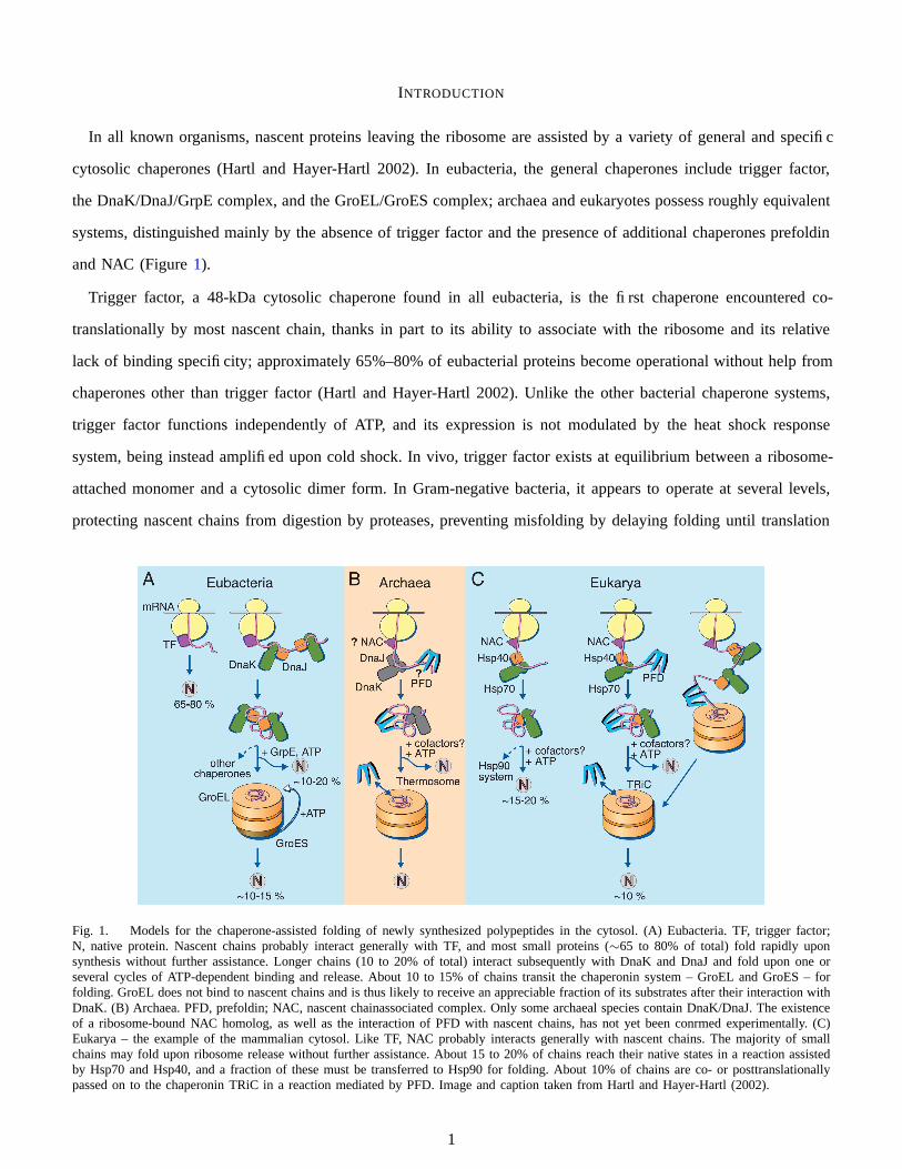
In all known organisms, nascent proteins leaving the ribosome are assisted by a variety of general and specific
cytosolic chaperones (Hartl and Hayer-Hartl 2002). In eubacteria, the general chaperones include trigger factor,
the DnaK/DnaJ/GrpE complex, and the GroEL/GroES complex; archaea and eukaryotes possess roughly equivalent
systems, distinguished mainly by the absence of trigger factor and the presence of additional chaperones prefoldin
and NAC (Figure 1).
Trigger factor, a 48-kDa cytosolic chaperone found in all eubacteria, is the first chaperone encountered co-
translationally by most nascent chain, thanks in part to its ability to associate with the ribosome and its relative
lack of binding specificity; approximately 65%–80% of eubacterial proteins become operational without help from
chaperones other than trigger factor (Hartl and Hayer-Hartl 2002). Unlike the other bacterial chaperone systems,
trigger factor functions independently of ATP, and its expression is not modulated by the heat shock response
system, being instead amplified upon cold shock. In vivo, trigger factor exists at equilibrium between a ribosome-
attached monomer and a cytosolic dimer form. In Gram-negative bacteria, it appears to operate at several levels,
protecting nascent chains from digestion by proteases, preventing misfolding by delaying folding until translation
Fig. 1. Models for the chaperone-assisted folding of newly synthesized polypeptides in the cytosol. (A) Eubacteria. TF, trigger factor;N, native protein. Nascent chains probably interact generally with TF, and most small proteins (∼65 to 80% of total) fold rapidly uponsynthesis without further assistance. Longer chains (10 to 20% of total) interact subsequently with DnaK and DnaJ and fold upon one orseveral cycles of ATP-dependent binding and release. About 10 to 15% of chains transit the chaperonin system – GroEL and GroES – forfolding. GroEL does not bind to nascent chains and is thus likely to receive an appreciable fraction of its substrates after their interaction withDnaK. (B) Archaea. PFD, prefoldin; NAC, nascent chainassociated complex. Only some archaeal species contain DnaK/DnaJ. The existenceof a ribosome-bound NAC homolog, as well as the interaction of PFD with nascent chains, has not yet been conrmed experimentally. (C)Eukarya – the example of the mammalian cytosol. Like TF, NAC probably interacts generally with nascent chains. The majority of smallchains may fold upon ribosome release without further assistance. About 15 to 20% of chains reach their native states in a reaction assistedby Hsp70 and Hsp40, and a fraction of these must be transferred to Hsp90 for folding. About 10% of chains are co- or posttranslationallypassed on to the chaperonin TRiC in a reaction mediated by PFD. Image and caption taken from Hartl and Hayer-Hartl (2002).
1

completes, cooperating with other chaperones in facilitating proteolysis of aggregate-prone conformations, and
efficiently catalyzing peptidyl-prolyl cis-trans isomerization. For larger proteins, several trigger factor molecules
may associate with each chain. In Gram-positive bacteria, a trigger factor homologue has also been implicated in
the secretion and post-secretory folding of essential proteases.
The atomic structure of trigger factor in various configurations has been solved by X-ray crystallography, and
paints a fascinating picture of trigger factor’s operation. The trigger factor molecule binds to the chaperone and
apparently covers the exit of the ribosomal tunnel, protecting and directing the nascent protein as it leaves the
ribosome. Dynamics experiments have revealed that the molecule undergoes significant conformational changes at
different stages, favoring a more splayed conformation presenting a hydrophobic patch while on the ribosome, and
removing the hydrophobic area while in the cytosol.
Because trigger factor binds to the ribosome and therefore associates with nearly all nascent chains cotransla-
tionally, understanding its operation offers important insights into the protein folding process.
REVIEW OF RESULTS
Putative role in membrane translocation
Trigger factor was originally discovered in the context of translocation of secreted proteins across the inner
membrane of the Gram-negative bacterium E. coli. In their studies of outer membrane protein A (OmpA) and
its precursor form (proOmpA), Crooke and Wickner (1987) discovered that, following dilution from 8 M urea,
proOmpA was competent for inner-membrane translocation only if cell extract (S100) was present during dilution.
Following the membrane trigger hypothesis (Wickner 1979), they named the necessary component of the extract, a
protein of approximately 60 kDa, “trigger factor” (Crooke and Wickner 1987). While trigger factor did not associate
with the fully folded native protein, it was found to bind proOmpA with a 1:1 stoichiometry in vitro (Lill et al.
1988; Lecker et al. 1989). Its copurification with the large unit of ribosomes, from which it can be dissociated by
1.5 M LiCl, suggests a role in the early stages of protein synthesis (Lill et al. 1988).
The hypothesis that trigger factor is crucial to membrane translocation in E. coli turned out to be false. Re-
naturation of proOmpA by rapid dilution in absence of trigger factor resulted in protein capable for membrane
translocation (Crooke et al. 1988), although trigger factor was found to aid long-term stability of the protein;
definitive evidence came as in-vivo experiments revealed that E. coli lacking the gene for trigger factor (∆tig) were
still viable and able to export proteins (Guthrie and Wickner 1990).
Surprisingly, however, a trigger factor homologue in the Gram-positive bacterium Streptococcus pyogenes, RopA,
appears to be involved in the export and post-translocational folding of secreted cysteine proteinase SpeB (Lyon
et al. 1998). The analysis of two mutants not displaying extracellular SpeB activity (as measured by casein digestion)
2
implicated a mutation in the ropA gene. Further exploration of RopA mutations revealed that different mutations
either render SpeB incompetent for secretion or prevent its post-secretory cleavage and refolding (vide ibidem).
Subsequently, Lyon and Caparon (2003) were able to tie the decreased activity of SpeB specifically to the absence
of the peptidyl-prolyl cis-trans isomerization activity of trigger factor.
Indeed, Wen et al. (2005) discovered that a trigger factor homologue (RopA) in Streptococcus mutans, the primary
etiological agent of dental cavities in humans and also a Gram-positive bacterium, was involved in resistance to
acid and reductive stress. A strain with a lacking RopA took significantly longer to grow on media adjusted to
pH 5.0 and on media containing the pesticide paraquot; when cultures were exposed to 0.2% hydrogen peroxide,
the mutant was over 28 times less viable than wild type. The mutant strain was also less competent for genetic
transformation and its ability to form biofilms on abiotic surfaces was severely impaired. The affected functions
are unified by their dependence on correct folding and export of secretory and membrane proteins, and indeed,
Wen et al. found markedly reduced levels of glucosyltransferases GtfB and GtfD, which synthesize extracellular
adhesives from glucose.
Binding with ribosome-bound nascent chains
Trigger factor was rediscovered by Valent et al. (1995) in studies seeking to clarify the binding of the E. coli
signal recognition particle (SRP) to nascent secreted proteins. They employed photo-cross-linking to associate
ribosome-bound nascent chains of the precursor outer membrane porin PhoE, generated with truncated messenger
RNA in a wheat-germ translation system, to investigate their binding with SRP. In addition to the GTPase (P48)
component of SRP, pre-PhoE cross-linked with another protein identified by immunoprecipitation as trigger factor.
Like SRP, it only bound polypeptides in the context of the ribosome (the complex failed to bind nascent chains
released via puromycin and high-salt treatment, or when already associated, to remain bound after release). Unlike
SRP, however, trigger factor was found to bind to pre-PhoE lacking its secretion signal sequence (PhoE-∆ss) as
well as to nascent firefly luciferase.
Independently, Hesterkamp et al. (1996) identified trigger factor in their search for chaperones involved in the
early stages of translation. Using β-galactosidase (lacZ) in a similar wheat-germ translation system, they found that,
unlike other cytosolic chaperones DnaK and GroEL, trigger factor bound to preprolactin in the context of translating
ribosomes when translation was stopped with chloramphenicol but efficiently dissociated when the nascent chain was
instead released with puromycin (Figure 2). Intriguingly, a competition experiment revealed that trigger factor did
in fact bind to non-translating ribosomes, provided a four-fold molar excess over translating ribosomes (Hesterkamp
et al. 1996), suggesting that its association with ribosomes does not require but is stabilized by translation.
3

Fig. 2. Trigger factor binds to ribosomes only during translation. Image taken from Hesterkamp et al. (1996).
Ribosomal protein L23 binding site
Searching for the specific binding site between trigger factor and the large ribosomal subunit, Kramer et al. (2002)
noticed a highly conserved “Gly-Phe-Arg-x-Gly-x-x-Pro” motif near the N-terminus of numerous homologues.
Observing that an FRK/AAA mutant exhibited reduced ribosome association when compared to wild type, and
finding the motif region susceptible to proteolysis, they suggested that these 8 residues formed a “trigger signature”
that is involved in ribosomal binding.
To locate the corresponding ribosomal binding site, crosslinking trigger factor and ribosomes were exposed to
ultraviolet irradiation, resulting in pellets containing 68 and 75 kDa molecules. These two crosslinked products
were found to be the ribosomal proteins L23 and L29, which propitiously have homologues in all kingdoms.
Copurification of trigger factor and ribosomes containing an inactive L29 protein showed little change in the level
of associativity compared with wild type. Mutations of L23, however, resulted in drastically reduced trigger factor-
L23 association in vitro and cell death in vivo. Further crosslinking experimentation showed that L23 Glu-18 is
required for trigger factor binding.
A later study by Ullers et al. (2003) used crosslinking experiments to report that L23 also associates with SRP, a
complex involved in transporting nascent inner membrane proteins. In fact, trigger factor and SRP likely compete
for interaction with L23, with a slight advantage going to SRP. This exposes the likelihood that the binding of
trigger factor to L23 is a highly complex interaction since the same ribosomal protein is used to attach to two
different molecules dependent on the translating polypeptide chain.
Interaction with other chaperones
Trigger factor has been found to cooperate and compete with other cytosolic chaperones. Kandror et al. (1995)
discovered that it formed a complex with GroEL and an abnormal bacterial fusion protein CRAG in vivo, and upon
4

dissociation from CRAG by addition of ATP, remained bound to GroEL. Varying tig expression levels implicated
trigger factor as a rate-limiting reactant in the hydrolysis of CRAG, and cells with very low expression of tig were
not viable when also expressing the fusion protein. Interestingly, while CRAG was almost entirely eliminated upon
very high expression of tig, a new proteolytic fragment was found in abundance, suggesting that superphysiological
concentrations of trigger factor help expose some sections of CRAG to protease activity while protecting others.
In subsequent experiments, Kandror et al. (1999) demonstrated that the degradation of CRAG is brought about by
repeated dissociation and association with the GroEL and trigger factor complex.
The DnaK/DnaJ chaperone system and trigger factor appear to be able to compensate for each other in vivo.
By replacing the tig gene with a kanamycin cassette (∆tig::kan), Deuerling et al. (1999) showed that, while cells
expressing DnaK were unaffected, cells lacking the dnaK52 gene were not viable, and turning off IPTG-regulatable
expression of dnaK/J caused massive aggregation of mostly cytosolic proteins. By analyzing aggregated proteins
from cells without, respectively, DnaK/J or trigger factor via two-dimensional gel electrophoresis, Deuerling et al.
(2003) found that the two chaperone systems overlap almost perfectly in terms of substrates; indeed, library scanning
revealed that 77% of the peptides binding trigger factor also bound DnaK. In ∆tig::kan cells, levels of σ32-regulated
heat shock proteins like DnaK were found to be two- to three-fold that of wild-type cells at 37 ◦C.
In a narrow temperature range around 20 ◦C, however, Genevaux et al. (2004) were able to grow viable E. coli
with the ∆tig∆dnaKdnaJ allele. Strains expressing mutant dnaJ unable to bind to DnaK were not viable, suggesting
that, in absence of trigger factor, DnaJ is critical in mediating DnaK binding to nascent polypeptide chains.
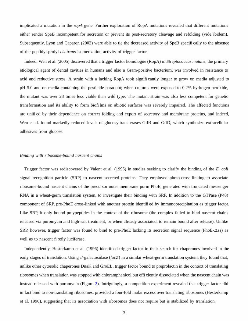
Trigger factor cooperates with the DnaK/DnaJ/GrpE (KJE) system in folding some large multidomain proteins,
although it appears to assist only cotranslational folding (Agashe et al. 2004). In vitro, the KJE system promoted
efficient refolding of denatured firefly luciferase, increasing efficiency from 10% to 70%–90%, while adding trigger
factor with or without KJE had no effect. Cotranslationally, while addition of KJE significantly improved the
solubility of luciferase, adding trigger factor with low levels of KJE increased the activity two- to three-fold.
Strikingly, observing the kinetics of folding revealed that this increase in efficiency comes at the cost of delaying
folding until the full protein has been translated. While the yields of firefly luciferase obtained in a chaperone-
free in-vitro translation system are low (ca. 5%), activity in the correctly folded fraction of the protein appeared
almost immediately upon completion of translation. When trigger factor and KJE were present, however, refolding
was markedly delayed (t 1
2
∼ 10 min), though yields were significantly improved (Figure 3). This delay is also
present in vivo: when in-vivo translation of luciferase in wild-type E. coli was arrested using chloramphenicol, the
amount of active protein continued to climb, whereas in ∆tig∆dnaK cells, luciferase activity level became constant
immediately after the addition of the antibiotic (ibidem).
Having also shown that ribosomes translating firefly luciferase recruit additional trigger factor molecules during
5
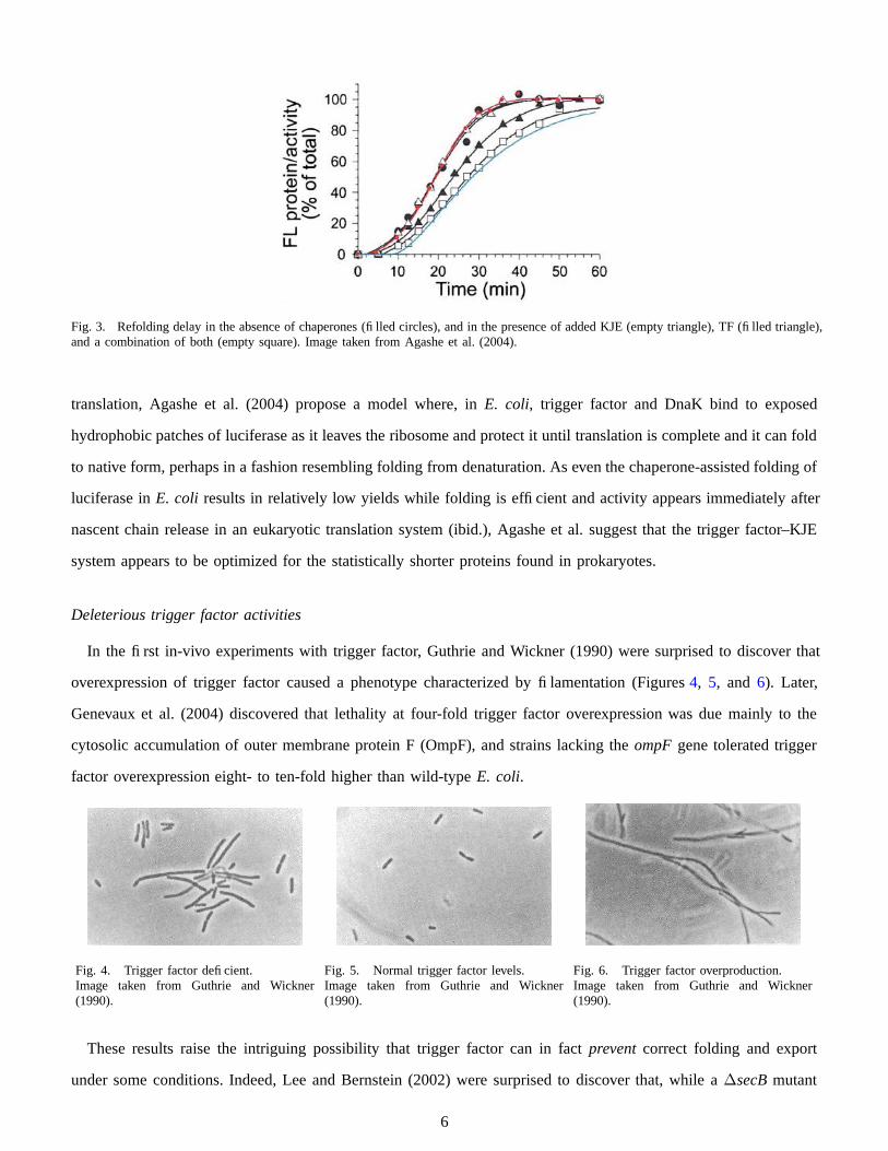
Fig. 3. Refolding delay in the absence of chaperones (filled circles), and in the presence of added KJE (empty triangle), TF (filled triangle),and a combination of both (empty square). Image taken from Agashe et al. (2004).
translation, Agashe et al. (2004) propose a model where, in E. coli, trigger factor and DnaK bind to exposed
hydrophobic patches of luciferase as it leaves the ribosome and protect it until translation is complete and it can fold
to native form, perhaps in a fashion resembling folding from denaturation. As even the chaperone-assisted folding of
luciferase in E. coli results in relatively low yields while folding is efficient and activity appears immediately after
nascent chain release in an eukaryotic translation system (ibid.), Agashe et al. suggest that the trigger factor–KJE
system appears to be optimized for the statistically shorter proteins found in prokaryotes.
Deleterious trigger factor activities
In the first in-vivo experiments with trigger factor, Guthrie and Wickner (1990) were surprised to discover that
overexpression of trigger factor caused a phenotype characterized by filamentation (Figures 4, 5, and 6). Later,
Genevaux et al. (2004) discovered that lethality at four-fold trigger factor overexpression was due mainly to the
cytosolic accumulation of outer membrane protein F (OmpF), and strains lacking the ompF gene tolerated trigger
factor overexpression eight- to ten-fold higher than wild-type E. coli.
Fig. 4. Trigger factor deficient.Image taken from Guthrie and Wickner(1990).
Fig. 5. Normal trigger factor levels.Image taken from Guthrie and Wickner(1990).
Fig. 6. Trigger factor overproduction.Image taken from Guthrie and Wickner(1990).
These results raise the intriguing possibility that trigger factor can in fact prevent correct folding and export
under some conditions. Indeed, Lee and Bernstein (2002) were surprised to discover that, while a ∆secB mutant
6
of E. coli failed to export a majority of its outer membrane proteins OmpA, OmpC, and OmpF, as well as most
of the periplasmic protein MPB, a ∆secB∆tig mutant showed no such defect. Further, these cells showed none of
the preOmpA, preOmpC, and preOmpF precursors present in the cytosol of wild-type E. coli, which explains the
puzzling preOmpF aggregation. Supplemental experiments excluding other possible causes led Lee and Bernstein
(2002) to hypothesize that the binding of trigger factor to nascent prosecretory proteins prevents their interaction
with the SecY translocon system, and that SecB may well have evolved specifically to compete with trigger factor
and deliver the nascent chain to the translocon.
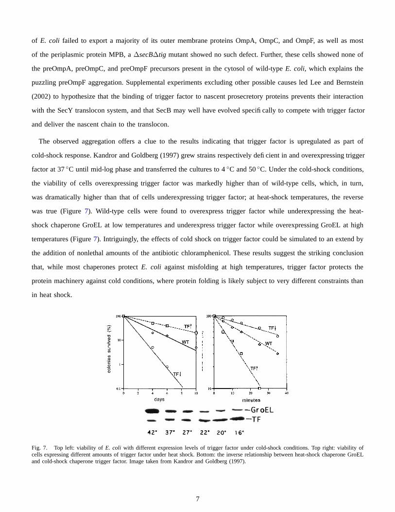
The observed aggregation offers a clue to the results indicating that trigger factor is upregulated as part of
cold-shock response. Kandror and Goldberg (1997) grew strains respectively deficient in and overexpressing trigger
factor at 37 ◦C until mid-log phase and transferred the cultures to 4 ◦C and 50 ◦C. Under the cold-shock conditions,
the viability of cells overexpressing trigger factor was markedly higher than of wild-type cells, which, in turn,
was dramatically higher than that of cells underexpressing trigger factor; at heat-shock temperatures, the reverse
was true (Figure 7). Wild-type cells were found to overexpress trigger factor while underexpressing the heat-
shock chaperone GroEL at low temperatures and underexpress trigger factor while overexpressing GroEL at high
temperatures (Figure 7). Intriguingly, the effects of cold shock on trigger factor could be simulated to an extend by
the addition of nonlethal amounts of the antibiotic chloramphenicol. These results suggest the striking conclusion
that, while most chaperones protect E. coli against misfolding at high temperatures, trigger factor protects the
protein machinery against cold conditions, where protein folding is likely subject to very different constraints than
in heat shock.
Fig. 7. Top left: viability of E. coli with different expression levels of trigger factor under cold-shock conditions. Top right: viability ofcells expressing different amounts of trigger factor under heat shock. Bottom: the inverse relationship between heat-shock chaperone GroELand cold-shock chaperone trigger factor. Image taken from Kandror and Goldberg (1997).
7

Nascent Chain Cradling
With the crystallization of the ribosome (Yusupov et al. 2001), ribosome protein L23 was seen to abut the very
exit of the ribosomal tunnel. Since trigger factor must associate with L23 for chaperone activity to persist, it became
apparent that trigger factor likely covered the aperture of the ribosomal tunnel. Hoffmann et al. (2006) investigated
this possibility, showing that trigger factor biochemically shields nascent polypeptide chains from proteolysis as they
exit the ribosomal tunnel. Studies on the DnaK/DnaJ/GrpE complex showed a similar potential to shield nascent
chains, but at significantly lower capacity, thus making trigger factor unique.
To demonstrate nascent chain shielding, in vitro degradation of substrate proteins was performed by adding
proteinase K co- and posttranslationally. In the absence of trigger factor, the ICDH protein (from E. coli, 417 aa,
46 kDa, with an added 41 aa C-terminus linker) and m10 protein (mutation of SH3 domain of α-spectrin designed
to assume a random coil structure, 62 aa, 7 kDa, with the same C-terminus linker added) were quickly degradated,
while in the presence of wild-type trigger factor these proteins remained intact for much longer periods. Because
of the C-terminus linker, both proteins were fully exposed from the ribosomal tunnel, implying that trigger factor
had shielded polypeptides of 7 and 15 kDa. Further, the same experiment was applied using a FRK/AAA mutant of
trigger factor, which does not associate to the ribosome, and both proteins were degradated quickly. This suggests
that trigger factor must be bound to the ribosome to associate with substrate chains and shield them from proteolysis.
To determine if larger proteins can be shielded by trigger factor Hoffmann et al. repeated the m10 mutant to
produce chains that are 2, 3, 4, and 5 times as long (19 kDa, 26 kDa, 34 kDa, and 41 kDa). Figure 8 shows the
effect of proteolysis on the various sized chains. After two minutes of proteinase K treatment the control has
degraded nearly all of the protein while one can still populations of very high weight chains (up to 41 kDa) that
were protected by trigger factor. The fact that such large molecules can be protected by trigger factor, which itself
is 48 kDa, leaves open the possibility that either multiple trigger factor molecules associate with each individual
chain (cotranslationally), or that trigger factor allows the larger molecules to fold domains that are protease-resistant
(the latter being less likely given that m10 was designed to generate a random coil structure).
Finally, it is worth note that all substrate chains eventually degrade within a reasonable amount of time, and that
trigger factor only slows the proteolysis process. This is is consistent with an earlier model that allows for rapid
binding and release of trigger factor to the substrate to speed chaperone assistance and prolyl-peptidyl isomerization
(Maier et al. 2001).
Peptidyl-Prolyl Isomerase Activity
The prolyl-peptidyl isomerase (PPIase) activity of trigger factor was initially discovered by Stoller et al. (1995),
who specifically searched for a ribosome-associated PPIase, and having discovered one, identified it as trigger
8

Fig. 8. Trigger factor shields large polypeptides from proteolysis. “BSA” column does not use trigger factor in the reaction (control), “TF”uses wild-type trigger factor, and “NC” uses a trigger factor mutant missing its PPIase domain. Image taken from Hoffmann et al. (2006).
factor. In contrast to other known PPIases, whose catalysis was significantly more efficient for short, unstructured
oligopeptides (tetrapeptide-4-nitroanilides) than for proteins, trigger factor was found to be exceptionally efficient on
unfolded proteins: the catalysis efficiency of 10 nM trigger factor in the refolding of RNase T1 (rate-limited by the
isomerization of its sole cis-proline) was equivalent to that of cyclosporin, previously thought to be a very efficient
PPIase, at 120 nM (Stoller et al. 1995). Hesterkamp et al. (1996) independently identified trigger factor as a PPIase
via sequence homology with FK506-binding proteins (FKBP) and confirmed its activity on short oligopeptides.
While trigger factor is overall distinct from the unrelated PPIase families of cyclophilins, and parvulins, Stoller
et al. (1995) found its subsite specificity to be reminiscent of FKBPs, with which (particularly FKBP12) trigger
factor also shares weak sequence homology, and from which it is distinguished by a missing FK506-binding
loop domain and consequent insensitivity to FK506 (Callebaut and Mornon 1995). Using limited proteolysis with
subtilisin, Stoller et al. (1996) located the PPIase domain in the 11.8 kDa fragment of trigger factor composed of
residues 145–251, an area homologous to FKBP, while Hesterkamp and Bukau (1996) isolated a region comprising
residues 132–247 using endoproteinase GluC (V8) and verified its PPIase activity.
By studying the kinetics of trigger factor isomerization activity and binding to its substrates, Scholz et al. (1997)
found that the exceptional isomerization efficiency in unfolded proteins is a result of trigger factor’s tight binding
to an unfolded protein; indeed, unfolded α-lactalbumin competitively inhibited trigger factor catalysis in RNase
T1 while native α-lactalbumin had no such effect. Further, the catalysis of short oligopeptides was unaffected by
competition with α-lactalbumin, indicating that PPIase activity is localized in a physically separate domain from
the substrate binding site.
Conversely, the PPIase activity of trigger factor is relatively independent of its other chaperone activities for
proteins lacking cis prolines. Li et al. (2001) reduced PPIase activity to 1% by independently replacing Tyr-221
9
and Phe-233, to obtain Y221G and F233Y, respectively, and found that, in vivo, the mutants increased the fraction
of soluble adenylate kinase from 5% to 15% and 20%, respectively, as compared to an increase to 25% for wild-
type trigger factor. Kramer et al. (2004) engineered a trigger factor mutant lacking PPIase activity by replacing
Phe-198 (F198A) and, having verified that it does not exhibit PPIase activity in the folding of RNase T1, decisively
demonstrated that the F198A variant preserves the non-PPIase chaperone functions: it showed similar binding
specificities in a library scan, its additional efficiently prevents aggregation of glyceraldehyde-3-phosphate dehy-
drogenase (GAPDH) diluted from 3 M GdnHCl, it can be crosslinked to nascent isocitrate dehydrogenase (ICDH),
and it successfully complements lethality in ∆dnaK mutants of E. coli.
In some cases, it appears that trigger factor is the only PPIase employed. Lyon and Caparon (2003) demonstrated
that the PPIase activity of trigger factor homologue RopA in Streptococcus pyogenes is involved in the extracellular
conversion of cysteine protease SpeB. Each of several strains expressing RopA mutants with confirmed severe
decrease in PPIase activity displayed a folding defect whereby conversion to mature SpeB required eight hours
longer than in wild-type strains.
Indeed, in Mycoplasma genitalium, the self-reproducing organism with the smallest known genome, trigger factor
is the only PPIase (Bang et al. 2000). Searching the sequenced genome revealed no regions homologous to any other
known PPIases, and PPIase activity remained unaffected by the addition of immunosuppressive drugs cyclosporin
A and FK506, which inhibit the cyclophilin and FKBP families of PPIases, respectively. In addition, observed
substrate specificity of the M. genitalium PPIase activity in vitro could only be accounted for by trigger factor.
Functional domains
The trigger factor molecule is composed of three separate structural domains which grossly correspond with
the protein’s three major functions: PPIase activity, ribosome binding, and chaperone activity. Although its initial
discovery estimated trigger factor to be a 60 kDa molecule, shortly afterward Guthrie and Wickner (1990) sequenced
E. coli trigger factor exactly (encoded by the tig gene), finding 432 residues (48 kDa). As mentioned earlier, Stoller
et al. (1996) located the PPIase domain around residues 145–251 while Hesterkamp and Bukau (1996) suggested
residues 132–247.
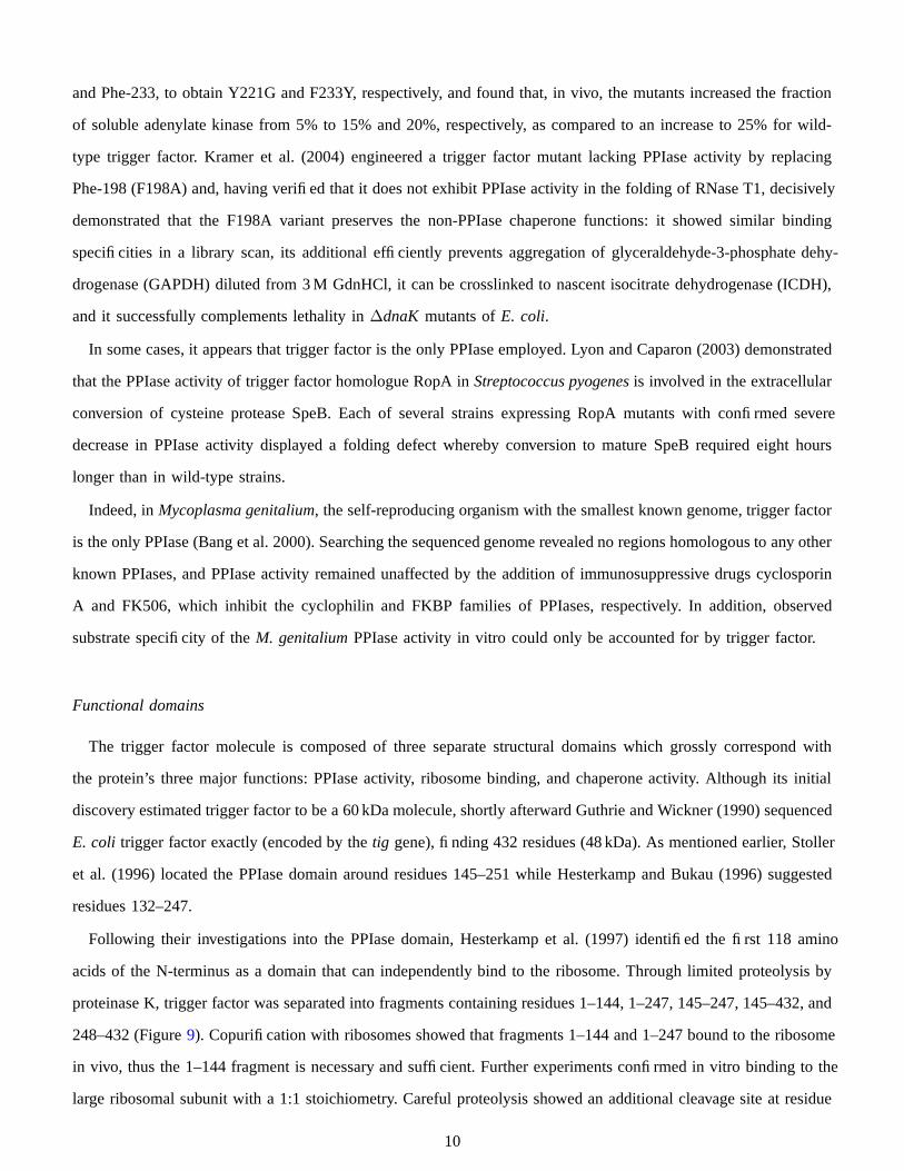
Following their investigations into the PPIase domain, Hesterkamp et al. (1997) identified the first 118 amino
acids of the N-terminus as a domain that can independently bind to the ribosome. Through limited proteolysis by
proteinase K, trigger factor was separated into fragments containing residues 1–144, 1–247, 145–247, 145–432, and
248–432 (Figure 9). Copurification with ribosomes showed that fragments 1–144 and 1–247 bound to the ribosome
in vivo, thus the 1–144 fragment is necessary and sufficient. Further experiments confirmed in vitro binding to the
large ribosomal subunit with a 1:1 stoichiometry. Careful proteolysis showed an additional cleavage site at residue
10
119 (overlooked by Stoller et al. (1996)) identifying a compact 118 amino acid binding domain and a 26 residue
linker to the known PPIase domain.
Fig. 9. Copurification of trigger factor fragments with ribosomes from cell extracts. KCl concentrations of 100 and 500 nM with supernatant(S) and pellet (P) applied to SDS-PAGE. Image taken from Hesterkamp et al. (1997).
At the time when the PPIase and ribosome binding N-terminus domains were first identified, the structure
and precise function of the ∼ 185 residue C-terminus domain still remained an enigma. Lill et al. (1988) had
suggested, and Hesterkamp et al. (1996) and many others more fully demonstrated, that trigger factor bound to
nascent polypeptide chains, however, the PPIase and ribosome binding N-terminus domains show very little sign of
involvement in this activity (although, indeed, the N-terminus domain has shown minor chaperone activity on its own
(Genevaux et al. 2004)). Therefore, the best hypothesis required the C-terminus domain to be involved association
with nascent polypeptide chains. The sequence of residues 248–432 (forming the the C-terminus domain) did
contain a large number of interspersed hydrophobic amino acids (Patzelt et al. 2001), and lends credence to this
hypothesis. However, the actual structure of the C-terminus domain, and thus the orientation of these residues,
remained unknown until the molecule was crystallized and solved by Ferbitz et al. (2004) and Ludlam et al. (2004).
Exciting recent work by Merz et al. (2006) has gone on to assert that the C-terminus domain of trigger factor is
critical to its chaperone activity. To show this, trigger factor variants were carefully designed that maintained stable,
independently folded molecules of the five combinations of the three individual domains (“N”, “P”, “C”, “NC”, and
“PC”). In vitro, the addition of wild-type trigger factor as well as the “C” variants prevented the aggregation of the
protein GAPDH during denaturation, whereas the “N” and “P” variants did nothing. A growth analysis was also
done in vivo, with a ∆tig∆dnaK strain of E. coli, exhibiting similar results: all plates with trigger factor variants
with the C-terminus domain saw less aggregation that those without (although the “NC” and “PC” variants proved
more successful at reducing aggregates than the “C” variant).
11

Atomic structures
Ferbitz et al. (2004) were able to crystallize E. coli trigger factor as a monomer as well as the 118 amino
acid N-terminus domain of E. coli trigger factor bound to the large ribosome subunit of Haloarcula marismortui.
Superimposition of X-ray data shows the N-terminus domain binding to ribosomal protein L23 and the C-terminus
domain forming a cradle over the exit of the ribosomal tunnel. In agreement with Kramer et al. (2002), the N-
terminus domain binds directly over the exit to the ribosomal tunnel, near Glu18 of ribosomal protein L23. Judging
from crystallization uncertainly, amongst other things, it could also be hypothesized that the full-length trigger
factor is able to swing, on average, 10 degrees in all directions around its attachment point, around the “trigger
factor signature” association with L23. Thus, although the N-terminus is bound to the ribosome, the remainder of
the molecule remains flexible, allowing for varied interactions with the nascent chain.
Lars Ferbitz (2005) further identifies a structural homologue between the two “arms” of the C-terminus domain
and the integral outer membrane chaperone SurA. When associated with the ribosome, this domain “cradles” the
exit to the ribosomal tunnel, exposing hydrophobic amino acids toward the tunnel, and providing space for up
to a 15 kDa molecule (Figure 10). However, a later study by Schlunzen et al. (2005), asserted that this pocket is
actually a much smaller crevice due to the conformation of the nearby ribosomal protein L24 interacting with the
C-terminus domain arms.
Around the same time as Ferbitz et al., Ludlam et al. (2004) crystallized near complete Vibrio cholerae trigger
factor in its dimer form, to investigate prior observations by Patzelt et al. (2002) that trigger factor can exist as
both a monomer and dimer in equilibrium. Their X-ray data shows the burial of the N-terminal domain of each
individual molecule when in dimer form, supporting the conclusion that dimers cannot bind to the ribosome. The
PPIase substrate-binding site is also blocked by its own C-terminus loop, suggesting that the loop disassociates on
ribosome binding, allowing for PPIase activity.
Baram et al. (2005) were able to crystallized the N-terminus domain of eubacterium Deinococcus radiodurans
trigger factor bound to the large ribosome subunit of the same organism (as opposed to E. coli trigger factor bound
to a Haloarcula marismortui ribosome) revealing a much different binding configuration than found by Ferbitz
et al.. The N-terminus domain featured a binding site that connected to both the L23 and L29 ribosomal proteins.
Comparison with monomeric structures suggests that a conformational change must occur upon association. It is
also shown that the N-terminus domain becomes splayed across the exit of the ribosomal tunnel, exposing a path
to its hydrophobic region. This helps support later conclusions made by Hoffmann et al. 2006 that trigger factor
shields nascent chains from the cytosol, as earlier discussed.
Interestingly, it is also possible that since the N-terminus domain binds to two separate regions of L23, on both
sides of the extended loop that is exposed to the ribosomal tunnel in eubacterial L23, that the binding of trigger
12

Fig. 10. Trigger factor (TF) forms a cradle over the ribosomal tunnel exit. Image taken from Ferbitz et al. (2004).
factor may cause an allosteric conformational alteration varying the inner tunnel shape (Baram and Yonath 2005).
This may even work in both directions, providing a dynamic communication path to control rate and directionality
of nascent proteins. This is an attractive hypothesis when also noted that only the eubacterial kingdom contains
both trigger factor and a ribosomal protein L23 with this kind of elongated loop exposed to the ribosomal tunnel.
Protein Dynamics
In recent years, techniques for real time observations of trigger factor molecules have been cleverly conceived
and demonstrated. Maier et al. (2003) were one of the first to labeled E. coli trigger factor with fluorophores to
observe the kinetics of association and dissociation with the ribosome. By introducing an R14C mutation (located in
the N-terminus ribosome binding domain) into E. coli trigger factor and labeling it with BADAN, the rate constants
of ribosome binding were estimated through the comparison of relative fluorescence over time (where association
corresponds to a decrease in fluorescence). Using this technique, the rate of ribosome binding was found to be
slow, with a two-step process of association followed by isomerization. (Later, Kaiser et al. (2006) supported this
claim by using fluorescence to show a difference in binding time between molecules with and without the PPIase
domain).
The lifetime of the trigger factor-ribosome complex was shown to be upwards of 30 seconds (at 20 ◦C), drastically
longer than earlier experiments that found the lifetime of association between trigger factor and protein substrate
to be 100 milliseconds (Maier et al. 2001). This suggests a mechanism by which a ribosome bound trigger factor
protein rapidly binds and releases different segments of a single nascent chain many times. This may be to prevent
aggregation, to “scan” for prolyl bonds in need of catalysis, and to avoid interference with rapid protein folding
reactions.
Most recently, Kaiser et al. (2006) have assembled a more comprehensive view of the kinetics of E. coli trigger
13

factor function. Through a similar fluorescence labeling technique (FRET), a conformation change was observed
when trigger factor first associates with the ribosome. After incorporating donor (D) and acceptor (A) fluorophores at
specific sites in the sequence (residues 14, 150, 326, and 376, see Figure 11), a marked difference in intramolecular
emission was seen between sites 14 and 150 when trigger factor bound to the ribosome. As can be seen in Figure
12, a decrease in fluorescence occurs once trigger factor is bound to the ribosome, suggesting that trigger factor
is in a compacted form when unbound and a more open and elongated form when bound (separating donors and
acceptors at sites 14/150).
Fig. 11. FRET labeling of trig-ger factor. Image taken from Kaiseret al. (2006).
Fig. 12. Intramolecular FRET emissions intensity. Image taken from Kaiser et al. (2006).
Kaiser et al. further demonstrated that trigger factor can remain associated with a nascent chain after disassociation
from the ribosome, but cannot initiate substrate protein binding independently. This allows a second trigger factor to
associate to a single nascent chain (after binding with the ribosome), resulting in multiple trigger factor molecules
chaperoning a single chain, and agreeing with prior biochemical experiments suggesting such (Agashe et al. 2004).
Two fluorescence experiments were performed to confirm these kinetics. In the first experiment, the middle plot
of Figure 13 shows intramolecular compaction fluorescence (FRET) and ribosome disassociation fluorescence
(BADAN) unaffected by ongoing translation, implying that the elongated trigger factor molecule stays bound
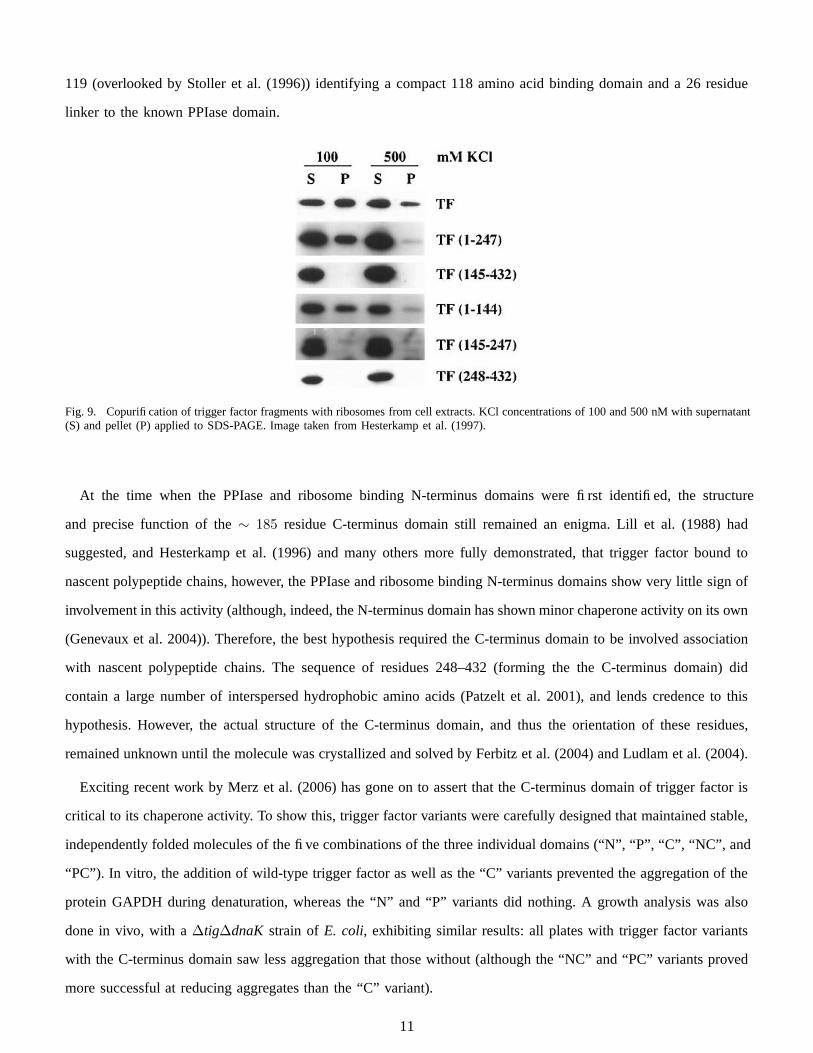
to the chain even after disassociation. In a separate experiment the translation of the titin I27 domain was stalled
at various points, exposing none, one, or two hydrophobic residue sequence regions to be associated with trigger
factor (Figure 14). Two large relative jumps in BADAN fluorescence again suggests multiple independent trigger
factor binding events with the two hydrophobic regions.
The current model of trigger factor function and reaction rate determined by Kaiser et al. can be seen in Figure
15. In reaction 1, free trigger factor exists in a rapid monomer-dimer equilibrium. Trigger factor monomers elongate
and associate with the ribosome with t 1
2
∼ 10.3 ±2.2 seconds (reaction 2) and remain associated during translation
for a similar amount of time (reaction 3). Upon ribosome disassociation, trigger factor may remain bound to the
nascent chain with t 1
2
of up to 35 seconds (reaction 4), during which another trigger factor molecule might associate
14

Fig. 13. Fluorescence emission of labeled and unlabeled trigger factor. Top: compaction occurs at the same time as disassociation (comparedwith non-ribosome-binding FRK/AAA). Middle: multiple trigger factor molecules binding to nascent chain. Bottom: compaction is slowerthan disassociation when EDTA added. Image taken from Kaiser et al. (2006).
with the ribosome and a different region of the nascent chain (reaction 5). Once trigger factor disassociates from
the substrate chain, it quickly resumes a compact conformation in both monomeric and dimer form (reaction 6).
DISCUSSION
Taken together, the literature to date outlines a tantalizing picture of how trigger factor operates. It associates
cotranslationally with the large unit of the ribosome, possibly because the nascent chain inside the ribosomal tunnel
causes allosteric changes to the trigger factor binding site on L23. Attached to the ribosome, trigger factor covers the
exit of the tunnel to protect nascent chains from degradation and possibly creates a hydrophobic cradle in which
the translated protein can partially or entirely fold. As the nascent polypeptide grows, trigger factor molecules
remain temporarily attached to its hydrophobic patches; as one trigger factor molecule thus leaves its location
on the ribosome, another is recruited in its place. The precise nature of the interaction of trigger factor with the
ribosomal tunnel and the nascent chain present there, possibly mediated via L23, and of any chaperone-like role
of the ribosome itself, await investigation.
The role and origin of the peptidyl-prolyl-cis-trans-isomerase (PPIase) activity of trigger factor remains clouded
15
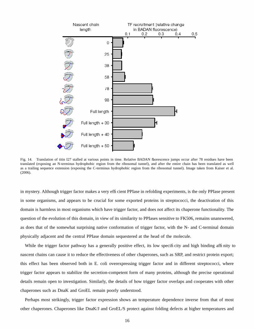
Fig. 14. Translation of titin I27 stalled at various points in time. Relative BADAN fluorescence jumps occur after 78 residues have beentranslated (exposing an N-terminus hydrophobic region from the ribosomal tunnel), and after the entire chain has been translated as wellas a trailing sequence extension (exposing the C-terminus hydrophobic region from the ribosomal tunnel). Image taken from Kaiser et al.(2006).
in mystery. Although trigger factor makes a very efficient PPIase in refolding experiments, is the only PPIase present
in some organisms, and appears to be crucial for some exported proteins in streptococci, the deactivation of this
domain is harmless in most organisms which have trigger factor, and does not affect its chaperone functionality. The
question of the evolution of this domain, in view of its similarity to PPIases sensitive to FK506, remains unanswered,
as does that of the somewhat surprising native conformation of trigger factor, with the N- and C-terminal domain
physically adjacent and the central PPIase domain sequestered at the head of the molecule.
While the trigger factor pathway has a generally positive effect, its low specificity and high binding affinity to
nascent chains can cause it to reduce the effectiveness of other chaperones, such as SRP, and restrict protein export;
this effect has been observed both in E. coli overexpressing trigger factor and in different streptococci, where
trigger factor appears to stabilize the secretion-competent form of many proteins, although the precise operational
details remain open to investigation. Similarly, the details of how trigger factor overlaps and cooperates with other
chaperones such as DnaK and GroEL remain poorly understood.
Perhaps most strikingly, trigger factor expression shows an temperature dependence inverse from that of most
other chaperones. Chaperones like DnaK/J and GroEL/S protect against folding defects at higher temperatures and
16
17
Fig. 15. Current model of trigger factor function. Image taken from Kaiser et al. (2006).
are therefore upregulated as part of heat shock response; in contradistinction, trigger factor prevents or repairs
defects caused by colder conditions, and is therefore upregulated in cold shock. Not much is known about the
biochemical mode of this chaperone activity, as the details of protein folding defects in heat shock and cold shock
are poorly understood; the relative abundance of trigger factor research may make it an excellent model cold-shock
chaperone.
REFERENCES
Agashe, V. R., S. Guha, H.-C. Chang, P. Genevaux, M. Hayer-Hartl, M. Stemp, C. Georgopoulos, F. U. Hartl,and J. M. Barral (2004). Function of Trigger Factor and DnaK in Multidomain Protein Folding Increase inYield at the Expense of Folding Speed. Cell 117(2), 199–209.
Bang, H., A. Pecht, G. Raddatz, T. Scior, W. Solbach, K. Brune, and A. Pahl (2000). Prolyl isomerases in aminimal cell. Catalysis of protein folding my trigger factor from Mycoplasma genitalium. European Journalof Biochemistry 267, 3270–3280.
Baram, D., E. Pyetan, A. Sittner, T. Auerbach-Nevo, A. Bashan, and A. Yonath (2005). Structure of triggerfactor binding domain in biologically homologous complex with eubacterial ribosome reveals its chaperoneaction. Proc. Natl. Acad. Sci. USA 102(34), 12017–12022.
Baram, D. and A. Yonath (2005). From peptide-bond formation to cotranslational folding: dynamic, regulatoryand evolutionary aspects. FEBS Letters 579(4), 948–954.
Callebaut, I. and J.-P. Mornon (1995). Trigger factor, one of the Escherichia coli chaperone proteins, is anoriginal member of the FKBP family. FEBS Letters 374, 211–215.
Crooke, E., L. Brundage, M. Rice, , and W. Wickner (1988). ProOmpA spontaneously folds in a membraneassembly competent state which trigger factor stabilizes. The EMBO Journal 7(6), 1831–1835.
Crooke, E. and W. Wickner (1987). Trigger Factor: A Soluble Protein that Folds pro-OmpA into a Membrane-Assembly-Competent Form. Proc. Natl. Acad. Sci. USA 84(15), 5216–5220.
Deuerling, E., H. Patzelt, S. Vorderwulbecke, T. Rauch, G. Kramer, E. Schaffitzel, A. Mogk, A. Schulze-Specking,H. Langen, and B. Bukau (2003). Trigger factor and DnaK possess overlapping substrate pools and bindingspecificities. Molecular Microbiology 47, 1317–1328.
Deuerling, E., A. Schulze-Specking, T. Tomoyasu, A. Mogk, and B. Bukau (1999). Trigger factor and DnaKcooperate in folding of newly synthesized proteins. Nature 400, 693–696.
Ferbitz, L., T. Maier, H. Patzelt, B. Bukau, E. Deuerling, and N. Ban (2004). Trigger factor in complex with theribosome forms a molecular cradle for nascent proteins. Nature 431, 590–596.
18
Genevaux, P., F. Keppel, F. Schwager, P. S. Langendijk-Genevaux, F. U. Hartl, and C. Georgopoulos (2004). Invivo analysis of the overlapping functions of DnaK and trigger factor. EMBO Reports 5(2), 195–200.
Guthrie, B. and W. Wickner (1990). Trigger Factor Depletion or Overproduction Causes Defective Cell Divisionbut Does Not Block Protein Export. Journal of Bacteriology 172, 5555–5562.
Hartl, F. U. and M. Hayer-Hartl (2002). Molecular Chaperones in the Cytosol: from nascent chain to foldedprotein. Science 295(5561), 1852–1858.
Hesterkamp, T. and B. Bukau (1996). Identification of the prolyl isomerase domain of Escherichia coli triggerfactor. FEBS Letters 385, 67–71.
Hesterkamp, T., E. Deuerling, and B. Bukau (1997). The Amino-terminal 118 Amino Acids of Escherichia coliTrigger Factor Constitute a Domain That Is Necessary and Sufficient for Binding to Ribosomes. J. Biol.Chem. 272(35), 21865–21871.
Hesterkamp, T., S. Hauser, H. Lutcke, and B. Bukau (1996). Escherichia coli trigger factor is a prolyl isomerasethat associates with nascent polypeptide chains. Proc. Natl. Acad. Sci. USA 93(9), 4437–4441.
Hoffmann, A., F. Merz, A. Rutkowska, B. Zachmann-Brand, E. Deuerling, and B. Bukau (2006). Trigger FactorForms a Protective Shield for Nascent Polypeptides at the Ribosome. J. Biol. Chem. 281(10), 6539–6545.
Kaiser, C. M., H.-C. Chang1, V. R. Agashe, S. K. Lakshmipathy, S. A. Etchells, M. Hayer-Hartl, F. U. Hartl, andJ. M. Barral (2006). Real-time observation of trigger factor function on translating ribosomes. Nature 444,455–460.
Kandror, O. and A. L. Goldberg (1997). Trigger factor is induced upon cold shock and enhances viability ofEscherichia coli at low temperatures. Proc. Natl. Acad. Sci. USA 94, 4978–4981.
Kandror, O., M. Sherman, and A. Goldberg (1999). Rapid Degradation of an Abnormal Protein in Escherichiacoli Proceeds through Repeated Cycles of Association with GroEL. Journal of Biological Chemistry 274,37743–37749.
Kandror, O., M. Sherman, M. Rhode, and A. L. Goldberg (1995). Trigger factor is involved in GroEL-dependentprotein degradation in Escherichia coli and promotes binding of GroEL to unfolded proteins. The EMBOJournal 14, 6021–6027.
Kramer, G., H. Patzelt, T. Rauch, T. A. Kurz, S. Vorderwulbecke, B. Bukau, and E. Deuerling (2004). Triggerfactor’s peptidyl-prolyl cis/trans isomerase activity is not essential for the folding of cytosolic proteins inEscherichia coli. J. Biol. Chem. 279(14), 14165–14170.
Kramer, G., T. Rauch, W. Rist, S. Vorderwulbecke, H. Patzelt, A. Schulze-Specking, N. Ban, E. Deuerling, andB. Bukau (2002). L23 protein functions as a chaperone docking site on the ribosome. Nature 419, 171–174.
Lars Ferbitz (2005). Structure of trigger factor in complex with the ribosome defines the molecular environmentof the emerging nascent polypeptide chain. Ph. D. thesis, Swiss Federal Institute of Technology Zurich.
Lecker, S., R. Lill, T. Ziegelhoffer, C. Georgopoulos, J. Philip J. Bassford, C. A. Kumamoto, and W. Wickner(1989). Three pure chaperone proteins of Escherichia coli—SecB, trigger factor, and GroEL—form solublecomplexes with precursor proteins in vitro. The EMBO Journal 8, 2703–2709.
Lee, H. C. and H. D. Bernstein (2002). Trigger Factor Retards Protein Export in Escherichia coli. Journal ofBiological Chemistry 277(45), 43527–43535.
Li, Z.-Y., C.-P. Liu, L.-Q. Zhu, G.-Z. Jing, and J.-M. Zhou (2001). The chaperone activity of trigger factor isdistinct from its isomerase activity during co-expression with adenylate kinase in Escherichia coli. FEBSLetters 506, 108–112.
Lill, R., E. Crooke, B. Guthrie, and W. Wickner (1988). The “trigger factor cycle” includes ribosomes,presecretory proteins, and the plasma membrane. Cell 54, 1013–1018.
Ludlam, A. V., B. A. Moore, and Z. Xu (2004). The crystal structure of ribosomal chaperone trigger factor fromVibrio cholerae. Proc. Natl. Acad. Sci. USA 101(37), 13436–13441.
Lyon, W. R. and M. G. Caparon (2003). Trigger Factor-Mediated Prolyl Isomerization Influences Maturation ofthe Streptococcus pyogenes Cysteine Protease. J. of Bacteriology 185(12), 3661–3667.
Lyon, W. R., C. M. Gibson, and M. G. Caparon (1998). A role for Trigger Factor and an Rgg-like regulator inthe transcription, secretion and processing of the cysteine proteinase of Streptococcus pyogenes. The EMBOJournal 17, 6263–6275.
Maier, R., E. Barbara, S. Christian, L. Hauke, and S. Franz-Xaver (2003). Interaction of trigger factor with theribosome. J. Mol. Biol. 326, 585–592.
19
Maier, R., C. Scholz, and F. X. Schmid (2001). Dynamic association of trigger factor with protein substrates. J.Mol. Biol. 314, 1181–1190.
Merz, F., A. Hoffmann, A. Rutkowska, B. Zachmann-Brand, B. Bukau, and E. Deuerling (2006). The C-terminalDomain of Escherichia coli Trigger Factor Represents the Central Module of Its Chaperone Activity. J. Biol.Chem. 281(42), 31963–31971.
Patzelt, H., G. Kramer, T. Rauch, H. J. Schonfeld, B. Bukau, and E. Deuerling (2002). Binding specificity ofEscherichia coli trigger factor. Biol. Chem. 383(10), 1611–1619.
Patzelt, H., S. Rudiger, D. Brehmer, G. Kramer, S. Vorderwulbecke, E. Schaffitzel, A. Waitz, T. Hesterkamp,L. Dong, J. Schneider-Mergener, B. Bukau, and E. Deuerling (2001). Binding specificity of Escherichia colitrigger factor. Proc. Natl. Acad. Sci. USA 98(25), 14244–14249.
Schlunzen, F., D. N. Wilson, P. Tian, J. M. Harms, S. J. McInnes, H. A. Hansen, R. Albrecht, J. Buerger, S. M.Wilbanks, and P. Fucini (2005). The Binding Mode of the Trigger Factor on the Ribosome: Implications forProtein Folding and SRP Interaction. Structure 13(11), 1685–1694.
Scholz, C., G. Stoller, T. Zarnt, G. Fischer, and F. X. Schmid (1997). Cooperation of enzymatic and chaperonefunctions of trigger factor in the catalysis of protein folding. The EMBO Journal 16, 54–58.
Stoller, G., K. P. Rucknagel, K. H. Niederhaus, F. X. Schmid, G. Fischer, and J.-U. Rahfeld (1995). A ribosome-associated peptidyl-prolyl cis/trans isomerase identified as the trigger factor. The EMBO Journal 14, 4939–4948.
Stoller, G., T. Tradler, J.-U. Rucknagel, and G. Fuscher (1996). An 11.8 kDa proteolytic fragment of theE. coli trigger factor represents the domain carrying the peptidyl-prolyl cis/trans isomerase activity. FEBSLetters 384, 117–122.
Ullers, R. S., E. N. Houben, A. Raine, C. M. ten Hagen-Jongman, M. Ehrenberg, J. Brunner, B. Oudega,N. Harms, and J. Luirink (2003). Interplay of signal recognition particle and trigger factor at L23 near thenascent chain exit site on the Escherichia coli ribosome. J. Cell Biol. 161(4), 679–684.
Valent, Q. A., D. A. Kendall, S. High, R. Kusters, B. Oudega, and J. Luirink (1995). Early events inpreprotein recognition in E. coli: interaction of SRP and trigger factor with nascent polypeptides. The EMBOJournal 14(22), 5494–5505.
Wen, Z. T., P. Suntharaligham, D. G. Cvitkovitch, and R. A. Burne (2005). Trigger Factor in Streptococcusmutans Is Involved in Stress Tolerance, Competence Development, and Biofilm Formation. Infection andImmunity 73(1), 219–225.
Wickner, W. (1979). The assembly of proteins into biological membranes: The membrane trigger hypothesis.Annu. Rev. Biochem. 48, 23–45.
Yusupov, M. M., G. Z. Yusupova, A. Baucom, K. Lieberman, T. N. Earnest, J. H. D. Cate, and H. F. Noller(2001). Crystal Structure of the Ribosome at 5.5 A Resolution. Science 292(5518), 883–896.