the tricorythidae of the oriental region - ephemeroptera
TRANSCRIPT

313
THE TRICORYTHIDAE OF THE ORIENTAL REGION
Pavel Sroka1and Tomáš Soldán2
1 Biological Faculty, the University of South Bohemia, Branišovská 31,
370 05 České Budějovice, Czech Republic 2 Institute of Entomology, Branišovská 31, 370 05 České Budějovice, Czech Republic
Abstract Based on detailed taxonomic revision of predominantly larval material of the family Tricorythidae (Ephemeroptera) so far available from the Oriental Region, a new genus, Sparsorythus gen. n., is established to include six new species: S. bifurcatus sp. n. (larva, imago male and female), S. dongnai sp. n. (larva, imago male and female), S. gracilis sp. n. (larva), S. grandis sp. n. (larva), and S. ceylonicus sp. n. (larva), and S. multilabeculatus sp. n. (imago male), respective differential diagnoses are presented. S. jacobsoni (Ulmer 1913) comb. n. is transferred from the genus Tricorythus, now supposed to cover only a part of Afrotropic species of this family. Further five species are described but left unnamed since the larval stage is still unknown. The egg stage (a single polar cap and usually hexagonal exochorionic structures) is described for the first time, relationships of Sparsorythus gen. n. to all other genera of the family and their composition are discussed with regard to classical extent of knowledge and rather confusing data in the past. Available data on biology of this new genus are summarized and its distribution with regard to historical biogeography id briefly discussed.
Key words: Tricorythidae; Oriental region; Sparsorythus gen. n; new species; taxonomy; biogeography.
Introduction
Eaton (1868) established the genus Tricorythus on the basis of Caenis varicauda Pictet, 1843–1845 described in adult stage from the Upper Egypt. The same author (Eaton 1884) mentioned adults of two species, T. varicauda and T. discolor ranging the genus to his Section 7 of Third Series of Group II. of the Genera. Moreover, Eaton (1884: Table 15, Fig. 25) figured the wing of a species called Tricorythus (Malay sp.), but he did not describe or mention this material in his text. Ulmer (1913) noted the venation figured by Eaton (1884) corresponds to his new species (T. jacobsoni), described after adult material from Java. Later, he revised the adult description (Ulmer 1925) and published a description of larval stage assigned just to this species (Ulmer 1940).
The genus has been classified in the Caenidae and Ephemerellidae and, finally, in the separate family with apparently polyphyletic components by Lestage (1942) to include also the present families Leptohyphidae, Ephemerythidae and Machadorythidae
314 Pavel Sroka and Tomáš Soldán
defined later (Edmunds and Traver 1954, Edmunds et al. 1963, Landa and Soldán 1985, McCafferty and Wang 2000).
Three additional genera, namely Dicercomyzon, Tricorythurus and Neurocaenis, had been established from the continental Afrotropic region. The genus Dicercomyzon Demoulin, 1954 constitutes the monotypic subfamily Dicercomyzinae Edmunds and Traver, 1954.
The genus Tricorythurus Lestage, 1942 (type species Tricorythus latus Ulmer, 1916, by monotypy, type locality the Congo River, Zaire, Kingshasa, other locality, Bahr-el-Djebel) seems to be disputable. According to Kimmins (1960), the genus Tricorythurus was erected on the basis of possibly 3-segmented forceps (Ulmer 1916, Lestage 1942), female and larvae were unknown. Type species was reexamined by Demoulin (1954). He found the forceps to be actually 2-segmented and also other characters are congeneric with the genus Tricorythus. Consequently, he synonymized Tricorythurus Lestage 1942 with Tricorythus Eaton, 1868 (cf. also Demoulin 1970). But some authors, e.g., Hubbard (1990) maintain the opinion on Tricorythurus to be a valid taxon, the subgenus of the genus Tricorythus in this case.
The genus Neurocaenis Navás, 1936 (type species N. fuscata Navás, 1936: by original designation, type locality Zaire, Beni, male and larva unknown) was originally defined mainly on the basis of minor differences in the arrangement of cross veins (Navás 1936; Demoulin 1954, 1970) to include, besides type species, 5 species originally described in the genus Tricorythus. The only Oriental species known so far, T. jacobsoni Ulmer (1913) was also transferred to the genus Neurocaenis (Demoulin 1954). Later Demoulin (1970) admitted subgeneric status of this genus. Demoulin (1970) also summarized all Afrotropic taxa of the Tricorythidae: There are 11 species (6 in Tricorythus and 5 in Neurocaenis) described mostly according to adult stage with the exception of T. reticulatus Barnard, 1932 and T. discolor (Burmeister 1938), the larval characters of which are mentioned by Barnard (1932) and Crass (1947).
Generic status of Neurocaenis was followed by Edmunds and Traver (1954), Edmunds et al. (1963), Hubbard and Peters (1978), Hubbard and Pescador (1978), Hubbard (1990), the latter author also mentioned the occurrence of this genus in Madagascar most probably on the basis of Demoulin’s (1958) record on unidentified species of Neurocaenis from Madagascar. Although the synomyny of Tricorythus-Neurocaenis has been intuitively supposed for a long time (cf., e.g., Soldán 1983, 1991, Sivaramakrishnan and Venkataraman 1987), the respective formal taxonomic act was conducted by Oliarinony et al. (1998). These authors, besides discussing the problems of cross veins character value, also described nine new species of Tricorythus from Madagascar.
Recently, new genera Madecassorythus, Spinirythus and Ranorythus were established by Elouard and Oliarinomy (1997), Oliarinomy and Elouard (1998a), and Oliarinomy and Elouard (1998b).

Tricorythidae of the Oriental Region 315
After description of Tricorythus jacobsoni (Ulmer 1913) only sparse literature data exist about occurrence of Tricorythids in the Oriental region. Hubbard and Peters (1978) and Hubbard and Pescador (1978) mentioned the genus Neurocaenis (not identified species) from Srí Lanka. Under the generic name Tricorythus, the Oriental Tricorythidae were mentioned by Soldán (1983, 1991) from southern part of Vietnam, Sivaramakrishnan and Venkataraman (1987) from southern India (Madras State), and McCafferty and Wang (2000) from Indonesia.
Based on a relatively extensive material from the Oriental region, the principal objectives of this study are, as follows: (i) to determine the proper generic identity of Tricorythus jacobsoni Ulmer; (ii) to compare all the other specimens available from the Oriental region with this species and find possible differences in both adult and larval arrangement of morphological characters and (iii) to discuss in detail the relationships of the Oriental species to other Tricorithidae genera (and subfamilies).
Systematic Part
Sparsorythus gen. n. Mature larva (in alcohol): Head – Apparently wider than long. Antennae longer than head length. Scape and pedicle well differentiated, pedicle about twice longer than scape. Labrum – Oval, about twice as wide as long. A single row of medialy diminishing bristles at the anterior margin. Uniformly scattered bristles on dorsal surface. Two submarginal groups of shorter dense tiny bristles on the ventral side of labrum. Hypopharynx – Lingua rounded, ellipsoidal, with medial incurvation. Lingua longer than superlinguae. Superlinguae triangular, rounded or bluntly pointed at apex, with a row of bristles in distal half of outer margin, diminishing apically. Mandibles – Outer incisors triangular, with numerous bristles on the ventral side. Apex simple or apically with a pair of short rounded projections. Inner incisors approximately on the same shape and length, with bristles on the vental side and tiny branched setae on the dorsal side. Right prostheca shorter by 1/3 than left one, expanded apically, bifurcated or with several pointed teeth. A group of branched setae longer than prostheca inserted at its base. Left prostheca as long as or slightly shorter than the inner incisor, rod-like, with simple bluntly pointed apex or apex bearing several bluntly pointed projections. A group of branched setae as long as or shorter than prostheca inserted at its base. Outer margin of mandibles with a row of long filtering setae. Short transversal row of setae on the ventral side near mandible base. Maxillae – Suture of stipes and galeolacinia apparent. Maxillae roughly oblong-shaped or elipsoidal. Apical part of maxilla nearly truncate, the outer apical (galeal) lobe well apparent, produced. Maxilla about by 1/3 longer than wide. Outer margin of maxilla convex without any setation. Anterolateral part of maxilla with a group of long setae. Similar, but smaller setae also on the anterior and medial margin of the
316 Pavel Sroka and Tomáš Soldán
galeolacinia. A regular oblique ventral transversal row of stout setae situated at distal third of galeolacinia. Maxillary palps completely missing. No sclerotised structure at the place of insertion of maxillary palp recognizable. Labium – Glossae and paraglossae fused into rounded triangular plate with two groups of lateral submarginal setae. The whole plate surrounded with a regular row of setae diminishing apically. Labial palps three-segmented. First segment oblong-shaped, about by 1/3 shorter than the second one, without any setation. Second segment curved, apically bluntly pointed, with a row of stout marginal setae at its outer margin and tiny submarginal setae at its inner margin. Third segment very small, bluntly pointed at apex, without any setation. Pronotum – Oblong-shaped, as wide as head, about twice longer than wide and about by 1/4 shorter than mesonotum. Legs – Femora flat, shorter than tibiae. Fore femora with a conspicuous transversal row of flat rounded articulated spines and concave posteromedial margin. Foretibiae with a longitudinal row of spines or bristles near their inner margin. Claws strongly hooked, with two teeth approximately in the middle and one or two subapical spines. Surface of the middle and hind femora covered by spines of various sizes. Posterior margins of the middle and hind femora with spines and setae. Abdomen – Abdominal segments bearing gills only moderately compressed. Segments VIII, IX and X only slightly longer than segments I–VII. Posterolateral spines of abdominal segments well apparent, as long as 1/3 or 1/4 of segment length. Anlagen of male external genitalia (penis and forceps) well apparent in larvae of the last instar. Gills – Six or five pairs of gills on abdominal segments II – VII or II – VI. Gills on segments II – VI alike, with rounded or elipsoidal plates and two branched ventral membranous parts with rich filaments. Plates simple, thin, not enforced, with only several tiny and short marginal bristles. Gills on abdominal segment VII strongly reduced (if present). Dorsal plate always missing, ventral membranous part reduced to a single or bifurcated filament. Caudal filaments – Paracercus always apparently longer than cerci. Sexual dimorphism in arrangement of caudal filaments well visible, cerci and paracercus of males much wider and compressed at base than those in females. Segment of cerci and paracercus without hires and bristles. Spines of different length and shape only round the posterior margin of individual segments. Imago male (in alcohol): Body smaller and slimmer than female. Head apparently wider than long. Composed eyes large. Antennal pedicle much longer than scape. Pronotum approximately as long as head. Fore wings translucent, colourless or coloured mainly in basal half with dark grey smudges. Costal field with maximally 15 crossweins (if present, mostly badly visible), pterostigma not developed. Posterior margin covered with fine hairs, diminishing distally. Otherwise the venation follows the general tricorythid plan, including the typical “tricorythid fork”.

Tricorythidae of the Oriental Region 317
Posterior margin covered with fine hairs, diminishing distally. Hindwings absent. Fore legs with two rounded claws, other pairs with claws dissimilar, one hooked and one rounded. Subgenital plate entire, not divided. Forceps evidently two-segmented. Basal segment of forceps always shorter than distal one. Last segment of forceps provided with numerous attached structures. Penis lobes completely fused, usually forming a rod-like structure, moderately extending basal segment of forceps. The apex of penis rounded, often with apparent medial nick indicating the original separation of mesomeres. Caudal filaments longer than body, without hairs. Paracercus longer than cerci. Imago female (in alcohol): Body large and robust. Head apparently wider than long. Composed eyes smaller than those in males. Antennal pedicle much longer than scape. Pronotum approximately as long as head. Fore wings translucent, colourless or coloured mainly in basal half with dark grey smudges. Costal field with maximally 15 crossweins (if present, mostly badly visible), pterostigma not developed. Posterior margin covered with fine hairs, diminishing distally. Hindwings absent. Legs slender, long. All pairs with claws dissimilar, one hooked and one rounded. Caudal filaments shorter than body, covered with fine hairs. Paracercus longer than cerci. Subimago (in alcohol): Similar to imago, with darker wing coloration. Egg (dissected from mature female, critical point dried, gold-coated, and electronmicrograms taken by scanning microscope Jeol JSM 6300 at 10–15 kV): Generally oval-shaped, always apparently longer than wide, about 150–200 �m in length and 70–130 �m in width. A single polar cap (type I - noncoiled, single unit cap according to Koss and Edmunds 1974) of about from 1/4 to 1/2 of the egg length always present. Egg pole opposite to the polar cap rounded or bluntly pointed like. Polar cap itself always rounded at apex. Egg surface regularly covered with numerous polygonal (usually hexagonal) exochorionic structures of about 25–30 �m in diameter. Micropyle unknown. Etymology: Sparsorythus (m.), from Latin sparsus meaning spotted or blotched and Tricorythus, related genus. Named after common presence of dark spots or smudges on wings. Type species: Sparsorythus bifurcatus sp. n. Species included: Sparsorythus bifurcatus sp. n., S. jacobsoni (Ulmer 1913) comb. n., S. dongnai sp. n., S. gracilis sp. n., S. grandis sp. n., S. ceylonicus sp. n., S. multilabeculatus sp. n.

318 Pavel Sroka and Tomáš Soldán
Biology and distribution: Eggs adapted to be attached to substrate (polar caps well developed). Larvae highly rheophilous, at the localities always at places with the highest or very high current velocities preferring stony bottom, passive filtrators, life cycle unknown. Generally known from the Oriental region (Indian subcontinent, South East Asia, Sunda Islands and Philippines).
Sparsorythus bifurcatus sp. n.
(Figs. 1–6, 9–14, 35–40, 52, 67)
Tricorythus sp.1 (partim): Soldán, 1991: 8. Tricorythus sp. 2 (partim): Soldán, 1991: 8.
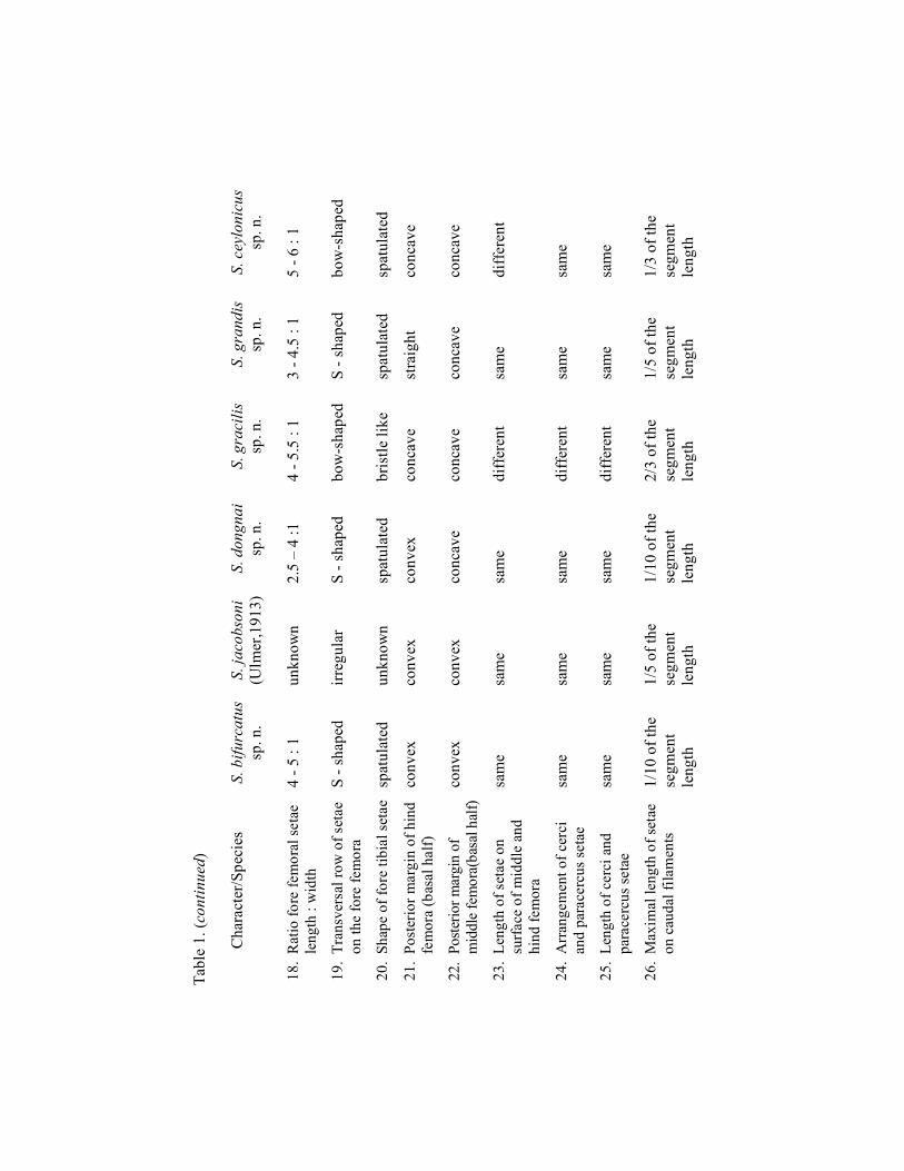
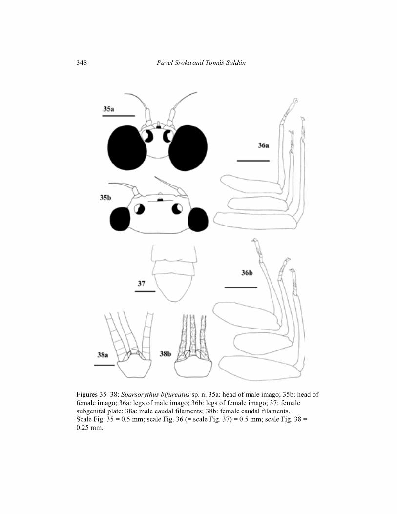
Mature larva (in alcohol): General coloration of body pale yellowish with black markings on dorsal side. Uniformly pale yellowish without any markings on ventral side. Body length 4–5 mm. Cerci approximately 1.1 x longer than body, paracercus approximately 1.3 x longer than body. Head apparently wider than long (ratio length : width 1 : 1.4). Eyes black, ocelli grayish. Composed eyes of males considerably larger than those in females (Figs. 5, 6). Distance between composed eyes in males as long as or slightly larger than the eye width. The ratio of distance between composed eyes in females to the eye width 2.2: 1. Hypopharyngeal lingua approximately as wide as long, divided by a short rill in the middle (Fig. 13). Right prostheca (Fig. 14b) notched, triangular, with concave margins and several short pointed teeth, bearing several setae on the inner side. Length of right prostheca about by 1/3 shorter than that of the inner incisor. Left prostheca (Fig. 14a) rod-like, pointed at apex, as long as the inner incisor. Two stout long setae, subequal to prostheca, inserted on its base. Labial plate without a small nick at the middle of anterior margin. Posterior margin of mesonotum overlapping at most the first abdominal segment. Rudimental gill on abdominal segment VII bifurcated, Y-shaped (Fig. 9f). Legs (Fig. 10) relatively robust. Length ratio femur : tibia : tarsus - 1.9 : 2.2 : 1 (fore legs); 2 : 2 : 1 (middle legs); 2.4 : 2.5 : 1 (hind legs). Ratio of femur length : width 1.8 : 1 in all leg pairs. Posterior margin of the middle and hind femora convex with rounded or bluntly pointed setae, irregularly alternating with tiny hairs. Transversal row of setae on fore femora slightly S-shaped. Fore femoral setae rounded at apex and about 4–5 times longer than wide (Fig. 11a). Foretibiae with conspicuous inner submarginal oblique row of setae, narrower and longer than femoral ones (Fig. 11b). Surface of the middle and hind femora sparsely covered by very small spines. Caudal filaments (Fig. 12) with a circles of sparse and small setae on the rounded posterior margins of individual segments. Setae are smaller than one-tenth of the length of segments.

Tricorythidae of the Oriental Region 319
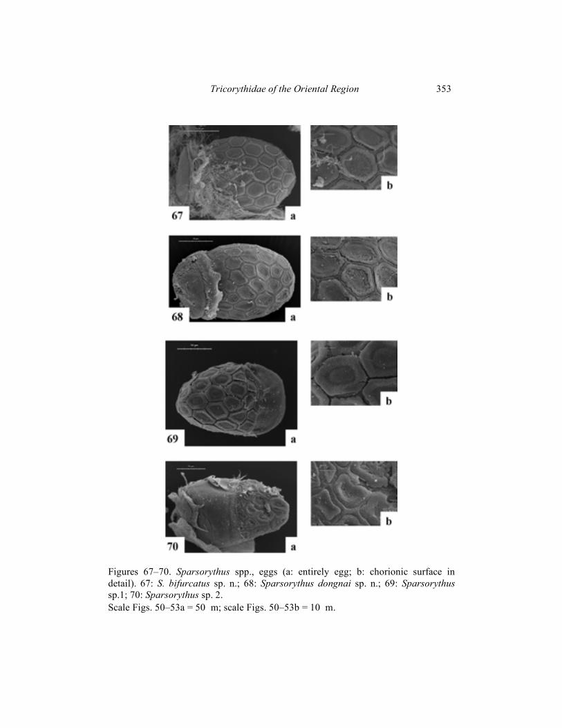
Imago male (in alcohol): Body length 3.5–5.5 mm. Cerci approximately 1.9 times longer than body, paracercus approximately 2.6 times longer than body. Head dark blackish-brown. Prothorax yellowish-brown with black markings. Mesothorax and metathorax brown. Abdomen pale, brownish, with pale black markings. Legs pale brown-yellowish. Cerci pale, yellowish, posterior margins of segments darker, grey. Composed eyes much larger than those in females (Fig. 35). Distance between composed eyes in males slightly shorter than the eye width. Pedicle longer than scape (ratio length of scape : length of pedicle is 1 : 1.8). Penis lobes apparently constricted subapically (Fig. 52). Penis extending the basal segment of forceps and reaching approximately the 1/4 of the second forceps segment. Penis with apparent medial nick indicating the original separation of mesomeres. Venation of the forewing (Fig. 39) relatively variable in number and pattern of cross veins. Forewing basal half dark coloured and distal half translucent. Femora narrower than those in females (Fig. 36). Length ratio femur : tibia : tarsus – 2.5 : 2.5 : 1 (fore legs); 2.9 : 3 : 1 (middle legs); 3.7 : 3.8 : 1 (hind legs). Imago female (in alcohol): Body length 5–6 mm. Length of cerci approximately 0.92 x body length, length of paracercus approximately 0.96 x body length. Head dark blackish-grey. Prothorax brownish-grey. Mesothorax and metathorax brown. Abdomen and legs brownish-grey. Composed eyes much smaller than those in males (Fig. 35). The ratio of distance between composed eyes to the eye width 2.8 : 1. Scape very small (ratio length of scape : length of pedicle is 1 : 2.3). Venation of the forewing (Fig. 40) relatively variable, similar to male. Forewing dark coloured in their basal half, distal half of wing translucent. Femora wider than those in males (Fig. 36). Length ratio femur : tibia : tarsus - 3 : 3 : 1 (fore legs); 2.9 : 3.2 : 1 (middle legs); 3.5 : 3.4 : 1 (hind legs). Subimago: Unknown. Egg (Fig. 67): 175 �m long, 74 �m wide. Surface with apparent polygonal (mainly hexagonal) structures. Polar cap covers approximately 1/5 of the surface. Egg pole opposite to the polar cap rounded. Material examined: Holotype: mature larva, Vietnam, Kinh-Dinh River, Nha-Ho, 16. IV. – 4. V. 1982, T. Soldán leg.; paratypes (parts on slides): 7 mature larvae, 12 immature larvae, 132 male imagines and 283 female imagines, same data as holotype, 1 mature larva, Vietnam, Tuan Hai prov., Kinh-Dinh R., Nha-Ho, 2. XI. 1984, T. Soldán leg, 4 mature larvae and 2 female imagines, Vietnam, Dong-Nai prov., Dong-Nai R., Nam Cat Tien res., 6. – 18. XI. 1989, T. Soldán leg.
Holotype and paratypes deposited in the Institute of Entomology, České Budějovice, Czech Republic.
320 Pavel Sroka and Tomáš Soldán
Etymology: Species is named according to a characteristic bifurcation of the last tracheal gill in larvae. Biology: The Kinh Dinh river at Nha-Ho, about 10 km W of Phan Rang is a large permanent lowland river (150–200 m across), 20–150 cm in mean depth during dry season and about 4–5 m water level fluctuation in the wet season (daily fluctuation of about 10 cm during dry season). The river is regulated in order to supply a system of artificial irrigation and forms a large number of rapids and backwaters. Judging from the primary plant succession on the river bed, this regulation originates from at least 50–70 years ago (Rejmánek, pers. comm.). Water is very turbid (transparency at most 15–20 cm), slightly alkaline (pH = 7.2–8.0) and relatively warm (maximal temperatures 24.6–29.8°C by night and day, respectively, in dry season).
Larvae of S. bifurcatus sp. n. were collected in various habitats by kicking technique, or occasionally by Surber sampler in April and May 1982 (dry season) and October–November 1984 (wet season).
They evidently prefer gravel bottom riffles or stones from small to medium size (up to 10–15, or 30–40 cm in diameter, respectively). They were never found on pure coarse sand bottom, at mixed sandy and clayey habitats and organic debris or in plat roots or submerged vegetation of Elodea sp. and Polygonum tomentosum. They were collected only at places with fast to very fast current velocities being never found at habitats with the current lower than 30–40 cm s-1. Most larvae were collected at gravel bottom riffle with more than 60 cm s-1, however some specimens occurred also at places with about 40–50 cm s-1 current velocity. They can easily survive fluctuation of current velocity up to more than 100 cm s-1 as well as fluctuation of water level (fluctuation observed by 1.5 m during dry season). On the other hand, their survival is apparently limited by gradual drying up of respective habitats. They were never found in temporary backwaters or pools isolated temporarily for more than one day. Larvae are always solitary to rare at habitats in question, their standing crop never reached more than about 5%, contrary to, e.g., Rhoenanthus distafurcus Bae et McCafferty, 1991 (up to 10%), Potamanthus (Potamanthodes) formosus Eaton, 1892 (up to 10%), Baetidae (mostly Baetis spp. and Pseudocloeon, up to 25%), Leptophlebiidae (mostly Choroterpes [Euthraulus] and Choroterpides sp., up to 15%), Heptageniidae (mostly Cinignina spp. up to 10%), and Ephemerellidae and Caenidae (mostly Ephemerella, Torleya and Drunella, and Caenis and Clypeocaenis, respectively, up to 20%). Life cycle is generally unknown. During dry season, first subimagines emerged about half an hour before sunset, submarine molting occurred shortly after the emergence. Mating flight followed immediately and was finished shortly after the sunset. Differential diagnosis: Combination of larval characters distinguishing S. bifurcatus sp. n. from other species of Sparsorythus gen. n. is apparent from Table 1 in Appendix A. Unique characters no. 9, 11 (reach of mesonotum and shape of the last

Tricorythidae of the Oriental Region 321
tracheal gill) distinguish it from all other species of the genus. The species has united characters 10, 21, 22 (number of tracheal gills, shape of middle and hind femoral margins) with S. jacobsoni comb. n. and 7, 26 (shape of right prostheca, setation on caudal filaments) with S. dongnai sp. n. Adults can be compared only with S. dongnai sp. n., S. jacobsoni comb. n. and S. multilabeculatus sp. n. From these, males can be generally distinguished by wing coloration, penis shape and eye size. Eggs can be distinguished by shape and arrangement of hexagonal structures and relative size of the polar cap.
Sparsorythus jacobsoni comb. n.
Tricorythus jacobsoni Ulmer, 1913: 105, fig. 5, 6. Tricorythus jacobsoni: Ulmer, 1924: 50, fig. 23, 24. Tricorythus jacobsoni: Ulmer, 1939: 521, 638, figs. 336–344.
Mature larva (in alcohol): General coloration of body brownish-yellow. Body length 5–6 mm. Cerci approximately 1.5 x longer than body, paracercus approximately 1.6 x longer than body. Head apparently wider than long. Hypopharyngeal lingua approximately as wide as long, without a rill in the middle, with large U-shaped medial incurvation. Superlinguae pointed at apex. Right prostheca notched, with one long curved projection at distal part, bearing several setae on the inner side. Right prostheca about by 1/3 shorter than the inner incisor. Distal part of the left prostheca extended, with several short pointed teeth. Left prostheca about by 1/3 shorter than the inner incisor. Some stout long setae, subequal to prostheca, inserted on its base. Labial plate with a small nick at the middle of anterior margin. Rudimental gill on abdominal segment VII filamentous. Legs relatively robust. Length ratio femur : tibia : tarsus - 2 : 2.7 : 1 (fore legs); 2.6 : 2.8 : 1 (middle legs); 2.2 : 2.6 : 1. (hind legs). Ratio of femur length : width 2 : 1 in all leg pairs. Posterior margins of the middle and hind femora convex with rounded or bluntly pointed setae, irregularly alternating with tiny hairs. Arrangement of setae on the fore femoral dorsal surface irregular. Foretibiae with conspicuous inner submarginal row of setae (shape of this setation unknown). Surface of the middle and hind femora sparsely covered by very small spines. Caudal filaments with circles of sparse setae rounded the posterior margins of individual segments. Setae are smaller than 1/5 of the length of segments. Imago male (in alcohol): Body length 5–5.5 mm. Cerci approximately 2.5 times longer than body, paracercus approximately 2.6 times longer than body. Head and prothorax dark blackish. Mesothorax yellowish-brown. Metathorax yellowish-brown. Abdomen pale, greyish. Legs pale greyish, femora darker. Cerci pale, greyish, with darker blackish stripes. Penis lobes apparently only slightly constricted
322 Pavel Sroka and Tomáš Soldán
subapically. Penis extending the basal segment of forceps and reaching approximately the 1/4 of the second forceps segment. Penis without apparent medial nick indicating the original separation of mesomeres. Venation of the forewing relatively variable in cross veins number and pattern. Cross veins in costal field present, badly visible. Forewing dull, coloured dark grey, with blackish-grey veins. Imago female (in alcohol): Body length 6 mm. Length of cerci is approximately 0.8 x body length, length of paracercus is approximately 0.9 x body length. General coloration of body dark greyish-yellow with black markings. Prothorax lighter. Ventral side of body greyish-yellow. Venation of the forewing is relatively variable, similar to male. Forewing dull, coloured dark grey, with blackish-grey veins. Subimago female (in alcohol): Similar to imago, with darker wing coloration. Egg: Unknown. Type locality: Wonosobo, Java. Distribution: Java, Sri Lanka, Sumatra, Philippines.
The more detailed description of this species in: Ulmer G. (1913) Note V. Ephemeriden aus Java, gesammelt von Edw. Jacobson.
Notes Leyden mus. 35:102–120. [male described, figured; type: male, Wonosobo, Java].
Ulmer, G. (1924) Ephemeropteren von den Sunda-Inseln und den Philippinen. Treubia 6:28–91. [male, female described, figured].
Ulmer G. (1939–1940) Eintagsfliegen (Ephemeroptera) von den Sunda Inseln. Arch. Hydrobiol., Suppl. 16:443–692. [larva described, figured]. Differential diagnosis: Combination of larval characters distinguishing S. jacobsoni comb. n. from other species of Sparsorythus gen. n. is apparent from Tab.1. Unique characters no. 8, 11, 19 (presence of nick on labium, shape of the last tracheal gill and irregular arrangement of fore femoral setae) distinguish it from all other species of the genus. The species has united characters mainly with S. grandis sp. n. (5, 6 – absence of rill on hypopharynx, shape of apex of the left prostheca), and with S. bifurcatus sp. n. (10, 21, 22 – number of tracheal gills, shape of middle and hind femoral margins). Adults can be compared only with S. bifurcatus sp. n., S. dongnai sp. n. and S. multilabeculatus sp. n. From these, males can be distinguished by coloration of wings and penis shape.

Tricorythidae of the Oriental Region 323
Sparsorythus dongnai sp. n.
(Figs. 7, 8, 15–19, 41, 42, 51, 53–55, 68)
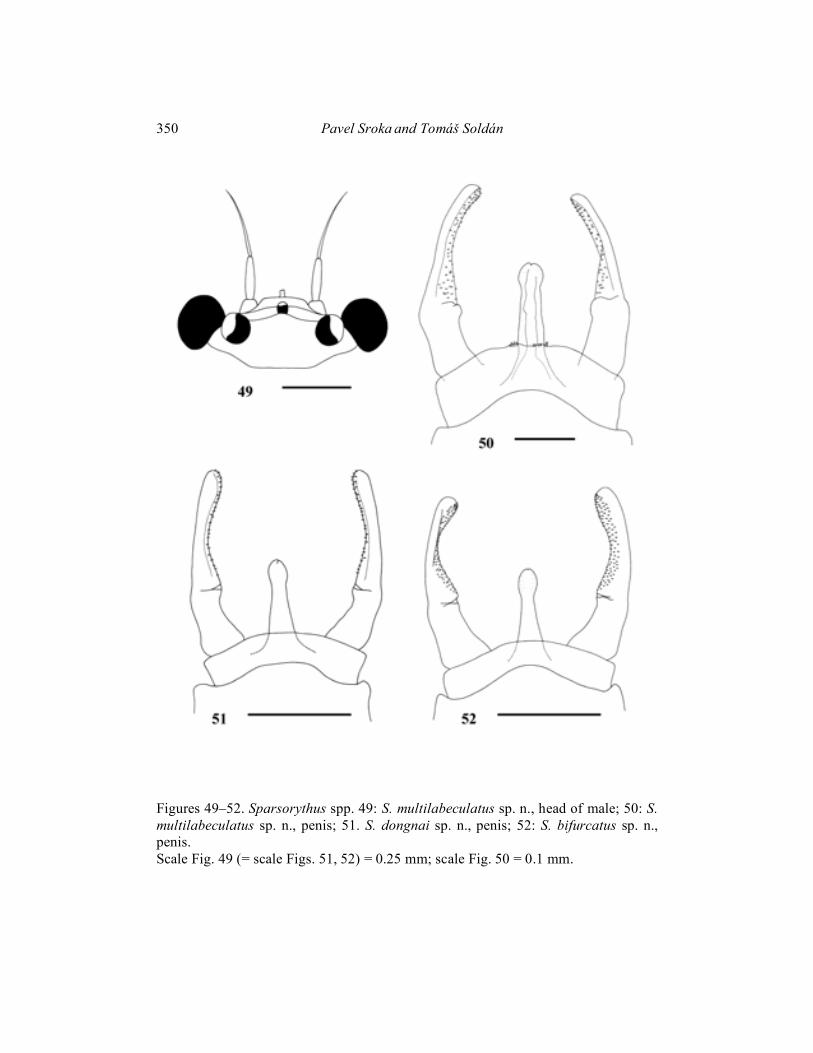
Mature larva (in alcohol): General coloration of body pale yellowish-brown with black markings on dorsal side similar to S. bifurcatus sp. n. Uniformly pale yellowish without any markings on ventral side. Body length 4–5 mm. Cerci are approximately 1.2 times longer than body, paracercus is approximately 1.3 times longer than body. Head apparently wider than long (ratio length : width is 1 : 1.6). Eyes black, ocelli greyish. Composed eyes of males considerably larger than those in females (Figs. 7, 8). Distance between composed eyes in males shorter than the eye width. The ratio of distance between composed eyes in females to the eye width 2.6: 1. Hypopharyngeal lingua rounded, approximately as wide as long, divided by a short rill in the middle (Fig. 18). Right prostheca (Fig. 19b) notched, triangular, with several long pointed teeth, bearing several setae on the inner side. Length of right prostheca is about 2/3 of the length of inner incisor. Left prostheca (Fig. 19a) rod-like, pointed at apex, slightly shorter than the inner incisor. Several stout long setae, subequal to prostheca, inserted on its base. Labial plate without a small nick at the middle of anterior margin. Posterior margin of mesonotum overlapping at most the fourth abdominal segment in females and the fifth one in males. Rudimental gill on abdominal segment VII absent. Legs (Fig. 15) relatively robust. Length ratio femur : tibia : tarsus – 2.1 : 2.6 : 1 (fore legs); 2.2 : 2.2 : 1 (middle legs); 2.8 : 3 : 1 (hind legs). Ratio of femur length : width 1.9 : 1 (fore legs); 2.1 : 1 (middle and hind legs). Fore femoral setae about 2.5–4 times longer than wide, with blunt apex (Fig. 16a). Fore tibial setae narrower and longer (Fig. 16b). Dorsal surface of the middle and hind femora sparsely covered by very small spines. Middle femoral posterior margins slightly concave, hind femoral ones convex. Individual segments of caudal filaments sparsely rounded at its posterior margins with very small setae. Setae are always smaller than 1/10 of the length of segments (Fig. 17). Imago male (in alcohol): Body length 5–5.5 mm. Length of cerci approximately 2 x body length, length of paracercus approximately 2.4 x body length. Head pale brown. Prothorax pale yellowish with dark brown markings. Meso- and metathorax pale brown. Abdomen, legs and cerci whitish. Composed eyes are much larger than those in females. Distance between composed eyes in males slightly shorter than the eye width. Pedicle longer than scape (ratio length of scape : length of pedicle is 1 : 2). Penis (Fig. 51) extending to the basal segment of forceps and extending to approximately 1/4 of the second forceps segment. Penis with apparent medial nick indicating the original separation of mesomeres. Wings (Fig. 41) translucent, very slightly coloured in proximal part. Venation of the forewing relatively variable in cross veins number and pattern. Legs see Fig. 53. Length ratio femur : tibia : tarsus – 2.7 : 3 : 1 (fore legs); 2.6 : 2.7 : 1 (middle legs); 3.4 : 3.4 : 1 (hind legs).

324 Pavel Sroka and Tomáš Soldán
Imago female (in alcohol): Body length 5–6 mm. Head, prothorax, legs and abdomen brownish-yellow. Mesothorax and metathorax brown. Cerci whitish. Length of cerci is approximately 0.5 x body length, length of paracercus is approximately 0.6 x body length. Composed eyes much smaller than those in males. The ratio of distance between composed eyes to the eye width 2.7 : 1. Venation of the forewing (Fig. 42) relatively variable, similar to male. Forewings dark coloured in their basal half, distal half of wing translucent. Coloration of basal wing part is much darker than in males of this species. Legs see Fig. 54. Length ratio femur : tibia : tarsus – 2.6 : 3 : 1 (fore legs); 3 : 3 : 1 (middle legs); 3 : 2.8 : 1 (hind legs). Egg (Fig.68): 175 �m long, 73 �m wide. Surface with exserted polygonal (mainly hexagonal) structures. Polar cap covers approximately 1/3 of the surface. Egg pole opposite to the polar cap rounded. Subimago: Unknown. Material examined: Holotype: mature larva, Vietnam, Dong-Nai R., Nam Cat Tien res., 6. – 18. xii. 1989, T. Soldán leg.; paratypes (parts on slides): 8 mature larvae, 5 immature larvae, 6 male imagines and 4 female imagines, same data as holotype.
Holotype and paratypes deposited in the Institute of Entomology, České Budějovice, Czech Republic. Etymology: Species is named after its type locality – the Dong-Nai River. Differential diagnosis: Combination of larval characters distinguishing S. dongnai sp. n. from other species of Sparsorythus gen. n. is apparent from Tab.1. Unique character no. 9 (reach of mesonotum) distinguish it from all species of the genus. The species has united characters 7, 26 (shape of right prostheca, setation on caudal filaments) with S. bifurcatus sp. n. Adults can be compared only with S. bifurcatus sp. n., S. jacobsoni comb. n. and S. multilabeculatus sp. n. From these, males can be generally distinguished by wing coloration, penis shape and eye size. Eggs can be distinguished by shape and arrangement of hexagonal structures and relative size of the polar cap.
Sparsorythus gracilis sp. n.
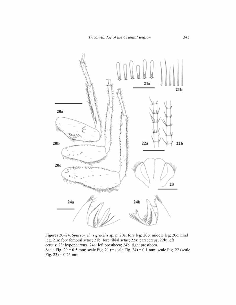
(Figs. 20–24) Mature female larva (in alcohol): Body length 4.9 mm. Length of cerci is approximately 0.8 x body length, length of paracercus is approximately 0.9 x body length. General coloration of body surface brownish-yellow. Gills very pale yellowish. Ventral side of body pale yellowish. Head apparently wider than long

Tricorythidae of the Oriental Region 325
(ratio length : width is 1 : 1.4). Eyes black, ocelli grayish. The ratio of distance between composed eyes to the eye width 2.6 : 1. Hypopharyngeal lingua approximately as wide as long, divided by a long rill in the middle. Small nick at the anterior margin of the lingua (Fig. 23). Right prostheca (Fig. 24b) notched, with several pointed teeth, bearing several setae on the inner side. Length of right prostheca is about 2/3 of the length of the inner incisor. Left prostheca (Fig. 24a) rod-like, bluntly pointed at apex, as long as the inner incisor. Two stout long setae, subequal to prostheca, inserted on its base. Labial plate without a small nick at the middle of anterior margin. Posterior margin of mesonotum overlapping at most the third abdominal segment. Rudimental gill on abdominal segment VII absent. Legs (Fig. 20) with regard to other species of this genus very slim. Length ratio femur : tibia : tarsus – 1.9 : 2.3 : 1 (fore legs); 2 : 2.3 : 1 (middle legs); 2.5 : 2.9 : 1 (hind legs). Ratio of femur length : width 2.5 : 1 (fore legs); 2.6 : 1 (middle legs), 2.9 : 1 (hind legs). Posterior margin of the middle and hind femora slightly concave at its basal half, with rounded or bluntly pointed setae, irregularly alternating with tiny hairs. Transversal row of setae on the fore femora bow-shaped, with a group of chaotically inserted setae near fore femoral posterior margin. Fore femoral setae about 5–6 times longer than wide, rounded apically (Fig. 21a). Setae on the fore tibiae very thin, spiky (Fig. 21b). Dorsal surface of the middle and hind femora sparsely covered by spines of the small and medium size. Individual segments of caudal filaments rounded at its posterior margin with setae approximately as long as 1/3 of the length of segments. Lateral margins with spiky setae as long as 2/3 of the length of segments. These long spiky setae on both sides of paracercus and only on the inner sides of cerci (Fig. 22). Male larvae, imagines and subimagines unknown. Material examined: Holotype: 1 mature larva, India, Madras state, Poona R., Poona, ix. 1962, V. Landa leg.; paratypes (parts on slides): 2 mature larvae, same data as holotype.
Holotype and paratypes deposited in the Institute of Entomology, České Budějovice, Czech Republic.
Etymology: From Latin gracilis meaning slim, the species is named after tenuous body structures (legs, fore tibial setae). Differential diagnosis: Combination of larval characters distinguishing S. gracilis sp. n. from other species of Sparsorythus gen. n. is apparent from Tab.1. Unique characters no. 7 (shape of right prostheca), 15, 16, 17 (shape of legs), 20 (bristle like setae on fore tibiae), and 24, 25, 26 (specific setation on caudal filaments) distinguish it from all species of the genus. The species seems to be well separated from all other species of Sparsorythus gen. n. Most united characters to S. gracilis sp. n. can be found in S. ceylonicus sp. n. (4, 19, 23 – presence of nick on hypopharynx,
326 Pavel Sroka and Tomáš Soldán
arrangement of fore femoral setae, shape of setae on middle and hind femoral surface).
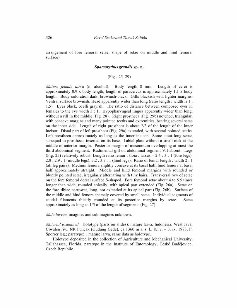
Sparsorythus grandis sp. n.
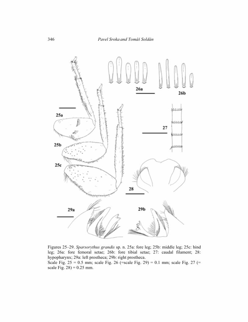
(Figs. 25–29) Mature female larva (in alcohol): Body length 8 mm. Length of cerci is approximately 0.9 x body length, length of paracercus is approximately 1.1 x body length. Body coloration dark, brownish-black. Gills blackish with lighter margins. Ventral surface brownish. Head apparently wider than long (ratio length : width is 1 : 1.5). Eyes black, ocelli grayish. The ratio of distance between composed eyes in females to the eye width 3 : 1. Hypopharyngeal lingua apparently wider than long, without a rill in the middle (Fig. 28). Right prostheca (Fig. 29b) notched, triangular, with concave margins and many pointed teeths and extremities, bearing several setae on the inner side. Length of right prostheca is about 2/3 of the length of the inner incisor. Distal part of left prostheca (Fig. 29a) extended, with several pointed teeths. Left prostheca approximately as long as the inner incisor. Some stout long setae, subequal to prostheca, inserted on its base. Labial plate without a small nick at the middle of anterior margin. Posterior margin of mesonotum overlapping at most the third abdominal segment. Rudimental gill on abdominal segment VII absent. Legs (Fig. 25) relatively robust. Length ratio femur : tibia : tarsus – 2.4 : 3 : 1 (fore legs); 2.8 : 2.9 : 1 (middle legs); 3.2 : 3.7 : 1 (hind legs). Ratio of femur length : width 2 : 1 (all leg pairs). Medium femora slightly concave at its basal half, hind femora at basal half approximately straight. Middle and hind femoral margins with rounded or bluntly pointed setae, irregularly alternating with tiny hairs. Transversal row of setae on the fore femoral dorsal surface S-shaped. Fore femoral setae about 4 to 5.5 times longer than wide, rounded apically, with apical part extended (Fig. 26a). Setae on the fore tibiae narrower, long, not extended at its apical part (Fig. 26b). Surface of the middle and hind femora sparsely covered by small setae. Individual segments of caudal filaments thickly rounded at its posterior margins by setae. Setae approximately as long as 1/5 of the length of segments (Fig. 27). Male larvae, imagines and subimagines unknown. Material examined: Holotype (parts on slides): mature larva, Indonesia, West Java, Ciwalen riv., NR Puncak (Gudung Gede), ca 1360 m a. s. l., 8. iv. – 3. ix. 1983, P. Sporrer leg.; paratype: 1 mature larva, same data as holotype.
Holotype deposited in the collection of Agriculture and Mechanical University, Tallahassee, Florida, paratype in the Institute of Entomology, České Budějovice, Czech Republic.

Tricorythidae of the Oriental Region 327
Etymology: From Latin grandis meaning large, the species is named after its relatively robust body. Differential diagnosis: Combination of larval characters distinguishing S. grandis sp. n. from other species of Sparsorythus gen. n. is apparent from Tab.1. Unique characters no. 3, 7 (shape of hypopharyngeal lingua, shape of right prostheca) distinguish it from all species of the genus. Most united characters to S. grandis sp. n. can be found in S. jacobsoni comb. n.(5, 6 – absence of hypopharyngeal rill, shape of apex of the right prostheca).
Sparsorythus ceylonicus sp. n.
(Figs. 30–34)
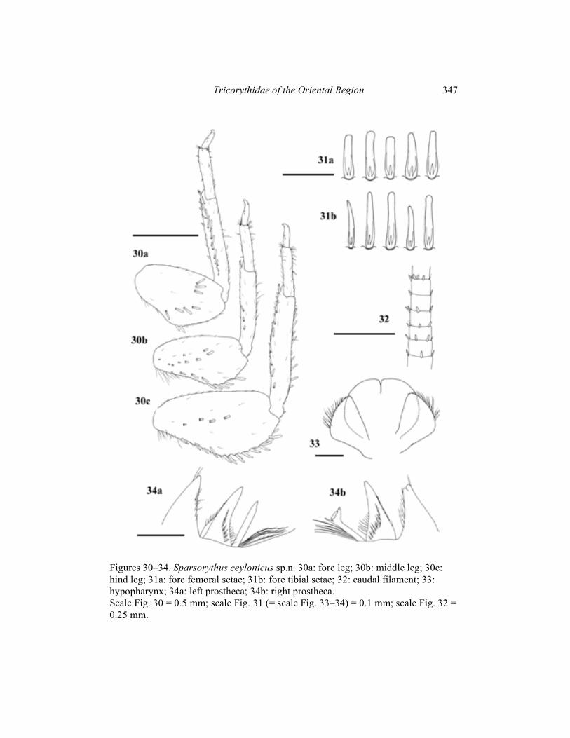
Mature male larva (in alcohol): Body length 4–5 mm. Length of cerci is approximately 0.9 x body length, length of paracercus is approximately 1.1 x body length. General coloration of body yellowish-brown. Uniformly pale yellowish on ventral side. Head apparently wider than long (ratio length : width is 1 : 1.4). Eyes black, ocelli grayish. The ratio of distance between composed eyes to the eye width 2.8 : 1. Size of composed eyes comparable with female larvae of other species of the genus. Hypopharyngeal lingua rounded, approximately as long as wide, divided by a short rill in the middle (Fig. 33). Right prostheca (Fig. 34b) bifurcated, with a small number of projections, bearing several setae on the inner side. Length of right prostheca is about 2/3 of the length of the inner incisor. Left prostheca (Fig. 34a) rod-like, pointed apically, subequal to the inner incisor. Some stout long setae, as long as prostheca, inserted on its base. Labial plate without a small nick at the middle of anterior margin. Posterior margin of mesonotum overlapping at most the second abdominal segment. Rudimental gill on abdominal segment VII absent. Legs (Fig. 30) relatively robust. Length ratio femur : tibia : tarsus – 1.7 : 2.2 : 1 (fore legs); 2 : 2 : 1 (middle legs); 2.6 : 1.9 : 1 (hind legs). Ratio of femur length : width 1.6 : 1 (fore legs); 2 : 1 (middle and hind legs). Posterior margins of the middle and hind femora slightly concave at its basal half, with sparse rounded or bluntly pointed setae, irregularly alternating with tiny hairs. Transversal row of setae on the fore femoral dorsal surface bow-shaped. Setae on the fore femora (Fig. 31a) relatively wide near its base, narrowing apically. Apex blunt. Fore femoral setae about 3 to 4.5 times longer than wide. Fore tibial setae (Fig. 31b) narrower and longer. Dorsal surface of the middle and hind femora very sparsely covered by some setae of various sizes, including relatively big ones. Individual segments of caudal filaments sparsely rounded at its posterior margins by small setae. Setae are smaller than 1/3 of the length of segments (Fig. 32). Female larvae, imagines and subimagines unknown.

328 Pavel Sroka and Tomáš Soldán
Material examined: Holotype (parts on slides): mature larva, Sri Lanka, Ratnapura dist., Kukula Ganga, Waddagala, 17. iv. 1973, Dawis and Rowe leg.
Holotype deposited in the collection of Agriculture and Mechanical University, Tallahassee, Florida. Etymology: The species is named after the type locality (Sri Lanka, formerly Ceylon). Differential diagnosis: Combination of larval characters distinguishing S. ceylonicus sp. n. from other species of Sparsorythus gen. n. is apparent from Tab. 1. United characters to S. ceylonicus sp. n. can be found mainly in S. gracilis sp. n. (4, 19, 21, 22, 23 – presence of nick on hypopharynx, arrangement of fore femoral setae, shape of middle and hind femoral posterior margins, shape of setation on the middle and hind femoral dorsal surface).
Sparsorythus multilabeculatus sp. n.
(Figs. 43, 49, 50, 56) Imago male (in alcohol): Body length 3 mm. Cerci approximately 3.3 x longer than body, paracercus approximately 5 x longer than body. Head, prothorax, abdomen, legs and cerci pale greyish-brown. Meso and metathorax brown. Composed eyes (Fig. 49) moderately enlarged (the ratio of distance between composed eyes to the eye width 2.4 : 1.). Pedicle much longer than scape (ratio length of scape : length of pedicle is 1 : 2.6) Penis extending the basal segment of forceps and reach to approximately 1/3 of the second forceps segment. Some small thorn-like structures on the subgenital plate near penis base. Penis (Fig. 50) with apparent medial nick indicating the original separation of mesomeres. Wing (Fig. 43) with typically organised smudges of various intensity. Most dark smudges in fields C, Sc, A, and near MA1 – MA2 fork. Lighter blotches in fields R1 and MP1. Legs see Fig. 56. Length ratio femur : tibia : tarsus – 2.5 : 2.6 : 1 (fore legs); 2.9 : 2.7 : 1 (middle legs); 3.5 : 3.3 : 1 (hind legs). Larvae, female imagines and subimagines unknown. Material examined: Holotype: imago male, Vietnam, Dong-Nai Prov., Dong-Nai R., Nam Cat Tien res., 6. – 18. xi. 1989, T. Soldán lgt.; paratypes (parts on slides): 27 male imagines, same data as holotype.
Holotype and paratypes deposited in the Institute of Entomology, České Budějovice, Czech Republic. Etymology: The species is named after the presence of more dark spots on its wings.

Tricorythidae of the Oriental Region 329
Differential diagnosis: Adults of this species can be compared only with S. bifurcatus sp. n., S. dongnai sp. n. and S. jacobsoni comb. n. From these, males can be distinguished by coloration of wings, penis shape and eye size.
Sparsorythus sp. 1
(Figs. 44, 57, 58, 69) Imago female (in alcohol): Body length 3.5–4 mm. Head, prothorax and abdomen greyish-brown. Mesothorax, metathorax and legs brownish-yellow. Dorsal side of body and cerci whitish. Forewing (Fig. 44) dark coloured in their basal half, distal half of wing translucent. Legs see Fig. 57. Length ratio femur : tibia : tarsus – 2.8 : 3.1 : 1 (fore legs); 2.5 : 2.6 : 1 (middle legs); 3.3 : 3.7 : 1 (hind legs). Egg (Fig. 69): 160 �m long, 100 �m wide. Surface with exserted polygonal (mainly hexagonal) structures. Polar cap covers approximately 1/4 of the surface. Egg pole opposite to the polar cap bluntly pointed like. Larvae, male imagines and subimagines unknown. Material examined: Female imagines (parts on slides), Vietnam, Dong-Nai prov., Dong-Nai riv., Nam Cat Tien res., 6. – 18. xii. 1989, T. Soldán leg.
Ca 100 ex. deposited in the Institute of Entomology, České Budějovice, Czech Republic.
Sparsorythus sp. 2
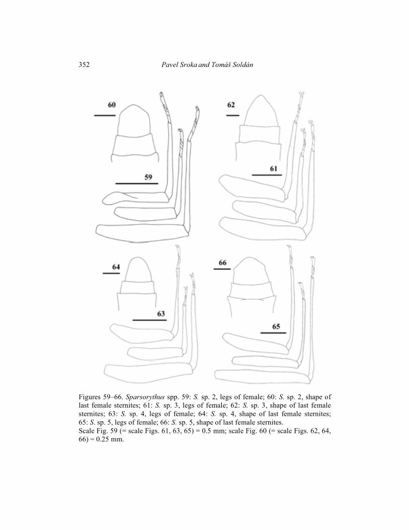
(Figs. 45, 59, 60, 70) Imago female (in alcohol): Body length 4 mm. General coloration of body brownish-black. The ratio of distance between composed eyes to the eye width 1 : 2.5. Cerci pale yellowish with darker grey stripes. Wings (Fig. 45) very lightly brownish, mainly in fields C, Sc. Legs see Fig. 59. Length ratio femur : tibia : tarsus – 2.3 : 2.7 : 1 (fore legs); 2.4 : 2.4 : 1 (middle legs); 2.8 : 2.9 : 1 (hind legs). Egg (Fig. 70): 190 �m long, 115 �m wide. Surface with slightly exserted polygonal (mainly hexagonal) structures. Polar cap covers approximately 1/2 of the surface. Egg pole opposite to the polar cap bluntly pointed like. Larvae, male imagines and subimagines unknown.
330 Pavel Sroka and Tomáš Soldán
Material examined: Female imagines (parts on slides), Thailand, Chiengmai prov., Chiengmai, Mae Ping, 9. iv. 1964, W.L. & J.G. Peters leg.
Two ex. deposited in the Institute of Entomology, České Budějovice, Czech Republic.
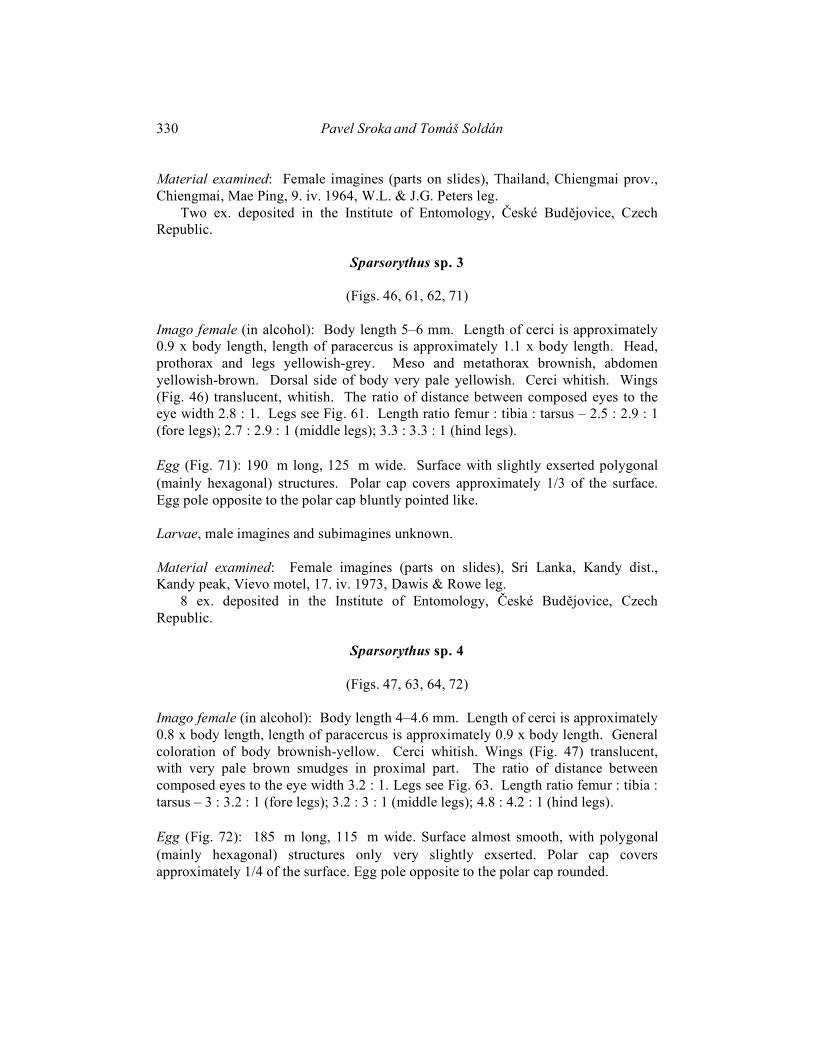
Sparsorythus sp. 3
(Figs. 46, 61, 62, 71) Imago female (in alcohol): Body length 5–6 mm. Length of cerci is approximately 0.9 x body length, length of paracercus is approximately 1.1 x body length. Head, prothorax and legs yellowish-grey. Meso and metathorax brownish, abdomen yellowish-brown. Dorsal side of body very pale yellowish. Cerci whitish. Wings (Fig. 46) translucent, whitish. The ratio of distance between composed eyes to the eye width 2.8 : 1. Legs see Fig. 61. Length ratio femur : tibia : tarsus – 2.5 : 2.9 : 1 (fore legs); 2.7 : 2.9 : 1 (middle legs); 3.3 : 3.3 : 1 (hind legs). Egg (Fig. 71): 190 �m long, 125 �m wide. Surface with slightly exserted polygonal (mainly hexagonal) structures. Polar cap covers approximately 1/3 of the surface. Egg pole opposite to the polar cap bluntly pointed like. Larvae, male imagines and subimagines unknown. Material examined: Female imagines (parts on slides), Sri Lanka, Kandy dist., Kandy peak, Vievo motel, 17. iv. 1973, Dawis & Rowe leg.
8 ex. deposited in the Institute of Entomology, České Budějovice, Czech Republic.
Sparsorythus sp. 4
(Figs. 47, 63, 64, 72) Imago female (in alcohol): Body length 4–4.6 mm. Length of cerci is approximately 0.8 x body length, length of paracercus is approximately 0.9 x body length. General coloration of body brownish-yellow. Cerci whitish. Wings (Fig. 47) translucent, with very pale brown smudges in proximal part. The ratio of distance between composed eyes to the eye width 3.2 : 1. Legs see Fig. 63. Length ratio femur : tibia : tarsus – 3 : 3.2 : 1 (fore legs); 3.2 : 3 : 1 (middle legs); 4.8 : 4.2 : 1 (hind legs). Egg (Fig. 72): 185 �m long, 115 �m wide. Surface almost smooth, with polygonal (mainly hexagonal) structures only very slightly exserted. Polar cap covers approximately 1/4 of the surface. Egg pole opposite to the polar cap rounded.

Tricorythidae of the Oriental Region 331
Larvae, male imagines and subimagines unknown. Material examined: Female imagines (parts on slides), Philippines, Mindanao, Mt. Apo School, 15 km SW Davao, ca 500 m a. s. l., 22. – 31. x. 1965, D. Davis leg.
Fifteen ex. deposited in the Institute of Entomology, České Budějovice, Czech Republic.
Sparsorythus sp. 5
(Figs. 48, 65, 66, 73) Imago female (in alcohol): Body length 4–4.5 mm. Length of cerci approximately 0.5 x body length, length of paracercus approximately 0.6 x body length. General coloration of body black. Cerci greyish with darker grey stripes. Wings (Fig. 48) black. The ratio of distance between composed eyes to the eye width 3.3 : 1. Legs see Fig. 65. Length ratio femur : tibia : tarsus – 3.2 : 4 : 1 (fore legs); 3.9 : 4 : 1 (middle legs); 4.5 : 5 : 1 (hind legs). Egg (Fig. 73): 190 �m long, 120 �m wide. Surface with only slightly exserted areas between polygonal structures. Polar cap covers approximately 1/4 of the surface. Egg pole opposite to the polar cap rounded. Larvae, male imagines and subimagines unknown. Material examined: Female imagines (parts on slides), Indonesia, Sulawesi – Utara, Dumoga-Bone, NP Sungai Tupah, 5. viii. 1985, D. Dudgeon leg.
26 ex. deposited in the Institute of Entomology, České Budějovice, Czech Republic.
Discussion Systematics. Altogether, six genera of the family Tricorythidae have been described from the Afrotropic (Ethiopian) Region (including Madagascar) so far.
The genus Dicercomyzon Demoulin, 1954 (separate subfamily Dicercomyzinae) is undoubtedly well characterized by its prominent apomorphies mainly in the larval stage: highly expanded femora, dorsoventrally flattened body, pro- and mesosternum with a disc of friction hairs, superlinguae of hypopharynx highly developed laterally (Demoulin 1954, 1970, Kimmins 1957, Edmunds and Traver 1954, Edmunds et al. 1963, Landa and Soldán 1985, McCafferty and Wang 2000). In adults, penes are slightly to deeply divided, associated with auxilliary processes (McCafferty and Wang 2000).
332 Pavel Sroka and Tomáš Soldán
Genera Madecassorythus, Spinirythus and Ranorythus represent either the most plesiomorphic lineage within the Tricorythidae (Madecassorythus and Spinirythus), or intermediary lineage to that represented by afrotropical tricorythids (Ranorythus). Genera Madecassorythus and Spinirythus were separated into the subfamily Madecassorythinae by Oliarinomy and Elouard (1997). The same authors placed genus Ranorythus into the separate subfamily Ranorythinae (Oliarinomy and Elouard 1998). This subfamilial classification was discussed by McCafferty and Wang (2000) who point out, e.g., lack of some synapomorphies in Tricorythus and Ranorythus, even suggesting by default that Ranorythus even belongs to the Tricorythinae lineage.
The genus Spinirythus differs from Sparsorythus gen. n. in imaginal stage (larvae of Spinirythus remain unknown) by penis shape and degree of paracercus reduction. Spinirythus has entirely separated penial lobes associated with lamellar auxilliary processes. Penis and auxilliary processes are approximately of the same size and shape, paracercus reduced.
Second genus of the subfamily Madecassorythinae, Madecassorythus, is characterized also by separated penial lobes with auxilliary processes (penis and auxilliary processes differently formed, penis lobes much longer than gonostyles), paracercus well developed. Both genera of the subfamily Madecassorythinae have big sexual dimorphism in eye size (composed eyes in males much larger than in females).
On the other hand, Sparsorythus gen. n. has completely fused penial lobes without auxilliary processes, paracercus well developed. Sexual dimorphism in eye size often pronounced but probably do not represent a truly consistent character (see below). Larvae of Madecassorythus have following characters, which separate them from the Sparsorythus gen. n.: presence of maxillar palp, absence of regular row of setae on the fore tibiae. In Madecassorythus larvae, tracheal gill on the abdominal segment VII always missing. (But in some Sparsorythus gen. n. species also present only five pairs of tracheal gills like in Madecassorythus).
The genus Ranorythus is characterized in imaginal stage (larvae remain unknown) by partly (in proximal part) fused penial lobes, distally separated. Auxilliary processes are absent. Sexual dimorphism in eye size well apparent. Paracercus reduced in males only, in females paracercus normally developed.
Difference between Sparsorythus gen. n. and Tricorythus is mainly completely reduced maxillar palp in the larval stage of Sparsorythus gen. n. Other characters are not consistent throughout all species of this genus. Sexual dimorphism in eye size in Tricorythus always absent, in Sparsorythus gen. n. present, but probably in a variable degree. In some species, difference in eye size between sexes is really big (S. bifurcatus sp. n., S. dongnai sp. n.). On the other hand, some species have relatively small eyes of males (S. ceylonicus sp. n., S. multilabeculatus sp. n.). Unfortunately, there we have not any comparison with females of these species. Tracheal gill on

Tricorythidae of the Oriental Region 333
abdominal segment VII in Tricorythus absent, in Sparsorythus gen. n. sometimes present, but not in all species.
As noted above, Sparsorythus jacobsoni comb. n. was originally described by Ulmer (1913) after male adult stages as Tricorythus jacobsoni. Later, the more detailed description of male adults was given by the same author (Ulmer, 1925). In this study, female adults from the same type locality as males were also described. Fifteen years later, Ulmer described a larva, which in his opinion belongs to T. jacobsoni. Larvae were collected in a different localities than imagines and there is not any evidence, which associates larvae described by Ulmer in 1940 with imagines described by him before. It is a question if this larva really belongs to S. jacobsoni comb. n. The nymph figured by Edmunds et al. (1963), collected in the Philippines, and called “Neurocaenis jacobsoni Ulmer ?“ is evidently a different species than Ulmer’s larva, described in 1940 and declared to be a larva of T. jacobsoni. In larva, figured by Edmunds et al. (1963), the tracheal gill on abdominal segment VII is absent and also the shape of its legs is different from Ulmer’s larva, similar to S. gracilis sp. n.
Establishing Sparsorythus bifurcatus sp. n. as a type species of Sparsorythus gen. n. (instead of S. jacobsoni comb. n.) rests upon our better knowledge of S. bifurcatus sp. n. In this species, that the described larva really belongs to imago (the same type locality of these with the only tricorythid species founded) is a greater probability. Another occasion is nonavailability of any comparative material of S. jacobsoni comb. n.
However, within the “true” African Tricorythus are still open questions. Quite recently, Barber-Jones (2004) indicates there are only two species, namely T. reticulatus Barnard, 1932 and T. discolor (Burmeister, 1938) among the representatives of the genus Tricorythus in South Africa, which really can be classified within this genus. The others might represent an undescribed genus, possibly related to the genus Ranorythus.
Oriental Tricorythidae significantly differ from African ones. The most explicit difference is completely reduced maxillar palp in all oriental species. All African Tricorythidae have big, well-developed maxillar palp. Total reducing of maxillar palp is a big morphological change and it was very probably unique event, which happened to one collective ancestor of all present oriental Tricorythidae. Within the genus Sparsorythus gen. n. can be found a big variability (in some species present six pairs of tracheal gills, in some only five; in some species is difference in eye size between sexes big and in some species is small). But absence of maxillar palp shows their collective origin.
Genus Sparsorythus gen. n. is an advanced group from the Tricorythidae family with many apomorphies (reduced hind wings, completely fused penial lobes without auxilliary processes, missing maxillar palp, reduced or missing gill on abdominal segment VII, big sexual dimorphism in eye size in some species). Diversity of the family Tricorythidae in Asia is very probably much bigger than species described in
334 Pavel Sroka and Tomáš Soldán
this study. Establishing of the genus Sparsorythus gen. n. like a taxon involving all Asian Tricorythidae is not certainly the final status.
Distribution and Biogeography The family Tricorythidae appear to have evolved primarily in Gondwanaland. All the subfamilies occur in Africa or Madagascar. Only the genus Sparsorythus is found in Sri Lanka and Southeast Asia. These oriental Tricorythidae are geographically isolated from African species for more than 100 mil. years. After seceding of Indian subcontinent and Africa, Indian species developed separately. After connecting of Indian subcontinent and Asia (about 45 mil. years ago), mayflies from India expanded into the rest of the South-east Asia (Edmunds 1979), Jacob (2003).
The genus Sparsorythus was found in almost all regions of the South-East Asia (Sri Lanka, India, Thailand, Vietnam, Indonesia and Philippines).
Acknowledgments We are indebted to Janice G. and the late professor William L. Peters, A & M University, Tallahassee for providing us with some material. We also thank K. Bláhová for technical assistance. This study was partly supported by Reasearch Project No. 1QS500070505 by Grant Agency of the Academy of Sciences.
Literature Cited Barber-James, H. M. In press. A synpopsis of the Afrotropical Tricorythidae. Pages
187–20 in F. R. Hauer, J. A. Stanford, and R. L. Newell (editors). International Advances in the Ecology, Zoogeography and Systematics of Mayflies and Stoneflies. University of California Press. Berkeley, California, USA.
Barnard, K. H. 1932. South African May-flies (Ephemeroptera). Transactions of the Royal Society of South Africa 20:201–259.
Corbet, P. S. 1960. Larvae of certain East African Ephemeroptera. Revue de zoologie et de botanique africaines 61:119–129.
Crass, R. S. 1947. The May-Flies (Ephemeroptera) of Natal and the Eastern Cape. Annals of the Natal Museum 11:37–110.
Demoulin, G. 1954. Recherches critiques sur les Ephéméroptères Tricorythidae d’Afrique et d’Asie. Bulletin et Annales de al Societe Royale d’Entomologie de Belgique 90:264–277.
Demoulin, G. 1954. Description préliminaire d’un type larvaire nouveau d’Ephéméroptères Tricorythidae du Congo Belge. Bulletin de l'Institut Royale des Sciences Naturelles de Belgique 30(6):1–4.

Tricorythidae of the Oriental Region 335
Demoulin, G. 1957. Le type larvaire probable des Tricorythus Eaton (Ephemeroptera: Tricorythidae). Bulletin de l’Institut Royale des Sciences Naturelles de Belgique 33(19):1–4.
Demoulin, G. 1958. Un curieuse larve d’Ephéméroptère de l’Angola portugais. Bulletin et Annales de al Societe Royale d’Entomologie de Belgique 95 (7-8):249–252.
Demoulin, G. 1970. Ephemeroptera des faunes éthiopienne et malgache. South African Animal Life, 14:24–170.
Eaton, A. E. 1868. An outline of a re-arrangement of the genera of Ephemeridae. Entomologist's Monthly Magazine 5:82–91.
Eaton, A. E. 1883–1888. A Revisional Monograph of Recent Ephemeridae or Mayflies. Transactions of the Linnean Society of London, Second Series, Zoology 3:1-352, 65 pl.
Edmunds Jr., G. F., R. K. Allen, and W. L. Peters. 1963. An Annotated Key to the Nymphs of the Families and Subfamilies of Mayflies (Ephemeroptera). Univ. Utah Biol. Ser. 13(1), pp. 53.
Edmunds Jr., G. F., and J. R. Traver. 1954. An outline of a reclassification of the Ephemeroptera. Proceedings of the Entomological Society of Washington 56:236–240.
Edmunds Jr., G. F. 1979. Biogeografical relationships of the Oriental and Ethiopian Mayflies. Pages 11–14 in K. Pasternak, and R. Sowa, editors. Proceedings of the Second International Conference on Ephemeroptera.. Państwowe Wydawnictwo Naukowe, Warszawa – Kraków, 312 pp.
Elouard, J.-M., and R. Oliarinony. 1997. Biodiversité aquatique de Madagascar. 6 - Madecassorythus un nouveau genre de Tricorythidae définissant la nouvelle sous-famille des Madecassorythinae (Ephemeroptera, Pannota). Bulletin de la Société entomologique de France 102(3):225–232.
Fernando, C. H. 1965. A Guide to the Freshwater Fauna of Ceylon. Supplement 2. Bulletin of the Fisheries Research Station of Ceylon 17:177–211.
Hubbard, M. D., and W. L. Peters. 1984. Ephemeroptera of Sri Lanka: An introduction to their ecology and biogeography. Pages 257–274 in C. H. Fernando, editor. Ecology and Biogeography in Sri Lanka. Dr. W. Junk, The Hague.
Hubbard, M. D. 1990. Mayflies of the World. A Catalog of the Family and Genus Group Taxa (Insecta: Ephemeroptera), Flora and Fauna Handbook No. 8, Sandhill Crane Press, Gainesville, 119 pp.
Jacob, U. 2003. Africa and its Ephemeroptera: Remarks from a biogeographical view. Pages 317–325 in E. Gaino, editor. Research Update on Ephemeroptera and Plecoptera. University of Perugia, Perugia, Italy.
Kimmins, D. E. 1960. Notes on East African Ephemeroptera, with descriptions of newBulletin of the British Museum (Natural History), Entomology 9:337–355.
336 Pavel Sroka and Tomáš Soldán
Koss, R. W., and G. F. Edmunds Jr. 1974. Ephemeroptera eggs and their contribution to phylogenetic studies of the order. Zoological Journal of the Linnean Society 55(4):267–349.
Lestage, J. A. 1942. Contribution à l’étude des Ephéméroptères. XXV. Notes critiques sur les anciens Caenidiens d’Afrique et sur l’indépendance de l’évolution tricorythido-caenidienne. Bull. Mus. R. Hist. Nat. Belg., XVIII, 48, 20 pp.
McCafferty, W. P., and T.-Q. Wang. 2000. Phylogenetic systematics of the major lineages of Pannote Mayflies (Ephemeroptera: Pannota). Transactions of the American Entomological Society 126(1):9–101.
Navás, L. 1936. Insectes du Congo Belge, Série IX. Revue de Zoologie et de Botanique Africaines 28:333–368.
Oliarinony, R., and J.-M. Elouard. 1998b. Biodiversité aquatique de Madagascar. 8 - Spinirythus un nouveau genre de Tricorythidae (Ephemeroptera, Pannota). Bulletin de la Société entomologique de France 103(3):237–244.
Oliarinony, R., and J.-M. Elouard. 1998a. Biodiversité aquatique de Madagascar. 7 - Ranorythus un nouveau genre de Tricorythidae définissant la nouvelle sous-famille des Ranorythinae (Ephemeroptera, Pannota). Bulletin de la Société entomologique de France 102(5):439–447.
Oliarinony, R., J.-M. Elouard, and N. Raberiaca. 1998. Biodiversité aquatique de Madagascar 19 - neuf nouvelles espèces de Tricorythus Eaton [ephemeroptera, Pannota, Tricorythidae]. Revue français d'entomologie 20:73–90.
Sivaramakrishnan, K. G., and K. Venkataraman. 1987. Biosystematic studies of south Indian Leptophlebiidae and Heptageniidae in relation to egg ultrastructure and phylogenetic interpretations. Proceedings of the Indiana Academy of Science (Animal Sciences) 96:637–646.
Soldán, T. 1983. Two new species of Clypeocaenis (Ephemeroptera, Caenidae) with a description of adult stage and biology of the genus. Acta Entomologica Bohemoslovaca 80:196–205.
Soldán, T. 1991. An annotated list of mayflies (Ephemeroptera) found in the Nam Cat Tien National Park. Pages 4–9 in K. Spitzer, J. Lepš, and M. Zacharda, editors. Nam Cat Tien Czechoslovak Vietnamese Expedition, November 1989, Research Report. Institute of Entomology, Czechoslovak Acad. Sci., České. Budějovice, 45 pp.
Soldán, T., and V. Landa. 1991. Two new species of Caenidae (Ephemeroptera) from Srí Lanka. Pages 235–246 in J. Alba-Tercedor, and A. Sanchez-Ortega, editors. Overview and Strategies of Ephemeroptera and Plecoptera. The Sandhill Crane Press, Gainesville, Florida.
Soldán, T., and M. Putz. 2000. The larva of Rhoenanthus distafurcus Bae et McCafferty (Ephemeroptera, Potamanthidae) with notes on distribution and biology. Aquatic Insects 22:9–17.
Tricorythidae of the Oriental Region 337
Ulmer, G. (1913) Ephemeriden aus Java, Gesammelt von Edw. Jacobson, Note V., Notes from the Leyden Museum, 35:102–120.
Ulmer, G. 1916. Ephemeropteren von Aequatorial-Afrika. Archiv für Naturgeschichte 81(A) 7 (1915):1–19.
Ulmer, G. 1925. Ephemeropteren von den Sunda-Inseln und den Philippinen. Treubia;Recueil de Travaux Zoologiques, Hydrobiologiques et Oceanographiques 6(1):28–91.
Ulmer, G. 1940. Eintagsfliegen (Ephemeropteren) von den Sunda-Inseln. Archiv für Hydrobiologie 16:443–692, p. 690.

App
endi
x A
Ta
ble
1. C
ritic
al c
hara
cter
s di
stin
guis
hing
larv
ae o
f the
gen
us S
pars
oryt
hus g
en. n
.
Cha
ract
er/S
peci
es
S. b
ifurc
atus
sp
. n.
S. ja
cobs
oni
(Ulm
er,1
913)
S.
don
gnai
sp
. n.
S. g
raci
lis
sp. n
. S.
gra
ndis
sp
. n.
S. c
eylo
nicu
s sp
. n.
1.
Dis
tanc
e be
twee
n co
mpo
sed
eyes
in m
ales
as
long
as o
r sl
ight
ly la
rger
th
an th
e ey
e w
idth
unkn
own
shor
ter t
han
the
eye
wid
th
unkn
own
unkn
own
muc
h la
rger
th
an th
e ey
e w
idth
2.
Rat
io o
f dis
tanc
e be
twee
n co
mpo
sed
eyes
in
fem
ales
to e
ye w
idth
2.2
: 1
unkn
own
2.6
: 1
2.6
: 1
3 : 1
un
know
n
3.
Shap
e of
hy
poph
aryn
geal
ling
ua
as w
ide
as
long
as
wid
e as
lo
ng
as w
ide
as
long
as
wid
e as
lo
ng
wid
er th
an
long
as
wid
e as
lo
ng
4.
Pres
ence
of m
edia
l nic
k on
hyp
opha
rynx
ab
sent
ab
sent
ab
sent
pr
esen
t ab
sent
pr
esen
t
5.
Pres
ence
of m
edia
l rill
on
hyp
opha
rynx
pr
esen
t ab
sent
pr
esen
t pr
esen
t ab
sent
pr
esen
t
6.
Ape
x of
the
left
pros
thec
a po
inte
d ex
tend
ed, w
ith
seve
ral t
eeth
po
inte
d bl
untly
po
inte
d ex
tend
ed, w
ith
seve
ral t
eeth
bl
untly
po
inte
d
7.
Rat
io ri
ght p
rost
heca
le
ngth
: w
idth
2
: 1
2.4
: 1
2 : 1
3.
3 : 1
1.
8 : 1
2.
4 : 1
8.
Med
ial n
ick
on la
bium
ab
sent
pr
esen
t ab
sent
ab
sent
ab
sent
ab
sent

Tabl
e 1.
(con
tinue
d)
C
hara
cter
/Spe
cies
S.
bifu
rcat
us
sp. n
. S.
jaco
bson
i (U
lmer
, 191
3)
S. d
ongn
ai
sp. n
. S.
gra
cilis
sp
. n.
S. g
rand
is
sp. n
. S.
cey
loni
cus
sp. n
.
9.
Win
g pa
ds re
achi
ng to
th
e ab
dom
inal
segm
ent
I un
know
n IV
(fem
ales
); V
(mal
es)
III
III
II
10.
Rud
imen
tal g
ill o
n ab
dom
inal
segm
ent V
II
pres
ent
pres
ent
abse
nt
abse
nt
abse
nt
abse
nt
11.
Shap
e of
rudi
men
tal g
ill
on a
bdom
inal
seg
men
t V
II
bifu
rcat
ed
filam
ento
us
- -
- -
12.
Rat
io fe
mur
: tib
ia :
tars
us (f
ore
leg)
1.
9 : 2
.2 :
1
2 : 2
.7 :
1
2.1
: 2.6
: 1
1.
9 : 2
.3 :
1
2.4
: 3 :
1
1.7
: 2.2
: 1
13.
Rat
io fe
mur
: tib
ia :
tars
us (m
iddl
e le
g)
2 : 2
: 1
2.6
: 2.8
: 1
2.2
: 2.2
: 1
2 : 2
.3 :
1 2.
8 : 2
.9 :
1 2
: 2 :
1
14.
Rat
io fe
mur
: tib
ia :
tars
us (h
ind
leg)
2.
4 : 2
.5 :
1 2.
2 : 2
.6 :
1 2.
8 : 3
: 1
2.5
: 2.9
: 1
3.2
: 3.7
: 1
2.6
: 1.9
: 1
15.
Rat
io fo
re fe
mur
leng
th :
wid
th
1.8
: 1
2 : 1
1.
9 : 1
2.
5 : 1
2
: 1
1.6
: 1
16.
Rat
io m
iddl
e fe
mur
le
ngth
: w
idth
1.
8 : 1
2
: 1
2.1
: 1
2.6
: 1
2 : 1
2
: 1
17.
Rat
io h
ind
fem
ur le
ngth
: w
idth
1.
8 : 1
2
: 1
2.1
: 1
2.9
: 1
2 : 1
2
: 1
Con
tinue
d on
nex
t pag
e
Tabl
e 1.
(con
tinue
d)
C
hara
cter
/Spe
cies
S.
bifu
rcat
us
sp. n
. S.
jaco
bson
i (U
lmer
,191
3)
S. d
ongn
ai
sp. n
. S.
gra
cilis
sp
. n.
S. g
rand
is
sp. n
. S.
cey
loni
cus
sp. n
.
18.
Rat
io fo
re fe
mor
al se
tae
leng
th :
wid
th
4 - 5
: 1
un
know
n 2.
5 –
4 :1
4
- 5.5
: 1
3 - 4
.5 :
1 5
- 6 :
1
19.
Tran
sver
sal r
ow o
f set
ae
on th
e fo
re fe
mor
a S
- sha
ped
irreg
ular
S
- sha
ped
bow
-sha
ped
S - s
hape
d bo
w-s
hape
d
20.
Shap
e of
fore
tibi
al se
tae
spat
ulat
ed
unkn
own
spat
ulat
ed
bris
tle li
ke
spat
ulat
ed
spat
ulat
ed
21.
Post
erio
r mar
gin
of h
ind
fem
ora
(bas
al h
alf)
co
nvex
co
nvex
co
nvex
co
ncav
e st
raig
ht
conc
ave
22.
Poste
rior m
argi
n of
m
iddl
e fe
mor
a(ba
sal h
alf)
conv
ex
conv
ex
conc
ave
conc
ave
conc
ave
conc
ave
23.
Leng
th o
f set
ae o
n su
rface
of m
iddl
e an
d hi
nd fe
mor
a
sam
e sa
me
sam
e di
ffer
ent
sam
e di
ffer
ent
24.
Arr
ange
men
t of c
erci
an
d pa
race
rcus
seta
e
sam
e sa
me
sam
e di
ffer
ent
sam
e sa
me
25.
Leng
th o
f cer
ci a
nd
para
cerc
us se
tae
sam
e sa
me
sam
e di
ffer
ent
sam
e sa
me
26.
Max
imal
leng
th o
f set
ae
on c
auda
l fila
men
ts
1/10
of t
he
segm
ent
leng
th
1/5
of th
e se
gmen
t le
ngth
1/10
of t
he
segm
ent
leng
th
2/3
of th
e se
gmen
t le
ngth
1/5
of th
e se
gmen
t le
ngth
1/3
of th
e se
gmen
t le
ngth
Tricorythidae of the Oriental Region 341
Figures
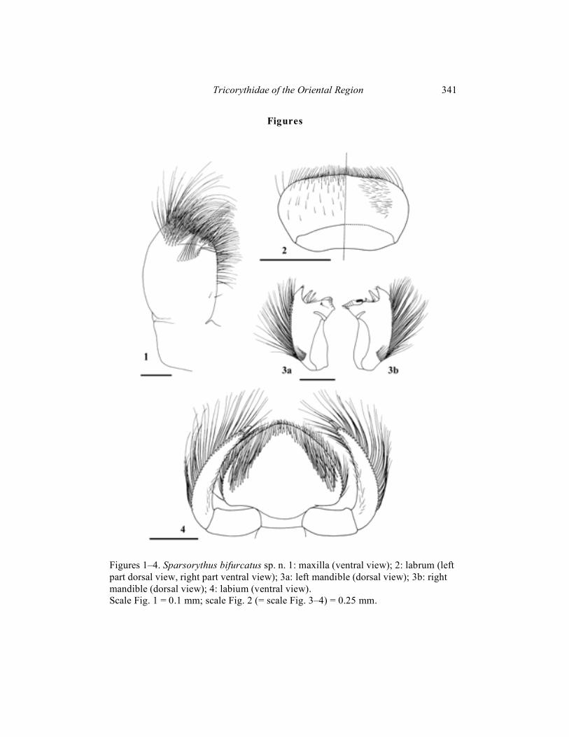
Figures 1–4. Sparsorythus bifurcatus sp. n. 1: maxilla (ventral view); 2: labrum (left part dorsal view, right part ventral view); 3a: left mandible (dorsal view); 3b: right mandible (dorsal view); 4: labium (ventral view). Scale Fig. 1 = 0.1 mm; scale Fig. 2 (= scale Fig. 3–4) = 0.25 mm.

342 Pavel Sroka and Tomáš Soldán
Figures 5–9. Sparsorythus spp. 5, 6, 9: S. bifurcatus sp. n.; 7, 8: S. dongnai sp. n.; 5: head of male larva; 6: head of female larva; 7: head of male larva; 8: head of female larva; 9a–9f: gills on abdominal segments II–VII. Scale Figs. 5–8 = 0.5 mm; scale Fig. 9 = 0.25 mm.

Tricorythidae of the Oriental Region 343
Figures 10–14. Sparsorythus bifurcatus sp. n. 10a: fore leg; 10b: middle leg; 10c: hind leg; 11a: fore femoral setae; 11b: fore tibial setae; 12: caudal filament; 13: hypopharynx; 14a: left prostheca; 14b: right prostheca. Scale Fig. 10 (= scale Fig. 12) = 0.25 mm; scale Fig. 11 (= scale Fig. 13–14) = 0.1 mm.

344 Pavel Sroka and Tomáš Soldán
Figures 15–19. Sparsorythus dongnai sp. n. 15a: fore leg; 15b: middle leg; 15c: hind leg; 16a: fore femoral setae; 16b fore tibial setae; 17: caudal filament; 18: hypopharynx; 19a: left prostheca; 19b: right prostheca. Scale Fig. 15 = 0.5 mm; scale Fig. 16 (= scale Fig. 19) = 0.1 mm; scale Fig. 17 (= scale Fig. 18) = 0.25 mm.
Tricorythidae of the Oriental Region 345
Figures 20–24. Sparsorythus gracilis sp. n. 20a: fore leg; 20b: middle leg; 20c: hind leg; 21a: fore femoral setae; 21b: fore tibial setae; 22a: paracercus; 22b: left cercus; 23: hypopharynx; 24a: left prostheca; 24b: right prostheca. Scale Fig. 20 = 0.5 mm; scale Fig. 21 (= scale Fig. 24) = 0.1 mm; scale Fig. 22 (scale Fig. 23) = 0.25 mm.
346 Pavel Sroka and Tomáš Soldán
Figures 25–29. Sparsorythus grandis sp. n. 25a: fore leg; 25b: middle leg; 25c: hind leg; 26a: fore femoral setae; 26b: fore tibial setae; 27: caudal filament; 28: hypopharynx; 29a: left prostheca; 29b: right prostheca. Scale Fig. 25 = 0.5 mm; scale Fig. 26 (=scale Fig. 29) = 0.1 mm; scale Fig. 27 (= scale Fig. 28) = 0.25 mm.
Tricorythidae of the Oriental Region 347
Figures 30–34. Sparsorythus ceylonicus sp.n. 30a: fore leg; 30b: middle leg; 30c: hind leg; 31a: fore femoral setae; 31b: fore tibial setae; 32: caudal filament; 33: hypopharynx; 34a: left prostheca; 34b: right prostheca. Scale Fig. 30 = 0.5 mm; scale Fig. 31 (= scale Fig. 33–34) = 0.1 mm; scale Fig. 32 = 0.25 mm.
348 Pavel Sroka and Tomáš Soldán
Figures 35–38: Sparsorythus bifurcatus sp. n. 35a: head of male imago; 35b: head of female imago; 36a: legs of male imago; 36b: legs of female imago; 37: female subgenital plate; 38a: male caudal filaments; 38b: female caudal filaments. Scale Fig. 35 = 0.5 mm; scale Fig. 36 (= scale Fig. 37) = 0.5 mm; scale Fig. 38 = 0.25 mm.

Tricorythidae of the Oriental Region 349
Figures 39–48. Sparsorythus spp. 39: S. bifurcatus sp. n. (male); 40: S. bifurcatus sp. n. (female); 41: S. dongnai sp. n. (male); 42: S. dongnai sp. n. (female); 43: S. multilabeculatus sp.n.; 44: S. sp. 1 (female); 45: S. sp. 2 (female); 46: S. sp. 3 (female); 47: S. sp. 4 (female); 48: S. sp. 5 (female). Scale = 1 mm.
350 Pavel Sroka and Tomáš Soldán
Figures 49–52. Sparsorythus spp. 49: S. multilabeculatus sp. n., head of male; 50: S. multilabeculatus sp. n., penis; 51. S. dongnai sp. n., penis; 52: S. bifurcatus sp. n., penis. Scale Fig. 49 (= scale Figs. 51, 52) = 0.25 mm; scale Fig. 50 = 0.1 mm.

Tricorythidae of the Oriental Region 351
Figures 53–58. Sparsorythus spp. 53: S. dongnai sp. n., legs of male; 54: S. dongnai sp. n., legs of female; 55: S. dongnai sp. n., shape of last female sternites; 56: S. multilabeculatus sp. n., legs of male; 57: S. sp. 1, legs of female; 58: S. sp. 1, shape of last female sternites. Scale Fig. 53 (= scale Fig. 54, 57) = 0.5 mm; scale Fig. 55 (= scale Fig. 56, 58) = 0.25 mm
352 Pavel Sroka and Tomáš Soldán
Figures 59–66. Sparsorythus spp. 59: S. sp. 2, legs of female; 60: S. sp. 2, shape of last female sternites; 61: S. sp. 3, legs of female; 62: S. sp. 3, shape of last female sternites; 63: S. sp. 4, legs of female; 64: S. sp. 4, shape of last female sternites; 65: S. sp. 5, legs of female; 66: S. sp. 5, shape of last female sternites. Scale Fig. 59 (= scale Figs. 61, 63, 65) = 0.5 mm; scale Fig. 60 (= scale Figs. 62, 64, 66) = 0.25 mm.
Tricorythidae of the Oriental Region 353
Figures 67–70. Sparsorythus spp., eggs (a: entirely egg; b: chorionic surface in detail). 67: S. bifurcatus sp. n.; 68: Sparsorythus dongnai sp. n.; 69: Sparsorythus sp.1; 70: Sparsorythus sp. 2. Scale Figs. 50–53a = 50 �m; scale Figs. 50–53b = 10 �m.

354 Pavel Sroka and Tomáš Soldán
Figures 71–73. Sparsorythus spp., eggs (a: entirely egg; b: chorionic surface in detail). 71: Sparsorythus sp. 3; 72: Sparsorythus sp. 4; 73: Sparsorythus sp. 5. Scale Figs. 54–56a = 50 �m; scale Figs. 54–56b = 10 �m.