the transcription factor fgstuap influences spore ... development, ... involved in cell-cycle...
TRANSCRIPT

54 / Molecular Plant-Microbe Interactions
MPMI Vol. 24, No. 1, 2011, pp. 54–67. doi:10.1094 / MPMI-03-10-0075. © 2011 The American Phytopathological Society
The Transcription Factor FgStuAp Influences Spore Development, Pathogenicity, and Secondary Metabolism in Fusarium graminearum
Erik Lysøe,1 Matias Pasquali,2,3 Andrew Breakspear,2 and H. Corby Kistler2,4 1Department of Plant Health and Plant Protection, Bioforsk–Norwegian Institute of Agricultural and Environmental Research, 1432 Ås, Norway; 2Department of Plant Pathology, University of Minnesota, St. Paul 55108, U.S.A.; 3Centre de Recherché Public, Département Environment et Agro-biotechnologies, Gabriel Lippmann, L-4422 Belvaux, Luxembourg 4United States Department of Agriculture–Agricultural Research Service Cereal Disease Laboratory, St. Paul, MN 55108, U.S.A.
Submitted 25 March 2010. Accepted 21 September 2010.
Fusarium graminearum is an important plant-pathogenic fungus and the major cause of cereal head blight. Here, we report the functional analysis of FgStuA, the gene for a transcription factor with homology to key developmental regulators in fungi. The deletion mutant was greatly reduced in pathogenicity on wheat heads and in production of sec-ondary metabolites. Spore production was significantly im-paired in ΔFgStuA, which did not develop perithecia and sexual ascospores, and lacked conidiophores and phialides, leading to delayed production of aberrant macroconidia. FgStuAp appears to act as a global regulator that may affect many diverse aspects of the life cycle of F. graminea-rum. Transcriptome analysis shows that thousands of genes are differentially expressed in the mutant during asexual sporulation and infection of wheat heads and under condi-tions that induce secondary metabolites, including many that could account for the mutant phenotypes observed. The primary regulatory targets of FgStuAp are likely genes involved in cell-cycle control, and the predicted FgStuAp sequence has an APSES domain, with homology to helix-loop-helix proteins involved in cell-cycle regulation. The As-pergillus StuAp response element (A/TCGCGT/ANA/C) was found highly enriched in the promoter sequences of cell-cycle genes, which was upregulated in the ΔFgStuA deletion mutant.
The APSES proteins are a conserved class of transcription factors that are unique to fungi, and are known to regulate key developmental processes in ascomycetes. These transcription factors are often involved in developmental programs, such as sexual maturation in Neurospora crassa (Asm1p), conidio-phore morphogenesis with formation of metulae and phialides in Aspergillus nidulans (StuAp), dimorphic switching and chlamydospore formation in Candida albicans (Efg1p), and pseudohyphal growth in Saccharomyces cerevisiae (Phd1p and Sok2p) (Ramirez-Zavala and Dominguez 2008). The original members (Asm1p, Sok2p, Phd1p, Efg1p, and StuAp) were
used to designate the group, and they behave as developmental regulators in their respective fungal species (Aramayo et al. 1996; Gimeno and Fink 1994; Miller et al. 1992; Stoldt et al. 1997; Ward et al. 1995). Developmental regulators can be part of many cellular processes, and the APSES transcription factors have been connected to expression of genes involved in metabo-lism (Doedt et al. 2004), secreted enzymes (Korting et al. 2003), cell wall (Sohn et al. 2003), targets for the cAMP signal trans-duction pathway (Tong et al. 2007), virulence and pathogenicity (Staib et al. 2002; Tong et al. 2007), and secondary metabolism (Twumasi-Boateng et al. 2009).
APSES proteins contain a highly conserved domain of ap-proximately 100 amino acids, the so-called APSES domain. The flanking sequences differ significantly within the fungal kingdom and have no known function. The APSES domain has structural similarity to the DNA-binding domain of eukaryotic basic helix-loop-helix (bHLH) proteins, and displays homol-ogy with fungal transcription factors involved in cell-cycle regulation (Dutton et al. 1997). The bHLH-type transcription factors are known to form homo- and heterodimers and are able to interact with numerous regulatory cofactors. In A. nidu-lans, StuAp has been found to bind to MluI cell-cycle box (MCB) sequences by in vitro complex formation and by one-hybrid experiments (Dutton et al. 1997). MCB sequences is a target DNA consensus for the MCB-binding factor (MBF) composed of heterodimers of bHLH proteins, such as Mbp1p/Swi6p in S. cerevisiae; this MBF complex regulates cell-cycle genes (Koch et al. 1993). APSES proteins could po-tentially act as both activator and repressor, because of reversi-ble transitions between spherical and filamentous cells (Doedt et al. 2004). The APSES domains is extremely similar among fungi and probably emerged early in fungal evolution from a viral KilA-N precursor that was acquired by the host cell (Iyer et al. 2002). The KilA-N domain is found in a wide range of proteins of large bacterial and eukaryotic DNA viruses.
Fusarium graminearum (sexual stage Gibberella zeae) is an economically important plant pathogen, causing head blight on wheat and barley and stalk and ear rot disease of corn (Goswami and Kistler 2004). The damage caused by the fun-gus is twofold: in addition to yield and quality losses due to sterility of the florets and formation of discolored, withered and light test-weight kernels, infected grains may contain sig-nificant levels of the mycotoxins trichothecene and zearale-none (McMullen et al. 1997), thus making the grain unfit for food or feed due to the regulatory limits on mycotoxin con-sumption worldwide. These characteristics cause price reduc-
E. Lysøe and M. Pasquali contributed equally to this work.
Corresponding author: E. Lysøe; Telephone: +47 92609123; Fax: +4764946110; E-mail: [email protected]
*The e-Xtra logo stands for “electronic extra” and indicates that five sup-plementary tables and three supplementary figures are published online.
e-Xtra*

Vol. 24, No. 1, 2011 / 55
tion and difficulties for marketing, exporting, and processing of infected grain. With the goal of reducing damage caused by this plant pathogen, the genome of F. graminearum has been sequenced, annotated, and compared with other organisms (Cuomo et al. 2007).
During the annotation process of F. graminearum, we found four predicted genes with similarity to APSES proteins. Here, we report the functional analysis of FgStuA, an F. graminearum gene encoding a protein with homology to the best-described APSES transcription factor in other fungi, StuAp. We wanted to see whether some of the conserved phenotypes of stuA mu-tants reported in other fungi also were found in F. graminea-rum, and to find functions specific to F. graminearum. Tar-geted deletion suggests that FgStuAp is involved in regulation of several developmental processes, especially necessary for spore development, pathogenicity, and secondary metabolism in F. graminearum; mechanisms which also where found in some of the other stuA mutants in other fungal species. We did global gene expression studies under three conditions to see whether we were able to find genes directly regulated by FgStuAp, and common and unique regulatory processes of FgStuAp compared with other fungi. Microarray analysis on the wild type and ΔFgStuA deletion mutant during asexual sporulation (liquid carboxymethylcellulose [CMC]), after wheat head inoculation, and on a secondary metabolite-inducing me-dia (secondary metabolism [SecMet]) show that thousands of genes are directly or indirectly affected by FgStuAp. The high number of differentially regulated genes makes it impossible to separate direct and indirect effects from the lack of FgStuAp. In S. cerevisiae, transcriptional regulators within a functional category (for example, cell cycle) were often bound to genes encoding other transcriptional regulators (Lee et al. 2002), and this cascade could be an explanation to the high number of genes differentially expressed in ΔFgStuA. We also studied the StuAp response element in A. nidulans “A/TCG CGT/ANA/C”, which was found significantly enriched in cell-cycle promoters in the F. graminearum genome sequence.
RESULTS
FgStuA encodes an APSES-type transcription factor. The APSES proteins, like FgStuAp, are key regulators of
fungal development in several species (Ramirez-Zavala and Dominguez 2008). The open reading frame of FgStuA (FGSG_10129) consists of 2,487 bp (2,031 bp in the coding sequence), comprising four exons and three introns (Supple-mentary Fig. S1a). The predicted FgStuAp protein is 676 amino acids in length, with a predicted mass of 74.2 kDa and an isoelectric point of 6.47. Pfam database searches pointed to the presence of a conserved APSES-type DNA binding domain (pfam02292), and showed the existence of putative homo-logues in several filamentous fungi, including A. fumigatus, A. nidulans, Botryotinia fuckeliana, C. albicans, Chaetomium globosum, Exophiala dermatitidis, F. oxysporum, Glomerella cingulata, Magnaporthe grisea, N. crassa, N. africana, Po-dospora anserina, Penicillium marneffei, S. cerevisiae, Scle-rotinia sclerotiorum, and Yarrowia lipolytica. The FgStuAp protein sequence showed the highest homology with FoStuAp in F. oxysporum, with 72% identical amino acids and 78% positives (E = 0).
We compared gene expression levels of the FgStuA gene and three other genes (FGSG_04220, FGSG_05283, and FGSG_ 10384) with interpro (IPR003163) APSES protein in F. graminearum, using published expression data from Plexdb (www.plexdb.org) and unpublished data from a PH-1 wheat infection series and germinating ascospores in liquid complete medium (CM) (Supplementary Fig. S2). We found that FgStuA
is constitutively expressed during all conditions tested, with high expression levels during nitrogen starvation. Expression of the other three APSES genes was coregulated under some conditions but was not significantly affected by the FgStuA deletion.
Morphology and sporulation. In order to study the function of FgStuA, we deleted the
gene using the split marker method (Catlett et al. 2003) (Fig. 1A). Transformants were verified by Southern (Fig. 1B and C) and polymerase chain reaction (PCR) (Fig. 1D). Deletion of FgStuA produced radical phenotypic changes in sporulation, pathogenicity, and secondary metabolite production. In cul-ture, the wild-type PH-1 strain produced red pigment on V8 and potato dextrose agar (PDA) agar, large quantities of mac-roconidia, and aerial mycelia. In contrast, the ΔFgStuA strain showed a white or yellow colony phenotype (on V8 medium or PDA agar medium, respectively), with stunted mycelium em-bedded in the solid media. The ΔFgStuA had a radial growth of 3.12 ± 0.23 cm on minimal media (MM) after 3 days at room temperature whereas wild-type PH-1 measured 4.72 ± 0.16 cm. Cultures of the ΔFgStuA mutant produced few macroco-nidia on sporulation medium (CMC). After 62 h, only 90 ± 60 macroconidia/ml were obtained from the ΔFgStuA mutant, in contrast to 1.1 ± 0.38 × 10⁶ macroconidia/ml in PH-1. The wild type produced macroconidia on solitary phialides or on multiple phialides borne on conidiophores (Fig. 2A); the mu-tant failed to form conidiophores or phialides and, instead, ap-peared to produce spores directly from hyphae (Fig. 2B). The mutant macroconidia germinated at a slower rate than the wild type, approximately 4 h later than the wild type at 25°C in liq-uid CM without shaking. In addition, under conditions where the wild type produced conidiophores (Fig. 2C), ΔFgStuA hyphae developed bulbous, chitin-rich regions in the hyphae
Fig. 1. Deletion of the FgStuA gene from Fusarium graminearum. A, Illustration of the split marker protocol for constructing the FgStuAdeletion. Restriction sites, hybridization probes, and screening primers are indicated. B and C, Southern blot of genomic DNA from ΔFgStuA mutants (M7 and M8) digested with PstI, using B, a hph probe amplified with primers HY1F and YG2R and C, a right flank probe amplified with primers 10129-3F and 10129-4R. D, Polymerase chainreaction of the ΔFgStuA M7 mutant (used for all subsequent experi-ments) using primers 10129f and 10129r indicates deletion of the FgStuA gene.

56 / Molecular Plant-Microbe Interactions
(Fig. 2D). The mutant produced neither perithecia nor asco-spores by selfing, nor was it able to sexually cross with wild-type or other strains (results not shown). The summary of all ΔFgStuA phenotypes is described in Table 1.
Loss of pathogenicity. To investigate the role of FgStuAp on pathogenicity, the
ability to cause symptoms was evaluated on wheat cv. Norm. No symptoms could be identified in the ΔFgStuA or mock-inoculated plants whereas wild-type PH-1 showed tissue bleaching and deformed awns (Fig. 3A). On 10 replicate heads, the disease level from the wild type scored 6.2 ± 2.7 (10 is maximum disease) 14 days after inoculation, whereas the ΔFgStuA mutant and the mock-inoculated control scored zero on all 10 replicates. The ΔFgStuA strain was isolated from the infected spike, proving its ability to survive inside the plant tis-sue but inability to cause symptoms on it. The ΔFgStuA mu-tant also showed reduced colonization on apple (Fig. 3B and C). Together, this illustrates that disruption of FgStuA impairs the pathogenic ability of F. graminearum.
Secondary metabolism is reduced. One of the most noticeable phenotypes of the ΔFgStuA mu-
tant was the reduction in the red pigment aurofusarin on both V8 and PDA agar (Fig. 4A). The wild type showed the typical red pigmentation but the mutant was white (on V8) or yellow (on PDA). The ability of ΔFgStuA to produce trichothecenes also was assayed both in culture and in planta. On SecMet me-dium, the amount of 15-acetyldeoxynivalenol (15ADON) pro-duced by ΔFgStuA was measured to be <1% of the wild-type level (Fig. 4B) whereas deoxynivalenol (DON) was not de-tected in the mutant (Fig. 4C). Under the same conditions, the wild-type strain produced 15ADON at approximately 525 ppm and DON at approximately 7.5 ppm in the culture media. Dur-ing wheat infection, the wild-type strain produced 15ADON at
approximately100 ppm (Fig. 4D) and DON at approximately 300 ppm (Fig. 4E), whereas no trichothecenes were detected in the mutant in any of the 10 replicate spikelets.
Gene expression experiments during sporulation, toxin production, and wheat infection.
Sporulation (CMC). To try to identify genes regulated by FgStuAp during sporulation, gene expression levels were moni-tored for the mutant and the wild type on a medium conducive
Fig. 2. Spore production in ΔFgStuA. A, Wild-type strain PH-1 produces abundant macroconidia borne on phialides while B, the ΔFgStuA mutant fails to form conidiophores or phialides and produces aberrant macroconidia directly from hyphae. C, At the time when the wild type produces conidiophores and phialides, the ΔFgStuA mutant produces swollen chitin-rich cells in the mycelia, D, reflecting an anomalous developmental program.
Table 1. Summary of ΔFgStuA phenotypesa
Parametersb ΔFgStuA PH-1
Macroconidiac 90 ± 60 1.1 ± 0.38 × 10⁶ Germination of conidia (h) 12 8 Radial growthd 3.12 ± 0.23 4.72 ± 0.16 Conidiophores nd Yes Perithecia and ascospores nd Yes Mycelia on V8 and PDA Stunted Aerial V8 media color White Red PDA color Yellow Red Wheat disease levele Zero on all heads 6.2 ± 2.7 Apple colonization <Wild type Yes 15ADON <1% than wild type ≈525 ppm DON nd ≈300 ppm Chitin contentf Reduced Control No. of septa in macroconidia Reduced Control Catalase activity Reduced Control Hydrophobicity Reduced Control a nd = Not detected. b PDA = potato dextrose agar, 15ADON = 15-acetyldeoxynivalenol pro-
duction in media, and DON = deoxynivalenol production in wheat. c Number of spores/ml after 62 h of growth on carboxymethylcellulose
media. d Radial growth on minimal media in cm after 3 days at room temperature.e On 10 replicate heads 14 days after inoculation (0 to 10). f In macroconidia and germlings.

Vol. 24, No. 1, 2011 / 57
to spore production, CMC. After 24 h, PH-1 produced large numbers of macroconidia in all three replicates (1.9 ± 0.2 × 10⁵ macroconidia/ml), whereas no conidia could be found in any of the mutant replicates. Global gene expression was com-pared on total RNA obtained from PH-1 and ΔFgStuA mycelia. To enrich for expression differences in conidiogenous hyphae rather than simply identify genes expressed in spores them-selves, the cultures were filtered through Miracloth and washed extensively to get rid of spores prior to RNA isolation. The total catalog of expressed genes and those differentially expressed greater than twofold between the mutant and the wild type were noted and categorized according to predicted function. At P value = 0.04, the total number of expressed probesets in PH-1 was 9,932 and, in ΔFgStuA, 10,169 (Table 2). There was a surprisingly high number of differentially expressed genes on the CMC media, where 2,952 genes had a higher expression in ΔFgStuA and 2,471 had a higher expression in the wild-type strain. From those with higher expression in the wild-type strain, putative genes involved in macroconidia production can be found, such as the trehalose synthase (FGSG_04456) and putative orthologs to the Aspergillus genes FluG (FGSG_-10043), FlbB (FGSG_01313), and FldD (FGSG_01915).
Compared with the genome as a whole, the probesets with higher expression in PH-1 on CMC were especially enriched (P = 0) in MIPS (Munich Information Center for Protein Se-quences) category 99, “Unclassified proteins”. Of 2,952 probe-sets, 1,544 probesets (74.1%) were unclassified, reflecting the limited knowledge of genes involved in spore production. Other categories of probesets with significantly higher expres-sion in PH-1 were genes involved in “C-compound and carbo-hydrate metabolism” (MIPS category 01.05; P = 9.43E-5), “Polysaccharide binding” (MIPS category 16.05; P = 0.0005), “Extracellular metabolism” (MIPS category 01.25; P = 0.001), and “Disease, virulence, and defense” (MIPS category 32.05; P = 0.0026). Expression data suggest that carbohydrate sources
in the CMC media were utilized for production of macroconidia, where several putative enzymes were found with higher ex-pression in the wild type, such as amylase (FGSG_03842), maltase (FGSG_03703 and FGSG_03890), mannosidase (FGSG_00807, FGSG_02314, FGSG_04679, FGSG_04930, and FGSG_09931), galactosidase (FGSG_02059, FGSG_ 03904, and FGSG_11032), and glucosidase (FGSG_02632, FGSG_03387, FGSG_03462, FGSG_03703, FGSG_03890, FGSG_04913, FGSG_04953, FGSG_05292, FGSG_06278, FGSG_06605, FGSG_07274, FGSG_08757, and FGSG_ 11326).
Probesets with higher expression in ΔFgStuA on CMC were especially enriched in the functional categories “Metabolism” (MIPS category 01; P = 0), “Protein synthesis” (MIPS category 12; P = 8.04E-93), “Proteins with binding function or cofactor requirement” (MIPS category 16; P = 2.34E-28), and “Tran-scription” (MIPS category 11; P = 1.11E-22) (Table 3). These categories are very similar to gene expression profiles during swelling and spore activation in macroconidia after 2 h of incubation in liquid CM (Seong et al. 2008), and suggest that ΔFgStuA is delayed in development because the CMC data are from 24 h. The slow radial growth and spore germination in ΔFgStuA also supports this observation.
Secondary metabolite production also was affected on CMC medium for the ΔFgStuA strain. The mutant was reduced in pigmentation on both V8 and PDA media (Fig. 4A), and this was reflected in gene expression on CMC, where 17 adjoining probesets, including all known aurofusarin biosynthetic genes (Frandsen et al. 2006), showed very low expression or were not detected in ΔFgStuA compared with PH-1 (Fig. 4F). The ΔFgStuA mutant also produced few or no spores on PDA and V8 media compared with the wild type, similar to the effect on CMC.
Pathogenicity—inoculated wheat heads. To explain differ-ences in gene expression that could account for loss of patho-genicity in the ΔFgStuA mutant, genes expressed during plant colonization were analyzed comparing expression patterns with the wild type. At P value = 0.04, the total number of ex-pressed probesets in wheat was 6,184 for PH-1 and 2,638 for ΔFgStuA (Table 2), likely reflecting the poor growth of the mutant in planta, as seen in the pathogenicity assays. The high number of expressed PH-1 probesets reflects the complex bio-logical machinery needed to cause disease in wheat. Only 242 probesets showed higher expression in ΔFgStuA during sur-vival on wheat, in contrast to 4,293 that were higher in PH-1. The probesets with higher expression in ΔFgStuA on wheat where especially enriched (P = 4.17E-6) in MIPS category 99, “Unclassified proteins” (82.3%). The probesets with higher ex-pression in PH-1 were significantly enriched in most func-tional categories (Table 3) such as: “Metabolism” (MIPS cate-gory 01, P = 0), “Protein with binding function or cofactor requirement” (MIPS category 16; P = 0), and “Cellular trans-port, transport facilities and transport routes” (MIPS category 20; P = 0) as well as “Protein synthesis” (MIPS category 12; P = 3.54E-67), “Protein fate” (MIPS category 14; P = 7.15E-43), and “Transcription” (MIPS category 11; P = 5.29E-32).
Fig. 3. Pathogenicity of ΔFgStuA. A, Pathogenicity test on the wheat cv.Norm. The wild type causes tissue bleaching and deformed awns on in-fected spikes. No symptoms could be found in plants inoculated with the ΔFgStuA mutant or mock inoculated plants. B and C, Apple tissue inocu-lated with mutant and wild type after 3 days of incubation.
Table 2. Number of genes expressed in the three experimentsa
Parameters CMC Wheat SecMet
Total expressed ΔFgStuA 10,169 2,638 10,499 Total expressed PH-1 9,932 6,184 10,250 Higher expressed ΔFgStuA (>twofold) 1,586 39 417 Higher expressed PH-1 (>twofold) 1,342 538 462 Expressed only in ΔFgStuA 1,366 203 746 Expressed only in PH-1 1,129 3,755 495 a CMC = carboxymethylcellulose and SecMet = secondary metabolism.

58 / Molecular Plant-Microbe Interactions
The gene expression levels of the known trichothecene-related genes were also highly affected in ΔFgStuA. In wheat, the wild type showed high expression levels in most of the genes but no activity in any of the genes could be found in the mutant (Fig. 4G). This correlates with the chemical analysis, where no DON or 15ADON could be found in the ΔFgStuA-inoculated wheat spikelets.
SecMet. On SecMet media, the total number of expressed probesets was 10,250 for PH-1 and 10,499 for ΔFgStuA, at P value = 0.04 (Table 2). There were 1,163 probesets with higher expression in ΔFgStuA and 957 with higher expression in PH-1. When sorting the functional categories according to P value, the genes with higher expression in ΔFgStuA were weakly en-riched in “Amino acid/amino acid derivatives transport” (MIPS
Fig. 4. Secondary metabolite production in ΔFgStuA. A, Comparison of pigment production by PH-1 and ΔFgStuA on V8 and potato dextrose agar (PDA) media. B to E, Production of the trichothecenes B, 15-acetyldeoxynivalenol (15ADON) and C, deoxynivalenol (DON) on the secondary metabolism(SecMet) media and D and E, on wheat heads; nd = not detected. Expression map of F, aurofusarin- and G, trichothecene-related genes during growth on carboxymethylcellulose (CMC), SecMet, and wheat; gray signals = not detected.
Table 3. Presence of the StuAp-binding motif A/TCGCGT/ANA/C from Aspergillus spp. in promoter sequences of genes belonging to MIPS Functional Category 10 “Cell cycle and DNA processing” in Fusarium graminearum, Neurospora crassa, and Saccharomyces cerevisiaea
Motifs present (%)
Species, motifs In cell-cycle promoters In all promoters P valueb
F. graminearum 296/652 (45) 4,440/13,332 (33) 5.56E-11 N. crassa 291/622 (47) 3,367/9,825 (34) 1.69E-11 S. cerevisiae 279/1012 (28) 1,122/5,881 (19) 3.68E-12 Sequences with StuAp-binding motifsc F. graminearum (4–8 motifs) 22/652 (3.37) 204/13,332 (1.53) 1E-4 N. crassa (4–9 motifs) 22/622 (3.54) 153/9,825 (1.56) 7E-5 S. cerevisiae (4–8 motifs) 11/1012 (1.09) 35/5,881 (0.595) 4E-2
a Promoter sequences = 1,000 nucleotides upstream of genes for F. graminearum and N. crassa and 500 nucleotides upstream for S. cerevisiae. MIPS = Munich Information Center for Protein Sequences.
b Calculated using a χ2 test to find if promoter sequences of genes in the MIPS Functional Category “Cell cycle and DNA processing” are enriched in theStuAp binding motif A/TCGCGT/ANA/C from Aspergillus.
c Promoter sequences with the highest number of StuAp-binding motifs, and corresponding genes found in the functional category “Cell cycle and DNA processing”.

Vol. 24, No. 1, 2011 / 59
category 20.01.07; P = 0.002), “Unclassified proteins” (MIPS category 99; P = 0.003), “Non-vesicular cellular import” (MIPS category 20.09.18.07; P = 0.0046), and “Sugar trans-port” (MIPS category 20.01.03.01; P = 0.0056) (Table 3). The genes with higher expression in PH-1 were enriched in “Unclas-sified proteins” (MIPS category 99; 2.41E-7), “Virulence, dis-ease factors” (MIPS category 32.05.05; P = 6.76E-5), “Fatty acid metabolism” (MIPS category 01.06.05; P = 0.0002), “Toxins” (MIPS category 32.05.05.01; P = 0.002), and “Sec-ondary metabolism” (MIPS category 01.20; P = 0.0056). This, together with the trichothecene toxin data, indicates that
SecMet medium could be useful for secondary metabolite screening in Fusarium spp. According to MIPS FunCat, the number of genes associated with secondary metabolites (in-cluding toxins) was 36 in PH-1 and 22 in ΔFgStuA on the SecMet media. It seems that secondary metabolite production in all three experiments was reduced in ΔFgStuA.
Summary expression data. Enriched functional categories for each experiment are shown in Figure 5A. Principal compo-nent analyses (PCA) show that the difference in expression profile between the wild type and mutant is greatest during sporulation and pathogenicity (Fig. 5B and C). The detailed
Fig. 5. MIPS FunCat and principal component analyses (PCA) of the experiments. A, MIPS functional analysis of the genes differentially expressed in ΔFgStuA and PH-1 on carboxymethylcellulose (CMC), secondary metabolism (SecMet), and wheat. Number of genes used: on CMC (ΔFgStuA = 2,952, PH-1 = 2,471), SecMet (ΔFgStuA = 1,163, PH-1 = 957), and wheat (ΔFgStuA = 242, PH-1 = 4293). B and C, PCA of the items (probesets) comparing the relationship between the three experiments.

60 / Molecular Plant-Microbe Interactions
expression data for all genes and functional categories can be found in Supplementary Tables S1, S2, and S3.
Chitin and glucan metabolism is affected. Among the genes significantly more highly expressed in the
wild type than mutant on CMC were those related to chitin
(Fig. 6A) and glucan synthesis. We found 25 genes associated with glucan metabolism more highly expressed in PH-1 than in ΔFgStuA on CMC, in contrast to only 5 that were more highly expressed in the mutant. Genes encoding chitin-binding proteins were not significantly expressed in the mutant, and several chitin synthase genes and a chitinase were expressed at
Fig. 6. Expression of chitin-related genes and staining of chitin in spores and germlings. A, Expression heat map of genes related to chitin metabolism in ΔFgStuA and PH-1 on carboxymethylcellulose (CMC), secondary metabolism (SecMet), and wheat; gray signal = not detected. B and C, Chitin-binding compound calcoflour was used to stain spores and germlings of mutant and wild-type strains. A 10-fold longer exposure time was needed for the ΔFgStuAmutant to achieve levels of florescence similar to the wild-type conidia. Mutant spores form fewer septa at more irregularly spaced intervals.

Vol. 24, No. 1, 2011 / 61
significantly lower levels. Chitin and glucan are major compo-nents of the conidial cell wall (Schmit and Brody 1976), and the lower expression levels of genes involved in wall synthesis might be expected for the mutant, which did not produce spores. To determine whether reduced gene expression for chi-tin metabolism was reflected in reduced chitin content of walls, spores and germlings were treated with the chitin-bind-ing stain calcofluor. There was a clearly diminished calcofluor affinity for both spores and germlings of the ΔFgStuA mutant compared with the wild type (Fig. 6B and C), suggesting that macroconidial cell walls in ΔFgStuA contain a lower amount of chitin. The staining also showed that the mutant conidia had a diminished number of septa, which resulted in irregular-sized conidial cells, which became more pronounced in germi-nating conidia compared with the relatively uniform size of wild-type cells. Chitin metabolism seems to be affected as a result of deletion of the FgStuA gene in F. graminearum.
Catalase activity and hydrophobicity. Catalase production has been reported to be deficient in stuA
mutants of A. nidulans and A. fumigatus (Scherer et al. 2002; Sheppard et al. 2005). In ΔFgStuA, the genes encoding several putative catalases (catalase A peroxisomal and catalase isozyme P) were not expressed at all on CMC media and showed lower expression on SecMet media (Fig. 7A). Furthermore, the ca-pacity of ΔFgStuA to break down hydrogen peroxide (H2O2) is reduced. The concentration of H2O2 decreased more rapidly
over time for PH-1 spores than for ΔFgStuA (Fig. 7B and C). In the linear phase, the wild type had a slope of –1E-5 and ΔFgStuA had a slope of –6E-6, suggesting that catalase activ-ity is negatively affected by lack of the FgStuAp protein.
Fig. 7. Expression of catalase genes and degradation of H2O2 using fungal conidia. A, Expression of catalase genes in PH-1 and ΔFgStuA during three differ-ent conditions; on carboxymethylcellulose (CMC) medium, on secondary metabolism (SecMet) medium, and during wheat infection; gray signals = not de-tected. B, Degradation of H2O2 over time inferred by reduced light absorbance at 240 nm. Fungal spores (105/ml) of PH-1 and ΔFgStuA were suspended in a solution of 5 mM H2O2. Absorbance of the solution was read every minute for a total of 120 min, and the graph shows an average of three replicates. C, In the linear phase, the wild type had a slope of –1E-5 and ΔFgStuA had a slope of –6E-6. Results show that PH-1 catalyses H2O2 more rapidly than ΔFgStuA.
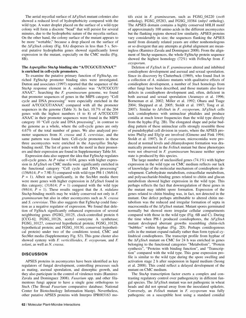
Fig. 8. Hydrophobicity of ΔFgStuA. A, Hydrophobic character of the mu-tant and wild-type cultures grown on V8 agar, measured 15 s after deposi-tion of the water droplet. B, Expression of putative hydrophobin genes on carboxymethylcellulose (CMC), secondary metabolism (SecMet), and wheat; gray signals = not detected.
62 / Molecular Plant-Microbe Interactions
The aerial mycelial surface of ΔFgStuA mutant colonies also showed a reduced level of hydrophobicity compared with the wild type. A water droplet placed on the surface of a wild-type colony will form a discrete “bead” that will persist for several minutes, due to the hydrophobic nature of the mycelia surface. On the other hand, the colony surface of the mutant appears to be more “wettable,” because a drop placed on the surface of the ΔFgStuA colony (Fig. 8A) disperses in less than 5 s. Sev-eral putative hydrophobin genes showed significantly lower expression in the mutant, especially on the CMC media (Fig. 8B).
The Aspergillus StuAp binding site “A/TCGCGT/ANA/C” is enriched in cell-cycle promoters.
To examine the putative primary function of FgStuAp, en-riched FgStuAp promoter binding sites were investigated. Dutton and associates (1997) reported that the sequence of the StuAp response element in A. nidulans was “A/TCGCGT/ ANA/C”. Searching the F. graminearum genome, we found that promoter sequences of genes in the MIPS category “Cell cycle and DNA processing” were especially enriched in the motif A/TCGCGT/ANA/C compared with all the promoter sequences in the genome (Table 3). In fact, 10.7% of the 204 genes with the highest numbers (4 to 8) of A/TCGCGT/ ANA/C in their promoter sequences were found in the MIPS category 10 “Cell cycle and DNA processing”, in contrast to the genome as a whole, where the cell-cycle genes are only 4.67% of the total number of genes. We also analyzed pro-moter sequences from N. crassa and S. cerevisiae, and the same pattern was found here. Cell-cycle promoters in these three ascomycetes were enriched in the Aspergillus StuAp-binding motif. The list of genes with the motif in their promot-ers and their expression are found in Supplementary Table S4.
Expression data also support the idea that FgStuAp regulates cell-cycle genes. At P value = 0.04, genes with higher expres-sion in ΔFgStuA on CMC media were significantly enriched in the functional category “Cell cycle and DNA processing” (156/614; P = 7.9E-5) compared with wild-type PH-1 (36/614; P = 1). Albeit not significantly, in the SecMet media there were more genes with higher expression in ΔFgStuA found in this category; (31/614; P = 1) compared with the wild type (9/614; P = 1). These results suggest that the A. nidulans StuAp-binding motifs may be widely conserved not only in F. graminearum but also in other ascomycetes such as N. crassa and S. cerevisiae. This also suggests that FgStuAp could func-tion as a negative regulator of expression. We found that dele-tion of FgStuA significantly enhanced expression of several neighboring genes (FGSG_10125, clock-controlled protein 6 [CCG-6]; FGSG_10126, acetyl coenzyme A synthetase; FGSG_10127, conserved hypothetical protein; FGSG_10128, hypothetical protein; and FGSG_10130, conserved hypotheti-cal protein) under two of the conditions tested, CMC and SecMet media (Supplementary Fig. S3). This gene cluster also showed synteny with F. verticillioides, F. oxysporum, and F. solani, as well as N. crassa.
DISCUSSION
APSES proteins in ascomycetes have been identified as key regulators of fungal development, controlling processes such as mating, asexual sporulation, and dimorphic growth, and they also participate in the control of virulence traits (Ramirez-Zavala and Dominguez 2008). Fusarium spp. and other fila-mentous fungi appear to have a single gene orthologous to StuA (The Broad Fusarium comparative database; National Center for Biotechnology Information blastp). Nevertheless, other putative APSES proteins with Interpro IPR003163 mo-
tifs exist in F. graminearum, such as FGSG_04220 (swi6 ortholog), FGSG_05283, and FGSG_10384 (mbp1 ortholog). The APSES domain contains a highly conserved bHLH motif of approximately 100 amino acids in the different ascomycetes but the flanking regions showed low similarity. APSES proteins vary considerably in size; the sequences flanking the APSES motif from distantly related yeasts are either nonhomologous or so divergent that any attempts at global alignment are mean-ingless (Ramirez-Zavala and Dominguez 2008). From the align-ment of StuAp sequences, the whole FgStuAp protein sequence showed the highest homology (72%) with FoStuAp from F. oxysporum.
Deletion of FgStuA in F. graminearum altered and inhibited conidiophore development and asexual and sexual sporulation. Since its discovery by Clutterbuck (1969), who found StuA in a collection of A. nidulans mutants with qualitative effects of conidiophore development, a number of StuA homologs from other fungi have been described, and those mutants also have defects in conidiophore development and, often, deficient in both asexual and sexual sporulation (Aramayo et al. 1996; Borneman et al. 2002; Miller et al. 1992; Ohara and Tsuge 2004; Sheppard et al. 2005; Stoldt et al. 1997; Tong et al. 2007). Similar to ΔFoStuA in F. oxysporum, ΔFgStuA cells lack conidiophores with uninuclear phialides and produce conidia at much lower frequencies than the wild type directly from the hypha (Fig. 2B). The elongated shape and polar bud-ding pattern of these uninuclear cells strongly resemble those of pseudohyphal cell division in yeasts, where the APSES pro-teins Phd1p and Efg1p are involved (Gimeno and Fink 1994; Stoldt et al. 1997). In F. oxysporum, microconidia were pro-duced at normal levels and chlamydospore formation was dra-matically promoted in the FoStuA mutant but these phenotypes were not observed in F. graminearum because neither spore state is produced by this species.
The large number of unclassified genes (74.1%) with higher expression in the wild type on CMC medium reflects our lack of knowledge of the molecular mechanism of conidiophore de-velopment. Carbohydrate metabolism, extracellular metabolism, and polysaccharide-binding genes related to chitin and glucan metabolism showed higher expression in the wild type. This perhaps reflects the fact that downregulation of these genes in the mutant may inhibit spore formation. Expression of the genes related to chitin binding was, in fact, not detected in the mutant. One defect perhaps attributable to altered chitin me-tabolism was the reduced and irregular formation of septa in macroconidia of the ΔFgStuA mutant. Mutant spores germinated normally but showed more irregular cellular compartments compared with those in the wild type (Fig. 6B and C). During the time when PH-1 produced conidiophores, the ΔFgStuA mutant developed aberrant growths resembling chitin-rich “bubbles” within hyphae (Fig. 2D). Perhaps conidiogenous cells in the mutant expand radially rather than form typical cy-lindrical condiophores. The transcript profile from hyphae of the ΔFgStuA mutant on CMC for 24 h was enriched in genes belonging to the functional categories ”Metabolism”, “Protein synthesis”, “Proteins with binding function”, and “Transcrip-tion” compared with the wild type. This gene expression pro-file is similar to the wild type during the spore swelling and activation stage 2 h after suspension in liquid medium (Seong et al. 2008). This could reflect a delayed development of the mutant on CMC medium.
The StuAp transcription factor exerts a complex and con-trasting regulatory control over pathogenicity in different fun-gal species. The ΔFgStuA mutant was not pathogenic in wheat heads and did not spread away from the inoculated spikelets. Conversely, an FoStuA mutant in F. oxysporum was fully pathogenic on a susceptible host using a standard conidial

Vol. 24, No. 1, 2011 / 63
inoculation of roots (Ohara and Tsuge 2004). Such a great dif-ference in pathogenicity for stuA mutants in different fungal pathogens may be due to different infection routes or strategies of the different Fusarium spp. Where F. oxysporum invades its host (in tomato) by direct hyphal penetration of the root host cells (Olivain and Alabouvette 1999), F. graminearum pro-duces high concentrations of trichothecene toxins during infec-tion of wheat heads through the epicarp, which leads to early cell death and rapid fungal growth through the different layers of the fruit coat (Jansen et al. 2005). In G. cingulata, a fungus that invades its host by means of an appressorium, the FgStuA homolog GcStuA was shown to be required for pathogenicity on apple (Tong et al. 2007). GcStuA was essential for genera-tion of normal turgor pressure within the appressorium but the mutant without a functional appressorium was still able to in-fect wounded but not unwounded apple fruit. For Candida spp., the APSES protein Efg1 was shown to be involved in ad-herence to and invasion of host cells and virulence in murine models of disseminated and oral candidiasis (Lo et al. 1997; Stoldt et al. 1997). Thus, APSES proteins may be involved in disease development in unique and unpredictable ways, because fungal development genes can impact key virulence factors in many different ways (Lengeler et al. 2000).
Gene expression during infection of wheat was greatly influ-enced by FgStuA. Only 2,638 genes were detected during infec-tion of the ΔFgStuA mutant compared with 6,184 expressed for wild-type PH-1. The lower number of genes detected for the mutant likely reflects a lower proportion of fungal RNA in infected plant tissue, with fungal genes having lower expres-sion levels being below detection limits in the RNA mixtures. Nevertheless, mutants and the wild type had distinct transcript profiles, and expression levels of some genes in the mutant had higher levels than in the wild type. Most of the genes with higher expression levels in the ΔFgStuA mutant (82.3%) were in the category “Unclassified Proteins” (MIPS category 99; P = 4.17E-6). Trichothecene mycotoxins are essential for full virulence and systemic infection of wheat by F. graminearum (Jansen et al. 2005); therefore, a major factor in reduced patho-genicity of the ΔFgStuA mutant is its inability to produce these toxins in planta (Fig. 4D and E) and the complete loss of tricho-thecene biosynthetic gene expression during infection (Fig. 4G). However, loss of toxin production probably is not the only rea-son for loss of pathogenicity because the mutant could not even infect the inoculated spikelet, unlike toxin nonproducing mutants (Jansen et al. 2005). Thousands of genes are ex-pressed at higher levels in wheat by the wild-type strain com-pared with the ΔFgStuA mutant, including genes for predicted pathogenicity factors such as FSR1 (FGSG_01665), SID1 (FGSG_05371), and GzGPA2 (FGSG_09614). However, the sheer number of genes differentially regulated by FgStuA makes it very difficult to distinguish cause from effect. Among the enriched functional categories differentially expressed in PH-1 (Fig. 5) was the category for “Cell rescue, defense, and virulence” (MIPS category 32), where 290/655 genes with higher expression in the wild type are found (P = 1.18E-16) in contrast to 7/655 of the genes with higher expression in the mutant (P = 0.9). Therefore, differences in pathogenicity be-tween the mutant and the wild type may be the cumulative effect of differential regulation of hundreds of genes.
Similar to the case of A. fumigatus, where gene clusters for several secondary metabolites, such as sterigmatacystin, glio-toxin, fumigaclavine, fumitremorgin, pseurotin A, and other unknowns, showed lower expression in an stuA mutant (Gravelat et al. 2008; Sheppard et al. 2005; Twumasi-Boateng et al. 2009), deletion of FgStuA in F. graminearum greatly reduced produc-tion of secondary metabolites. As previously mentioned, tricho-thecenes 15ADON (<1%) and DON (not detected) are greatly
reduced or absent in culture, and do not accumulate in wheat spikelets inoculated with the mutant. Loss of toxins is also strongly supported by gene expression data, especially in wheat, where the entire trichothecene cluster is not detected in ΔFgStuA, in contrast to high expression of these genes in the wild type. Additionally, the red pigment aurofusarin is reduced in the ΔFgStuA mutant on both PDA and V8 agar. In all, 17 ad-jacent genes, including all known aurofusarin-related genes, were highly expressed in the wild type during sporulation (CMC) but were mostly absent or reduced in the ΔFgStuA mu-tant. There is an interesting connection between loss of pig-ment and loss of sporulation in the mutant. A similar connec-tion between sporulation and secondary metabolism has been noted previously (Calvo et al. 2002); however, deletion of the polyketide synthase (PKS12) required for aurofusarin produc-tion in F. graminearum increased conidia production 10-fold (Malz et al. 2005). Here, conidia production and the red pig-ment probably are only stimulated by the CMC medium. All three gene-expression experiments reported here support the idea that secondary metabolism, in general, is reduced in the ΔFgStuA mutant.
Catalase activity was reduced in the spores of the ΔFgStuA mutant, which showed a higher sensitivity to oxidative stress and had lower catalase activity than the wild type. Catalase gene expression also is reduced in the mycelia of the ΔFgStuA mutant, and this might influence its ability to infect by reduc-ing its ability to overcome oxidative stress defense responses by the plant. These results are consistent with reports from others that catalase activity could be regulated by StuAp. In A. fumigatus, the stuA mutant was reduced in expression of cata-lase gene CAT1 in hyphae, and the mutant was markedly more susceptible to oxidative stress (Sheppard et al. 2005). The pu-tative CAT1 homolog in F. graminearum (FGSG_06733) was not found to be differentially expressed (data not shown). Also, the A. nidulans catalase gene cpeA was shown to be reduced in expression in an stuA mutant (Scherer et al. 2002). Two ho-mologous catalase genes in F. graminearum (FGSG_02974 and FGSG_12369; Fig. 7A) were actually increased in expres-sion in the ΔFgStuA mutant during growth in culture but, dur-ing wheat infection, were greatly reduced in the mutant.
Hydrophobins are cell wall proteins required for the produc-tion of aerial hyphae or normal conidiophores in filamentous fungi, and help to overcome surface tension and to prevent desiccation to aerial hyphae (Fuchs et al. 2004). The hydro-phobicity of the ΔFgStuA mutant was clearly reduced because the surface of the mutant mycelia was more wettable than the wild type. It is currently unclear whether the reduced hydro-phobicity reflects differences in the mycelium itself, due to lack of conidia borne on the mycelial surface, or is a result of the stunted aerial hyphae. Several predicted hydrophobin-related genes showed lower expression in the ΔFgStuA mutant, especially on sporulation medium. A G. cingulata StuA dele-tion mutant also showed a more wettable phenotype (Tong et al. 2007), and a StuA deletion mutant in A. fumigatus showed reduced expression of the conidial hydrophobin gene rodB (Sheppard et al. 2005). Reduced accumulation of hydropho-bins also may contribute to attenuation of pathogenicity of the ΔFgStuA mutant by reducing adherence to host tissue, as seen for mutants of the hydrophobin gene MPG1 in M. grisea (Talbot et al. 1996).
A link between nitrogen-limiting conditions and asexual sporulation has been previously observed, and this effect may be mediated by StuAp (Adams et al. 1998; Dahlberg and Vanetten 1982). The FgStuA gene showed constitutive expres-sion in the wild-type strain under all conditions for which there are microarray data, including expression in planta, dur-ing germination of both ascospores and conidia, during peri-

64 / Molecular Plant-Microbe Interactions
thecia development, on complete medium, and during C and N starvation. However, expression levels are especially high dur-ing N starvation. FgStuA also showed increased expression at 72 and 96 h in the wild-type strain in a wheat infection time course, time points which coincide with the appearance of dis-ease symptoms and asexual sporulation on infected plants (Anderson 1948). There is also an overlapping set of genes more highly expressed during sporulation on CMC medium and during growth on N starvation medium (data not shown). In fact, 72% of the probesets with higher expression in wild-type PH-1 during asexual spore production on CMC medium also have higher expression during N starvation.
StuAp appears to exert developmental control by way of negative regulation of genes involved in cell-cycle control. All known StuAp homologues regulate reversible transitions be-tween a spherical cell types (e.g., a budding yeast cell, conidio-phores, ascospores, and chlamydospores) and an elongated filamentous cells (e.g., true hyphae, pseudohyphae, and opaque-form cell) (Doedt et al. 2004). The APSES domain of StuAp in A. nidulans shares significant homology with DNA-binding domains of transcription factors controlling the critical G1/S phase cell-cycle transition in both S. cerevisiae and S. pombe (Dutton et al. 1997). In eukaryotic cells, genes required for DNA replication are periodically transcribed in the cell cycle, peaking at G1-S phase under control of specific transcription factors, such as the Mbp1/Swi6 complex in S. cerevisiae (Koch et al. 1993; Verma et al. 1992). The DNA-binding motif re-quired for StuA response in Aspergillus spp. was also found enriched in the promoter sequences of genes belonging to the functional category “Cell cycle and DNA processing” in the three ascomycetes F. graminearum, N. crassa, and S. cere-visiae. Genes in the MIPS category “Cell cycle and DNA proc-essing” were also found enriched among those genes more highly expressed in the ΔFgStuA mutant compared with the wild type. Because expression was elevated for those genes in the mutant, FgStuAp appears to act as a repressor, similar to what has been found for A. nidulans (Doedt et al. 2004; Dutton et al. 1997).
The profound and pervasive impact of FgStuAp on the de-velopment of F. graminearum has been clearly documented in the ΔFgStuA strain. Under conditions conducive to sporulation or secondary metabolite synthesis or during plant infection, the mutant has a drastically altered phenotype. These pheno-types—reduced sporulation, altered spore morphology, reduced toxin accumulation, and so on—may be explained by modula-tion of gene expression for particular enzymes, structural ele-ments, or biosynthetic pathways that will impact such develop-ment. Because FgStuA affects such a large number of genes and pathways, it makes it difficult to distinguish primary and secondary effects of this developmental regulator. Further work will be required to determine the primary set of genes directly regulated by FgStuAp.
MATERIALS AND METHODS
Strains and culture conditions. The F. graminearum PH-1 and the ΔFgStuA deletion mutant
generated from PH-1 were used in this study. For char-acterization of vegetative growth and asexual development, the following media were used: V8 agar (20% V8 juice, 0.2% CaCO3, and 1.5% Bacto-agar), PDA (Difco, BD, Franklin Lakes, NJ, U.S.A.), CM (Correll et al. 1987), CMC (Cappellini and Peterson 1965), liquid mung bean medium (Bai and Shaner 1996), and MM (Pontecorvo et al. 1953). Carrot agar (20% carrots and 1.5% agar) was used for sexual development and crossings using 1 ml of 2.5% Tween 60 per 20-ml petri dish, as previously described (Pasquali and Kistler 2006). To examine
colony surface hydrophobicity, a 30-µl droplet of dH2O was added to the top of 4-day-old fungal colonies grown on V8 agar and the persistence of the water droplet was examined over time.
Fungal transformation and generation of the ΔFgStuA mutant.
The DNA sequence was analyzed using the MIPS F. graminearum Genome DataBase (Guldener et al. 2006a). The split marker recombination procedure (Catlett et al. 2003), as described by Goswami and associates (2006), was used for gene replacement of FgStuA (FGSG_10129) in the F. grami-nearum PH-1 (NRRL 31084) (Fig. 1A). In total, 22 colonies were selected, 4 had the stunned phenotype common for stuA mutants in other fungi, and 2 of these were selected for South-ern and DNA analysis (Fig. 1B and C). For DNA isolation, the strains were grown on CM and DNA was isolated according to a protocol described by Pasquali and associates (2004). PCR was performed, and the PCR products were purified with QIAquick PCR Purification kit (Qiagen, Valencia, CA, U.S.A.). Protoplast preparation and fungal transformation were performed as described previously (Hou et al. 2002). Transformants were cultivated on V8 juice agar with hygromy-cin B at 250 µg/ml (Calbiochem, La Jolla, CA, U.S.A.) and single-spore isolates were analyzed by Southern (Fig. 1B and C) and PCR (Fig. 1D). Southern hybridization was performed according to a protocol described by Goswami and associates (2006) using 5 µg of DNA digested with PstI. The hph gene fragment amplified with primers HY1F and YG2R and a right-flanking probe amplified with primers 10129-3F and 10129-4R were used to confirm gene deletion of FG10129 and single integration of the hph gene. In addition, PCR was also used for verification of mutants with primers 10129f/r (Fig. 1D) using a rapid microwave treatment of the fungal samples for DNA ex-traction (Tendulkar et al. 2003).
Microarray experiments. Three microarray experiments were performed with the
ΔFgStuA mutant to establish gene expression profiles during asexual (macroconidium) sporulation in CMC medium, during infection of wheat, and during induction of secondary metabo-lites on SecMet medium as described by Miller and MacKenzie (2000). For RNA extraction during sporulation, the cultures were grown in 100 ml of liquid CMC media at 25°C, with con-stant light (60 Lux) and 150 rpm shaking for 24 h. The cul-tures were filtered through Miracloth and the mycelia were washed 10 times with 50 ml of dH2O to remove all spores. Total RNA was then extracted from the filtered sporulating mycelia. For RNA extraction during wheat (Triticum aestivum) infection, cv. Bobwhite was grown as previously described (Goswami and Kistler 2005). All spikelets were point inoculated at anthesis with 10 µl of conidial suspension of PH-1 or the ΔFgStuA mu-tant in 0.01% Triton 60 solution (105 spores/ml). After inocula-tion, the plants were placed in a growth chamber at 16°C for 8 h (night) and 18°C for 16 h (day), and the heads were covered with plastic bags to increase the humidity. The plastic bags were removed after 48 h and the heads were collected after an additional 24 h of growth (total 72 h), and immediately frozen at –80°C prior to RNA extraction. Four heads were pooled per biological replicate. For RNA extraction during SecMet pro-duction, the wild type or ΔFgStuA mutant were cultivated in two consecutive media by a previously described method (Miller and MacKenzie 2000). Total RNA was extracted from the fungal tissue after 3 days of growth on the second medium. The same fungal tissue also was used to analyze trichothecene production (see below: “Mycotoxin and ergosterol analysis”). All the experiments were replicated three times.

Vol. 24, No. 1, 2011 / 65
Total RNA was isolated using TRIzol reagent (Invitrogen, Carlsbad, CA, U.S.A.) and purified with an RNeasy Mini kit (Qiagen) according to the manufacturers’ instructions. Total RNA (10 µg) was labeled according to Affymetrix eukaryotic RNA-labeling protocols (Affymetrix, Santa Clara, CA, U.S.A.). The labeled RNA was hybridized with F. graminea-rum Affymetrix GeneChip (Guldener et al. 2006b), using three biological replications for each experiment. The processing and data acquisition from chips followed standard Affymetrix procedures in use at the Biomedical Image Processing Facility at the University of Minnesota. The resulting CEL files were analyzed with Genedata Expressionist software. The CEL files were normalized using a robust multichip analysis algorithm and all experiments were performed at P = 0.04. A t test was used to analyze differential expression, and only probe sets ex-pressed at greater than or equal to twofold were considered. A present-or-absent test was also performed, where “present” indicated significant signal from the probe sets above back-ground in at least two of the three biological replicate chips and “absent” indicated signal not significantly above back-ground in at least two out of three chips. Genes showing dif-ferential expression patterns based on either the t test or the present-or-absent test (the unique probe sets) are considered as differentially expressed genes. MIPS FunCat was used to ana-lyze functional categories from differentially expressed genes (Ruepp et al. 2004). PCA was performed with correlations of items (probesets) using covariance matrix and 50% valid values in Genedata Expressionist software. Data from the microarray experiments are stored as experiment FG13 at PLEXdb (Wise et al. 2006).
Mycotoxin and ergosterol analysis. Determinations of trichothecene accumulation were per-
formed after growth on SecMet media and after wheat inocula-tions, using cv. Bobwhite. For the SecMet experiments, the fungal cultures were grown as described above, the mycelium suspension was filtered, and 20 ml of the liquid phase were mixed with ethyl acetate on a shaker at 150 rpm for 2 h prior to analysis. For determination of trichothecene production on inoculated wheat heads, the PH-1 or ΔFgStuA mutant were point inoculated on a single spikelet at anthesis, using 10 µl of conidial suspension (106 spores/ml) in 0.01% Triton 60 solu-tion. After inoculation, wheat plants were placed in a humidity chamber for 48 h, then kept in a greenhouse at 20°C for an additional 12 days (total = 14 days). The single inoculated spikelet was analyzed for trichothecenes and was replicated on 10 heads. To determine DON and 15ADON concentration both on SecMet and in wheat, the samples were processed by gas chromatography, mass spectrometry analysis following the method described by Mirocha and associates (1998). Ergo-sterol content was measured on the lyophilized filtered myce-lia obtained after the SecMet growth, according to the protocol described by Dong and associates (2006).
Pathogenicity test. The pathogenicity of PH-1 and the ΔFgStuA mutant on
wheat (cv. Norm) was evaluated as previously described (Goswami and Kistler 2005). One spikelet was either point inoculated at anthesis with 10 µl of conidial suspension (103 spores/µl) or with a 2-mm2 mycelial mat of PH-1 or the ΔFgStuA mutant in 0.01% Triton 60 solution. Mock inocula-tions were conducted in a similar manner with the Triton 60 solution alone. After inoculation, wheat plants were placed in a humidity chamber for 48 h, then kept in a greenhouse at 20°C for an additional 12 days (total = 14 days). Thirty plants were inoculated and the experiment was repeated three times. The pathogenicity was scored by counting the number of spikelets
showing disease symptoms (necrosis or bleaching of palea or lemma) as previously described (Goswami and Kistler 2005). In total, 10 spikelets on each wheat head were evaluated for the presence of disease symptoms by scoring 5 spikelets above and 4 spikelets below the point of inoculation. ΔFgStuA-inocu-lated spikelets were plated on V8 to verify viability of the mu-tant inside the plant tissue.
To examine the ability to colonize apple, 2-mm V8 agar plugs of PH-1 and ΔFgStuA were inoculated on 1-cm apple discs and 2-by-1-by-2-cm pieces of cv. Gala. The spread of the fungus was measured in diameter and the color and softness of the lesion were evaluated to identify degrading activity by the fungus.
Catalase spectrophotometer assay. To analyze catalase activity in macroconidia, spores (1 ×
105/ml) of PH-1 or ΔFgStuA were suspended in 5 mM H2O2. The extinction coefficient of H2O2 at 240 nm is 43.6 M–1 cm–1 (Noble and Gibson 1970). The absorbance at 240 nm was measured every minute for 2 h using a Beckman DU 640 spec-trophotometer (Beckman Coulter, Fullerton, CA, U.S.A.).
Promoter analysis. To investigate enrichment of a specific motif in the promoter
regions for selected genes in F. graminearum and N. crassa, 1 kb upstream of each gene (“genes_upstream_1000”) was ob-tained from the Broad Institute database. For pattern matching, the RSAT program (van Helden 2003), available online, was used with the tool “dna-pattern”. For promoter analysis of S. cerevisiae, we used the tool “genome-scale dna-pattern” (using only 500 nucleotides upstream included in the software). The background frequency of a motif was calculated on the basis of the 1-kb (or 500-bp for S. cerevisiae) nucleotide upstream sequences for all predicted genes in the genome. Enrichment of a motif in the promoter sequences of selected genes, com-pared with the background frequency, was calculated using a χ2 test using the software “R”.
ACKNOWLEDGMENTS
We thank W. Xu and the University of Minnesota, Supercomputing In-stitute for advice and computing resources that made this study possible; K.-Y. Seong for technical suggestions; K. Hilburn (United States Depart-ment of Agriculture [USDA]–Agricultural Research Service Cereal Dis-ease Laboratory) for outstanding technical assistance; S. S. Klemsdal (Bioforsk) for support; H. Divon (Bioforsk) for a critical read through; U. Güldener (Institute of Bioinformatics and Systems Biology, German Re-search Center for Environmental Health) for assistance with MIPS FunCat analysis; and A. di Pietro (University of Cordoba) for providing the apple infection protocol. This project was supported by the National Research Initiative of the USDA Cooperative State Research, Education and Exten-sion Service, grant 2004-35604-14327, and the BILAT project 173277 supported by the Research Council of Norway. M. Pasquali was finan-cially supported by the “Branco Weiss—Society in Science” Fellowship.
LITERATURE CITED
Adams, T. H., Wieser, J. K., and Yu, J. H. 1998. Asexual sporulation in Aspergillus nidulans. Microbiol. Mol. Biol. Rev. 62:35-54.
Anderson, A. L. 1948. The development of Gibberella zeae head blight of wheat. Phytopathology 38:595-611.
Aramayo, R., Peleg, Y., Addison, R., and Metzenberg, R. 1996. Asm-1+, a Neurospora crassa gene related to transcriptional regulators of fungal development. Genetics 144:991-1003.
Bai, G. H., and Shaner, G. 1996. Variation in Fusarium graminearum and cultivar resistance to wheat scab. Plant Dis. 80:975-979.
Borneman, A. R., Hynes, M. J., and Andrianopoulos, A. 2002. A basic he-lix-loop-helix protein with similarity to the fungal morphological regu-lators, Phd1p, Efg1p and StuA, controls conidiation but not dimorphic growth in Penicillium marneffei. Mol. Microbiol. 44:621-631.
Calvo, A. M., Wilson, R. A., Bok, J. W., and Keller, N. P. 2002. Relation-

66 / Molecular Plant-Microbe Interactions
ship between secondary metabolism and fungal development. Micro-biol. Mol. Biol. Rev. 66:447-459.
Cappellini, R. A., and Peterson, J. L. 1965. Macroconidium formation in submerged cultures by a non-sporulating strain of Gibberella zeae. My-cologia 57:962-966.
Catlett, N. L., Lee, B. N., Yoder, O. C., and Turgeon, B. G. 2003. Split-marker recombination for efficient targeted deletion of fungal genes. Fungal Genet. Newsl. 50:9-11.
Clutterbuck, A. J. 1969. A mutational analysis of conidial development in Aspergillus nidulans. Genetics 63:317-327.
Correll, J. C., Klittich, C. J. R., and Leslie, J. F. 1987. Nitrate non-utilizing mutants of Fusarium oxysporum and their use in vegetative compatibil-ity tests. Phytopathology 77:1640-1646.
Cuomo, C. A., Gueldener, U., Xu, J. R., Trail, F., Turgeon, B. G., Di Pietro, A., Walton, J. D., Ma, L. J., Baker, S. E., Rep, M., Adam, G., Antoniw, J., Baldwin, T., Calvo, S., Chang, Y. L., DeCaprio, D., Gale, L. R., Gnerre, S., Goswami, R. S., Hammond-Kosack, K., Harris, L. J., Hilburn, K., Kennell, J. C., Kroken, S., Magnuson, J. K., Mannhaupt, G., Mauceli, E., Mewes, H. W., Mitterbauer, R., Muehlbauer, G., Munsterkotter, M., Nelson, D., O’Donnell, K., Ouellet, T., Qi, W. H., Quesneville, H., Roncero, M. I. G., Seong, K. Y., Tetko, I. V., Urban, M., Waalwijk, C., Ward, T. J., Yao, J. Q., Birren, B. W., and Kistler, H. C. 2007. The Fu-sarium graminearum genome reveals a link between localized polymor-phism and pathogen specialization. Science 317:1400-1402.
Dahlberg, K. R., and Vanetten, J. L. 1982. Physiology and biochemistry of fungal sporulation. Annu. Rev. Phytopathol. 20:281-301.
Doedt, T., Krishnamurthy, S., Bockmuhl, D. P., Tebarth, B., Stempel, C., Russell, C. L., Brown, A. J. P., and Ernst, J. F. 2004. APSES proteins regulate morphogenesis and metabolism in Candida albicans. Mol. Biol. Cell 15:3167-3180.
Dong, Y. H., Steffenson, B. J., and Mirocha, C. J. 2006. Analysis of ergo-sterol in single kernel and ground grain by gas chromatography-mass spectrometry. J. Agric. Food Chem. 54:4121-4125.
Dutton, J. R., Johns, S., and Miller, B. L. 1997. StuAp is a sequence-spe-cific transcription factor that regulates developmental complexity in Aspergillus nidulans. EMBO (Eur. Mol. Biol. Organ.) J. 16:5710-5721.
Frandsen, R. J. N., Nielsen, N. J., Maolanon, N., Sorensen, J. C., Olsson, S., Nielsen, J., and Giese, H. 2006. The biosynthetic pathway for auro-fusarin in Fusarium graminearum reveals a close link between the naphthoquinones and naphthopyrones. Mol. Microbiol. 61:1069-1080.
Fuchs, U., Czymmek, K. J., and Sweigard, J. A. 2004. Five hydrophobin genes in Fusarium verticillioides include two required for microconid-ial chain formation. Fungal Genet. Biol. 41:852-864.
Gimeno, C. J., and Fink, G. R. 1994. Induction of pseudohyphal growth by overexpression of Phd1, a Saccharomyces cerevisiae gene related to transcriptional regulators of fungal development. Mol. Cell. Biol. 14:2100-2112.
Goswami, R. S., and Kistler, H. C. 2004. Heading for disaster: Fusarium graminearum on cereal crops. Mol. Plant Pathol. 5:515-525.
Goswami, R. S., and Kistler, H. C. 2005. Pathogenicity and in planta my-cotoxin accumulation among members of the Fusarium graminearum species complex on wheat and rice. Phytopathology 95:1397-1404.
Goswami, R. S., Xu, J. R., Trail, F., Hilburn, K., and Kistler, H. C. 2006. Genomic analysis of host-pathogen interaction between Fusarium graminearum and wheat during early stages of disease development. Microbiology 152:1877-1890.
Gravelat, F. N., Doedt, T., Chiang, L. Y., Liu, H., Filler, S. G., Patterson, T. F., and Sheppard, D. C. 2008. In vivo analysis of Aspergillus fumigatus developmental gene expression determined by real-time reverse tran-scription-PCR. Infect. Immun. 76:3632-3639.
Guldener, U., Mannhaupt, G., Munsterkotter, M., Haase, D., Oesterheld, M., Stumpflen, V., Mewes, H. W., and Adam, G. 2006a. FGDB: A com-prehensive fungal genome resource on the plant pathogen Fusarium graminearum. Nucleic Acids Res. 34:456-458.
Guldener, U., Seong, K. Y., Boddu, J., Cho, S., Trail, F., Xu, J. R., Adam, G., Mewes, H. W., Muehlbauer, G. J., and Kistler, H. C. 2006b. Devel-opment of a Fusarium graminearum Affymetrix GeneChip for profiling fungal gene expression in vitro and in planta. Fungal Genet. Biol. 43:316-325.
Hou, Z. M., Xue, C. Y., Peng, Y. L., Katan, T., Kistler, H. C., and Xu, J. R. 2002. A mitogen-activated protein kinase gene (MGV1) in Fusarium graminearum is required for female fertility, heterokaryon formation, and plant infection. Mol. Plant-Microbe Interact. 15:1119-1127.
Iyer, L. M., Koonin, E. V., and Aravind, L. 2002. Extensive domain shuf-fling in transcription regulators of DNA viruses and implications for the origin of fungal APSES transcription factors. Genome Biol. 3(3).
Jansen, C., von Wettstein, D., Schafer, W., Kogel, K. H., Felk, A., and Maier, F. J. 2005. Infection patterns in barley and wheat spikes inocu-lated with wild-type and trichodiene synthase gene disrupted Fusarium graminearum. Proc. Natl. Acad. Sci. U.S.A. 102:16892-16897.
Koch, C., Moll, T., Neuberg, M., Ahorn, H., and Nasmyth, K. 1993. A role for the transcription factors Mbp1 and Swi4 in progression from G1 to S phase. Science 261:1551-1557.
Korting, H. C., Hube, B., Oberbauer, S., Januschke, E., Hamm, G., Albrecht, A., Borelli, C., and Schaller, M. 2003. Reduced expression of the hyphal-independent Candida albicans proteinase genes SAP1 and SAP3 in the efg1 mutant is associated with attenuated virulence during infection of oral epithelium. J. Med. Microbiol. 52:623-632.
Lee, T. I., Rinaldi, N. J., Robert, F., Odom, D. T., Bar-Joseph, Z., Gerber, G. K., Hannett, N. M., Harbison, C. T., Thompson, C. M., Simon, I., Zeitlinger, J., Jennings, E. G., Murray, H. L., Gordon, D. B., Ren, B., Wyrick, J. J., Tagne, J. B., Volkert, T. L., Fraenkel, E., Gifford, D. K., and Young, R. A. 2002. Transcriptional regulatory networks in Sac-charomyces cerevisiae. Science 298:799-804.
Lengeler, K. B., Davidson, R. C., D’Souza, C., Harashima, T., Shen, W. C., Wang, P., Pan, X. W., Waugh, M., and Heitman, J. 2000. Signal transduction cascades regulating fungal development and virulence. Mi-crobiol. Mol. Biol. Rev. 64:746-785.
Lo, H. J., Kohler, J. R., DiDomenico, B., Loebenberg, D., Cacciapuoti, A., and Fink, G. R. 1997. Nonfilamentous C. albicans mutants are aviru-lent. Cell 90:939-949.
Malz, S., Grell, M. N., Thrane, C., Maier, F. J., Rosager, P., Felk, A., Albertsen, K. S., Salomon, S., Bohn, L., Schäfer, W., and Giese, H. 2005. Identification of a gene cluster responsible for the biosynthesis of aurofusarin in the Fusarium graminearum species complex. Fungal Genet. Biol. 42:420-433.
McMullen, M., Jones, R., and Gallenberg, D. 1997. Scab of wheat and barley: A re-emerging disease of devastating impact. Plant Dis. 81:1340-1348.
Miller, J. D., and MacKenzie, S. 2000. Secondary metabolites of Fusarium venenatum strains with deletions in the Tri5 gene encoding trichodiene synthetase. Mycologia 92:764-771.
Miller, K. Y., Wu, J. G., and Miller, B. L. 1992. Stua is required for cell pattern formation in Aspergillus. Genes Dev. 6:1770-1782.
Mirocha, C. J., Kolaczkowski, E., Xie, W. P., Yu, H., and Jelen, H. 1998. Analysis of deoxynivalenol and its derivatives (batch and single kernel) using gas chromatography mass spectrometry. J. Agric. Food Chem. 46:1414-1418.
Noble, R. W., and Gibson, Q. H. 1970. The reaction of ferrous horseradish peroxidase with hydrogen peroxide. J. Biol. Chem. 245:2409-2413.
Ohara, T., and Tsuge, T. 2004. FoSTUA, encoding a basic helix-loop-helix protein, differentially regulates development of three kinds of asexual spores, macroconidia, microconidia, and chlamydospores, in the fungal plant pathogen Fusarium oxysporum. Eukaryot. Cell 3:1412-1422.
Olivain, C., and Alabouvette, C. 1999. Process of tomato root colonization by a pathogenic strain of Fusarium oxysporum f. sp. lycopersici in com-parison with a non-pathogenic strain. New Phytol. 141:497-510.
Pasquali, M., and Kistler, H. C. 2006. Gibberella zeae ascospore produc-tion and collection for microarray experiments. J. Vis. Exp. Published online. doi: 10.3791/115.
Pasquali, M., Marena, L., Fiora, E., Piatti, P., Gullino, M. L., and Garibaldi, A. 2004. Real-time polymerase chain reaction for identification of a highly pathogenic group of Fusarium oxysporum f. sp. chrysanthemi on argyranthemum frutescens L. J. Plant Pathol. 86:53-59.
Pontecorvo, G., Roper, A. B., Hemmons, L. M., MacDonald, K. D., and Bufton, A. W. 1953. The genetics of Aspergillus nidulans. Adv. Genet. 5:141-238.
Ramirez-Zavala, B., and Dominguez, A. 2008. Evolution and phylogenetic relationships of APSES proteins from Hemiascomycetes. FEMS (Fed. Eur. Microbiol. Soc.) Yeast Res. 8:511-519.
Ruepp, A., Zollner, A., Maier, D., Albermann, K., Hani, J., Mokrejs, M., Tetko, I., Guldener, U., Mannhaupt, G., Munsterkotter, M., and Mewes, H. W. 2004. The FunCat, a functional annotation scheme for systematic classification of proteins from whole genomes. Nucleic Acids Res. 32:5539-5545.
Scherer, M., Wei, H. J., Liese, R., and Fischer, R. 2002. Aspergillus nidu-lans catalase-peroxidase gene (cpeA) is transcriptionally induced during sexual development through the transcription factor StuA. Eukaryot. Cell 1:725-735.
Schmit, J. C., and Brody, S. 1976. Biochemical genetics of Neurospora crassa conidial germination. Bacteriol. Rev. 40:1-41.
Seong, K. Y., Zhao, X., Xu, J. R., Guldener, U., and Kistler, H. C. 2008. Conidial germination in the filamentous fungus Fusarium graminea-rum. Fungal Genet. Biol. 45:389-399.
Sheppard, D. C., Doedt, T., Chiang, L. Y., Kim, H. S., Chen, D., Nierman, W. C., and Filler, S. G. 2005. The Aspergillus fumigatus StuA protein governs the up-regulation of a discrete transcriptional program during the acquisition of developmental competence. Mol. Biol. Cell 16:5866-5879.
Sohn, K., Urban, C., Brunner, H., and Rupp, S. 2003. EFG1 is a major

Vol. 24, No. 1, 2011 / 67
regulator of cell wall dynamics in Candida albicans as revealed by DNA microarrays. Mol. Microbiol. 47:89-102.
Staib, P., Kretschmar, M., Nichterlein, T., Hof, H., and Morschhauser, J. 2002. Transcriptional regulators Cph1p and Efg1p mediate activation of the Candida albicans virulence gene SAP5 during infection. Infect. Im-mun. 70:921-927.
Stoldt, V. R., Sonneborn, A., Leuker, C. E., and Ernst, J. F. 1997. Efg1p, an essential regulator of morphogenesis of the human pathogen Candida albicans, is a member of a conserved class of bHLH proteins regulating morphogenetic processes in fungi. EMBO (Eur. Mol. Biol. Organ.) J. 16:1982-1991.
Talbot, N. J., Kershaw, M. J., Wakley, G. E., de Vries, O. M. H., Wessels, J. G. H., and Hamer, J. E. 1996. MPG1 encodes a fungal hydrophobin involved in surface interactions during infection-related development of Magnaporthe grisea. Plant Cell 8:985-999.
Tendulkar, S. R., Gupta, A., and Chattoo, B. B. 2003. A simple protocol for isolation of fungal DNA. Biotechnol. Lett. 25:1941-1944.
Tong, X. Z., Zhang, X. W., Plummer, K. M., Stowell, K. M., Sullivan, P. A., and Farley, P. C. 2007. GcSTUA, an APSES transcription factor, is required for generation of appressorial turgor pressure and full patho-genicity of Glomerella cingulata. Mol. Plant-Microbe Interact. 20:1102-1111.
Twumasi-Boateng, K., Yu, Y., Chen, D., Gravelat, F. N., Nierman, W. C., and Sheppard, D. C. 2009. Transcriptional profiling identifies a role for BrlA in the response to nitrogen depletion and for StuA in the regula-
tion of secondary metabolite clusters in Aspergillus fumigatus. Eukaryot. Cell 8:104-115.
van Helden, J. 2003. Regulatory sequence analysis tools. Nucleic Acids Res. 31:3593-3596.
Verma, R., Smiley, J., Andrews, B., and Campbell, J. L. 1992. Regulation of the yeast DNA replication genes through the MluI cell cycle box is dependent on Swi6. Proc. Natl. Acad. Sci. U.S.A. 89:9479-9483.
Ward, M. P., Gimeno, C. J., Fink, G. R., and Garrett, S. 1995. Sok2 may regulate cyclic Amp-dependent protein kinase-stimulated growth and pseudohyphal development by repressing transcription. Mol. Cell. Biol. 15:6854-6863.
Wise, R., Caldo, R., Hong, L., Wu, S., Cannon, E., and Dickerson, J. 2006. PLEXdb: A unified expression profiling database for plants and plant pathogens. (Abstr.) Phytopathology 96:S161.
AUTHOR-RECOMMENDED INTERNET RESOURCES
The MIPS Fusarium graminearum genome database: mips.gsf.de/genre/proj/FGDB
Genedata website: www.genedata.com Plant Expression database (PLEXdb): www.plexdb.org Broad Institute website: www.broad.mit.edu Regulatory Sequence Analysis Tools (RSAT) website: rsat.ulb.ac.be/rsat The R Project for Statistical Computing website: www.r-project.org