the toxicity of paraquat - occupational and …oem.bmj.com/content/oemed/23/2/126.full.pdf ·...
TRANSCRIPT

Brit. J. industr. Med., i966, 23, i26
The Toxicity of ParaquatD. G. CLARK, T. F. McELLIGOTT, and E. WESTON HURST
From Imperial Chemical Industries Limited, Industrial Hygiene Research Laboratories,Alderley Park, Nr. Macclesfield, Cheshire
Samples of paraquat dichloride and paraquat dimethosulphate are equitoxic when the LD50 is expressedas mg. paraquat ion/kg. body-weight. There are wide species variations in the LD5o and, of course,variations according to the route of administration in a single species.The pathological lesions attributable to paraquat are described in some detail. Among the most unusual
is a peculiar proliferative condition in the lungs, which in an extreme case and in many parts can hardlybe recognized as consisting of pulmonary tissue. With slight variations, the same microscopical picturemay be seen in the rat, mouse, dog, and man, and less often in the rabbit. The experimental evidencesuggests that once the condition is initiated it often proceeds in the absence of further exposure to paraquatuntil it becomes lethal.There is evidence that much of the mortality resulting from dermal application of paraquat in the rabbit
is caused not by percutaneous absorption but by oral contamination from the stratum corneum. Thisleads to glossitis and oesophagitis and an inability or unwillingness to eat.
In this paper we report the effects of paraquatadministered to laboratory animals as the dichlorideor dimethosulphate, either as a single dose by one orother route, or over a short term of repeated doses.Observations made by others on the effects of veryexceptional cases of poisoning in man, arising fromaccidental imbibition of a solution of the substancerather than from its use in the field, suggest thatsimilar pathological changes may be expected inhuman beings.
Chemical Constitution and Uses
Paraquat is a dipyridylium compound. Theformula and chemical nomenclature of the di-chloride are given below.
H3C-N+ +N-CH 3 [2.C1-]
I,I'-dimethyl-4,4'-dipyridylium dichloride
One main use of the dichloride and dimethosulphateis as weed-killers. They are effective only if sprayedon to the leaves of plants (more so in light than inthe dark), so that by judicious management weedsmay be eradicated while adjacent taller plants
remain unaffected. They also destroy the foliage ofcrops such as cotton and potatoes, thus renderingsimpler their harvest by modem techniques. Incontact with the soil they are rapidly inactivated andare no longer available to plants. They can there-fore be used for the control of weeds in a greatvariety of row-crops and have extensive applicationin pasture renovation. Their use can eliminate theneed for ploughing in the cultivation of annualcrops. Consideration of their physico-chemicalcharacteristics suggests that they are dissociated atall pH values; the paraquat ion alone, therefore,should determine the toxic effects, and in fact thisis the case.
Methods
Usually we used samples of paraquat dichloride ordimethosulphate, some of them 99 9% pure, dissolvedin water or isotonic saline and administered by a varietyof routes to animals which had free access to food andwater. On occasion we incorporated the substance in thediet, from which we could achieve quantitative recovery.In the studies on the effects of paraquat on the skin,however, we used a commercial preparation suchas isemployed in the field. In subacute toxicity tests, weattempted to examine histopathologically every organ inthe body, including not fewer than five levels of thealimentary canal.When administered in the food, we dissolved the salt
of paraquat in water and mixed it with the usual dietI26
Received for publication September I4, I965.
copyright. on 17 S
eptember 2018 by guest. P
rotected byhttp://oem
.bmj.com
/B
r J Ind Med: first published as 10.1136/oem
.23.2.126 on 1 April 1966. D
ownloaded from
The Toxicity of Paraquat
obtained from the manufacturers in the form of apowder. After adding 20% extract of malt, we extrudedthe diet through a sausage-meat machine and dried it ina vacuum-oven. We prepared a control diet similarly,except that we did not add a salt of paraquat.When applied to the skin, the toxicity of paraquat is
complicated by several factors best considered in thedescription of the results obtained. We followed astandard technique throughout. After clipping the backsof the animals on the day before beginning the test,avoiding of course any damage to the skin, we appliedthe material in a volume of i ml./kg. body-weight. Asthis volume remained constant, the concentration ofparaquat in mg./ml. is equal to the dose rate in mg./kg.We calculated LDsos and the confidence limits by
the moving-average interpolation method (Thompson,I947). Throughout this paper they appear as mg.paraquat ion/kg. body-weight.
Results
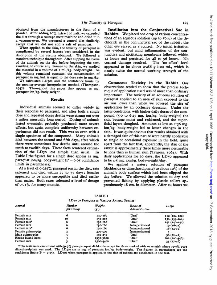
Individual animals seemed to differ widely intheir response to paraquat, and after both a singledose and repeated doses deaths were strung out overa rather unusually long period. Dosing of animalsfasted overnight probably produced more severeeffects, but again complete uniformity between ex-periments did not result. This was so even with asingle specimen of the compound. Many animalsdied between the second and fifth days, after whichthere were sometimes few deaths until around thetenth to twelfth days. These facts rendered estima-tion of the LD5o less simple than usual. InTable I the figures for a single dose appear as mg.paraquat ion/kg. body-weight (P = o0o5 confidencelimits in parentheses).At a level of o0025% paraquat ion in the diet, rats
sickened and died within 27 to 57 days; femalesappeared to be more susceptible and died earlierthan males. Both sexes tolerated a level of dosageof o-oi% for many months.
Instillation into the Conjunctival Sac inRabbits We placed one drop of various concentra-tions of an aqueous solution (up to 50%) of the di-chloride in the conjunctival sac of the rabbit; theother eye served as a control. No initial irritationwas evident, but mild inflammation of the con-junctiva and nictitating membrane followed withinI2 hours and persisted for 48 to 96 hours. Nocorneal damage resulted. The 'no-effect' levelappeared to be above o-286 g./ioo ml., which isnearly twice the normal working strength of thesolution.
Cutaneous Toxicity in the Rabbit Ourobservations tended to show that the precise tech-nique of application used was of more than ordinaryimportance. The toxicity of an aqueous solution ofparaquat applied to an area of skin exposed to theair was lower than when we covered the site ofapplication by an occlusive dressing. Under thelatter conditions, with higher daily doses of the com-pound (5-o to 6-25 mg. ion/kg. body-weight) theskin became moist and reddened, and the super-ficial layers sloughed. Amounts as low as I-6 mg.ion/kg. body-weight led to lesser changes in theskin. It was quite obvious that results obtained witha damaged skin of this nature were hardly applicableto single or occasional exposure in the field, quiteapart from the fact that, apparently, the skin of therabbit is approximately three times more permeableto ions than is human skin (Tregear, I964).. Withdaily applications for 20 days, the LDso appearedto be 4-5 mg. ion/kg. body-weight/day.We applied a watery solution of paraquat
(dichloride or dimethosulphate) to about io% of theanimal's body surface which had been clipped theday before. We allowed the solution to dry andprevented licking by applying plastic collars ap-proximately i8 cm. in diameter. After 24 hours we
TABLE ILD50 OF PARAQUAT IN VARIous ANIMAL SPECIES
Animal
Female ratsFemale ratsFemale rats*Female ratsFemale rats*Female guinea-pigsMale guinea-pigsRhode Island hensFemale cats
Numberper Group
6I06663553
Weight(g.)I30-I60150-205130-I60I30-160130-I60400-500190-250
1950-30002500-4400
Route ofAdministration
'Oral''Oral''Oral'IntraperitonealIntraperitonealIntraperitoneal'Oral''Oral''Oral'
LD5o
112 (IO4-122)I50 (139-I62)I4I ( I40-I42)I9 (I6-2I)i6 (14-I9)330 (22-4I)
262 (200-346)35 (27-46)
*The tests were carried out with 99.9% pure paraquat dichloride except for those marked with an asterisk where 99 9% puredimethosulphate was used. The LDsos are in mg. of paraquat ion/kg. body-weight. Tne figures in parentheses are theconfidence limits (P = o os). LD os when paraquat is applied to the skin of rabbits are considered in the text.
I27
copyright. on 17 S
eptember 2018 by guest. P
rotected byhttp://oem
.bmj.com
/B
r J Ind Med: first published as 10.1136/oem
.23.2.126 on 1 April 1966. D
ownloaded from

D. G. Clark, T. F. McElligott, and E. Weston Hurst
washed the area of application with water and driedit carefully. We then removed the collar. In sub-acute tests applications were continued for 20 days,and we observed the animals for a further two weeks.An approximate LD5o for a single application was236 mg. ion/kg. body-weight, and the LD50 in thesubacute test was 7 to I4 mg. ion/kg. body-weight/day. Neither clinical nor pathological abnormalitieswere apparent after repeated applications of2-8 mg. ion/kg., more than twice the normal workingconcentration of 0-I2%.
Signs of Poisoning after IntraperitonealDosing After a single large intraperitoneal dose(30 tO 75 mg. ion/kg.) in rats, the signs of poisoningvaried somewhat from animal to animal: mostpointed to an action of the substance on the centralnervous system. In the earlier stages, hyper-excitability, violent forced movements flinging theanimal about the cage, or a stiff and incoordinategait might be present. Spasms might occur, or thelimbs might be widely splayed. A rolling gait mightcontinue up to the time of death which, at the levelsof dosage employed, usually occurred on or beforethe fifth day. Additionally, in the earlier stagesbreathing might be gasping or, alternatively, deepand fast. Some days after a dose respiration becameincreasingly laboured. Over-secretion of theharderian glands was usually a feature.
In subacute experiments, tremor and hyper-aesthesia, and less frequently excessive harderiansecretion, might be apparent during and soon afterthe period of dosing. In animals which survived fora number of days after the last of a series of ascend-ing doses, the later signs of illness were chieflydifficult breathing, accompanied on occasion by aclicking or similar sound and by soiling of the hairaround the mouth and nares by a brownish fluid.
Signs of acute poisoning in the mouse were notdissimilar.
Sick rabbits did not eat or drink: they might behyperaesthetic or show a creamy exudate around thereddened eyelids. More often than not, however,the animal was dead in its cage one morning withouthaving shown more than inappetence on the pre-vious day. The histological abnormalities in a verylimited number of animals were not sufficientlyconstant or severe to account for death.
Post-mortem Findings after Intraperitonealor Oral Dosing At necropsy, after a single largeor a few smaller doses by whatever route, the lungsmight be very congested or show early 'consolida-tion'; four rabbits examined, however, showed slightor no evidence of severe pulmonary disorder. Somerats had a thymus smaller than normal for their age
and a small, pale spleen. Two of the four rabbitsexamined had small, pale spleens; they were too oldto have had a large thymus at the beginning of theexperiment.The rats and mice with laboured respiration four
or five days after a single dose, or at the end of asubacute experiment, all showed various degrees ofconsolidation of the lungs, which usually were plum-coloured and often sank in water. If they floated,there often escaped from a cut surface long redthreads which sank to the bottom of the bowl; thesewere composed of blood which seemed to be morethan normally coherent. Sometimes a frothy fluidexuded from the trachea.
After continued dosing with a concentration of0-025% paraquat ion in the diet, rats began to diewith respiratory signs after about 27 days. In someof these animals the appearance of the lungs wasthat described above. In others there was less con-gestion, and, instead of being plum-coloured,affected areas of the lung were greyish and fleshy;they gave the impression of being airless and theysank in water.
Histological Appearances
In the Rat, Mouse and Rabbit after Intraperitonealor Oral Dosing We directed the histological studychiefly to the lesions in the rat. Those in the mousewere reasonably similar, but in only one rabbit didwe observe definite changes (other than vascularcongestion) in the lungs, changes which were socharacteristic a feature in the other species.The most characteristic lesion found in the rats
which died after the administration of paraquat wasthat in the lungs. We followed all stages of thislesion up to the final state of near-solidity of muchof the lung substance. The symptomatology and themicroscopical picture both suggested that inanimals which died five to nine days after a dose ordoses of paraquat, impaired pulmonary functioncontributed materially to the fatal outcome.
In the majority of animals the lungs were verycongested, with oedema fluid in many of the alveoliand an excess ofmacrophages in others. Perivascularand peribronchial oedema also occurred, particularlyin the earlier stages; later, proliferating cells were sonumerous that they left no space for fluid in thesesituations. This cellular proliferation began asearly as the third day, and at first the cells appearedas more or less typical fibroblasts arising from theadventitia of the vessels and the fibrous tissuearound the bronchi (Fig. i). Polymorphonuclearleucocytes, some karyorrhectic, mononuclears, andmacrophages were present, and in some animalslarge numbers of eosinophils.
128
copyright. on 17 S
eptember 2018 by guest. P
rotected byhttp://oem
.bmj.com
/B
r J Ind Med: first published as 10.1136/oem
.23.2.126 on 1 April 1966. D
ownloaded from

~~*a- -. Xi.& b.w.;.4 -...
g;,4 1 ttr* t,46~~~~ ^ ~ -;jF!K ..:u.s .,.Jk 4.:4~~~~~~~~~~~~~~~~~~~~~~~4
,Nr-4mV.~~~ ~ ~ ~ ~ ~ ~ ~ ~ ~ ~ 4
4~~** | < n _ 0 0 ~~~~~~~~~~~~~~~~~. s j d ,
P~~~~~~~~~~
41'1~~ ~ ~ ~ ~ 4
Jp!'~~~~~~~~~~~~V!*.4/L*'SiSe,iftstz, t tW i b x J t t
'.^ .i-
v.S w s; :zM % .R tx 's: e W.x. s dx-G. ts*;,~~~~~~~~~~~,awtlw ^;r; A a v i Su, ,i e k > X>~~~~~v-A
FIG. I. Proliferation of fibroblasts around the vessels and bronchi in the earlier stages of reaction toparaquat in rats. Haematoxylin and eosin, X 250.
':u. 2. rrouse.eosin, x 250.
ration and swelling of alveolar (or septal) cells in subacute poisoning with paraquat. Haematoxylin and
copyright. on 17 S
eptember 2018 by guest. P
rotected byhttp://oem
.bmj.com
/B
r J Ind Med: first published as 10.1136/oem
.23.2.126 on 1 April 1966. D
ownloaded from

~~~~~~~~~ VFIG. 3. Proliferation of the epithelium of the terminal bronchi. Haematoxylin and eosin, X 250.
.e M '0e..#. t'ti Xt''t'~~~~~~~
FIG. 4. Most of the lung of this animal was solid and sank in water. ematoxylin and eosin., X 250.
copyright. on 17 S
eptember 2018 by guest. P
rotected byhttp://oem
.bmj.com
/B
r J Ind Med: first published as 10.1136/oem
.23.2.126 on 1 April 1966. D
ownloaded from

The Toxicity of Paraquat
As the days went by, the cellular proliferationincreased and spread into the alveolar walls, whereit was both diffuse and accentuated in whorls. Thecells were now much smaller and spindle-shaped orelongated and no longer characteristically fibro-blastic. Mitoses were very numerous. The epi-thelial cells of the alveoli (or the septal cells) alsoparticipated in the proliferative process (Fig. 2), andsome animals showed tiny duct-like structuresapparently derived from these cells. In otheranimals larger spaces were lined by proliferatedepithelium from the terminal bronchioles (Fig. 3).The extreme picture was seen in rats on long-termfeeding; large areas of the lung were solid and con-sisted of massed cells with no air-containing cavitiesbetween (Fig. 4). Both at this late stage and at anearlier stage with much pulmonary oedema, theremaining air-containing areas of the lungs tendedto show distended alveoli and alveolar ducts.The foregoing describes the appearances in the
majority of animals. In some, however, the epi-thelial proliferation, whether of alveolar cells or ofterminal bronchial epithelium, predominated in theabsence of great fibroblastic overgrowth. In someanimals also, a few giant cells of different types werepresent. Over a series of animals, therefore, thehistological pictures were very diverse.Once started, the consolidation of the lungs
appeared to be progressive in the absence of furtherdosing with paraquat: in some subacute tests theanimals appeared reasonably well at the time dosingceased, only to develop embarrassed respiration andto die five to seven days later.By comparison with the lungs, other organs were
relatively lightly affected.The liver was usually that of a fasting animal.
There might be some vacuolation of liver cellsaround the central veins, and a very few isolatednecrotic cells here or mid-zonally (five out of I3 ratsexamined histologically).The kidney of one rat showed occasional necrotic
cells in the proximal convoluted tubules. In IO ofI3 rats a variable number of other tubules, probablythe distal convoluted tubules, were lined by greatlyswollen cells with 'empty' cytoplasm and pyknoticnuclei. The remains of such cells sometimes layloose in some of the tubules of Henle.The adrenals were congested. The thymus (eight
out of I3 rats) and the spleen (one out of I2 rats)showed some lympholysis.
In the testis some seminiferous tubules showeddegenerative changes, with the formation of smallmultinucleated giant cells lying free in the lumen(two out of five rats).Mice which died at six or seven days, after the last
of a series of doses, showed a not dissimilar picturein the lungs, but the leucocytic element in theexudate was more pronounced. In many of thelarger arteries and veins polymorphs existed in verylarge numbers; they tended to collect as a peripherallayer several cells deep, leaving the red blood cor-puscles free ofleucocytes in the central stream. Theyalso occurred in large numbers around some of thevessels and bronchi, in the alveolar walls, and freein the alveoli. The last might suggest an infectiveprocess, but the overall picture in a number ofanimals was quite different. In the most severelyaffected animal, many polymorphs were karyor-rhectic, and in one or two places the alveolar wallsseemed to have disintegrated. Around some, butnot all, of the blood vessels oedema was marked, andearly proliferation of fibroblasts was seen. All thelungs were intensely congested.
Degeneration of occasional renal tubules occurredin the only mouse examined from this point of viewand was similar to that already described in the rat.By contrast with the rat and the mouse, the rabbit
displayed much less tendency to involvement of thelungs; indeed, only one of four animals examinedshowed any sign of cellular proliferation, althoughthe lungs uniformly were greatly congested in theseclinically severely affected or moribund animals. In-constant and not very severe changes included somenecrosis in the liver, necrosis of a few cells in theproximal convoluted tubules in the kidney, degen-eration of a few renal tubules similar to that observedin rats and mice, necrosis of cells in the outermostzone of the adrenal cortex, a mild degree of testiculardegeneration, and some lympholysis in the thymus.
In the Dog Experiments on dogs carried out inthe United States by Industrial BIOTEST Labora-tories, Inc. led in the lungs of some animals to acondition described as 'fibrosing pneumonitis'.Through the courtesy of Dr. J. C. Calandra we wereprivileged to see sections from these animals. Inour opinion, the condition present was essentiallysimilar to that in our rats fed for a long period witha low concentration of paraquat in the diet.
In Man Three cases of poisoning in man, none'occupational', were brought to our notice. Allfollowed accidental ingestion of a solution of para-quat. Through the courtesy of Dr. E. E. Doyle andMr. G. A. McL. Lee we were able to examine thelungs of a patient who died in Ireland, and throughthe courtesy of Dr. C. M. Bullivant we examinedlungs from patients who died in New Zealand.Here again the pulmonary lesions closely resembledthose in the rats fed paraquat. The precise amountof paraquat ingested and retained in the body in
131
copyright. on 17 S
eptember 2018 by guest. P
rotected byhttp://oem
.bmj.com
/B
r J Ind Med: first published as 10.1136/oem
.23.2.126 on 1 April 1966. D
ownloaded from

132 D. G. Clark, T. F. McEII
these cases was difficult to ascertain, but it mighthave been several hundred milligrams per kilogrambody-weight.
Signs of Poisoning and Pathology in theRabbit after Dermal Application Applied tothe skin of the rabbit paraquat led to a differentclinical picture. The most striking effect, after alatent period, was the secretion of copious amountsof brownish saliva. Rabbits so affected refused toeat, and death occurred in a state of cachexia. Insome of these animals renal tubular damage andpulmonary changes appeared as before. In others,the pulmonary changes were pronounced; thealveolar walls were thickened and contained a mixedcell infiltrate in which plump histiocytes were con-spicuous. In a minority the pulmonary changesresembled those in the rat but, although greatlythickened alveolar walls obliterated alveoli andcaused consolidation in some areas, there was littleevidence at this stage that the initial change wasperivascular or peribronchial; the interstitial tissueof the lung was diffusely involved. At low doses thepulmonary changes were often of doubtful signifi-cance and in some circumstances could reasonablybe accepted as being within normal limits.At first the significance of the profuse salivation
was not recognized, but it was soon evident that thisphenomenon was associated with glossitis and evenulceration of the tongue and oesophagus. Oral con-tamination did not appear to be taking place, butfurther investigations, not reported here in full,showed that excess salivation and glossitis did notoccur when oral contamination was absolutely ex-cluded by leaving the plastic collars in position afterwashing the sites of application and, in addition, bycovering the washed and dried skins with dryporous dressings. Conversely, when the rabbitswere given drinking water containing 286 p.p.m. ofparaquat dichloride (i:I000 dilution of the com-mercial preparation supplied) they developed glos-sitis within two days and then rapidly lost weight.
In order to assess the effect of early decontamina-tion of the skin following exposure to paraquat, wewashed the rabbits' skins with water after an applica-tion of 280 mg. ion/kg. body-weight so as to give anexposure varying from I5 minutes to four hours. Wethen returned these rabbits to their cages withouttheir plastic collars. Lesions definitely attributableto paraquat were not apparent either in the lungs orin the kidneys, but glossitis and oesophagitisoccurred, and anorexia and loss of weight werepronounced. It was assumed that, even after decon-tamination, there was a reservoir of paraquat in theskin and that persistent oral contamination occurredthrough grooming during the period of observation.
igott, and E. Weston Hurst
Consequently, it is believed that following skinapplication death, in many cases, was due to wastingfollowing an inability to eat, and that this resultedfrom lingual and oesophageal ulceration or inflam-mation caused by continued oral contaminationfrom a skin reservoir. Even during the subacute20-day test animals were exposed daily to thepossibility of oral contamination because, afterwashing and drying the skin, we left them withoutcollars until the time of the next application.
Discussion
For the most part this report concerns thetoxicity of paraquat in small laboratory animals, andparticularly in the rat and mouse. The pathologicalfeatures described seem also to be common topoisoning in the dog and in man. Apart from theeffect of large doses on the central nervous system,two points merit attention.One is that often the substance gives rise in the
lungs to a peculiar lesion which, betraying at firstbut little evidence of its presence, progresses in theabsence of further dosing to a point incompatiblewith life. As this condition may result from a singledose of paraquat, and as Daniel and Gage (I966),using the substance labelled with 14C, have found itpoorly absorbed and in any case quickly excreted inthe urine, we cannot consider the characteristiclesions as due to a cumulative effect of the herbicide.Rather, the compound initiates irreversible changesin the pulmonary tissues, but these are not recog-nized clinically until they are sufficiently advancedto bring the animal nearly to the point of death.Several analogous phenomena are known in thefield of experimental (and human) pathology.The second point is the behaviour of paraquat
when applied to the skin of the rabbit. It is wellknown that the stratum corneum can act as a skinreservoir for applied substances, as the experimentsof Vickers (I964) have so elegantly shown. This isespecially probable in the case of paraquat, which isreadily adsorbed by other materials. It is thus avail-able for continual application to the tongue duringnormal grooming, with consequent glossitis andfailure to eat. It is unlikely to be available forabsorption percutaneously, since the keratinouslayer is shed.
REFERENCES
Daniel, J. W., and Gage, J. C. (I966). Brit. J. industr. Med.,23, I33-
Thompson, W. R. (1947). Bact. Rev., iI, 15.Tregear, R. T. (I964). In Progress in the Biological Sciences
in Relation to Dermatology, Vol. 2, P. 275. Ed. Rook,A., and Champion, R. H. University Press, Cambridge.
Vickers, C. F. H. (I964). Ibid., p. 29I.
copyright. on 17 S
eptember 2018 by guest. P
rotected byhttp://oem
.bmj.com
/B
r J Ind Med: first published as 10.1136/oem
.23.2.126 on 1 April 1966. D
ownloaded from