the toxicity of dodax:mo nanocarriers supplementary ... · the toxicity of dodax:mo nanocarriers in...
TRANSCRIPT

1
Supplementary Information
Effect of counter ion and combination of constituents on
the toxicity of DODAX:MO nanocarriers in vitro and in vivo
Ana Cristina Norberto Oliveiraa,b, Marisa Passos Sárriac, Pedro Moreiraa, Joana
Fernandesa, Lisandra Castroa, Ivo Lopesa,b,d, Manuela Côrte-Reala, Artur Cavaco-
Pauloe, Maria Elisabete Cunha Dias Real Oliveirab, Andreia Castro Gomesa,f
aCBMA (Center of Molecular and Environmental Biology), Department of Biology, University of
Minho, Campus of Gualtar, 4710-057 Braga, Portugal
bCFUM (Center of Physics), Department of Physics, University of Minho, Campus of Gualtar, 4710-057
Braga, Portugal
cINL - International Iberian Nanotechnology Laboratory, Av. Mestre José Veiga, 4715-330 Braga,
Portugal
dNanodelivery-I&D em Bionanotecnologia Lda., Department of Biology, University of Minho, Campus
of Gualtar, 4710-057 Braga, Portugal
eCEB, Department of Biological Engineering, University of Minho, Campus of Gualtar, 4710-057
Braga, Portugal
(f) To whom correspondence should be addressed: E-mail: [email protected], Department of Biology,
University of Minho, Campus of Gualtar, 4710-057 Braga, Portugal. Tel: Tel: +351 253 601 511, Fax: +351
253 678 980
Electronic Supplementary Material (ESI) for Toxicology Research.This journal is © The Royal Society of Chemistry 2016

2
Zebrafish embryogenesis statistical analysis
One-way ANOVA results on zebrafish embryos epiboly percentage at 8 hpf showed no significant
interaction among different DODAX:MO ratios of liposomes. In fact, with exception of 9 µM of
DODAX, which caused a significant decrease of the epibolic arc percentage (comparatively with
control group: 0 µM DODAX), no significant interaction was observed among groups, for both the
free-compounds and liposomes (see Table S1).
When considering the effects of DODAX surfactants (administered as free lipids) on the HTA of 32
hpf zebrafish, the statistical results showed a significant interaction between the considered factors
(Table S1). An increase in DODAX concentration is translated into larger angles, causing atypical
acceleration of the embryonic development. On the contrary, MO did not affect the HTA normal
pattern. A similar pattern was also visible for the DODAX:MO liposomes.
The cardiac rhythm of zebrafish embryos exposed to the individual surfactants did not differed
significantly from the control groups up to 32 hpf (Table S1). Nevertheless, regarding DODAX:MO
liposomes, the heartbeat of pre-hatched larvae increased, particularly when exposed to C:M(2:1)
and C:M(4:1) liposomes. Interestingly, a normal cardiac rhythm appears to be restored after
exposure to increasing MO proportions for the molar fractions DODAX:MO (1:1), (1:2) and (1:4).
ANCOVA results on the effects of DODAX:MO liposomes in zebrafish embryos yolk volume
(adjusting for egg size), showed no significant interaction independently of DODAX:MO molar
fractions (Table S1). SNK showed that the tested DODAB concentrations did not cause significant
alterations in comparison to the control. Although a similar profile was visible with DODAC, the post-
hoc test (data not shown) detected differences between the concentration levels. An opposite trend
is observed for DODAC 9 µM, with an increasing yolk size. Presence of MO did not affect the
zebrafish yolk parameter.
The eye surface of zebrafish larvae exposed to DODAB up to 56 hpf did not differ statistically
from the control group, although a decreasing concentration-dependent tendency was recorded
(Table S1). At 56 hpf, only zebrafish larvae exposed to the lowest concentration of DODAC survived,

3
exhibiting a significant reduction of eye surface in comparison to the control group (Table S1). No
phenotypic alterations of the eyes were observed for zebrafish larvae exposed to either DODAX:MO
liposomes or MO alone. No differences in body size were recorded within the groups of exposed
zebrafish embryos at 56 hpf (SNK not shown).
When analyzing the effects of DODAX in spontaneous movements of zebrafish embryos at 32 hpf,
a significant interaction was observed (see Table S1), associated with an increased percentage of this
behavior, in a concentration-dependent manner. MO individual administration did not affect this
parameter (Table S1). For the DODAX:MO liposomes, increasing the molar fraction of DODAX
(without varying MO molar fraction) or increasing the molar fraction of MO (without varying DODAX
molar fraction) showed potential to lead to a reduction in spontaneous movements. The molar
fraction DODAX:MO (1:1) did not differ significantly from the control group (SNK not shown).
One-way ANOVA results on zebrafish larvae free-swimming behavior at 80 hpf, did show any
significant interaction among different DODAX:MO ratios of liposomes. In opposition, individual
exposure of DODAX led to high embryotoxicity, decreasing the percentage of larvae exhibiting free-
life, while MO did not cause any apparent toxic effect.
4
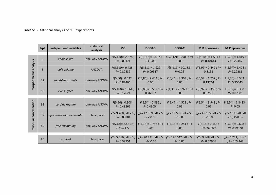
Table S1 - Statistical analysis of ZET experiments.
hpf independent variables statistical analysis MO DODAB DODAC M:B liposomes M:C liposomes
8 epipolic arc one-way ANOVA F(5,110)= 2.278 ; P= 0.05171
F(5,112)= 2.487 ; P< 0.05
F(5,112)= 3.900 ; P< 0.05
F(5,100)= 1.534 ; P= 0.18614
F(5,95)= 1.419 ; P=0.22447
8 yolk volume ANCOVA F(5,110)= 0.428 ; P= 0.82839
F(5,111)= 1.929; P= 0.09517
F(5,111)= 10.188 ; P<0.05
F(5,99)= 0.449 ; P= 0.8131
F(5.94)= 1.424 ; P=2.22281
32 head-trunk angle one-way ANOVA F(5,60)= 0.432 ; P= 0.82466
F(5,86)= 2.434 ; P< 0.05
F(5,44)= 7.303 ; P< 0.05
F(5,57)= 1.752 ; P= 0.13744
F(5,70)= 0.533 ; P= 0.75043
mor
phom
etric
ana
lysi
s
56 eye surface one-way ANOVA F(5,108)= 1.564 ; P= 0.17624
F(5,85)= 0.507 ; P= 0.76997
F(1,31)= 23.971 ; P< 0.05
F(5,92)= 0.358 ; P= 0.87581
F(5,92)= 0.358 ; P= 0.87581
32 cardiac rhythm one-way ANOVA F(5,54)= 0.908 ; P= 0.48266
F(5,54)= 0.896 ; P=0.49054
F(5.47)= 4.522 ; P< 0.05
F(5,54)= 3.948 ; P< 0.05
F(5,54)= 7.8433 ; P<0.05
32 spontaneous movements chi-square χ2= 9.268 ; df = 5 ; P= 0.09884
χ2= 12.369 ; df = 5 ; P< 0.05
χ2= 19.596 ; df = 5 ; P< 0.05
χ2= 45.165 ; df = 5 ; P< 0.05
χ2= 107.378 ; df = 5 ; P<0.05
80 free-swimming one-way ANOVA F(5,18)= 2.4619 ; P =0.7172
F(5,18)= 9.757 ; P< 0.05
F(5,18)= 3.251 ; P< 0.05
F(5,18)= 0.148 ; P=0.97809
F(5,18)= 0.608 ; P= 0.69520m
uscu
lar c
oord
inat
ion
80 survival chi-square χ2= 5.316 ; df = 5 ; P= 0.39951
χ2= 70.891 ; df = 5 ; P< 0.05
χ2= 176.042 ; df = 5 ; P< 0.05
χ2= 9.868; df = 5 ; P= 0.07906
χ2= 6.731; df = 5 ; P= 0.24142
5
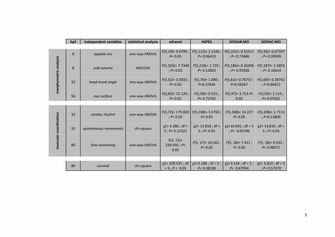
hpf independent variables statistical analysis ethanol HEPES DODAB:MO DODAC:MO
8 epipolic arc one-way ANOVA F(4,19)= 9.6799 ; P< 0.05
F(5,112)= 2.1546 ; P= 0.06413
F(5,101)= 0.55411 ; P= 0.73486
F(5,93)= 0,07397 ; P= 0.99599
8 yolk volume ANCOVA F(5,103)= 7.7348 ; P< 0.05
F(5,218)= 1.729 ; P= 0.12820
F(5,186)= 0.26206 ; P= 0.93326
F(5,187)= 1.5853 ; P= 0.16614
32 head-trunk angle one-way ANOVA F(3,32)= 3.3032 ; P< 0.05
F(5,76)= 1.080 ; P=0.37836
F(5,61)= 0.78757 ; P=0.56267
F(5,69)= 0.38742 ; P=0.86921
mor
phom
etric
ana
lysi
s
56 eye surface one-way ANOVA F(3,60)= 15.129 ; P< 0.05
F(5,99)= 0.525 ; P= 0.75702
F(5,97)= 3.753 P< 0.05
F(5,93)= 2.116 ; P= 0.07031
32 cardiac rhythm one-way ANOVA F(3,72)= 179.820 ; P< 0.05
F(5,108)= 3.5763 ; P< 0.05
F(5,108)= 14.227 P< 0.05
F(5,108)= 1.7113 ; P=0.13809
32 spontaneous movements chi-square χ2= 4.380 ; df = 3 ; P= 0.22323
χ2= 13.818 ; df = 5 ; P< 0.05
χ2=10.691 ; df = 5 ; P= 0.05786
χ2= 14,810 ; df = 5 ; P< 0.05
80 free-swimming one-way ANOVAF(3, 12)=
239.050 ; P< 0.05
F(5, 17)= 10.542 ; P< 0.05
F(5, 18)= 7.411 ; P< 0.05
F(5, 18)= 0.932 ; P= 0.48371m
uscu
lar c
oord
inat
ion
80 survival chi-square χ2= 229.535 ; df = 5 ; P < 0.05
χ2=5.286 ; df = 5 ; P= 0.38198
χ2=3.134 ; df = 5 ; P= 0.67934
χ2= 3.833 ; df = 5 ; P= 0.57370

6
Statistics
Prior to data analysis, all assumptions were met testing for normality (Shapiro-Wilk test)
and homogeneity of variances (Levene’s test).
To investigate the influence of DODAX:MO liposomes on ROS production and on
mitochondrial membrane potential of the BJ-5ta cell line, a one-way ANOVA (seven levels: NT -
non-treated cells; B:M(1:0), B:M(2:1), B:M(1:2), C:M(1:0), C:M(2:1) and C:M(1:2) liposomes
treated-cells) was conducted. Regarding ROS production, 40 µg mL-1 and 80 µg mL-1 liposomes
were analyzed separately, as were the timepoints considered for each test variable. Results
were expressed as mean ± standard error of the mean (S.E.M.).
In order to detect differences among concentrations in the overall survival of zebrafish
embryos, a chi-square test was conducted with the observed values for each test condition
(hpf were analyzed separately). The null hypothesis of “no differences among concentrations”
was considered for the establishment of the expected values (average survival of all
treatments, for a given hpf).
To investigate the effect of DODAX and MO exposure (as free-compounds and in
combination, as liposomes) on the epibolic arc (8 hpf), on the HTA (32 hpf) and on the cardiac
rhythm (32 hpf) of zebrafish embryos, six-level one-way ANOVA analysis were conducted.
To avoid biases associated with covariates, ANCOVA model was considered to determine
the influence of the DODAX:MO liposomes on zebrafish yolk volume at 8 hpf (egg volume was
used as co-variable). To investigate differences among groups on eye surface of zebrafish
larvae at 56 hpf, a six-level one-way ANOVA was applied. In order to guaranty that the
morphometric results emerging from the proposed experiments were not a result of larvae
size differences between groups, a six-level one-way ANOVA was also conducted on zebrafish
body size at 56 hpf.
In order to detect differences among groups in the percentage of spontaneous movements
of zebrafish embryos at 32 hpf, a chi-square test was conducted with the observed values for

7
each test condition. The null hypothesis of “no differences among groups” was considered for
the establishment of the expected values (average percentage of spontaneous movements of
all treatments). The percentage of zebrafish larvae exhibiting free-swimming behavior at 80
hpf, was used as dependent variable in a six-level one-way ANOVA design, to test for
differences between treatments.
In vitro data statistical analysis was performed with Prism 5 (GraphPad software Inc. v5),
while the remaining analyses were conducted in STATISTICA (StatSoft v7, Tulsa, OK). For in
vitro data, post hoc comparisons were conducted using Dunnett’s multiple comparison test,
while for in vivo data Student-Newman-Keuls analysis were considered. Statistical significance
is highlighted as follow: *p < 0.05, **p < 0.01, ***p < 0.001.
8
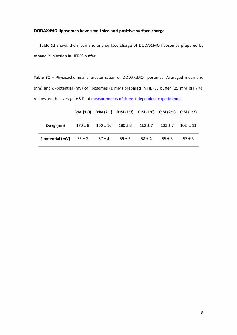
DODAX:MO liposomes have small size and positive surface charge
Table S2 shows the mean size and surface charge of DODAX:MO liposomes prepared by
ethanolic injection in HEPES buffer.
Table S2 – Physicochemical characterization of DODAX:MO liposomes. Averaged mean size
(nm) and ζ -potential (mV) of liposomes (1 mM) prepared in HEPES buffer (25 mM pH 7.4).
Values are the average ± S.D. of measurements of three independent experiments.
B:M (1:0) B:M (2:1) B:M (1:2) C:M (1:0) C:M (2:1) C:M (1:2)
Z-avg (nm) 170 ± 8 160 ± 10 180 ± 8 162 ± 7 133 ± 7 102 ± 11
ζ-potential (mV) 55 ± 2 57 ± 4 59 ± 5 58 ± 4 55 ± 3 57 ± 3
9
Effect of DODAX:MO liposomes on the cellular viability of several cell lines
The effects of DODAX:MO liposomes on cell viability were evaluated in five other cell lines,
in order to understand if the cytotoxic response was consistent along a variety of cells, and to
allow stronger conclusions about nanocarriers’ toxicity.
Human embryonic kidney cell line 293T (ATCC® CRL-3216™), human melanoma cell line
MDA-MB-435 and human breast carcinoma cell line MDA-MB-468 (ATCC® HTB-132™) were
routinely gown in DMEM® cell culture medium supplemented with 1 % L-Glu, 10 % FBS, 1 %
pyruvate sodium solution and 1 % P/S solution. Human myelogenous leukemia cell line K-562
(ATCC® CCL-243™) and human monocytic cell line THP1 (ATCC® TIB-202™) were grown in RPMI
1640® cell culture medium supplemented with 1 % L-Glu, 10 % FBS and 1 % P/S solution. All
cell lines were kept in a humidified incubator (37 °C and 5 % CO2) and sub-cultured every 3-4
days in order to maintain sub-confluency.
For the MTS assay 293T, MDA-MB-435, MDA-MB-468, K562 and THP1 cells were seeded
into 96-multiwell plates (TPP, Switzerland) at a density of 20 × 103, 5 × 10³, 7.5 × 10³, 20 × 103
and 50 × 103 cells per well, respectively, in complete cell culture medium. For the SRB assay
293T, MDA-MB-435, MDA-MB-468 were seeded in 96-multiwell plates (TPP, Switzerland) at a
density of 20 × 103, 5 × 10³, 7.5 × 10³ cells per well, respectively, in complete cell culture
medium. For the LDH assay cells were seeded at a density of 100 × 103 cells per well for 293T,
MDA-MB-435 and MDA-MB-468, and at a density of 200 × 103 cells per well for K562 and THP1
cells, in 24-multiwell plates (TPP, Switzerland).
MTS, LDH and SRB assays were performed as described in Materials and Methods.
Figure S1 shows the results of the MTS assay after a 48 h-period incubation of 293T, MDA-
MB-435, MDA-MB-468, K562 and THP1 cells with 5, 20, 40 and 80 µg mL-1 B:M(1:0), B:M(2:1),
B:M(1:2), C:M(1:0), C:M(2:1) and C:M(1:2) liposomes.

10
Figure S1 - Metabolic activity of different cell lines after incubation with DODAX:MO
liposomes. 293T (A), MDA-MB-435 (B), MDA-MB-468 (C), K562 (D) and THP1 (E) cells were
exposed to 5, 20, 40 and 80 µg mL-1 of B:M(1:0), B:M(2:1), B:M(1:2), C:M(1:0), C:M(2:1) and
C:M(1:2) liposomes, and MTS was performed after 48 h. The % of metabolic activity was
expressed in relation to non-treated cells (NT), set to 100 %. Results are expressed as mean ±
S.E.M. of three independent experiments.
The metabolic activity after exposure to DODAX:MO liposomes was cell-line dependent, but
similar in the five tested cell lines, with DODAC-based liposomes inducing higher cytotoxicity
than DODAB-based liposomes, for the same molar fraction. B:M(1:2) formulation had always a
less negative effect on cellular metabolism when compared to the other liposomes.
Figure S2 shows the results of the SRB assay after a 48 h-period incubation of 293T, MDA-
MB-435, and MDA-MB-468 cells with 5 and 40 µg mL-1 B:M(1:0), B:M(2:1), B:M(1:2), C:M(1:0),
C:M(2:1) and C:M(1:2) liposomes. For higher concentrations, liposomes interfered with the
assay.

11
Figure S2 - Cellular proliferation of different cell lines after 48 h exposure to DODAX:MO
liposomes. 293T (A), MDA-MB-435 (B), MDA-MB-468 (C) cells were incubated with 5 and 40 µg
mL-1 of B:M(1:0), B:M(2:1), B:M(1:2), C:M(1:0), C:M(2:1) and C:M(1:2) liposomes and SRB assay
was performed after 48 h. Time point 0 h (before addition of liposomes) was considered as 100
% cell proliferation, and the % of cell proliferation was expressed relative to non-treated cells
(NT). Results are expressed as mean ± S.E.M. of three independent experiments.
The effects of DODAX:MO liposomes on cellular proliferation were cell line-dependent, with
MDA-MB-435 being the most severely affected cell line. A clear correlation between DODAC
and DODAB, or the presence of MO and cellular proliferation, was not found although B:M(1:2)
seemed to generally interfere less with cells’ proliferation.

12
Figure S3 shows the results of the LDH assay after a 4 h-period incubation of 293T, MDA-
MB-435, MDA-MB-468, K562, THP1 and BJ5-ta cells with 80 and 160 µg mL-1 B:M(1:0),
B:M(2:1), B:M(1:2), C:M(1:0), C:M(2:1) and C:M(1:2) liposomes. This timepoint was chosen in
order to observe an immediate effect of the interaction of the nanocarriers with plasma cell
membranes.
Figure S3 – Plasma membrane integrity of different cell lines after contact with DODAX:MO
liposomes. 293T (A), MDA-MB-435 (B), MDA-MB-468 (C), K562 (D) THP1 (E) and BJ5-ta (F) cells
were incubated with 80 and 160 µg mL-1 of B:M(1:0), B:M(2:1), B:M(1:2), C:M(1:0), C:M(2:1)
and C:M(1:2) liposomes and LDH assay was performed after 4 h. The % of membrane integrity
was expressed as the percentage of the intracellular LDH activity relative to the total
(extracellular + intracellular) LDH activity. Results are expressed as mean ± S.E.M. of three
independent experiments.
The disruptive effects of DODAX:MO liposomes on plasma cell membranes were cell line-
dependent, but once again the same trend was observed: DODAC-based formulations strongly
affected the plasma membrane integrity of 293T, MDA-MB-435, MDA-MB-468, K562, THP1
and BJ5-ta, for the same molar fraction. B:M(1:0) liposomes induced less disruptive effects
when compared to the other nanoformulations.

13

14
ROS accumulation after incubation with DODAX:MO liposomes
The accumulation of reactive species of oxygen (ROS) was determined by the DCFH-DA
assay as described in Materials and Methods. Cells were seeded into 24-multiwell plates (TPP,
Switzerland) at a cell density of 100 × 103 cells per well for MDA-MB-468, and at a density of
200 × 103 cells per well for K562 cells.
Results shown on Figure 3 show that incubation of BJ5-ta cells with 80 µg mL-1 liposomes
gave a peak of ROS accumulation after 4 h incubation, which decreased after 8 h of exposure.
This was the result of the higher cytotoxicity associated with DODAC-based nanoformulations
(Fig. 1, S1, S2, and S3), that compromised cellular integrity, and therefore decreased ROS
detected. Therefore, for the DCF assays with MDA-MB-435 and K562 cell lines, 4 h incubation
with DODAX:MO liposomes was chosen.
Figure S4 shows that ROS accumulation was cell-line dependent, and MDA-MB-435 and
K562 seemed to be less sensitive than BJ5-ta (Fig. 3) to DODAX:MO liposomes.
Figure S4 - Reactive oxygen species (ROS) accumulation induced by DODAX:MO liposomes.
MDA-MB-468 (A) and K562 (B) cells were incubated with 40 and 80 µg/ml DODAX:MO
liposomes for 4 h, and DCFH-DA assay was performed to determine ROS accumulation. Results
are expressed mean ± S.E.M. of three independent experiments. NT - non-treated cells.