the three-dimensional structure of dna - arizona state university
TRANSCRIPT

Ann. Rev. Biochem. 1982.51:395-427
THE THREE-DIMENSIONAL
STRUCTURE OF DNA 1
Steven B. Zimmerman
Laboratory of Molecular Biology, National Institute of Arthritis, Diabetes, and Digestive and Kidney Diseases, National Institutes of Health, Bethesda, Maryland 20205
CONTENTS
Perspectives and Summary ........................................................................................ 395 Structural Models for DNA ...................................................................................... 396
Origins of polynucleotide models ... ........................................................................... 396 Terminology and conventions .................................................................................... 397 B form DNA .......................................................................................................... 397 Z form DNA .......................................................................................................... 407 Other forms of DNA ................................................................................................ 414
Helical Repeat of DNA ............................................................................................ 418 Heterogenity in DNA Structure ................................................................................ 420
Stotie heterogeneity .................................................................................................. 420 Dynamic heterogeneity ............................................................................................ 420
Concluding Remarks.................................................................................................. 421
Perspectives and Summary This is a particularly interesting time to review the three-dimensional structures of DNA. Among recent dramatic events are the demonstration of a
radically new type of DNA structure-the left-handed Z-helix-as well as the determination of the detailed structure of a segment of B form DNA. The latter is now especially relevant, as an increasing number of heretical candidates are being proposed to replace several familiar models for DNA, including the Watson-Crick model for the B form. Accordingly, the emphasis of this review is on the regular structures that have been proposed for double-stranded DNA.
The review starts by considering two of the major conformations (or "forms") that have been proposed for DNA, namely the B and Z forms,
IThe US Government has the right to retain a nonexclusive, royalty-free license in and to any copyright covering this paper.
395
Ann
u. R
ev. B
ioch
em. 1
982.
51:3
95-4
27. D
ownl
oade
d fr
om a
rjou
rnal
s.an
nual
revi
ews.
org
by A
RIZ
ON
A S
TA
TE
UN
IV. o
n 08
/18/
10. F
or p
erso
nal u
se o
nly.

396 ZIMMERMAN
with a brief mention of several others, including the A, C, and D forms. Although this survey contains some recent information on the interconversions between certain of these forms and a mention of studies on their dynamic behavior, no attempt is made to be comprehensive in these areas. The status of the structure of DNA-RNA hybrids is briefly reviewed. Obviously, a detailed review of all of these areas is impossible and considerable selection has been exercised. Studies of transfer RNA or of oligonucleotide or polynucleotide complexes with drugs or proteins are only discussed as they seem directly pertinent to DNA structure per se. Theoretical studies are deemphasized relative to experimental studies. The literature covered by this review extends through the middle of 1981.
DNA structure was last reviewed in this series by Jovin (1). A number of other reviews have appeared on nucleic acid structure (2-8) and physical properties (9) as well as on the properties of DNA-protein complexes (10, 11), chromatin (12), and topoisomerases (13-15), and complexes of nucleic acids or their components with metals (16, 17), with drugs (18), or with water (19). Related reviews have centered upon the data obtained from optical techniques (20) or NMR (21-25) (24 reviews nucleosides and nucleotides). Superhelical DNA has been also been reviewed (26-29).
Structural Models for DNA
ORIGINS OF POLYNUCLEOTIDE MODELS With the exception of the recently discovered Z form, the various conformations of DNA were all originally distinguished and defined by their X-ray fiber diffraction patterns. In all cases, this type of diffraction record has provided the richest source of data against which detailed structural proposals for polymeric DNA can be tested. Such fiber diffraction patterns provide two major types of information (30): the helical parameters (pitch, residue repeat distance, and residues/tum), which are often readily obtainable from spacings of the reflections in the X-ray patterns, and the intensity distribution pattern itself. The helical parameters are used to set limits on the type of detailed structural proposals ("models") that need to be considered, and trial structures are built within these constraints. Such provisional models are further subjected to the laws of structural chemistry: covalent bond angles or distances must be within the ranges determined by accurate structure determinations on small molecules. The coordinates of stereochemically acceptable models are used to calculate predicted diffraction patterns, which are then compared with the intensity distribution in the observed diffraction pattern. The model typically goes through a number of cycles of adjustment to improve the fit of its calculated diffraction pattern to the one observed.
Ann
u. R
ev. B
ioch
em. 1
982.
51:3
95-4
27. D
ownl
oade
d fr
om a
rjou
rnal
s.an
nual
revi
ews.
org
by A
RIZ
ON
A S
TA
TE
UN
IV. o
n 08
/18/
10. F
or p
erso
nal u
se o
nly.

THREE-DIMENSIONAL STRUCTURE OF DNA 397
It is in the nature of this train of argument that the best model so obtained is not necessarily unique. The coordinates are presented at atomic resolution because they must be so specified for testing of the model, not because they are necessarily a unique solution dictated by the diffraction data. Because of the shortage of diffraction data, assumptions are incorporated into the model building. For example, base-pairing schemes are commonly assumed from physicochemical or other sources. Sugar puckers or backbone dihedral angles may be fixed to certain values or constrained to be within certain ranges. Indeed, even the hand of the basic helix must be assumed, at least initially, although subsequently right- vs left-handed models can be tested against each other.
These limitations on DNA models derived from fiber data have stimulated major reinterpretations and have led to proposals for left-handed and "side-by-side" models. These proposals are discussed in some detail. In the case of Z DNA, the original structure was obtained from single crystal X-ray diffraction analysis of short oligomers. This approach does yield a unique structure; however, it must then be shown that polymers can assume the same structure as did the oligomers. This stage of the demonstration once again has rested heavily on fiber diffraction studies, in conjunction with other physical chemical evidence. Several models are compared in Figure I and their parameters are summarized in Table 1. Finally, it should be noted that the original structural proposals for the various forms of DNA stem from data on solid samples, i.e. fibers or crystals. A number of recent studies have concluded that the original models must be modified if they
are to portray realistically the structures present in solution.
TERMINOLOGY AND CONVENTIONS For .those interested in the definitions of specific conformational features such as the various sugar puckers and the ranges of dihedral angles the descriptions of Saenger (47) or of Arnott (48) are suggested.
A designation such as "the B form" of DNA is commonly used in the literature to refer to both the actual structure that gives rise to the characteristic diffraction pattern and to the hypothetical structural model that is proposed to rationalize the diffraction pattern. In this review, the former meaning is employed. Specific structural proposals such as types of sugar puckers and sets of atomic coordinates are considered to be attributes of models for the actual structure.
B FORM DNA The Watson-Crick proposal (49) provided the basis for the familiar model of B form DNA. Elegant studies (50) led to a stereochemically acceptable version of this model that was consistent with the observed
Ann
u. R
ev. B
ioch
em. 1
982.
51:3
95-4
27. D
ownl
oade
d fr
om a
rjou
rnal
s.an
nual
revi
ews.
org
by A
RIZ
ON
A S
TA
TE
UN
IV. o
n 08
/18/
10. F
or p
erso
nal u
se o
nly.

398 ZIMMERMAN
A DNA B DNA
& @ Y; -
� -
� -
--
� �
--
� --/
� � � �
0 @
eDNA D DNA
�� (5 � "
�< R$; (j)
Z DNA
---....! f
�-----
� /f-@
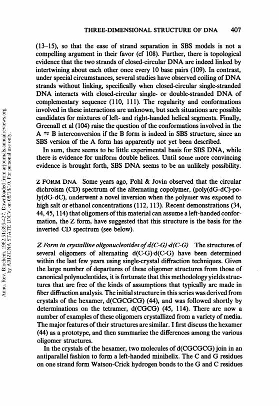
Figure 1 Models for various conformations of DNA. Segments containing 20 base pairs are shown for right-handed models of A (31), B (31), C (32) or D DNA (33) and for the left-handed Z, form of DNA (34). The upper views are perpendicular to the helical axes and the lower views look along the helical axes. The continuous helical lines are formed by linking the phosphorus atoms along each strand. The line segments indicate the positions of the base pairs and are formed by joining the Cl' atoms of each base pair. This simplified mode of representation emphasizes the differences in helical parameters and in positions of the base pairs among these models. The arrow in the lower view of Z DNA indicates the C}' of the deoxyguanosine residue, the base of which is relatively exposed in this structure (see text); the deoxycytidine residue is more centrally located.
diffraction pattern. Arnott & Hukins (31) refined this structure and provided a set of atomic coordinates that have been widely used in discussions of B DNA. Certain aspects of the B form are not presently controversial: all current models assume a double helix with antiparallel strands and with Watson-Crick base pairs oriented roughly at right angles to the helix axis (Figure 1). Controversy exists as to whether the helix is right-or left-handed or both, as to the exact number of base pairs/tum, and as to the disposition of the bases (coplanarity of bases within the pairs and their orientation with respect to the helix axis).
Most DNA can adopt the B form as defined by its characteristic X-ray fiber diffraction pattern: The bulk of the sequences of natural DNA over a wide range of base compositions (51, 52) as well as synthetic DNA of several simple base sequences (35) yields the B pattern under appropriate
Ann
u. R
ev. B
ioch
em. 1
982.
51:3
95-4
27. D
ownl
oade
d fr
om a
rjou
rnal
s.an
nual
revi
ews.
org
by A
RIZ
ON
A S
TA
TE
UN
IV. o
n 08
/18/
10. F
or p
erso
nal u
se o
nly.

DNA
conformation
A
B
Alternating
B
C
D
T
Z
THREE-DIMENSIONAL STRUCTURE OF DNA 399
Axial rise Base pairs
per base Base pairs per
Occurrence pair (A) per turn repeating unit Ref
Most n�tural and syn- 2.6 11.0 31,35,36
thetic DNA
Most natural and syn- 3.4 10.0a 31,35,37
thetic DNA
Several alternating 3.4 10.0 2 38 purine-pyrimidine
DNA
Most natural and syn- 3.3 7.9-9.6 33,35,39,40
thetic DNA
Several synthetic DNA 3.0 8.0 33,35,41
Glucosylated bacterio- 3.3-3.4 8.0--8.4 42,43 phage DNA
Several al ternating 3.6-3.8 12.0 2 34,44-46
purine-pyrimidine
DNA
a Alternative models with different parameters are discussed in text.
conditions. Further, the patterns are similar to each other in detail, which suggests a narrow range of variability in B form structure. This apparent homogeneity is in contrast to recent suggestions from nondiffraction techniques and from single-crystal diffraction studies on oligonucleotides, which indicate either static or dynamic structural heterogeneity (see below). Early indications of deviant structures in DNA of high AT-content have been correlated with the presence of non-DNA material (53). There are a few species of synthetic DNA, namely poly(dA)'poly(dT), poly(dI)-poly(dC), and poly(dA-dI)'poly(dC-dT), that have a B form that is significantly different from that of the other complementary deoxypolymers. These polymers have different intermolecular packing arrangements and a slightly changed value for the rise per residue (35, 54). A striking indication of altered structure or structures for the conformations of poly(dA)'poly(dT) and poly(dI)·poly(dC) is their refusal to undergo a transition from the B form to the A form (35, 54-56). These unusual properties assume particular interest given the existence of relatively long dA' dT sequences in vivo (57).
B form adopted by self-complementary oligodeoxynucleotides The structure of a long self-complementary oligodeoxynuc1eotide, d(CGCGAATTCGCG), has recently been solved by single-crystal diffraction techniques (58-61). The dodecanucleotide forms more than a full turn of a helix whose
Ann
u. R
ev. B
ioch
em. 1
982.
51:3
95-4
27. D
ownl
oade
d fr
om a
rjou
rnal
s.an
nual
revi
ews.
org
by A
RIZ
ON
A S
TA
TE
UN
IV. o
n 08
/18/
10. F
or p
erso
nal u
se o
nly.

400 ZIMMERMAN
overall structure is notably like that proposed for the canonical model for B DNA. This study therefore allows an unambiguous examination at high resolution of a prototype for the B form of DNA, and is certainly a most important addition to our knowledge.
The structure adopted by the dodecanucleotide is a right-handed antiparalIel double-stranded helix with the bases essentially perpendicular to the helix axis (58). While these major attributes of the B form model are present, the detailed structure of the oligomer departs from that of the familiar model in a number of significant ways.
The well-defined molecular axis is not straight; rather the axis traces a smooth bend of significant curvature (a total of 19° bend distributed over the entire length, which corresponds to a radius of curvature of 112 A; for reference, the diameter of the B helix itself is '" 21 A.) A clear basis for the bend is seen in the intermolecular contacts within the crystal (58). The terminal three G-C base pairs of each duplex form five H bonds with atoms of the next duplex "above" themselves in a manner that requires a bend in the helix axis. There are two formal possibilities: either the DNA is intrinsically bent and the crystal packing simply accommodates this innate tendency or, alternatively, the packing causes the bending. The authors provide several reasonable arguments for the latter interpretation, but ultimately we must await the results of similar studies with other oligomers. It is the terminal alternating G-C sequences that are involved in the canted interactions between duplexes, so that it will be important to see what structures form in oligomers lacking these terminal sequences or, alternatively, containing longer internal sequences. The bend in the dodecamer clearly poses a problem if we wish to know the structure of unbent DNA, since deconvoluting the bent structure requires some arbitrary decisions. Notwithstanding, it is most useful to examine this remarkable structure.
The crystal structure differs from the usual models derived from fiber diffraction in a fundamental way: The dodecamer duplex is not simply a set of 24-nucleotidyl residues of essentially identical conformation joined monotonously into 12 base pairs of essentially identical structure. Rather there are enormous variations in the helical relationships between successive base pairs, and often significant conformational differences between the residues (59, 60). The helical variations span the full range observed by fiber diffraction for structures as disparate as those of the A, B, and D forms of DNA. For example, the eleven local helical steps correspond to 9.4, 9.1, 10.8, 9.6, 9.6, 11.2, 10.0, 8.7, 11.1, 8.0, and 9.7 residues/turn (59). These values may be compared to the values from fiber diffraction of -- 11, 10, and 8 residues/turn for the A, B, and D helices, respectively (Table 1). Hence, although the average (local) helical parameters in the dodecanucleo-
Ann
u. R
ev. B
ioch
em. 1
982.
51:3
95-4
27. D
ownl
oade
d fr
om a
rjou
rnal
s.an
nual
revi
ews.
org
by A
RIZ
ON
A S
TA
TE
UN
IV. o
n 08
/18/
10. F
or p
erso
nal u
se o
nly.

THREE-DIMENSIONAL STRUcrURE OF DNA 401
tide (9.65 residues/turn and 3.33 A rise/residue) are within a few percent of those inferred from the B form diffraction, the structure has tremendous local variation. Indeed, while the base pair tilt and position relative to the helix axis for the central six base pairs is similar to that in the B DNA models from fiber diffraction studies (31), the adjacent base pair on either side has a distinctly A DNA conformation, and another peripheral base pair is like that of the D form. The other backbone conformational angles are generally similar to those inferred from earlier fiber diffraction or theoretical studies, with a few values in unusual ranges.
The bases of a given base pair are not coplanar in the dodecanucleotide (59,60). The average propellor twist (i.e. total dihedral angle between base planes in one H-bonded base pair) is 17.3° ± 0.4° for the central four A-T residues and 11.5° ± 5.1° for the G-C pairs at the ends. [In contrast, an earlier model for B DNA from fiber diffraction studies (31) has an almost coplanar arrangement of the bases within a base pair (twist = 4°).]
The variation in sugar conformations within the dodecamer has provided several insights (59, 59a). First, the conformations span essentially the whole range possible for deoxyfuranose rings. They show a correlation between the glycosidic torsion angle and the sugar ring conformation. The purine residues tend toward the C2'-endo sugar pucker and a high value for their glycosidic angle, while the pyrimidine residues tend toward a lower glycosidic angle and their sugars are more like C3'-endo puckered sugars. This behavior was rationalized in terms of steric contacts between the sugars and the 02 of the pyrimidines. The authors note a further striking fact, which they formalize as the "principle of anticorrelation:" the two sugars of a given base pair tend to have values of their internal torsion angles (specifically about C4'-C3') that are equidistant from that of a central value corresponding to the Cl '-exo conformation. In other words, if one sugar in a given base pair is C3'-endo, the other sugar in that pair tends to have a C2'-endo conformation. Several deoxyribosyl structures and a recent DNA-RNA hybrid structure seem to be consistent with this principle, whereas the sugars in yeast phenylalanine tRNA are not.
In addition to the striking static heterogeneity in local structure, the dodecanucleotide provides evidence of dynamic heterogeneity. The thermal vibrations of individual atoms were inferred from their temperature factors as obtained in the X-ray structure determination. Drew et al (59) indicate that the relatively larger vibrations of peripheral atoms are consistent with the rapid intramolecular motions ascribed to DNA from NMR and other measurements (see below)
DNA in solution is often presumed to be in the B form (see section on the helical repeat of DNA). While modeling studies of solvent accessibility
Ann
u. R
ev. B
ioch
em. 1
982.
51:3
95-4
27. D
ownl
oade
d fr
om a
rjou
rnal
s.an
nual
revi
ews.
org
by A
RIZ
ON
A S
TA
TE
UN
IV. o
n 08
/18/
10. F
or p
erso
nal u
se o
nly.

402 ZIMMERMAN
to the surfaces of the B form and of other polynucleotide structures have appeared (62, 63), experimental knowledge of the organization of water about duplex DNA has been sparse. [The single-crystal structure of the proflavine complex with dCpdG is a notable exception (64).] The dodecanucleotide structure provides a unique opportunity to visualize water structure in a sizeable unit ofB form DNA (61). There are 72 ordered molecules of water per duplex. Of these, 50 are either in the grooves or closely associated with the phosphate groups. The most striking arrangement of water molecules is in the region of the minor groove associated with the central AA IT sequence. Solvent is organized in layers up to three or four molecules deep. The innermost layer forms a regular "spine" of water bridging the hydrophilic groups of alternate bases. This backbone of water is compatible with any sequence of A-T or I-C base pairs, but not with G-C residues, due to the disrupting influence of the 2-amino group of guanine. This pattern suggests that guanine residues will destabilize the B form, an inference that is in general concurrence with the fiber diffraction survey of Leslie et al (35).
Finally, we note a study that suggests that the dodecanucleotide structure may represent a reasonable model for B DNA in solution. Lomonosoff et al [(65) and further discussion in (60)] have found a good correlation between the rates of nuclease cleavage of internucleotide bonds of the dodecamer in solution and their "exposure" (magnitude of the local helical rotation between base pairs) in the crystal.
A complex of a second sizable oligodeoxynucleotide, d(CGTACG), with daunomycin has been shown by single-crystal diffraction analysis to contain a short segment that adopts a structure similar to that of the canonical B form. This complex forms a self-complementary right-handed anti parallel mini-DNA helix, with a molecule of drug intercalated between the C-G base pairs at each end (66). The structure of the ends of the molecules is modified for intercalation, but the central A and T residues share many characteristics with the familiar B DNA model. The sugars of the central A and T residues conform to the principle of anticorrelation above (59). The authors indicate however that the backbone conformations of the central base pairs of the hexanucleotide have significant departures from those in the usual A or B form models.
B form model for alternating copolymers A single-crystal X-ray structure determination (67, 67a) on the tetranucleotide d(ATAT) has led to interesting proposals, (38, 67a) for a modified B conformation suitable for a regularly alternating copolymer. In the crystal, the two base pairs at each end of the tetranucleotide form H bonds to two different adjacent tetranucleotide molecules, which yield short segments of right-handed antiparallel
Ann
u. R
ev. B
ioch
em. 1
982.
51:3
95-4
27. D
ownl
oade
d fr
om a
rjou
rnal
s.an
nual
revi
ews.
org
by A
RIZ
ON
A S
TA
TE
UN
IV. o
n 08
/18/
10. F
or p
erso
nal u
se o
nly.

THREE-DIMENSIONAL STRUCTURE OF DNA 403
double helix. The most striking feature of the crystal structure is the regular alternation in sugar pucker and glycosidic dihedral angle, X. The adenosine residues both have a C3'-endo sugar conformation, while the thymidine residues have a C2'-endo pucker. This alternation has been incorporated into a model for the polymer poly(dA-dT)'poly(dA-dT), which is generally rather similar to the canonical B form model [cf Figure 10 in (68)]. This model is proposed as one of a family of basically similar alternating structures that could be built to maximize base-stacking interactions (38; see also 67a, 69). Such alternating B models are consistent with a number of the properties of poly(dA-dT)'poly(dA-dT) previously observed in solution [see (38) for references]. Shindo et al (70) have independently suggested a model with an alternating backbone conformation for this polymer in solution based on two resonance peaks in the 31p NMR spectrum. Recent 31p NMR studies have also detected the two resonance peaks expected for the alternating B model in fibers of poly(dA-dT)'poly(dA-dT) (71), as well as in solutions of poly(dA-dbr5U)'poly(dA-dbr5U) and poly(dI-dC)'poly(dIdC) (68). The tetramethylammonium ion has been suggested to favor an alternating conformation for poly(dA-dT)'poly(dA-dT) (72).
Lomonosoff et al (65) have provided evidence that poly(dG-dC)'Poly(dGdC) in low-salt solutions can also adopt an alternating type of conformation, presumably of the B form, based upon pancreatic DNase I digestion patterns of the polymer. [An alternating B model was proposed (73) for the high-salt form of (dG-dC)-oligomers in an earlier study; in hindsight, it seems as likely that the dinucleotide repeat inferred is related to the more recently delineated Z form (see below).] Less detailed models for alternating structures have been suggested from theoretical studies (74, 75).
Bent models/or B DNA Structural models for DNA have generally been built as unbent double helices. There have, however, been several suggestions of smoothly bent or discontinuously bent ("kinked") models. Smoothly bent DNA has actually been observed, in the form of the dodecanucleotide duplex (58) discussed in a previous section. There have been a number of theoretical studies of smoothly bent models for DNA wound around the histone cores of chromatin (76-79).
Various kinked models feature straight segments of B form DNA interspersed at regular intervals with residues having alterations in certain torsional angles of their sugar-phosphate backbones (80-83). A variety of superhelical arrangements can result, depending upon both the frequency of kinking and its detailed mechanism. Such kinked models form a conceptual framework for observations as diverse as the compaction of chromatin and its regular patterns of nuclease digestion, the "breathing" of DNA, or
Ann
u. R
ev. B
ioch
em. 1
982.
51:3
95-4
27. D
ownl
oade
d fr
om a
rjou
rnal
s.an
nual
revi
ews.
org
by A
RIZ
ON
A S
TA
TE
UN
IV. o
n 08
/18/
10. F
or p
erso
nal u
se o
nly.

404 ZIMMERMAN
the mechanism of drug intercalation. Solvent bombardment or thermal sources have been suggested as possible origins for kinks and other types of localized or traveling fluctuations in DNA structure (84-86). There is no direct evidence for kinking at present. 31p NMR studies of DNA in either chromatin or free in solution have not indicated the presence of species with an altered chemical shift as might be expected if kinking occurred and if the phosphodiester geometry was markedly changed at the kinked site (87-90).
Left-handed models for B DNA There has in the past generally been a consensus that B form DNA is a right-handed helix. As particularly emphasized by Sasisekharan and his collaborators, this assumption does not have a secure experimental basis. Let us review the arguments for left-handed DNA, reserving for the moment a discussion of mixtures of left- and righthanded DNA in the form of the side-by-side DNA model. It has long been known that DNA in the solid state can rapidly and reversibly be interconverted between the A, B, and C forms, simply by varying the relative humidity and salts present (37). The ease of these transitions in fibers was interpreted to mean that all of these forms are of the same hand. Since the original model building for the A form appeared to rule out a left-handed structure (cf section on A DNA below), the A, n, and C forms were all inferred to be right handed. This reasoning is subject to dispute at several points: First, it is only an intuitive conclusion at present that transitions in fibers can not change the hand of the structures involved. Second, even if this is accepted as a working hypothesis, several groups have shown that the B form of DNA interconverts in fibers with the Z form (46, 91), a presumably left-handed structure; hence, if anything, this line of argument suggests that the n form could be left handed. Third, satisfactory lefthanded models apparently can be built for the A form (and also for the B and D forms) (69, 92). Detailed sets of coordinates have been supplied by Gupta et al (69) that are stated to fulfill the usual stereochemical criteria. Several groups have noted that, surprisingly enough, only minor differences in the values of backbone dihedral angles need occur between left- and right-handed conformations, although the orientation of the base undergoes a significant shift (2, 7, 75, 92-94). Energy calculations have indicated to some a preference for right-handed conformations (2,7,94), although base stacking energies seem to be similar (95). A major criterion for an acceptable model is that it yield a predicted diffraction pattern consistent with the observed diffraction pattern. The left-handed B DNA model appears satisfactory in this respect (69). Hence, from the results of the fiber diffraction approach, there is no clear basis for preferring left- to right-handed DNA.
Ann
u. R
ev. B
ioch
em. 1
982.
51:3
95-4
27. D
ownl
oade
d fr
om a
rjou
rnal
s.an
nual
revi
ews.
org
by A
RIZ
ON
A S
TA
TE
UN
IV. o
n 08
/18/
10. F
or p
erso
nal u
se o
nly.

THREE-DIMENSIONAL STRUCTURE OF DNA 405
It may be noted that there are several unambiguous observations of right-handed helices in oligodeoxynucleotides of mixed base sequence in both the A and B forms and in RNA [the short A form-like helices of yeast phenylalanine tRNA (96)]. As yet, the only examples of a left-handed helix are seen in certain oligodeoxynucleotides and polydeoxynucleotides, which have a regularly alternating purine-pyrimidine sequence and a structure that is presumably not of general occurrence (see section on Z form DNA), or in short oligoribonucleotides that are constrained by a second covalent link between base and sugar (97). In sum, while there are indications that natural DNA is right handed, there seems no obvious reason why it should not be able to adopt a left-handed conformation.
Side-by-side models for DNA Two groups have independently proposed a new class of model for the B form of DNA (98-101). These so-called side-by-side or SBS models are basically different from either the uniformly right- or uniformly left-handed structures discussed above. All of the SBS models feature a regular alternation of short segments of left- and righthanded double-stranded DNA. The versions proposed by Sasisekharan and collaborators (98, 1(0) have alternating segments of five base pairs, so that the two strands do not undergo a net winding around themselves. The most recent SBS model of Millane & Rodley (101) has a slight right-handed bias so that the strands wind around each other, i.e. they are "linked" about once for every 77 base pairs. In contrast, the uniform left- or right-handed models previously discussed are linked once for every pitch length of approximately ten base pairs. A debate has developed in the literature between the proponents of the double helix and those favoring the SBS conformation. We first consider the arguments based on model building and X-ray diffraction studies and then consider topological and other types of evidence.
Models of SBS DNA have many more degrees of freedom available than do models of uniform helices, since the repeating unit of SBS DNA is long (ten base pairs rather than one or two base pairs) and heterogeneous (lefthanded, right-handed, and junction regions). Consequently, while modeling of such a large unit is a technically difficult problem, there seems little doubt that stereochemically acceptable SBS models can be built (101, 102). Detailed studies of the conformation and base stacking have appeared (95). It may be surprising to the reader that the expected diffraction patterns for such models are not totally different from those of the regularly helical models. Similarities arise because the fiber diffraction patterns are dominated by two features that appear similarly in SBS and uniform models. The first feature is the apparent helical repeat: canonical niodels have a pitch of "" 34 A, consistent with a tenfold helix and implying diffraction layer lines
Ann
u. R
ev. B
ioch
em. 1
982.
51:3
95-4
27. D
ownl
oade
d fr
om a
rjou
rnal
s.an
nual
revi
ews.
org
by A
RIZ
ON
A S
TA
TE
UN
IV. o
n 08
/18/
10. F
or p
erso
nal u
se o
nly.

406 ZIMMERMAN
at spacings of N /34 A (where N is an integer). Similarly spaced layer lines may be generated by SBS models with an exact or approximate structural repeat distance of 34 A, which corresponds to a unit of five left-handed and five right-handed base pairs. The second common feature of SBS and double-helical models is the centrally located Watson-Crick base pairs, which are situated approximately at right angles to the helix axis and spaced at an average of'" 3.4 A. This 3.4-A average spacing between base pairs results in the strong meridional reflection that is observed at N /3.4 A and that also dominates the form of an interatomic scattering function called the axial Patterson function, which has been applied to this problem (103). The general similarities in this function for the SBS and canonical B DNA models therefore do not form a basis for distinguishing between these structures as candidates for the B form. A more incisive test is to calculate the actual pattern expected from the atomic coordinates of the models in question. This has been done by two groups for several versions of SBS models (102, 104). In both studies, the authors concluded that the predicted diffraction patterns of the particular SBS models they examined do not fit the observed diffraction data nearly as well as do the predicted patterns of the best double-helical structures. In particular, SBS models predict several meridional reflections that are not observed. These studies have dealt with diffraction from fibers of noncrystalline DNA, the so-called continuous transform. There is also considerable diffraction data from fibers of semicrystalline B DNA. Amott (105) notes that the intensity distribution of the crystalline data agrees better with the double-helical rather than the SBS model. Further, a point originally made by Dover (106) is applied (105). The crystal lattice adopted by the lithium salt of DNA generates precisely regular intermolecular contacts between neighboring molecules for base pairs related by steps of 36.15° ± 0.25° of helical rotation, i.e. for a helix with precisely ten base pairs per tum of the helix. This relationship makes immediate sense in that it optimizes favorable lattice interactions for each base pair if the structure involved is that of a regular tenfold helix; however, it would be essentially a fortuitous result for an SBS type of structure, since SBS structures do not have a regular repeat at 36° intervals about the molecular axis.
As mentioned, the two strands of SBS DNA are relatively unlinked because of the alternation between left- and right-handed segments. A possible consequence of this feature, as noted a number of years ago (107), and presumably one of the motive forces behind SBS DNA, is that the relative lack of linking of the strands might facilitate the separation of the two strands during replication or other processes. However, the cell has developed elegant enzymatic mechanisms for winding and unwinding DNA
Ann
u. R
ev. B
ioch
em. 1
982.
51:3
95-4
27. D
ownl
oade
d fr
om a
rjou
rnal
s.an
nual
revi
ews.
org
by A
RIZ
ON
A S
TA
TE
UN
IV. o
n 08
/18/
10. F
or p
erso
nal u
se o
nly.
THREE-DIMENSIONAL STRUCTURE OF DNA 407
(13-15), so that the ease of strand separation in SBS models is not a compelling argument in their favor (cf 108). Further, there is topological evidence that the two strands of closed-circular DNA are indeed linked by intertwining about each other once every 10 base pairs (109). In contrast, under special circumstances, several studies have observed coiling of DNA strands without linking, specifically when closed-circular single-stranded DNA interacts with closed-circular single- or double-stranded DNA of complementary sequence (110, 111). The regularity and conformations involved in these interactions are unknown, but such situations are possible candidates for mixtures of left- and right-handed helical segments. Finally, Greenall et al (104) raise the question of the conformations involved in the A � B interconversion if the B form is indeed in SBS structure, since an SBS version of the A form has apparently not yet been described.
In sum, there seems to be little experimental basis for SBS DNA, while there is evidence for uniform double helices. Until some more convincing evidence is brought forth, SBS DNA seems to be an unlikely possibility.
Z FORM DNA Some years ago, Pohl & Jovin observed that the circular dichroism (CD) spectrum of the alternating copolymer, (poly(dG-dC)-poly(dG-dC), underwent a novel inversion when the polymer was exposed to high salt or ethanol concentrations (112, 113). Recent demonstrations (34, 44, 45, 114) that oligomers of this material can assume a left-handed conformation, the Z form, have suggested that this structure is the basis for the inverted CD spectrum (see below).
Z Form in crystalline oligonucleotides of d(C-G) -eI(C-G) The structures of several oligomers of alternating d(C-G)·d(C-G) have been determined within the last few years using single-crystal diffraction techniques. Given the large number of departures of these oligomer structures from those of canonical polynucleotides, it is fortunate that this methodology yields structures that are free of the kinds of assumptions that typically are made in fiber diffraction analysis. The initial structure in this series was derived from crystals of the hexamer, d(CGCGCG) (44), and was followed shortly by determinations on the tetramer, d(CGCG) (45, 114). There are now a number of examples of these oligomers crystallized from a variety of media. The major features of their structures are similar. I first discuss the hexamer (44) as a prototype, and then summarize the differences among the various oligomer structures.
In the crystals of the hexamer, two molecules of d(CGCGCG) join in an antiparallel fashion to form a left-handed miniheHx. The C and G residues on one strand form Watson-Crick hydrogen bonds to the G and C residues
Ann
u. R
ev. B
ioch
em. 1
982.
51:3
95-4
27. D
ownl
oade
d fr
om a
rjou
rnal
s.an
nual
revi
ews.
org
by A
RIZ
ON
A S
TA
TE
UN
IV. o
n 08
/18/
10. F
or p
erso
nal u
se o
nly.

408 ZIMMERMAN
on the other strand. The six base pairs so formed comprise one half of a left-handed helical tum.
The conformations of the residues also alternate in this structure of alternating base sequence. In general, the dC residues are similar to the residues in the canonical models for B DNA, while the dG residues are very different. For example, the sugars of the dC and dG residues display a regular alternation between the C2'-endo and C3'-endo conformations, respectively. Further, the dihedral angle around the C4'-C5' bond alternates markedly (between a gauche-gauche conformation in dC residues, as in B DNA, and a gauche-trans conformation in dG residues). Since the C and G residues have different conformations, the repeating unit on a given strand is a dinucleotide. These dinucleotide units are very regularly arrayed in the minihelix, although the symmetry of the lattice does not demand this regUlarity. The regularity even extends across the gap where duplexes stack upon each other except, of course, for the missing phosphate groups. The result is that the crystal structure approximates that of a continuous polymer, with 6 dinuc1eotides or 12 bases for each helical tum of a given strand. These alternating conformations produce an irregular zig-zag course of the sugar-phosphate backbone, hence the Z DNA designation for this structure (Figure I). The residues also alternate in the relationship of the base to its sugar. The dC residue has an anti relationship, which seems to be generally needed to relieve steric interactions between pyrimidine bases and their sugars (115). The guanine residues in contrast assume a syn conformation wherein the base closely approaches its sugar. This is the first example of a residue of a natural nucleotide that adopts the syn position in an oligomeric or larger system, although the bases in polynucleotides can be constrained toward this position by a bulky base substituent (116) or by a second covalent linkage between base and sugar (97).
As might be expected from the unique alternation of backbone and base-sugar conformations, the positioning of base pairs within the helix and the base-stacking relationships are remarkable. The base pairs are relatively peripheral in location in Z DNA. Because of the syn conformation of the guanosine, the reactive N7 and C8 positions of guanine are located at the margins of the structure and thereby made accessible to environmental insults (see below). The base pairs at the d(CpG) sequences are displaced laterally by 7 A relative to each other, so that ordinary base overlaps do not occur. Rather, the cytosine residue of one base pair stacks with a neighboring cytosine residue on the opposite strand. The guanine residues at this sequence do not stack with the adjacent bases, but rather overlap the furanose ring oxygens of the adjacent sugars. In contrast the stacks at the d(GpC) sequences are relatively similar to those of B DNA with overlap, of the bases that adjoin on the same strand. The Z form has a single very
Ann
u. R
ev. B
ioch
em. 1
982.
51:3
95-4
27. D
ownl
oade
d fr
om a
rjou
rnal
s.an
nual
revi
ews.
org
by A
RIZ
ON
A S
TA
TE
UN
IV. o
n 08
/18/
10. F
or p
erso
nal u
se o
nly.

THREE-DIMENSIONAL STRUCTURE OF DNA 409
deep helical "groove" that corresponds in location to the minor groove (i.e. the groove between the sugar-phosphate backbones) in the A or B forms.
Limited but significant variation has been observed in the structures solved to date for d(CGCG) or d(CGCGCG) crystallized under various conditions (34, 44, 45, 114, 117). The principal variation occurs in the orientation of the phosphate group of the GpC sequence. In the various crystals that have been solved, one or more of the phosphate residues has rotated from its position in the predominant conformation so that it lies further outwards and away from the minor groove (34, 45, 114, 117). Models for continuous polymers based upon each of these two phosphate conformations, labelled ZI and Zn, have been described (see below). A related change in the conformation of the phosphate group occurs in the crystalline tetramer solved by Drew et al (45). In this structure, labeled Z', the phosphate group of the GpC sequence again rotates outward, as in going from ZI to Zn. The reasons for the rotations in Z' and Zn are apparently different, being correlated with repulsion by a neighboring chloride ion for Z' (45) or, in some cases at least, with binding to a nearby hydrated magnesium ion for Zn (34, 114). The altered phosphate positioning in Z' is correlated with a change of sugar pucker of the internal deoxyguanosine residues, so that those sugars adopt a conformation (Cl'-exo) similar to that in the deoxycytidine residues. The result is that the Z' backbone has an almost uniform sugar pucker, which demonstrates that the characteristic dinucleotide repeat of Z DNA does not necessarily need to extend to a major involvement of the sugar conformation (45).
Z form in fibers of alternating polymers The demonstration of the Z structure in crystals of oligomers has led to attempts to identify such a structure in fibers or in solutions of polymeric DNA. X-ray fiber diffraction has provided relatively unambiguous evidence in the case of fibers. Arguments for the Z form in solution are summarized in the next section.
Fibers of certain alternating deoxypolymers yield a distinctive X-ray diffraction pattern identified with the Z form [which has also been called the S form in polymers (35)]. The original observation of Z form diffraction from fibers was made with poly(dG-dC)·poly(dG-dC) (46). Basically similar patterns have been collected from fibers of poly(dA-dC)-poly(dG-dT) (35) and poly(dG-dm5C)-poly(dG-dm5C) (118). A related pattern from poly(dA-ds4T)-poly(dA-ds4T) (119) has been reinterpreted in terms of the Z structure (46).
Models play an important role in fiber diffraction in validating proposed structures, as outlined earlier. In the case of polymeric DNA, the models for the Z form have followed the structures that were determined by single-
Ann
u. R
ev. B
ioch
em. 1
982.
51:3
95-4
27. D
ownl
oade
d fr
om a
rjou
rnal
s.an
nual
revi
ews.
org
by A
RIZ
ON
A S
TA
TE
UN
IV. o
n 08
/18/
10. F
or p
erso
nal u
se o
nly.

410 ZIMMERMAN
crystal diffraction of the oligomers. The initial model for poly(dG-dC)· poly(dG-dC) (46) differs significantly in backbone position and base-stacking arrangements from the oligomer structures; a more recent model (120) approaches the oligomer structure more closely. In neither case were predicted diffraction patterns presented for the models. The discussion of Wang et al (34) contains the most detailed polymeric models based on the oligomers. These authors have generated full sets of atomic coordinates for each of the two variations on the Z form described above, i.e. ZI and ZII, which they observed in their oligomer studies. The predicted diffraction pattern for their model of the predominant conformation (Zv (34) fits the observed pattern (46), which provides strong evidence for the Z conformation in fibers of this polymer.
Is an RNA backbone compatible with the Z form? In the initial description of the Z form, the authors (44) indicated that the 2'-hydroxyl position of the sugar would point outwards from the helix and so would not necessarily be sterically encumbered. There are, however, no spectral indications that the ribopolymer, poly(rG-rC)·poly(rG-rC), enters a high-salt form under conditions where the deoxypolymer does (113). The relationship between such high-salt conformations and the Z form is considered next.
The high-salt form of alternating polymers and its relationship to the Z form The studies ofPohl & Jovin on the high-salt form of poly(dG-dC)-poly(dGdC) were clearly an important factor in the choice of d(CGCG) and d(CGCGCG) for crystallization, which led to the elucidation of the Z structure. The original studies, which showed a requirement for high-salt concentrations (113) or intermediate levels of ethanol (112), have been extended to include a variety of cations that are able to elicit this form (based on spectral criteria) at much lower concentrations (121). For example, poly(dG-dm5C)-poly(dG-dm5C) undergoes an inversion in its circular dichroism spectrum in the presence of 5 /LM hexamine cobalt or 2 /LM spermine. This study also demonstrates a dramatic influence of polymer composition: the methylated polymer undergoes the spectral transition at '" 1 mM Mg2+, a concentration about lOoo-fold lower than that needed for the unmethylated polymer (121). Although I continue to use the high-salt designation for the conformation with inverted CD spectrum described by Pohl & Jovin (113), these results clearly show it can be induced under specific conditions at low ionic strength. As is described in the next section, binding of certain substituents also favors the occurrence of the high-salt form.
Other properties besides circular dichroism and ultraviolet absorbance (112, 113) have been used to measure the transition. The 3H-exchange
Ann
u. R
ev. B
ioch
em. 1
982.
51:3
95-4
27. D
ownl
oade
d fr
om a
rjou
rnal
s.an
nual
revi
ews.
org
by A
RIZ
ON
A S
TA
TE
UN
IV. o
n 08
/18/
10. F
or p
erso
nal u
se o
nly.

THREE-DIMENSIONAL STRUCfURE OF DNA 411
properties of the high-salt form are markedly different from those of the low-salt form. Two protons have half-times at least 50-fold longer than those in more usual polynucleotide conformations. Ramstein & Leng (122) note their potential use to assay for the high-salt conformation in natural DNA. The transition is also accompanied by marked changes in Raman scattering spectra (123).
There is considerable evidence that the high-salt form corresponds to the Z form. First, the laser Raman spectrum of crystalline d(COCOCO) is similar that of poly( dO-dC)· poly( dO-dC) in high-salt solution and different from that of the polymer in low-salt solution (123a). Second, the increase in molecular length going through the transition at intermediate ethanol concentrations corresponds to that expected for the conversion from the B to the Z form (123b). Third, the high-salt form (as evidenced by its CD spectrum) is observed for polymers with regularly alternating purinepyrimidine sequences, i.e. polymers of alternating (dO-dC) (113), (dO-dm 5C) (121), or (dI-dbr5C) (73), but not for the nonalternating polymer, poly(dG)'poly(dC) (113). This specificity is consistent with the basic structure of the Z form, which requires alternation of purines and pyrimidines. Incidentally, the inversion of the CD spectrum might seem to be evidence for the presumed conversion from a right-handed B form to a left-handed Z form. However, theoretical considerations indicate that inversion does not necessarily correspond to a change in helical sense (124). In addition, there are instances of similar inverted CD spectra for which there are viable alternative explanations. For example, the product of the annealing of complementary closed-circular DNA, Form V DNA, has a related spectrum ( l11), although its irregular base sequence seems incompatible with a regular Z conformation. A second inverted spectrum is that of poly(dIdC)'poly(dI-dC) in low-salt media. As described in the section on D form DNA, it has been variously interpreted by different groups as due to leftor right-handed helices quite different from the Z form.
Further evidence for correspondence between the high-salt and Z forms comes from NMR experiments. These studies suggest an alternating conformation for the high-salt form of alternating (dO-dC). The IH NMR spectrum of (dO-dC)g (73) indicates that one but not both glycosidic torsion angles and one but not both sugar puckers change in the Pobl-Jovin transition. The 31p NMR spectrum of this material (73) as well as that of a 145 base-pair length of alternating (dC-dO) (68, 125) splits into two resonances of approximately equal intensity, which indicates an alternating conformation about the phosphodiester linkage. Magnetic shielding constants calculated for poly( dO-dC)' poly( dO-dC) in high salt were in agreement with the presence of the Z form but not of the B form (126). These various NMR
Ann
u. R
ev. B
ioch
em. 1
982.
51:3
95-4
27. D
ownl
oade
d fr
om a
rjou
rnal
s.an
nual
revi
ews.
org
by A
RIZ
ON
A S
TA
TE
UN
IV. o
n 08
/18/
10. F
or p
erso
nal u
se o
nly.

412 ZIMMERMAN
results are consistent with but not uniquely diagnostic for the Z form. Adoption of the Z structure is also suggested by studies of derivatives of alternating sequence polymers (see next section).
There are two studies that raise the possibility that the high-salt form may not be the Z form. First, determination of the helical periodicity of either poly(dG-dC)·poly(dG-dC) or poly(dG-dm5C)·poly(dG-dm5C) in the highsalt form by nuclease digestion while the polymers are bound to absorbents (see section on the helical repeat of DNA) gives > 13 base pairs/ turn rather than the value of 12 expected for the Z structure. It is not clear whether the discrepancy reflects the effects of adsorption or whether a different structure is present. Second, the calculated CD spectrum for the Z form does not agree with that observed for the high-salt form of poly(dG-dC)·poly(dG-dC) (127). Whether the disagreement arises from assumptions in the calculations or in the choice of structure has not been resolved.
The structural transition of alternating (dG-dC) in solution generally occurs at a relatively high-salt concentration (1 13). Crawford et al (1 14) note that, despite this, the various hexamer and tetramer crystals containing segments of Z DNA were generally obtained from low-salt solutions. They therefore suggest that there is an equilibrium between left- and right-handed conformations, which shifts toward the Z form as crystallization proceeds. The crystals themselves nominally qualify as high-salt environments, having concentrations of charged groups of the order of several molar (44, 45). A form of d(CGCG) crystallized at lower salt concentrations has been described but not solved in detail; this form undergoes a reversible saltdependent transition to the Z' structure while still in crystalline form (128).
Reaction of alternating (dG-de) with ligands Several ligands favor the transition of poly(dG-dC) ·poly(dG-dC) to a form with an inverted CD spectrum similar to that induced by high-salt levels. Mitomycin C induces such a change with a polymer specifity like that for the salt-induced conversion: alternating deoxypolymers of (dG-dC) are affected, but not the corresponding ribopolymer or the nonalternating deoxypolymer. The drug also can cause a qualitatively similar but much less extensive change in the CD spectrum of natural DNA (129).
Extensive reaction of the carcinogen, N-acetoxy-N-acetyl-2-aminofluorene, can cause poly(dG-dC)·poly(dG-dC) to undergo an inversion in its CD spectrum even at low ionic strengths (130--132). At relatively low levels of derivatization, the polymer remains more prone to undergo the transition, . as judged by reduced levels of ethanol needed for the reaction. The major site of covalent attachment of the bulky fluorene derivative is at the
Ann
u. R
ev. B
ioch
em. 1
982.
51:3
95-4
27. D
ownl
oade
d fr
om a
rjou
rnal
s.an
nual
revi
ews.
org
by A
RIZ
ON
A S
TA
TE
UN
IV. o
n 08
/18/
10. F
or p
erso
nal u
se o
nly.

THREE-DIMENSIONAL STRUCTURE OF DNA 413
C8 position of guanine, which leads to very interesting speculation in terms of the Z structure. (The usual caveat applies : we do not know that the inverted spectrum of the fluorene derivative corresponds to that of the Z form, although it is certainly worth making this working hypothesis to entertain the implications.) Reactive sites on guanine (N7 and C8) are particularly exposed to the medium in the Z structure, and the potential importance of the Z form in reactions with chemicals in the environment was noted by Wang et al (44). Several consequences have been suggested. First, the guanine residues may be more reactive in the Z form. While this suggestion was difficult to test in high-salt or ethanolic media (131), it may be approachable in the aqueous media recently described (121). Second, once reacted, the altered residues may lock the sequence into the Z form.
Related studies have been performed with a second carcinogen, Nhydroxy-N-2-aminofluorene (130) or with dimethyl sulfate (132a). Also the intercalating dye, ethidium bromide, was early noted to be an effective inhibitor of the transition to the high-salt form, presumably due to a preference for binding to the low-salt form (133).
Z form in natural DNA? The unusual properties associated with Z DNA and with the high-salt form of DNA of alternating purine-pyrimidine sequence have prompted experiments that seek evidence for the occurrence of either or both states in DNA of heterogeneous sequence. As just outlined, ligands may induce limited amounts of the high-salt form in natural DNA. Several studies that do not depend on such ligands are available at the moment; more are expected.
Supercoiled phage PM2 DNA, a closed-circular DNA of heterogeneous sequence, undergoes a salt-induced cooperative transition to a form adsorbed by nitrocellulose filters (134). The basis of the adsorption is unknown; formation of a Z structure is proposed, with either the Z form itself or single-stranded regions between B and Z regions binding to the filter. (The degree of retention of poly(dG-dC) ·poly(dG-dC) under these conditions would be of interest.) The NaCI concentration required for the transition is similar to that for the high-salt transition of alternating d(G-C). A higher salt level is required for linear PM2 DNA, in accord with earlier suggestions (135, 136) that the excess energy of negative supercoiling would aid the transition from a right-handed to a left-handed structure. Prior treatment of the DNA with N-acetoxy-N-acetyl-2-aminofluorene (see preceding section) or with ultraviolet irradiation lowered the salt concentration needed to induce the transition. The authors speculate that an increased tendency to enter the proposed Z conformation as a result of DNA damage may provide a signal to DNA repair systems.
Ann
u. R
ev. B
ioch
em. 1
982.
51:3
95-4
27. D
ownl
oade
d fr
om a
rjou
rnal
s.an
nual
revi
ews.
org
by A
RIZ
ON
A S
TA
TE
UN
IV. o
n 08
/18/
10. F
or p
erso
nal u
se o
nly.

414 ZIMMERMAN
Klysik et al (137) have cloned plasmids containing inserts of various lengths of alternating d(G-C). Inserts oflonger than 40 base pairs of d(G-C) were unstable and suffered deletions. The authors generated a short segment (138-176 base pairs) of heterogeneous sequence DNA containing near its center about a third of its length as alternating d(G-C) residues [which were in tum interrupted by a single d(GATC) sequence]. This DNA segment underwent a salt-induced transformation to a form with a partially inverted CD spectrum. The high-salt spectrum was equivalent to that of a mixture of heterogeneous sequence DNA with an amount of alternating d(G-C) lower than that which was actually present, which lead the authors to suggest that d(G-C) residues near the edges of the insert might not be in a Z type of conformation. As they note, this argument makes several assumptions, including a lack of signal from the proposed junction regions. In contrast, the 31p NMR spectrum in high-salt media showed the appearance of a second resonance in about the amount expected for total conversion of the insert. Klysik et al further applied an important test of the proposed conversion from right- to left-handed regions. A DNA segment with alternating d(G-C) ends was inserted into a plasmid; the linking number of the DNA was estimated from the band pattern in gels run at salt concentrations that spanned the transition. A high-salt conversion clearly occurred that was formally equivalent to the unwinding of about half of the expected number of supercoils for a B to Z transition. The authors suggest that the relatively small change reflects a partial conversion. An alternative interpretation may be considered: Because of their self-complementary sequences, the d(G-C) inserts could "loop out" to make a cruciform structure, with an expected change in linking number about equal to that actually observed. This ambiguity can be avoided in principle by using inserts of a sequence that is not self-complementary but that forms the Z structure. Poly(dA-dC)'poly(dG-dT) appears to fit these requirements (35). It may be noted that a stretch of 62 base pairs of this latter sequence has been found in vivo (138), while comparably long sequences of alternating d(G-C) have apparently not been described. Dickerson & Drew (60) suggest that the tendency of alternating (dC-dG) sequences to adopt the Z form may indeed be small, given the absence of Z structure in their crystals of d(CGCGAATTCGCG). Those crystals were comparable in salt level to those of d(CGCG) or d(CGCGCG) containing the Z form.
Antibodies specific for the high-salt form have been elicited in several animals in response to injections of either brominated or unbrominated poly (dG-dC) 1>oly(dG-dC) (14Oa). The fluorescence staining patterns of these antibodies on the polytene chromosomes of Drosophila have provided the first evidence for the Z form in natural DNA (14Ob). A regulatory role has been suggested for such Z form regions (121, 14Ob).
Ann
u. R
ev. B
ioch
em. 1
982.
51:3
95-4
27. D
ownl
oade
d fr
om a
rjou
rnal
s.an
nual
revi
ews.
org
by A
RIZ
ON
A S
TA
TE
UN
IV. o
n 08
/18/
10. F
or p
erso
nal u
se o
nly.

THREE-DIMENSIONAL STRUCTURE OF DNA 415
Transition between B and Z forms Amott et al (35, 46) observed the Z form in fibers of alternating d(G-C) that earlier had given the B form diffraction pattern. Sasisekharan & Brahmachari (91) showed a relatively rapid conversion between these forms that was controlled by changes in the ambient relative humidity. The two groups reached opposite conclusions: one argued that B and Z forms are of different hand (46), while the other concluded that a ready transition under mild conditions ruled out a change in handedness of the helix (91). Whatever the outcome in fibers, Simpson & Shindo (125) note that the transition in solution between the high- and low-salt forms of the 145 base pair pieces of alternating d(G-C) can occur without total strand dissociation; it is, of course, not clear that the same forms are involved as in fibers.
The Z form (74, 139, 140) and the B � Z transition (136) have also been considered from a theoretical point of view. Possible model structures for the interface between B and Z helices have been discussed, both with unstacked (44) or completely stacked junctions (141).
OTHER FORMS OF DNA Studies of polymers of simple repeated sequence have been crucial in delimiting the variation expected from base sequences. A recent survey of many complementary deoxypolymers of defined repeating sequences, by Leslie et al (35), based on X-ray fiber diffraction methods should be noted.
A form DNA Most DNA will enter the A form. Notable exceptions are poly(dA) ·poly(dT) (see above), poly(dA-dG) ·poly(dC-dT) (35), and the glucosylated DNA from bacteriophage T2 ( 142). The A form of poly(dAdT)-poly(dA-dT) was originally reported to be unstable relative to the D form (described in the next section) (41), although exceptions to this behavior have been noted (35, 143). Fibers of different materials in the A form tend to be quite crystalline and to share the same space group and the same lattice parameters (35, 36), which suggests that the A conformation may be favored by specific packing interactions. The A form of DNA can apparently, however, occur in solution in ethanolic solvents (144, 145).
The original model for A DNA (36) and a refined model (31) are righthanded helices with 11 base pairs per tum (Figure 1). The structures of two self-complementary oligodeoxynucleotides, d(GGTATACC)(145a) and d(iodo-CCGG) ( 145b), have recently been determined by single-crystal methods to be of the A form. It is very significant that both form righthanded helices. Left-handed models for polymeric A form DNA which are stated to be stereochemically acceptable have been reported, but the details are not yet available (69, 92). Given the differences in the molecular transforms of left- and right-handed versions (69), a quantitative comparison
Ann
u. R
ev. B
ioch
em. 1
982.
51:3
95-4
27. D
ownl
oade
d fr
om a
rjou
rnal
s.an
nual
revi
ews.
org
by A
RIZ
ON
A S
TA
TE
UN
IV. o
n 08
/18/
10. F
or p
erso
nal u
se o
nly.

416 ZIMMERMAN
with the semi-crystalline fiber diffraction data must be awaited. NMR results indicate that the A and B conformations can exist in apposition in a single RNA-DNA duplex (146). A model-building study of this duplex has appeared (147).
DNA in solution can be induced to undergo a reversible cooperative transition in secondary structure over a small range of ethanol concentrations. The species involved have distinctive CD spectra that were early correlated with the presence of the B form at low ethanol levels and the A form at high ethanol levels (148). This assignment has been corroborated by a direct demonstration of the species involved in the ethanol-induced transition by diffraction techniques (149, 150).
C form DNA The characteristic C form X-ray diffraction pattern can be obtained from fibers of a wide variety of natural and synthetic DNAs held under relatively dehydrated conditions (35, 39, 40). The original model for the C form (40) as well as a subsequent refinement (32) correspond to a right-handed structure with 9.3 base pairs/turn (Figure 1). There is a widespread tendency in the literature to equate the C form with these models. However, unlike the forms so far discussed, the C form diffraction pattern is obtained from a family of structures possessing a wide range of helical parameters [7.9 to 9.6 residues/turn (39, 40), a range wider than that which separates the A and B forms].
DNA in concentrated salt solutions or in certain organic solvent-water mixtures adopts a distinctive CD spectrum. A similar spectrum is assumed by DNA in chromatin and in certain viruses. This spectrum had been correlated with the presence of the C form (148). However, a number of studies indicate that the conformation actually present is close to that of the B form (39, 15 1-157), which leaves the CD spectrum of the C form presently undefined.
D form DNA The characteristic X-ray diffraction pattern of the D form of DNA was originally obtained by Davies & Baldwin from fibers of poly (dA-dT) ·poly(dA-dT) (41). The closely related pattern from poly(dI-dC) -poly(dI-dC) (158) corresponded to an eightfold helix. In addition to those just mentioned, several other nonguanine-containing deoxypolymers have been found to adopt the D form (35, cf 33): poly(dA-dA-dT) ·poly(dA-dTdT), poly(dA-dI-dT) ·poly(dA-dC-dT), poly(dA-dI-dC)·poly(dI-dC-dT), and poly(dA-dC) ·poly(dI-dT). In general, the D form seems to occur in fibers under relatively dehydrated conditions, more like those favoring the A or C forms than those yielding the B form.
Mitsui et al (1 58) were unable to build a fully satisfactory right-handed
Ann
u. R
ev. B
ioch
em. 1
982.
51:3
95-4
27. D
ownl
oade
d fr
om a
rjou
rnal
s.an
nual
revi
ews.
org
by A
RIZ
ON
A S
TA
TE
UN
IV. o
n 08
/18/
10. F
or p
erso
nal u
se o
nly.

THREE-DIMENSIONAL STRUCTURE OF DNA 417
model consistent with the D form pattern from poly(dI-dC)·poly(dI-dC). They made the then startling suggestion that a left-handed model (with an unusual sugar conformation) was at least as likely as a right-handed model. Details of the models and their predicted diffraction patterns were not presented. The inverted CD spectrum of the polymer (which occurs at low ionic strengths) was taken as encouragement for the left-handed model. Whatever the handedness of the structure in solution, it is a substrate for a variety of enzymes that act on natural DNA (159). Such enzymatic action could be used to argue for a similar helical sense in solution for natural DNA and poly(dI-dC) 'poly(dI-dC) if we knew more about the binding sites of the enzymes. Amott et al (33) subsequently rejected the left-handed model of Mitsui et al on the grounds of its unusual nature and sugar conformation, and suggested a more usual right-handed model (Figure 1) to explain the diffraction pattern. Very recently, several residues of the dodecamer structure of Drew et at (59) (see section on B form DNA) have provided instances of the O'-endo sugar pucker suggested by Mitsui et aI, and certainly the left-handedness of their model cannot currently be used as a basis to reject their structure. What does prevent serious consideration of their proposal for D DNA is the lack of a detailed model to provide a basis for judging stereochemical acceptability and fit to the diffraction pattern. However, as part of their survey of left-handed structures, Sasisekharan and collaborators (69) have built a left-handed model for D DNA with an unexceptional sugar conformation (C2'-endo), as well as a righthanded version. They find their left-handed model agrees with the crystalline diffraction data about as well as does the previous right-handed model (33).
Extended DNA Amott et al (160) have recently interpreted the fiber diffraction pattern from a complex of DNA with a platinum-containing intercalator in terms of an unusual linear (i.e. nonhelical) model, which they call L DNA. The proposed structure is stabilized by intercalation between every second base pair. In general terms, the model is obtained by unwinding the helical turns of a double helix without unpairing the base pairs. The result is a ladder-like structure with the base pairs as rungs, generally much like the "cis-ladder" proposed by Cyriax & Oath (161) from a modeling study of nonintercalated DNA. These developments recall the venerable observation of Wilkins et al (162) of a reversibly altered phase of DNA formed by the mechanical extension of DNA fibers. The extension of the fibers was tentatively suggested to result from the actual extension of the molecules.
Ann
u. R
ev. B
ioch
em. 1
982.
51:3
95-4
27. D
ownl
oade
d fr
om a
rjou
rnal
s.an
nual
revi
ews.
org
by A
RIZ
ON
A S
TA
TE
UN
IV. o
n 08
/18/
10. F
or p
erso
nal u
se o
nly.

418 ZIMMERMAN
Miscellaneous models Two novel types of models have been proposed on theoretical grounds. In the "vertical helix" a relatively open duplex with its bases oriented parallel to the helical axis is generated by using the high anti range of the glycosidic torsion angle (163). In another series of models, a reversed backbone polarity has been suggested, which leads to the syn conformation for right-handed duplexes or the anti conformation for lefthanded duplexes (164).
RNA-DNA hybrids The early fiber diffraction studies of RNA-DNA hybrids (165-167) yielded RNA-like patterns with 11 or 12 base pairs/tum. These results are often cited to show that hybrids adopt only such RNA-like conformations. The NMR spectrum of a model system containing hybrid sequences [(rC)U-(dC)16 annealed to poly(dG)] is also consistent with an A form for the hybrid sequences (146).
A number of observations, however, indicate that at least some hybrids may rival DNA-DNA duplexes in structural capabilities. For example, Gray & Ratliff (168) have shown that certain synthetic hybrids undergo an ethanol-induced transition that is similar to the transition between the A and B forms of natural DNA (see section on A form DNA). Further, a diffraction study of highly solvated fibers of poly(rA) ' poly(dT) yielded a pattern similar to that of B DNA (169). A structural model has been proposed that has major similarities to the canonical B form model except that the backbone conformations of the two strands differ from each other and from those in the usual B form model (31, 50). Poly(rA) ' poly(dT) can also adopt A- or A'-like forms (6, 169) with 11 or 12 residues/tum, respectively, which emphasizes that at least some hybrid sequences are polymorphic. The hybrid poly(dI) ' poly(rC) also forms a tenfold helix in fibers (6); its structure has been suggested to be similar to that of the original Watson-Crick model for B DNA (49), which had the sugar pucker (C3'endo) most often associated with polyribonucleotide models.
Helical Repeat of DNA
The X-ray techniques that have had such a dominant role in deriving structural models for DNA require samples with considerable local order. The ordering may be as low as that in a rudimentary fiber where elongated molecules tend to pack with their axes in the same direction, or the ordering may be as high as that in a crystal with its precisely repeated units. In either case, order is usually obtained by repetitive intermolecular interactions in solid samples. Such interactions can influence the structure. Hence, studies in solution assume a double importance, being useful not only in themselves
Ann
u. R
ev. B
ioch
em. 1
982.
51:3
95-4
27. D
ownl
oade
d fr
om a
rjou
rnal
s.an
nual
revi
ews.
org
by A
RIZ
ON
A S
TA
TE
UN
IV. o
n 08
/18/
10. F
or p
erso
nal u
se o
nly.

THREE-DIMENSIONAL STRUCTURE OF DNA 419
but also to help in evaluation of the effect of the solid nature of the samples on the characteristics of the structure. Recently, two new techniques and a variation on an old technique have been used to determine one ofthe most basic characteristics of a model, its helical repeat, under conditions relatively free from intermolecular interactions (Table 2).
Wang and his collaborators developed an elegant technique employing dilute aqueous solutions of DNA. Sequences of known length were inserted into closed circular DNA. The resulting changes in linking number were evaluated from gel electrophoretic patterns and used to deduce the number of base-pairs/tum. Insets of heterogeneous sequences had a repeat of 10.6 ± 0. 1 residues/tum, while inserts of the homopolymer poly( dA)· poly( dT) were distinctly different (10. 1 ± 0. 1 residues/tum) (171 , 171a). Values for inserts of other sequences are summarized in Table 2.
A second approach is based on the observation of Liu & Wang (175) that when DNA that has been adsorbed to crystallites of calcium phosphate is digested with pancreatic DNase I, a regular pattern of single-strand cuts is found, presumably due to limited steric accessibility. The periodicity in lengths of the DNase products is equated by the authors with the helical repeat. Rhodes & KIug (172, 173) have extensively characterized this technique and shown the periodicity to be independent of the particular choice of adsorbent and nuclease. They argue that the helical repeats so obtained are indicative of solution values based on the indifference of the repeat to details of the adsorption or digestion conditions. Although the technique is likely to be free of DNA-DNA intermolecular interactions, the possibility of changes induced by surface adsorption must be kept in mind. There is good correspondence in the results of this and the preceding technique for the materials so far tested by both approaches. The adsorption technique gave helical repeats of 10.6 ± 0. 1 and 10.0 ± 0. 1 for DNA of heterogeneous sequence and for poly(dA)'poly(dT), respectively, in agreement with the insertion technique (Table 2). Values for several alternating copolymers, namely poly(dA-dT) ' poly(dA-dT), poly(dG-dC) ' poly(dG-dC) and poIY(dG-dm5C)'poly(dG-dm5C) are summarized in Table 2.
In comparing the helical repeats obtained by these techniques to those from fiber data, we must decide which fiber conformation is most appropriate. The canonical B form is an obvious, but not necessarily correct, choice to correspond to DNA in solution. The poly(dA)-poly(dT) periodicity does indeed agree exactly with that estimated from fiber data for the B form, perhaps reflecting the previous discussed reluctance of this polymer to leave the B form. In contrast, poly(dA-dT) ·poly(dA-dT), poly(dG) ' poly(dC), and DNA of heterogeneous sequence all had helical repeats between those of the classical A and B forms, i.e. between 1 1 and 10 residues/tum,
Ann
u. R
ev. B
ioch
em. 1
982.
51:3
95-4
27. D
ownl
oade
d fr
om a
rjou
rnal
s.an
nual
revi
ews.
org
by A
RIZ
ON
A S
TA
TE
UN
IV. o
n 08
/18/
10. F
or p
erso
nal u
se o
nly.

420 ZIMMERMAN
Table 2 Experimental estimates of the residues per turn of DNA duplexes
Base pairs per DNA sample Technique helical turn
Natural DNA B form fibers (high humidity) X-ray 1 0.0 ± 0. I S Wetted fibers X-ray 9.9 ± 0. 1 4 Solution Linking changes 1 0.6 ± 0. 1 Adsorbed on surface Nuclease digestion 1 0 .6 ± 0 . 1
d (CGCGAATTCGCG) Crystal X-ray 9 .65a, 1 0.lb
Poly (dA) • poly (dT) B form fibers X-ray 1 0 .0 Solution Linking changes 1 0 . 1 ± 0 . 1 Adsorbed t o surface Nuclease digestion 1 0.0 ± 0 . 1
Poly (dA-dT) • poly (dA-dT) B form fibers X-ray 10.0 Solution Linking changes 1 0 .7 ± 0 . 1 Adsorbed to surface Nuclease digestion 1 0 .5 ± < 0 . 1
Poly (dG) • poly (dC) B form fibers X-ray 10.0 Solution Linking changes 1 0 .7 ± 0 . 1
Poly (dG�C) • poly (dG�C) B form fibers X-ray 1 0.0
Adsorbed to surfacec Nuclease digestion 10 ± 1
Z form fibers X-ray 1 2.0 Adsorbed to surfaced Nuclease digestion 1 3 ± 1
Poly (dG-dmSC) ' poly (dG-dmsC) Adsorbed to surfacec Nuclease digestion 10.4 ± 0.4 Z form fibers X-ray 1 2 . 0
Adsorbed to surfaced Nuclease digestion 1 3.6 ± 0.4
a Average value using local helical axes. bUsing a single helical axis for all residues. c Adsorbed under conditions favoring low-salt fOrm. dAdsorbed under conditions favoring high-salt form.
Ref
1 7 0 1 7 0
1 7 1 , I 7 1 a 1 7 2 , 1 7 3
6 0
5 4 1 7 1 , 1 7 l a
1 7 3
4 1 1 7 1 a 1 7 3
1 7 4 1 7 1
35 1 1 8
46 1 1 8
1 1 8 1 1 8 1 1 8
respectively, while the repeats of the alternating d(G-C) and d(G-m5C) polymers were significantly greater than the fiber repeats for any duplex DNA, including Z DNA. Generally, then, the surface adsorption technique and the insertion technique give values for the residues per turn that are greater than those expected from the fiber or crystal structures. A model for DNA of heterogeneous sequence with 10.6 residues/tum has also been proposed based on energy calculations for DNA in the absence of water (76).
Ann
u. R
ev. B
ioch
em. 1
982.
51:3
95-4
27. D
ownl
oade
d fr
om a
rjou
rnal
s.an
nual
revi
ews.
org
by A
RIZ
ON
A S
TA
TE
UN
IV. o
n 08
/18/
10. F
or p
erso
nal u
se o
nly.

THREE-DIMENSIONAL STRUCTURE OF DNA 421
An alternative approach (170) attempted to bridge the gap between fiber and solution structures. Fibers were swollen with up to three volumes of water, and their X-ray fiber diffraction patterns were collected. Molecular orientation was sufficiently retained in this range to obtain helical parameters. The diffraction pattern remained of the B form, and the helical parameters were not significantly altered from those in fibers. There is therefore disagreement between the helical repeat of wetted DNA fibers (9.9 ± 0. 14 residues/turn) and that from the insertion or adsorption techniques (10.6 ± 0. 1 residues/tum). The behavior of the wetted fibers has been suggested to be influenced by remaining intermolecular interactions which either directly (172) or through multivalent cations (l72a) might constrain the helical repeat to stay near 10.0 residues/tum as in the fibers. Such interactions would have to extend through water layers which average at least several molecules in thickness. There is no evidence that multivalent ions occur in significant amounts in the wetted fiber samples.
Heterogeneity in DNA Structure
STATIC HETEROGENEITY DNA is highly polymorphic. Both natural DNA of heterogenous sequence and a variety of synthetic DNAs of simple repetitive sequence are capable of adopting more than one conformation. Base sequence rather than overall base composition often clearly influences the preferred conformation under a given set of conditions. Among many examples studied by fiber diffraction are the ability of poly(dA-dt) 'Poly(dAdT) to adopt the A,B,C, and D forms (41), while poly(dA)'poly(dT) has only been observed in a B form (54), or the ability ofpoly(dG-dC) 'poly(dGdC) to adopt the A,B, or Z forms (46), whereas poly(dG) 'poly(dC) is apparently restricted to the A and B forms (174). In addition, there are numerous instances of variation in physical, chemical, or other properties depending on base sequence, which undoubtedly involve conformational changes (3).
Examples of restricted variation in local nucleotide conformation are those observed in Z DNA or suggested for the alternating B DNA model; in each case two different nucleotidyl conformations alternate regularly. At a more complex level, the dodecanucleotide structure from Dickerson's group is a marvelous example of local structural variation, as discussed in the section on B form DNA. Considerable heterogeneity in backbone conformation is also suggested by the 31p NMR spectra of an oligodeoxynucleotide (1 76) and of fibers of natural DNA in the B form (177); such heterogeneity is not ascribed to the A form of natural DNA (177, 1 78).
Natural DNA sequences are not random. Analyses of the deviations from
Ann
u. R
ev. B
ioch
em. 1
982.
51:3
95-4
27. D
ownl
oade
d fr
om a
rjou
rnal
s.an
nual
revi
ews.
org
by A
RIZ
ON
A S
TA
TE
UN
IV. o
n 08
/18/
10. F
or p
erso
nal u
se o
nly.

422 ZIMMERMAN
randomness have revealed patterns that raise the possibility of more subtle influences of sequence upon structure (179-182).
DYNAMIC HETEROGENEITY The heterogeneity so far discussed is static in nature, i.e. under a given set of conditions a particular conformation is assumed to accompany a particular base sequence. An alternative source of heterogeneity is a dynamic variation of structure, not necessarily associated with particular base sequences. DNA in solution or in B form DNA fibers [but not in the A form (177, 178)] has often been suggested to undergo internal motions over a wide range of time constants and with significant amplitUdes [however, note (1 83)]. A detailed review of dynamic structural heterogeneity is beyond the scope of this review, but a few references are cited in this rapidly expanding field. Evidence concerning dynamic heterogeneity in DNA or in DNA-ligand complexes has been obtained by fluorescence depolarization (1 84, 1 85), by electron paramagnetic resonance (186), by NMR (87, 177, 1 83, 1 87-192); for review see 21), and, as mentioned in the section on B form DNA, by crystallography (59).
Concluding Remarks
A number of detailed models for DNA have been discussed. To the extent to which the models conform to reality, they provide snapshots of likely conformations for biological contexts. However, much is missing from the pictures, particularly the selection and distortion enforced by interaction with proteins or other molecules. Only recently have detailed structures for the classes of protein of interest begun to become available from singlecrystal diffraction results. The structures of two regulatory proteins that interact with DNA have been solved at high resolution (193). Neither crystal structure includes oligodeoxynucleotides or other representatives of DNA, so that the interactions of the proteins with DNA had to be inferred from modeling studies. In both cases, it was proposed that exposed a-helices protruding from the protein surface interact with the major grooves of a basically B form model for DNA. However, there is a dramatic difference between the two models: the catabolite gene activator protein of E. coli (194) is proposed to interact with left-handed B form DNA, while a right-handed B form DNA gives a neat fit for the ero repressor ofbaeteriophage A (195). These models of the DNA-protein complexes must be regarded as hypothetical, and may not prove to be representative. Nevertheless, these studies suggest the beginning of an exciting period wherein the functional importance of a variety of DNA structures can be judged not only by detailed studies of uncomplexed DNA but also by detailed studies of complexes with biologically important proteins or other molecules.
Ann
u. R
ev. B
ioch
em. 1
982.
51:3
95-4
27. D
ownl
oade
d fr
om a
rjou
rnal
s.an
nual
revi
ews.
org
by A
RIZ
ON
A S
TA
TE
UN
IV. o
n 08
/18/
10. F
or p
erso
nal u
se o
nly.

THREE-DIMENSIONAL STRUCTURE OF DNA 423
ACKNOWLEDGMENTS
The many editorial and logistic contributions of Barbara H. Pheitfer have been invaluable and are acknowledged with appreciation. I also thank the authors who sent preprints of unpublished work. Critical comments from my colleagues at the National Institutes of Health have been most valuable.
Literature Cited
1 . Jovin, T. M. 1976. Ann. Rev. Biochem 45:889-920
2. Sundaralingam, M. 1979. In Symposium on Biomolecular Structure. Conformation, Function and Evolution, ed. R. Srinivasan, pp. 259-83. New York: Pergamon
3. Wells, R. D., Goodman, T. C., Hillen, W., Hom, G. T., Klein, R. D., Larson, J. E., Miiller, U. R., Neuendorf, S. K., Panayotatos, N., Stirdivant, S. M. 1980. Prog. Nucleic Acid Res. Mol. Biol 24:167-267
4. Wells, R. D., Blakesley, R. W., Hardies, S. C., Hom, G. T., Larson, J. E., Selsing, E., Burd, J. F., Chan, H. W., Dodgson, J. B., Jensen, K. F., Nes, I. F., Wartell, R. M. 1977. Crit. Rev. Biochem 4:305-40
5. Amott, S., Chandrasekaran, R., Bond, P. J., Birdsall, D. L., Leslie, A. G. W., Puigjaner, L. C. 1980. 7th Ann. Katzir Katchaisky Can/. Struct. Aspects Recognition Assem. Biolog. Macramol Glen· side, Pa: Int. Sci. Servo
6. Chandrasekaran, R., Arnott, S., Banerjee, A., Campbell-Smith, S., Leslie, A. G. W., Puigjaner, L. 1980. ACS Symp. Ser. 141 :483-502
7. Sundaralingam, M., Westhof, E. 1979. Int. J. Quantum Chem Symp. 6:1 15-30
8. Jack, A. 1979. Int. Rev. Biochem: Chem Macromolecules II A 24:21 1-56
9. Record, M. T. Jr., Mazur, S. J., Melan«;on, P., Roe, J.-H., Shaner, S. L., Unger, L. 1981. Ann. Rev. Biochem. 50:997-1024
10. Champoux, J. J. 1978. Ann. Rev. Biochem 47:449-79
1 1 . von Hippel, P. 1979. BioI. Regul. Develop. 1 :279-347
12. McGhee, J. D., Felsenfeld, G. 1980. Ann. Rev. Biochem 49: 1 1 1 5-56
13. Cozzarelli, N. R. 1980. Cell 22:327-28 14. Cozzarelli, N. R. 1980. Science
207:953-60 15. Gellert, M. 198 1 . Ann. Rev. Biochem
50:879-910 16. Swaminathan, V., Sundaralingam, M.
1979. Crit. Rev. Biochem. 6:245-336
17. Lippard, S. J. 1978. Ace. Chem. Res. 1 1 :21 1-17
18. Patel, D. J. 1979. Ace. Chem. Res. 12: 1 1 8-25
19. Texter, J. 1978. Prog. Biophys. Mol. Bioi. 33:83-97
20. Tinoco, I. Jr., Bustamante, C., Maestre, M. F. 1980. Ann. Rev. Biophys. Bioeng. 9:107-41
21. Shindo, H. 1982. In Magnetic Resonance in Biology. Vol. 2, ed. J. Cohen, New York: Wiley. In press
22. Kearns, D. R. 1977. Ann. Rev. Biophys. Bioeng. 6:477-523
23. Cohen, J. S. 1980. Trends Biochem. Sci. 5:58-60
24. Davies, D. B. 1978. Prog. NMR Spectrosc. 12: 135-225
25. Schweizer, M. P. 1980. In Magnetic Resonance in Biology, ed. J. Cohen, 1:259-302. New York: Wiley
26. Bauer, W. R. 1978. Ann. Rev. Biophys. Bioeng. 7:287-3 13
27. Crick, F. H. C. 1976. Proc. Natl. Acad Sci. USA 73:2639-43
28. Wang, J. C. 1980. Trends BioL Sci. 5:219-21
29. Bauer, W. R., Crick, F. H. C., White, J. H. 1980. Sci. Am. 243:118-33
30. Wilson, H. R. 1966. Diffraction of xrays by Proteins, Nucleic Acids and Viruses. London: Arnold. 1 37 pp.
31 . Arnott, S., Hukins, D. W. L. 1972. Bio-chem. Biophys. Res. Commun. 47:1 504-9
32. Arnott, S., Selsing, E. 1975. J. Mol Bioi. 98:265-69
33. Amott, S., Chandrasekaran, R., Hukins, D. W. L., Smith, P. J. C., Watts, L. 1974. J. Mol. Bioi. 88:523-33
34. Wang, A. H.-J., Quigley, G. J., Ko1pak, F. J., van der Marel, G., van Boom, J. H., Rich, A. 1981. Science 2 1 1 :171-76
35. Leslie, A. G. W., Amott, S., Chandrasekaran, R., Ratliff, R. L. 1980. J. MoL BioL 143:49-72
36. Fuller, W., Wilkins, M. H. F., Wilson, H. R., Hamilton, L. D., Amott, S. 1965. J. Mol BioI. 12:60-80
37. Langridge, R., Wilson, H. R., Hooper,
Ann
u. R
ev. B
ioch
em. 1
982.
51:3
95-4
27. D
ownl
oade
d fr
om a
rjou
rnal
s.an
nual
revi
ews.
org
by A
RIZ
ON
A S
TA
TE
UN
IV. o
n 08
/18/
10. F
or p
erso
nal u
se o
nly.

424 ZIMMERMAN
C. W., Wilkins, M. H. F., Hamilton, L. D. 1960. J. Mol Biol 2:19-37
38. KIug, A., Jack, A., Viswamitra, M. A., Kennard, 0., Shakked, Z., Steitz, T. A. 1979. J. Mol Biol 13 1:669-80
39. Zimmerman, S. B., Pheilfer, B. H. 1980. J. Mol Biol 142:315-30
40. Marvin, D. A., Spencer, M., Wilkins, M. H. F., Hamilton, L. D. 1961. J. Mol. Biol 3:547-65
41. Davies, D. R, Baldwin, R L. 1963. J. Mol Biol 6:251-55
42. Mokul'skii, M. A., Kapitonova, K. A., Mokul'skaya, T. D. 1972. Mol Biol 6:883-901
43. Mokul'skaya, T. D., Smetanina, E. P., Myshko, G. E., Mokul'skii, M. A. 1975. Mol Biol 9:552-55
44. Wang, A. H.-J., Quigley, G. J., Kolpak, F. J., Crawford, J. L., van Boom, J. H., van der Marel, G., Rich, A. 1979. Nature 282:680--86
45. Drew, H., Takano, T., Tanaka, S., Itakura, K., Dickerson, R. E. 1980. Nature 286:567-73
46. Arnott, S., Chandrasekaran, R., Birdsall, D. L., Leslie, A. G. W., Ratliff, R. L. 1980. Nature 283:743-45
47. Saenger, W. 1973. Angew. Chem. 12:591-601
48. Arnott, S. 1970. Prog. Biophys. Malec. Biol 2 1:265-3 19
49. Crick, F. H. C., Watson, J. D. 1954. Proc. R. Soc. 223A:80--96
50. Langridge, R., Marvin, D. A., Seeds, W. E., Wilson, H. R., Hooper, C. W., Wilkins, M. H. F., Hamilton, L. D. 1960. J. Mol Biol 2:38-M
5 1 . Hamilton, L. D., Barclay, R. K., Wilkins, M. H. F., Brown, G. L., Wilson, H. R., Marvin, D. A., Ephrussi-Taylor, H., Simmons, N. S. 1959. J. Biophys. Biochem. Cytol 5:397-403
52. Premilat, S., Albiser, G. 1975. J. Mol. Biol 99:27-36
53. Selsing, E., Arnott, S. 1976. Nucleic Acids Res. 3:2443-50
54. Arnott, S., Selsing, E. 1974. J. Mol Biol 88:509-21
55. Pilet, J., Blicharski, J., Brahms, J. 1975. Biochemistry 14:1869-76
56. Langridge, R. 1969. J. Cell Physiol 74: Suppl. 1, pp. 1-20
57. Baralle, F. E., Shoulders, C. C., Goodbourn, S., Jeffreys, A., Proudfoot, N. J. 1980. Nucleic Acids Res. 8:4393-404
58. Wing, R., Drew, H., Takano, T., Broka, c., Tanaka, S., Itakura, K., Dickerson, R. E. 1980. Nature 287:755-58
59. Drew, H. R., Wing, R. M., Takano, T., Broka, C., Tanaka, S., ltakura, K.,
Dickerson, R. E. 1981. hoc. Natl Acad. Sci. USA 78:2179-83
59a. Dickerson, R E., Drew, H. R. 198 1 . Proc. Natl. Acad Sci. USA 78:7318-22
60. Dickerson, R. E., Drew, H. R. 198 1 . J. Mol Biol 149:761-86
61 . Drew, H. R., Dickerson, R. E. 198 1 . J. Mol Biol 1 5 1 :535-56
62. Alden, C. J., Kim, S.-H. 1979. J. Mol. Biol 132:41 1-34
63. Thlyagarajan, P., Ponnuswamy, P. K. 1979. Biopolymers 18:2233-47
64. Neidle, S., Berman, H. M., Shieh, H. S. 1980. Nature 288:129-33
65. Lomonossoff, G. P., Butler, P. J. G., Klug, A. 198 1 . J. Mol Biol 149:745-60
66. Quigley, G. J., Wang, A. H.-J., Ughetto, G., van der Marel, G., van Boom, J. H., Rich, A. 1980. Proc. Natl Acad Sci. USA 77:7204-8
67. Viswamitra, M. A., Kennard, 0., Jones, P. G., Sheldrick, G. M., Salisbury, S., Falvello, L. 1978. Nature 273:687-88
67a. Viswamitra, M. A., Shakked, Z., Jones, P. G., Sheldrick, G. M., Salisbury, S. A., Kennard, O. 1981 . Biopolymers. In press
68. Cohen, J. S., Wooten, J. B., Chatterjee, C. L. 198 1 . Biochemistry 20:3049-55
69. Gupta, G., Bansal, M., Sasisekharan, V. 1980. Int. J. BioL Macromol 2:368-80
70. Shindo, H., Simpson, R. T., Cohen, J. S. 1979. J. Biol Chem. 254:8125-28
71 . Shindo, H., Zimmerman, S. B. 1980. Nature 283:690--91
72. Marky, L. A., Patel, D., Breslauer, K. J. 1981 . Biochemistry 20:1427-31
73. Patel, D. J., Canuel, L. L., Pohl, F. M. 1979. hoc. Natl Acad. Sci. USA 76:2508-1 1
74. Sasisekharan, V., Gupta, G., Bansal. M. 198 1 . Int. J. Biol Macromol 3:2-8
75. Yathindra, N., Jayaraman, S. 1980. Current Sci. 49: 167-71
76. Levitt, M. 1978. Proc. Natl Acad. Sci. USA 75:640-44
77. Sussman, J. L., Trifonov, E. N. 1978. hoc. Natl Acad Sci. USA 75:103-7
78. Camerini-Otero, R. D., Felsenfeld, G. 1978. Proc. Natl Acad Sci. USA 75:1708-12
79. Olson, W. K. 1979. Biopolymers 18: 1213-33
80. Sobell, H. M., Tsai, C.-C., Gilbert, S. G., Jain, S. C., Sakore, T. D. 1976. Proc.
Natl Acad. Sci. USA 73:3068-72 8 1 . Sobell, H. M., Tsai, C.-C., Jain, S. C.,
Sakore, T. D. 1978. Philos. Trans. R. Soc. London Ser. B 283:295-98
82. Crick, F. H. C., KIug, A. 1975. Nature 255:530--33
Ann
u. R
ev. B
ioch
em. 1
982.
51:3
95-4
27. D
ownl
oade
d fr
om a
rjou
rnal
s.an
nual
revi
ews.
org
by A
RIZ
ON
A S
TA
TE
UN
IV. o
n 08
/18/
10. F
or p
erso
nal u
se o
nly.

THREE-DIMENSIONAL STRUCTURE OF DNA 425
83. Yathindra, N. 1979. Current Sci. 48:753-56
84. Sobell, H. M., Lozansky, E. D., Lessen, M. 1978. Cold Spring Harbor Symp. Quant. Biol 43:1 1-19
85. Lozansky, E. D., Sobell, H. M., Lessen, M. 1979. In Stereodynamics of Molecular Systems, ed. R. Sarma, pp. 265-82. New York: Pergamon
86. Englander, S. W., Kallenbach, N. R., Heeger, A. J., Krumhansl, J. A., Litwin, S. 1980. Proc. NatL Acad. Sci. USA 77:7222-26
87. Klevan, L., Armitage, I. M., Crothers, D. M. 1979. Nucleic Acids Res. 6:1607-16
88. Simpson, R. T., Shindo, H. 1979. Nucleic Acids Res. 7:481-92
89. Shindo, H., McGhee, J. D., Cohen, J. S. 1980. Biopolymers 19:523-37
90. Kallenbach, N. R., Appleby, D. W., Bradley, C. H. 1978. Nature 272: 134-38
91. Sasisekharan, V., Brahmachari, S. K. 1981. Curro Sci. 50:10-13
92. Gupta, G., Bansal, M., Sasisekharan, V. 1980. Proc. NatL Acad. Sci. USA 77:6486-90
93. Sasisekharan, V., Pattabiraman, N. 1978. Nature 275:159-62
94. Yathindra, N., Sundaralingam, M. 1976. Nucleic Acids Res. 3:729-47
95. Gupta, G., Sasisekharan, V. 1978. Nucleic Acids Res. 5:1655-73
96. Rich, A., RajBhandary, U. L. 1976. Ann. Rev. Biochem. 45:805-60
97. Ikehara, M., Uesugi, S., Yano, J. 1972. Nature New Biol 240:16-17
98. Sasisekharan, V., Pattabiraman, N., Gupta, G. 1978. Proc. NatL Acad. Sci. USA 75:4092-96
99. RodIey, G. A., Scobie, R. S., Bates, R. H. T., Lewitt, R. M. 1916. Proc. NatL Acad. Sci. USA 73:2959-63
100. Sasisekharan, V. 1981 . Curro Sci. 50:107- 1 1
101. Millane, R. P., Rodley, G. A. 1981. Nucleic Acids Res. 9:1765-73
102. Albiser, G., Premilat, S. 1980. Biochem. Biophys. Res. Commun. 95:123 1-37
103. Bates, R. H. T., McKinnon, G. C., Millane, R. P., Rodley, G. A. 1980. Pramaiia 14:233-52
104. Greenall, R. J., Pigram, W. J., Fuller, W. 1979. Nature 282:880-82
105. Amott, S. 1979. Nature 278:780-8 1 106. Dover, S. D. 1977. 1. MoL BioL
1 10:699-700 107. Pobl, F. M. 1967. Naturwissenschaften
54:616 108. Pobl, W. F., Roberts, G. W. 1978. 1.
Math. BioL 6:383-402
109. Crick, F. H. C., W1l!18, J. C., Bauer, W. R. 1979. J. Mol BIOL 129:449-61
1 10. DasGupta, C., Takehiko, S., Cunningham, R. P., Radding, C. M. 1980. Cell 22:437-46
1 1 1 . Stettler, U. H., Weber, H., Koller, T., Weissmann, C. 1919. J. Mol BioL 1 3 1 :21-40
1 12. Pobl, F. M. 1976. Nature 260:365-66 1 13. Pobl, F. M., Jovin, T. M. 1972. J. MoL
BioL 67:375-96 1 14. Crawford, J. L., Kolpak, F. J., Wang,
A. H.-J., Quigley, G. J., van Boom, J. H., van der Marel, G., Rich, A. 1980. Proc. NatL Acad. Sci. USA 77:4016-20
1 15. Haschemeyer, A. E. V., Rich, A. 1967. J. MoL BioL 27:369-84
1 16. Govil, G., Fisk, C. L., Howard, F. B., Miles, H. T. 198 1 . Biopolymers 20:573-603
1 17. Drew, H. R, Dickerson, R E. 1981. J. Mol BioL 152:723-36
1 18. Behe, M., Zimmerman, S., Felsenfeld, G. 198 1 . Nature 293:233-35
1 19. Saenger, W., Landmann, H., Lezius, A. G. 1973. Jerusalem Symp. Quantum Chem. Biochem. 5:457-66
120. Gupta, G., Bansal, M., Sasisekharan, V. 1980. Biochem. Biophys. Res. Commun. 95:728-33
121. Behe, M., Felsenfeld, G. 1981. Proc.
Natl Acad. Sci. USA 78: 1619-23 122. Ramstein, J., Leng, M. 1980. Nature
288:413-14 123. Pobl, F. M., Ranade, A., Stockburger,
M. 1973. Biochim. Biophys. Acta 335: 85-92
123a. Thamann, T. J., Lord, R. C., Wang, A. H. -J., Rich, A. 1981. Nucleic Acids Res. 9:5443-57
123b. Wu, H. M., Dattagupta, N., Crothers, D. M. 198 1 . Proc. Natl Acad. Sci. USA 78:6808-1 1
124. Bayley, P. M. 1973. Prog. Biophys. 27: 1-76
125. Simpson, R. T., Shindo, H. 1980. Nucleic Acids Res. 8:2093-2103
126. Mitra, C. K., Sarma, M. H., Sarma, R. H. 1981 . Biochemistry 20:2036-41
127. Vasmel, H., Greve, J. 1981. Biopolymers 20: 1329-32
128. Drew, H. R., Dickerson, R. E., Itakura, K. 1978. J. MoL BioL 125:535-543
129. Mercado, C. M., Tomasz, M. 1977. Biochemistry 16:2040-46
130. Sage, E., Leng, M. 1980. Proc. NatL Acad. Sci. USA 77:4597-601
131 . Santella, R M., Grunberger, D., Weinstein, I. B., Rich, A. 1981. Proc. Natl Acad. Sci. USA 18:1451-55
132. Sage, E., Leng, M. 1981. Nucleic Acids Res. 9:1241-50
Ann
u. R
ev. B
ioch
em. 1
982.
51:3
95-4
27. D
ownl
oade
d fr
om a
rjou
rnal
s.an
nual
revi
ews.
org
by A
RIZ
ON
A S
TA
TE
UN
IV. o
n 08
/18/
10. F
or p
erso
nal u
se o
nly.

426 ZIMMERMAN
132a. Moller, A., Nordheim, A., Nichols, S. R., Rich, A. 1981. hoc. Natl Acad. Sci. USA 78:4777-81
133. Pobl, F. M., Jovin, T. M., Baehr, W., Holbrook, J. J. 1972. Proc. Natl Acad. Sci. USA 69:3805-9
134. Kuhnlein, U., Tsang, S. S., Edwards, J. 1980. Nature 287:363-64
13:5. Gellert, M., Mizuuchi, K. 1980. As cited in Davies, D. R., Zimmerman, S. B. Nature 283:1 1-12
136. Benham, C. J. 1980. Nature 286:637-38 137. Klysik, J., Stirdivant, S. M., Larson, J.
E., Hart, P. A., Wells, R. D. 1981. Nature 290:672-77
138. Nishioka, Y., Leder, P. 1980. J. Biol Chem 255:3691-94
139. Zakrzewska, K., Lavery, R., Pullman, A., Pullman, B. 1980. Nucleic Acids Res. 8:3917-32
140. Jayaraman, S., Yathindra, N. 1980. Riochem Biophys. Res. Commun. 97: 1407-19
14Oa. Lafer, E. M., Moller, A., Nordheim, A., Stollar, B. D., Rich, A. 198 1 . Proc. Natl Acad. Sci. USA 78:3:546-S0
14Ob. Nordheim, A., Pardue, M. L., Lafer, E. M., Moller, A., Stollar, B. D., Rich, A. 198 1 . Nature 294:417-22
141. Gupta, G., Bansal, M., Sasisekharan, V. 1980. Biochem Biophys. Res. Commun. 97: 1258-67
142. Skuratovskii, I. Ya., Bartenev, V. N. 1978. MoL BioL 12:1359-76
143. Shindo, H., Wooten, J. B., Zimmerman, S. B. 1981. Biochemistry 20:74S-S0
144. Potaman, V. N., Bannikov, Yu. A., Sblyachtenko, L. S. 1980. Nucleic Acids Res. 8:635-42
145. Zavriev, S. K., Minchenkova, L. E., Frank-Kamenetskii, M. D., Ivanov, V. I. 1978. Nucleic Acids Res. 5:2657-63
14Sa. Shakked, Z., Rabinovich, D., Cruse, W. B. T., Egert, E., Kennard, 0., Sala, G., Salisbury, S. A., Viswamitra, M. A. 1981 . hoc. R. Soc. Ser. B. 213:479-87
14Sb. Conner, B. N., Takano, T., Tanaka, S., Itakura, K., Dickerson, R. E. 198 1 . Nature. 295:294-99
146. Selsing, E., Wells, R. D., Early, T. A., Kearns, D. R. 1978. Nature 275:249-50
147. Selsing, E., Wells, R. D., Alden, C. J., Arnott, S. 1979. J. BioL Chem 254::541 7-22
148. Tunis-Schneider, M. J. B., Maestre, M. F. 1970. J. MoL Biol 52:521-41
149. Zimmerman, S. B., Pheiffer, B. H. 1979. J. MoL Biol 135:1023-27
150. Gray, D. M., Edmondson, S. P., Lang, D., Vaughan, M. 1979. Nucleic Acids Res. 6:2089-2107
1 5 1 . Wang, J. C. 1969. J. MoL Biol 43: 25-39
152. Maniatis, T., Venable, J. H. Jr., Lerman, L. S. 1974. J. Mol Biol 84:37-64
153. Goodwin, D. C., Brahms, J. 1978. Nucleic Acids Res. 5:835-50
154. Gray, D. M., Taylor, T. N., Lang, D. 1978. Biopo/ymers 17:145-57
1 55. Baase, W. A., Johnson, W. C. Jr. 1979. Nucleic Acids Res. 6:797-814
1 56. Sprecher, C. A., Baase, W. A., Johnson, W. C. Jr. 1979. Biopoiymers 18: 1009-19
157. Lee, C.-H., Mizusawa, H., Kakefuda, T. 198 1 . Proc. Natl Acad. Sci. USA 78:2838-42
158. Mitsui, Y., Langridge, R., Shortie, B. E., Cantor, C. R., Grant, R. C., Kodama, M., Wells, R. D. 1970. Nature 228:1 166-69
1 59. Grant, R. c., Kodama, M., Wells, R. D. 1972. Biochemistry 1 1 :805-1 5
160. Arnott, S., Bond, P . J., Chandrasekaran, R. 1980. Nature 287:561-63
161. Cyriax, B., Gath, R. 1978. Naturwissenschaften 65:106-8
162. Wilkins, M. H. F., Gosling, R. G., Seeds, W. E. 1951. Nature 167:759-60
163. Olson, W. K. 1977. Proc. Natl. Acad. Sci. USA 74: 1775-79
164. Hopkins, R. C. 1981. Science 2 1 1 : 289-91
165. Milman, G., Langridge, R., Chamberlin, M. J. 1967. Proc. NatL Acad. Sci. USA 57:1804-10
166. O'Brien, E. J., MacEwan, A. W. 1970. J. Mol. Biol 48:243-61
167. Higuchi, S., Tsuboi, M., litaka, Y. 1969. BiopoJymers 7:909-16
168. Gray, D. M., Ratliff, R. L. 1975. BiopoJymers 14:487-98
169. Zimmerman, S. B., Pheiffer, B. H. 198 1 . hoc. Natl Acad. Sci. USA 78:78-82
1 70. Zimmerman, S. B., Pheiffer, B. H. 1979. Proc. Natl Acad. Sci. USA 76:2703-7
171 . Peck, L. J., Wang, J. C. 198 1 . Nature 292:375-78
1 71a. Strauss, F., Gaillard, C., Prunell, A. 1981 . Eur. J. Biochem 1 1 8:215-22
172. Rhodes, D., Klug, A. 1980. Nature 286:573-78
172a. Mandeikem, M., Dattagupta, N., Crothers, D. M. 1981. Proc. Natl Acad. Sci. USA 78:4294-98
173. Rhodes, D., KIug, A. 198 1 . Nature 292:378-80
1 74. Arnott, S., Selsing, E. 1974. J. Mol BioL 88:55 1-52
175. Liu, L. F., Wang, J. C. 1978. Cell 1 5:979-84
176. Patel, D. J., Canuel, L. L. 1979. Eur. J. Biochem. 96:267-76
Ann
u. R
ev. B
ioch
em. 1
982.
51:3
95-4
27. D
ownl
oade
d fr
om a
rjou
rnal
s.an
nual
revi
ews.
org
by A
RIZ
ON
A S
TA
TE
UN
IV. o
n 08
/18/
10. F
or p
erso
nal u
se o
nly.

THREE-DIMENSIONAL STRUCTURE OF DNA 427
177. Shindo, H., Wooten, J. B., Pheiffer, B. H., Zimmerman, S. B. 1980. Biochemistry 19:518-26
178. NaIl, B. T., Rothwell, W. P., Waugh, J. S., Rupprecht, A. 198 1 . Biochemistry 20:1881-87
179. NussinoY, R. 1980. Nucleic Acids Res. 8:4545-62
1 80. Zhurkin, V. B. 1981. Nucleic Acids Res. 9:1963-71
181 . Trifonoy, E. N. 1980. Nucleic Acids Res. 8:4041-53
182. Grantham, R. 1980. FEBS Lett. 121 : 193-99
183. DiVerdi, J. A., Opella, S. J. 1981. J. Mol. Biol 149:307-1 1
184. Wahl, Ph., Paoletti, J., LePecq, I.-B. 1970. Proc. Natl Acad. Sci USA 65: 417-21
185. Barkley, M. D., Zimm, B. H. 1979. J. Chem. Phys. 70:2991-3007
186. Robinson, B. H., Lerman, L. S., Beth,
A. H., Frisch, H. L., Dalton, L. R., Auer, C. 1980. J. Mol Biol 139:19-44
187. Hogan, M. E., Jardetzky, 0. 1980. Biochemistry 19:2079-85
188. Hogan, M. E., Jardetzky, O. 1980. Biochemistry 19:3460-68
189. Bolton, P. H., James, T. L. 1980. J. Am. Chem. Soc. 102:25-3 1
190. Rill, R. L., Hilliard, P. R., Jr., Bailey, J. T., Levy, G. C. 1980. J. Am. Chem. Soc. 102:418-20
191. Hogan, M. E., Jardetzky, O. 1979. Proc. Natl Acad. Sci. USA 76:6341-45
192. Early, T. A., Kearns, D. R. 1979. Proc. Natl Acad. Sci USA 76:4165-69
193. Davies, D. R. 1981. Nature 290:736-37 194. McKay, D. B., Steitz, T. A. 1981. Na
ture 290:744-49 195. Anderson, W. F., Ohlendorf, D. H.,
Takeda, Y., Matthews, B. W. 198 1 . Nature 290:754-58
Ann
u. R
ev. B
ioch
em. 1
982.
51:3
95-4
27. D
ownl
oade
d fr
om a
rjou
rnal
s.an
nual
revi
ews.
org
by A
RIZ
ON
A S
TA
TE
UN
IV. o
n 08
/18/
10. F
or p
erso
nal u
se o
nly.