the tegument and associated structures of fasciola...
TRANSCRIPT
5°5
The tegument and associated structures ofFasciola hepatica
By L. T. THREADGOLD
(From the Department of Zoology, The Queen's University, Belfast,Northern Ireland)
With 7 plates (figs, z to 8)
SummaryThe cuticle of light microscopy is shown by electron microscopy to be a surface layerof protoplasm which is an extension of areas of nucleated protoplasm lying deep in theparenchyma. The cuticle therefore exists at two levels. The external level is syncytial,consisting of plateaux separated by branching valleys. This level contains apicalpinocytotic vesicles, numerous mitochondria, endoplasmic membranes, large basaland other vacuoles, and dense spines. Tube-like evaginations from the base of theexternal level connect it to the individual areas of flask-shaped protoplasm whichcompose the internal level. Each of these areas of protoplasm contains a nucleus, greatnumbers of mitochondria, some vacuoles and diffuse inclusions, and the Golgi bodies.
The histochemistry and function of the cuticle is discussed in the light of this newknowledge of cuticular ultrastructure, and a comparison is made between the cuticleof Cestoda and Trematoda.
IntroductionT H E structure of the cuticle of parasitic Platyhelminthes has been a contro-versial subject during the last half-century. In Cestoda this controversy hasbeen resolved by the electron-microscope studies of the cuticle of Hymeno-lepis diminuta, H. rana, and Raillientina cesticulus (Kent, 1957), H. diminuta(Rothman, 1959, 1961), H. diminuta and R. cesticulus (Read, 1955), andDipylidium caninum (Threadgold, 1962). The cuticle was shown to be aprotoplasmic zone, containing mitochondria, membranes, and vacuoles, andto be connected by protoplasmic tubes to a nucleated area of protoplasm lyingdeep in the parenchyma. The cuticle was, therefore, a surface extension ofinterparenchymal cuticular cells.
Despite the electron-microscope study of Schistosoma mansoni by Senft andothers (1961), the real structure of the cuticle in Trematoda is not known.Senft and his colleagues stated that the schistosome cuticle is 'a vast spongylayer' and 'an acellular amorphous covering'. These statements are in sharpcontrast to the findings for Cestoda and leave the 5 major theories of cuticularstructure in Trematoda, as outlined by Hyman (1951), still extant. These5 theories can be condensed into 4, as follows:
1. The cuticle is an altered and degenerate epidermis.2. The cuticle is the basement membrane of a former epidermis.
[Quart. J. micr. Sci., Vol. 104, pt. 4, pp. 505-12, 1963.]

Threadgold—Tegument of Fasciola
FIG. I . Diagrammatic drawing of the structure of the tegument of F. hepatica. bm, basementmembrane; bv, basal vacuoles; cm, circular muscle; cv, cytoplasmic vacuoles; df, diffuse mass;er, endoplasmic reticulum; g, Golgi bodies; it, interstitial material; iv, imaginations of plasmamembrane; hn, longitudinal muscle; m, mitochondria; n, nucleus; pa, parenchymal cell; pt,
protoplasmic tubes; pv, pinocytotic vesicles; sp, spine; v, valley.

Threadgold—Tegument of Fasciola 507
3. The cuticle is the outer layer of an epidermis, the cells and nuclei ofwhich have sunk beneath the subcuticular musculature.
4. The cuticle is a secretion of specialized or ordinary mesenchymal cells(parenchyma).
A decision about these theories is impossible unless the structure of thecuticle is known in detail. This knowledge has importance beyond thesetheories, however, because it concerns the physiology of Trematoda, especiallytheir ability to resist destruction by their hosts. A further problem is thehomology of the cuticle. If this structure has a mesodermal origin, it wouldbe an exception to the general germ-layer hypothesis.
This electron-microscope study of Fasciola hepatica was undertaken toelucidate some of these points.
Materials and methodsLiving Fasciola were dissected from sheep livers, placed in a drop of Palade's
or Dalton's fixative (pH 7-4), and sliced into thin sections with a new razorblade. The slices were then left in fresh fixative at 0° C for 1 h. The tissuewas embedded in 15:85 butyl:methyl methacrylate and sectioned with glassknives on a Cambridge ultramicrotome.
Sections were mounted on carbon-coated grids and viewed in an Akashielectron microscope. Some sections were stained in lead hydroxide and sand-wiched with a methacrylate membrane. Negatives were made at X 2,000 toX 12,000 and enlarged as required.
ObservationsThe chief results of this investigation are summarized in fig. 1.The 'cuticle' is about 15 to 21 /x thick and consists of plateaux separated by
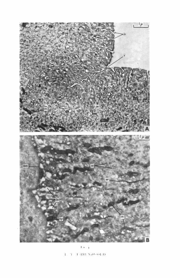
valleys (fig. 2). Its external surface has a definite plasma membrane which isinvaginated frequently along its length (fig. 3, A). Immediately below thesenarrow indentations are pinocytotic vesicles, apparently pinched off from theinvaginations. Around the larger vesicles are great numbers of small mem-brane-limited vacuoles and vesicles, and packed granules. The concentrationof vacuoles, vesicles, and granules gives the cuticle a superficial zone of in-creased electron density (figs. 2; 3, A; 4, A).
Occasionally the external cuticular surface is evaginated into balloon-shaped bodies containing fine granular material of low density. Theseevaginations are apparently nipped off into the environment (fig. 3, B).
Below the superficial dense zone the cuticle is composed of a backgroundmaterial containing the following:
1. Smooth-surfaced endoplasmic membranes, of variable length andperhaps derived from the external and internal plasma membranes.These membranes are usually aligned at right angles to the cuticularsurface (figs. 2; 4, B).
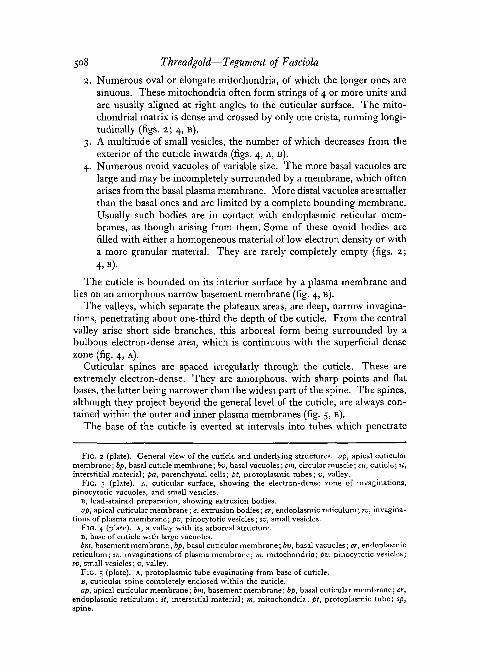
508 Threadgold—Tegument of Fasciola
2. Numerous oval or elongate mitochondria, of which the longer ones aresinuous. These mitochondria often form strings of 4 or more units andare usually aligned at right angles to the cuticular surface. The mito-chondrial matrix is dense and crossed by only one crista, running longi-tudinally (figs. 2; 4, B).
3. A multitude of small vesicles, the number of which decreases from theexterior of the cuticle inwards (figs. 4, A, B).
4. Numerous ovoid vacuoles of variable size. The more basal vacuoles arelarge and may be incompletely surrounded by a membrane, which oftenarises from the basal plasma membrane. More distal vacuoles are smallerthan the basal ones and are limited by a complete bounding membrane.Usually such bodies are in contact with endoplasmic reticular mem-branes, as though arising from them. Some of these ovoid bodies arerilled with either a homogeneous material of low electron density or witha more granular material. They are rarely completely empty (figs. 2;4 ,B) .
The cuticle is bounded on its interior surface by a plasma membrane andlies on an amorphous narrow basement membrane (fig. 4, B).
The valleys, which separate the plateaux areas, are deep, narrow invagina-tions, penetrating about one-third the depth of the cuticle. From the centralvalley arise short side branches, this arboreal form being surrounded by abulbous electron-dense area, which is continuous with the superficial densezone (fig. 4, A).
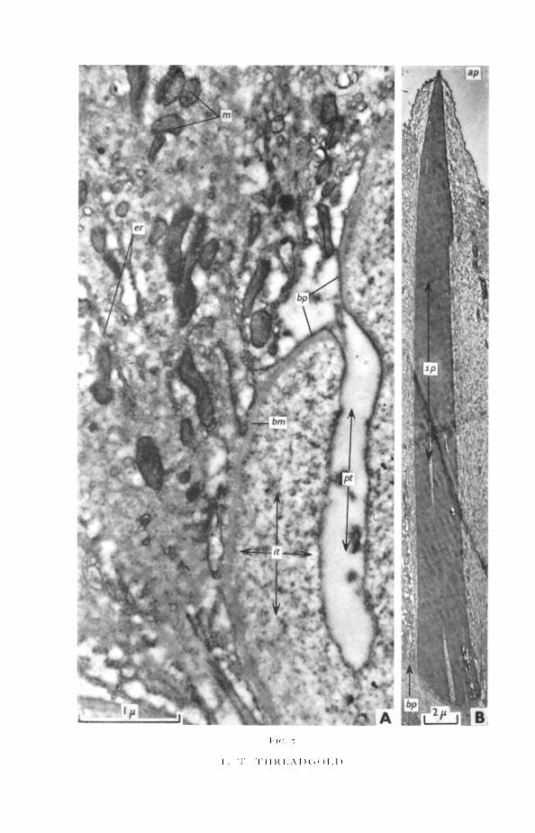
Cuticular spines are spaced irregularly through the cuticle. These areextremely electron-dense. They are amorphous, with sharp points and flatbases, the latter being narrower than the widest part of the spine. The spines,although they project beyond the general level of the cuticle, are always con-tained within the outer and inner plasma membranes (fig. 5, B).
The base of the cuticle is everted at intervals into tubes which penetrate
memFIG. 2 (plate). General view of the cuticle and underlying structures, ap, apical cuticular
membrane; bp, basal cuticle membrane; bv, basal vacuoles; cm, circular muscle; cu, cuticle; it,interstitial material; pa, parenchymal cells; pt, protoplasmic tubes; v, valley.
FIG. 3 (plate), A, cuticular surface, showing the electron-dense zone of invaginations,pinocytotic vacuoles, and small vesicles.
B, lead-stained preparation, showing extrusion bodies.ap, apical cuticular membrane; e, extrusion bodies; er, endoplasmic reticulum; iv, invagina-
tions of plasma membrane; pv, pinocytotic vesicles; sv, small vesicles.FIG. 4 (plate). A, a valley with its arboreal structure.B, base of cuticle with large vacuoles.bnt, basement membrane; bp, basal cuticular membrane; bv, basal vacuoles; er, endoplasmic
reticulum; iv, invaginations of plasma membrane; m, mitochondria; pv, pinocytotic vesicles;sv, small vesicles; v, valley.
FIG. 5 (plate), A, protoplasmic tube evaginating from base of cuticle.B, cuticular spine completely enclosed within the cuticle.ap, apical cuticular membrane; bm, basement membrane; bp, basal cuticular membrane; er,
:ndoplasmic reticulum; it, interstitial material; m, mitochondria; pt, protoplasmic tube: sp,spine

i . . i \ I 1 1 1 < [ : . \ I > c ; ( > i . i >

B
I ' I ' l l K l \ D l . ( i l l )
T . r i i R i ; . \ i ) < . ( ) i .
Threadgold—Tegument of Fasciola 509
the basement membrane, the muscle-layers, the interstitial material, and theparenchymal cells. The tubes bifurcate and anastomose so that sections showa confused picture of numerous ducts cut in various planes (figs. 2; 5, A).The walls of the tubes are extremely thin, consisting only of the basal plasmamembrane, except where accompanied by an inner layer of granular cyto-plasm. Near the cuticle the lumina of the tubes are clear, except for occasionalmitochondria and small areas of protoplasm (fig. 5, A). Deeper within theparenchyma and approaching the cuticular cells, the tubes are wider, and theamount of protoplasm and the number of mitochondria increases, whereasthe number and size of the vacuoles is reduced (fig. 6).
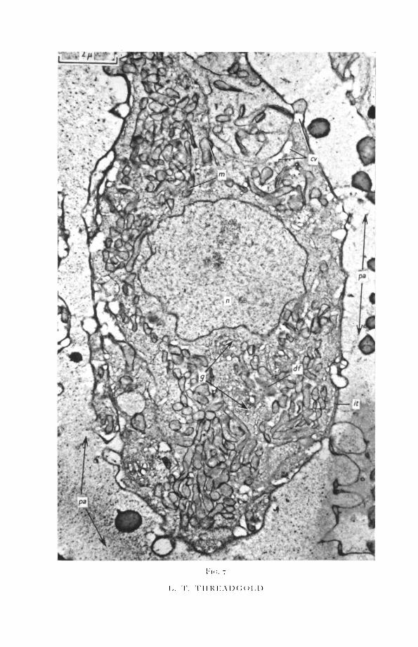
The cuticular cells occur either singly or in groups, often mixed with musclecells. Interstitial material surrounds and separates the cuticular cells from theparenchyma (figs. 6, 8). The individual cells are flask-shaped, the narrownecks tapering into the tubes leading to the cuticle (fig. 6). The nucleus liescentrally and has an undulating outline. The nuclear membrane is bulbousin places where the inner and outer membranes are separated by an expandedmiddle area (fig. 8). A single, very dense nucleolus is usual, but other largeand irregular electron-dense patches also occur.
The cytoplasm is filled with numerous mitochondria, which are mostlyshort or round (figs. 7, 8). The mitochondrial matrix is of medium electrondensity, with a fibrillar appearance. Cristae are rare, generally not more thanone in each mitochondrion, running in a longitudinal direction. A number ofGolgi bodies occur near the nucleus and consist of circular areas of mem-branes and vesicles (fig. 8).
In addition the cytoplasm contains first, endoplasmic membranes, bothrough, near the nuclear membrane, and smooth elsewhere; second, amorphouselectron-dense areas containing a few vacuoles and surrounded by a zone ofvacuoles; and finally vacuolated areas, mainly in contact with the plasmamembrane (figs. 9 and 10).
DiscussionThe results presented in this paper show clearly that the accepted structure
of the external covering of F. hepatica is incorrect. This external layer isneither a secreted inert structure, nor a much altered and degenerate epi-dermis. Theory 3 of the theories presented in the introduction is essentiallycorrect, although dismissed by Hyman (1951). The cuticle is a true cellulartegument, which is specialized in the following ways:
1. It is organized at two levels, external and internal. These two layers,although well separated, are connected together by protoplasmic strands.Such strands were described as canaliculi by Prenant (1928), but he didnot realize their significance.
2. The external layer, which functions as the tegument, is syncytial andwithout nuclei. Alvarado (1951) and Pantelouris and Gresson (i960)
2421.4 Li

51 o Threadgold— Tegument of Fasciola
claimed to have observed nuclei, but probably mistook massed mito-chondria (which are occasionally seen in electron micrographs), fornuclei, for such masses are very large.
This layer contains mitochondria, smooth endoplasmic membranes,numerous vacuoles, and electron-dense spines. The extreme surfacemembrane has either pinocytotic invaginations or bulbous evaginations.Despite the lack of nuclei, therefore, this external layer is protoplasmic.
3. The internal level is nucleated and composed of separate cells, which areeither well separated by intervening parenchymal cells or associated incompact groups. In addition to nuclei, the usual cytoplasmic organoidsare present, including the Golgi bodies.
4. The internal and external levels are connected by protoplasmic tubes,which are mainly hollow, but contain mitochondria and protoplasm inincreasing quantities as the interior level is approached.
Senft and his colleagues (1961) may be mistaken in concluding that thecuticle of Schistosoma is a vast spongy layer and an acellular amorphouscovering. Indeed, a careful examination of their electron micrographs sug-gests that the cuticle contains mitochondria, that evaginations arise basally,and that their epithelializing cells are really cuticular cells which are joinedto the cuticle by protoplasmic tubes. Positive confirmation of these pointscannot be made because of the low resolution of the photographs, but it istentatively suggested that the cuticle of Schistosoma is essentially similar tothat of Fasciola.
These findings have a bearing on the function of the cuticle. Mansour(1959) showed that a gut ligature did not prevent the uptake of glucose byFasciola. The cuticle must, therefore, be able to absorb glucose and probablydoes so by pinocytosis, for which there is now morphological evidence fromelectron micrographs. Pantelouris and Gresson (i960) demonstrated thatsome of the radioactive iron injected through the mouth eventually appearedin the cuticular cells (myoblasts) and the cuticle itself. They considered thecuticle either excretory or secretory in function, serving to eliminate excessmetabolites or to secrete mucus. The presence of evaginations and pinched-off extrusion bodies in electron micrographs may very possibly indicate theprocess whereby metabolites are secreted. Equally, such bodies might beevidence of the release of mucus over the cuticular surface, although no suchmucus layer was seen. Clearly the pinocytotic vesicles and extrusion bodies
FIG. 6 (plate). Protoplasmic tube projecting towards cuticle from cuticular cell. Stained withlead.
df, diffuse mass; it, interstitial material; m, mitochondria; n, nucleus; pa, parenchymal cell;pt, protoplasmic tube.
FIG. 7 (plate). Cuticular cell.cv, cytoplasmic vacuoles; df, diffuse mass; g, Golgi bodies; it, interstitial material; m,
mitochondria; «, nucleus;£a, parenchymal cells.FIG. 8 (plate). Cuticular cell with organelles.cv, cytoplasmic vacuoles; df, diffuse mass; g, Golgi bodies; it, interstitial material; m, mito-
chondria; n, nucleus; pa, parenchymal cell.
i.. T . T i l KKAD( ;o i . n

hi... i.
i. T . r i i u i - : . \ i ) c ; o i . n

I K . . S
1.. I . I l l K l . A l X i O l . l )
Threadgold—Tegument of Fasciola 511
are indicative of considerable cellular activity by the external layer of thecuticle.
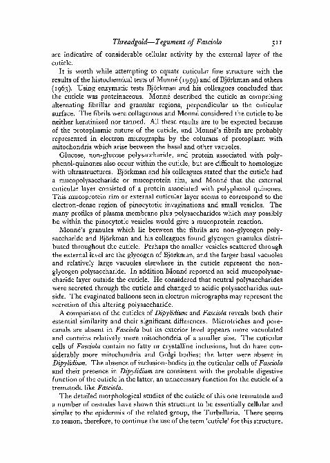
It is worth while attempting to equate cuticular fine structure with theresults of the histochemical tests of Monne (1959) and of Bjorkman and others(1963). Using enzymatic tests Bjorkman and his colleagues concluded thatthe cuticle was proteinaceous. Monne described the cuticle as comprisingalternating fibrillar and granular regions, perpendicular to the cuticularsurface. The fibrils were collagenous and Monne considered the cuticle to beneither keratinized nor tanned. All these results are to be expected becauseof the protoplasmic nature of the cuticle, and Monne's fibrils are probablyrepresented in electron micrographs by the columns of protoplasm withmitochondria which arise between the basal and other vacuoles.
Glucose, non-glucose polysaccharide, and protein associated with poly-phenol-quinones also occur within the cuticle, but are difficult to homologizewith ultrastructures. Bjorkman and his colleagues stated that the cuticle hada mucopolysaccharide or mucoprotein rim, and Monne that the externalcuticular layer consisted of a protein associated with polyphenol quinones.This mucoprotein rim or external cuticular layer seems to correspond to theelectron-dense region of pinocytotic invaginations and small vesicles. Themany profiles of plasma membrane plus polysaccharides which may possiblybe within the pinocytotic vesicles would give a mucoprotein reaction.
Monne's granules which lie between the fibrils are non-glycogen poly-saccharide and Bjorkman and his colleagues found glycogen granules distri-buted throughout the cuticle. Perhaps the smaller vesicles scattered throughthe external level are the glycogen of Bjorkman, and the larger basal vacuolesand relatively large vacuoles elsewhere in the cuticle represent the non-glycogen polysaccharide. In addition Monne reported an acid mucopolysac-charide layer outside the cuticle. He considered that neutral polysaccharideswere secreted through the cuticle and changed to acidic polysaccharides out-side. The evaginated balloons seen in electron micrographs may represent thesecretion of this altering polysaccharide.
A comparison of the cuticles of Dipylidium and Fasciola reveals both theiressential similarity and their significant differences. Microtriches and pore-canals are absent in Fasciola but its exterior level appears more vacuolatedand contains relatively more mitochondria of a smaller size. The cuticularcells of Fasciola contain no fatty or crystalline inclusions, but do have con-siderably more mitochondria and Golgi bodies; the latter were absent inDipylidium. The absence of inclusion-bodies in the cuticular cells of Fasciolaand their presence in Dipylidium are consistent with the probable digestivefunction of the cuticle in the latter, an unnecessary function for the cuticle of atrematode like Fasciola.
The detailed morphological studies of the cuticle of this one trematode anda number of cestodes have shown this structure to be essentially cellular andsimilar to the epidermis of the related group, the Turbellaria. There seemsno reason, therefore, to continue the use of the term 'cuticle' for this structure.
512 Threadgold— Tegument of Fasciola
The combined external and internal layers should be termed a tegument, theterm epidermis being avoided until the embryological origin of the tegumentis known for certain.
I wish to thank the Wellcome Trust and the Royal Society for the use of anAkashi electron microscope and ancillary equipment made available by a grantto Professor R. A. R. Gresson. Thanks are also due to Mr. W. Ferguson forenlarging the electron micrographs, and to Mr. A. Lyness for technicalassistance.
ReferencesALVARADO, F., 1951. Trabajos del Institute Jose de Acosta, serie biolbgica, III. Madrid.BJORKMAN, T. E., THORSELL, W., and LIENERT, E., 1963. Experientia, 19, 1.HYMAN, L., 1951. The Invertebrata, vol. 2. London (McGraw-Hill).KENT, H. N., 1957. Premier symposium sur la specificity parasitaire des parasites de vertebras.
University de NeuchStel.MANSOUH, T. E., 1959. Biochimica et biophysica Acta, 34, 457.MONNE, L., 1959. Ark. Zool., 12, 343.PANTELOURIS, E. M., and GRESSON, R. A. R., i960. Parasitology, 50, 165.PRENANT, M., 1928. Bull. Soc. zool. Fr.,53, 18.READ, C. P., 1955. Some physiological aspects and consequences of parasitism. New Brunswick
(University Press).ROTHMAN, A. H., 1959. J. Parasit., 45, 4 (Suppl.), 28.
i960. Ibid., 46, 5 (Suppl.), 10.SENFT, A. W., PHILPOTT, D. E., and PELOFSKY, A. H., 1961. Ibid., 47, 217.THREADGOLD, L. T., 1962. Quart. J. micr. Sci., 103, 135.